pneumocystis carinii karyotypes1786 honget al. in adjacent roomsin a different building from those...
TRANSCRIPT

JOURNAL OF CLINICAL MICROBIOLOGY, Aug. 1990, p. 1785-17950095-1137/90/081785-11$02.00/0Copyright C 1990, American Society for Microbiology
Pneumocystis carinii KaryotypesSUNG-TAE HONG,'12'3t PAUL E. STEELE,4 MELANIE T. CUSHION,2'3 PETER D. WALZER,2'3
SAUNDRA L. STRINGER,' AND JAMES R. STRINGER1*
Department of Molecular Genetics, Microbiology, and Biochemistry,l Department of Pathology and LaboratoryMedicine,4 and Department of Internal Medicine,2 College of Medicine, University of Cincinnati, and Veterans Affairs
Medical Center, Cincinnati, Ohio 45267-0524
Received 8 March 1990/Accepted 22 May 1990
Pulsed-field gel electrophoresis techniques were used to examine the chromosomes of Pneumocystis cariniiisolated from laboratory rats and two human subjects. P. carinii organisms isolated from each of four ratcolonies and from two patients each produced a distinct band pattern, but in all cases the bands ranged in sizefrom 300 to 700 kilobase pairs. P. carinùi from three rat colonies produced patterns containing 15 prominentbands. Of these 15 bands, 2 stained more intensely than would be expected of bands of their size, suggestingthat the P. carinii haploid genome contains 17 to 19 chromosomes. Summing the molecular sizes of the bandsand accounting for staining intensities suggested that the haploid genome of rat-derived P. carinii contains onthe order of 107 base pairs. Human-derived P. carinii produced patterns containing 10 to 12 bands whichappeared to be similar to the 15-band patterns seen in rat-derived P. caring with respect to the size range ofthe bands. P. carinii from the fourth rat colony produced a more complex band pattern containingapproximately 22 bands, most of which appeared to comigrate with the bands present in one of the 15-band P.carinii patterns, suggesting that these animals were simultaneously infected by two different varieties of P.carinii. Hybridization experiments using oligonucleotide probes specific for the P. carinu 18S rRNA gene
supported this possibility. The band pattern of P. carinii derived from a given rat colony was generally stableover time. P. carinii band patterns were not strictly rat strain specific and appeared to be transferable betweenanimals housed in the same room.
Pneumocystis carinii causes a pneumonia that is a princi-pal cause of death among patients with acquired immunode-ficiency syndrome (21, 28). Progress in characterizing thispathogen has been hampered by its fastidious growth re-quirements, which remain undefined. In vivo, the lungalveoli of immunocompromised mammals can accumulatevery large numbers of P. carinii organisms, which are oftenfound to be closely associated with type I pneumocytes (23,47).
P. carinii populations derived from infected lungs aremorphologically complex, containing many different formsof the organism which have been proposed to be stages in thelife cycle (1, 7, 25, 41). The principal forms of the organismare the trophic form, which is a small uninucleated cell, andthe cyst, which is a larger, spherical, multinucleated struc-ture resembling sporangenous structures of certain protozoaand fungi.
Until recently, P. carinii had been widely considered to bea protozoan, primarily on the basis of morphological obser-vations and drug susceptibilities. However, morphologicaland biochemical similarities between P. carinii and fungihave long been appreciated (32, 40). Recently, researchers inour laboratory and two others have examined the sequencesof rRNA genes of P. carinii (9, 37, 45). These moleculargenetic data suggest that P. carinii is more closely related tofungi than to protozoa.The organism appears to infect a range of vertebrates.
Over 75% of humans are seropositive for P. carinii by age 4,and many mammalian species host subclinical infections (16,31). It is not clear whether P. carinii organisms found inthese diverse hosts are the same organism or whether
* Corresponding author.t Present address: Department of Parasitology, Seoul National
University College of Medicine, Seoul 110-460, Korea.
genetic diversity exists among P. carinii organisms found ina given host species. Antigenic variability among P. carinhiisolates from different host species suggests that geneticvariants of the pathogen exist (13, 14, 22, 42). Animalpassage experiments and serological studies have suggestedthe presence of distinct strains (11, 18, 43). Furthermore,recent clinical studies have indicated that P. carinii antigenscan change in people with recurrent P. carinii pneumonia(29). To address the question of genetic variability among P.carinii isolates, we used pulsed-field gel electrophoresis(PFGE) to examine the genomes of P. carinii isolates fromimmunosuppressed rats and from two humans. The dataindicated that P. carinii populations isolated from a givensource are generally composed of organisms that contain thesame complement of chromosomes as judged by the size andnumber of bands resolved by PFGE. However, P. cariniipopulations isolated from four different colonies of infectedrats each displayed a distinct pattern of bands, as did the twosamples of P. carinii from humans. These findings indicatethat substantial karyotypic variability exists among popula-tions of the pathogen. Such karyotypic variability will be a
powerful tool with which to explore the epidemiology of P.carinii infections.
MATERIALS AND METHODS
Rat colonies. Male rats were obtained from three sources.Outbred Sprague-Dawley rats (viral antibody positive) wereexclusively from the colony kept in room 205H at HarlanSprague-Dawley, Madison, Wis. Upon arrival, Sprague-Dawley rats were placed in colonies A and B, which were
located in different buildings. Colony A had been establishedapproximately 1 year previously, and colony B had beenmaintained for 7 years. Lewis rats were placed into coloniesC and D (established 8 years previously), which were located
1785
Vol. 28, No. 8
on February 8, 2020 by guest
http://jcm.asm
.org/D
ownloaded from
1786 HONG ET AL.
in adjacent rooms in a different building from those of bothcolonies A and B. Two rats were housed per wire cageequipped with an automatic watering system. Absorbentpaper was placed underneath and changed on a biweeklybasis. Cages were held on double-sided stainless steel rackswith a 50-cage capacity. When a new shipment of rats wasreceived, the incoming animals were placed in separatecages and usually on separate racks; in some instances, newrats were placed on the same rack as infected animals but onthe opposite side. Incoming rats were never placed in thesame cage with immunosuppressed resident rats.
Provocation of P. carinùi pneumonitis. Rats were renderedimmunoincompetent by injection of methylprednisolone ac-etate (DepoMedrol; The Upjohn Co., Kalamazoo, Mich.) at4 mg/week for 8 to 16 weeks, as previously described (6). Toexacerbate P. carinii pneumonitis, malnutrition was inducedby administration of a low (8%)-protein diet (ICN Biochem-icals, Inc., Cleveland, Ohio). Water was supplemented withampicillin (1 mg/ml) to suppress secondary bacterial infec-tions.
Isolation of rat-derived P. carinii organisms. Immunosup-pressed rats were monitored for weight loss and other signsassociated with P. carinii pneumonitis, which usually devel-oped after 8 to 16 weeks of the regimen described above.Infected rats were sacrificed, the lungs were removed, andimpression smears were examined to determine the severityof infection. Grades of infection were assigned according toa semiquantitative grading system based on a previouslydescribed histological scoring technique (20). Lungs werehomogenized in a blender (Stomacher; Tekmar Inc., Cincin-nati, Ohio). The preparations were filtered through gauze toremove large particulates and then treated with a hypotoniclysis solution (0.85% ammonium chloride or 0.04 M KCl-0.025 M sodium citrate) to reduce contamination with hostcells. Organism preparations were again filtered at least fourto six times through 10-ktm-pore-size filters (Mitex; MilliporeCorp., Bedford, Mass.). Material that passed through thefilter was examined microscopically to determine the num-ber and purity of the organisms obtained. The preparationswere then treated with DNase at 8.5 ,ug/ml for 30 min at 37°Cin the presence of 10 mM MgCl2 to digest host cell nuclei.The DNase was inactivated by adding EDTA to a finalconcentration of 0.250 M. Organisms were pelleted by briefcentrifugation, and the pellet was suspended in 0.125 MEDTA (pH 7.4). The P. carinii organisms remained intactthroughout these procedures (37). Surveillance cultures ofeach isolate were performed by streaking 10 pt1 of homoge-nate onto Sabouraud dextrose and Mueller-Hinton agars inplates, followed by incubation for up to 2 weeks at 25 and35°C, respectively.
Isolation of P. carinii derived from human sources. P.carinii isolates from humans were obtained from four bron-choalveolar lavage fluid samples (026, 020, 009, and 133) andfrom two postmortem lung tissue specimens (samples 118and 142). All specimens were from human immunodeficiencyvirus-infected patients and were obtained through agree-ments with Robert Baughman, Pulmonary Division, Univer-sity of Cincinnati College of Medicine (bronchoalveolarlavage fluids), and the Department of Pathology and Labo-ratory Medicine, University of Cincinnati College of Medi-cine (postmortem samples). Bronchoalveolar lavage fluidswere treated with hypotonic lysis solution and DNase toreduce host cell contamination (as described above) and thenimmediately embedded in low-gelling agarose (see below).Postmortem specimens were processed as described abovefor isolation of P. carinii from rat lungs, except that all
procedures were performed in a BSL-2 laboratory andbiocontainment hood according to Centers for Disease Con-trol directives for handling of human immunodeficiencyvirus-infected specimens (F. A. Murphy, W. J. Martone,J. W. Curran, and J. D. Millar, Morbid. Mortal. WeeklyRep. 39[RR-1]:1-11, 1990). Human lung homogenates con-tained more host cell debris than did rat lungs and werefiltered extensively (at least 8 to 10 times).PFGE. P. carinii suspensions were mixed with a solution
of low-gelling agarose (Sigma Chemical Co., St. Louis, Mo.)in 0.125 M EDTA to achieve a parasite density of 5 x 10⁸nuclei per ml in 0.6% agarose as described previously (3).Titration of P. carinii densities from 5 x 10⁵ to 1 x 10⁹showed that this number of organisms was optimal forethidium bromide visualization. Gel-embedded cells weretreated with 0.25 mg of proteinase K (Boehringer MannheimBiochemicals, Indianapolis, Ind.) per ml and 1% Sarkosyl(CIBA-GEIGY Corp., Summit, N.J.) at 55°C for 18 h.Digested agarose plugs were stored in 0.5 M EDTA (pH 9.0)at 40C.Contour-clamped homogeneous electric field electropho-
resis (CHEF; 5) and field inversion gel electrophoresis(FIGE; 3) gels containing 1% agarose were prepared in 45mM Tris hydrochloride-45 mM boric acid-0.125 M EDTA(0.5x TBE) and electrophoresed at 14 to 160C in the samebuffer. CHEF was performed in a Bio-Rad CHEF DRIIapparatus. FIGE was performed in an apparatus constructedas described by Carle et al. (3). CHEF gels were generallyrun at 90 V for 96 h with 50- to 100-s gradual switching, whileFIGE gels were run at 105 V for 120 h with 50-s forward and25-s backward switching. Occasionally, gels were run withdifferent parameters, and these are noted in the figurelegends.
Isolation of rat-derived P. carinii DNA. For each prepara-tion of DNA, 2 x 10¹⁰ filtered, DNase-treated P. cariniiorganisms obtained from infected rat lung homogenates werelysed in 0.25 M EDTA containing 0.25 mg of proteinase Kper ml and 1% Sarkosyl at 550C. The lysate was thenincubated at 550C for 4 h. Digested lysates were extractedtwice with phenol plus chloroform and twice with chloro-form alone, and the DNA was precipitated with ethanol (24).Approximately 20 ,ug of DNA was obtained. The purity ofDNA preparations was assessed as described in Results.
Production of a rat-derived P. carinii genomic library.Three micrograms of P. carinii DNA obtained from fourLewis rats (two from colony C and two from colony D) waspartially cleaved with Sau3A (24). The ends of the DNAwere partially filled by treatment with Klenow polymerase inthe presence of dGTP and dATP. The DNA was fractionatedby electrophoresis through a 0.7% agarose gel. DNA thatmigrated between size markers of 23 and 16 kilobase pairs(kb) was excised, extracted from the agarose, and ligated to1 ,ug of the lambda Fix vector (Stratagene, La Jolla, Calif.).The ligated DNA was packaged in vitro by using Gigapack(Stratagene), and approximately 60,000 plaques were pro-duced by infection of Escherichia coli LE392. Approxi-mately 30% of these plaques were produced by chimericbacteriophage as judged by plaque formation on E. coliP2392 and by hybridization to a radiolabeled probe preparedby nick translation of a sample of the agarose gel-purifiedDNA used in the ligation to lambda Fix.
Hybridization probes. A synthetic 25-base oligonucleotideprobe designed to be specific for 18S rRNA sequencesunique to P. carinii had the sequence GGTAATCCAGGAGGGAAGGATCAGT. This sequence was complementaryto a sequence running from nucleotide residue 680 to residue
J. CLIN. MICROBIOL.
on February 8, 2020 by guest
http://jcm.asm
.org/D
ownloaded from

P. CARINII KARYOTYPES 1787
TABLE 1. Characteristics of P. carinii isolates studied
Sample Colony Date No. of Yield of Cultureb No. of bacteria/(mo/day/yr)a rats P. carinii nucici P. carinii nuclei
SD(1) A 11/29/88 7 4 x 10⁹ G+ cocci 2.4 x 10⁻⁶SD(3) B 12/1/88 8 13 x 109 G- rods 6.3 x 10⁻⁸SD(4) B 5/17/89 7 140 x 109 G+ cocci 2.2 x 10⁻⁸SD(6-4) B 6/6/89 1 6 x 10⁹ NDSD(8) A 6/21/89 1 2 x 10⁹ NDL(1) C 11/29/88 2 3 x 10⁹ NDL(3) C 1/3/89 4 2 x 10⁸ G+ cocci 1.6 x 10⁻²L(4) D 1/10/89 6 8 x 10⁸ ND 7 x 10⁻⁷L(6) D 5/18/89 4 28 x 109 ND 1 x 10⁻⁸L(7) C 6/5/89 4 3 x 10⁹ G- rods 1.3 x 1i-3L(8-1) D 6/5/89 1 6 x 10⁹ G- rods 1.6 x 1O-4L(8-2) D 6/5/89 3 3 x 10⁹ G+ cocci 9.1 x 10⁻⁴L(8-3) D 6/5/89 1 2 x 10⁸ G- rods 5.6 x 1(-7L(8-5) D 6/5/89 1 2 x 10⁹ NDL(8-7) D 6/5/89 1 9 x 10⁸ G+ cocci 1.2 x 10⁻⁶L(9-1) C 6/5/89 1 1 x 10⁷ G- rods 1.1 x 10⁻¹L(9-2) C 6/5/89 1 9 x 10⁷ G- rods 1.1 X 10⁻²L(9-5) C 6/5/89 3 9 x 10⁸ G+ cocci 3.8 x 1O-3L(10-1) C 6/7/89 1 2 x 10⁸ G+ cocci 5.9 x 10⁻⁷L(10-2) C 6/7/89 1 2 x 10⁸ G- rods 5.6 x 1O-3L(10-3) C 6/7/89 1 3 x 10⁸ G- rods 2.9 x 10⁻³L(10-4) C 6/7/89 1 5.2 x 10⁸ G+ cocci 1.9 x 10⁻⁷L(11-1) C 6/21/89 1 6 x 10⁷ G- rods 2.0 x 10⁻²L(11-2) C 6/21/89 1 2.9 x 10⁷ NDL(11-C) A 6/21/89 1 1.3 x 10⁸ NDL(11-D) A 6/21/89 2 5.0 x 10⁷ G+ rods 7.2 x 10⁻⁵
a Date when P. carinii was harvested.b G+, Gram positive; G-, gram negative; ND, none detected.
705 in the P. carinii 18S rRNA gene (9). This region is highlyvariable among 18S rRNA genes. The 25-mer was 50%identical to the corresponding sequence in the 18S rRNAgene of Neurospora crassa and 25% identical to the corre-sponding Saccharomyces cerevisiae sequence, the two 18Ssequences known to be most similar to that of P. carinii.Identity of the 25-mer with other 18S rRNA gene sequencesranged between 25 and 35% (Oxytricha nova, Acan-thamoeba castellani, Dictyostelium discoideum, corn, frog,and rat sequences were among those analyzed) (17). Asecond synthetic oligonucleotide 26 bases long and comple-mentary to nucleotide residues 636 to 662 of the P. carinii18S rRNA gene was also used. This 26-mer corresponded toone of the three probes reported by Edman et al. (9) tohybridize to human P. carinii in situ. The 26-mer performedidentically to the 25-mer described above in our hybridiza-tion experiments (data not shown).
Oligonucleotides were labeled by treatment with T4 poly-nucleotide kinase in the presence of [-y-32P]dATP. Total ratcell DNA was isolated from cultured Rat-3 cells (38) andlabeled by nick translation as described by Maniatis et al.(24). Radiolabeled cDNA corresponding to a segment of theP. carinii 18S rRNA was prepared by reverse transcriptionof total P. carinii RNA in the presence of universal 18SrRNA primer A as described previously (37). Other probeswere prepared by nick translation (24).
Southern blotting. CHEF and FIGE gels were prepared fortransfer by treatment with 0.25 M HCl for 20 min, followedby denaturation with 0.5 M NaOH and neutralization asdescribed by Schwartz and Cantor (33). DNA was trans-ferred by capillary flow onto nylon membranes (Nytran;Schleicher & Schuell, Inc., Keene, N.H.) as recommendedby the vendor. Prehybridization was done as described inreference 26 for oligonucleotide probes and as described inreference 36 for other probes. Hybridization and washing
conditions for oligonucleotide probes were as follows: hy-bridization in 6x SSPE (0.9 M NaCI, 0.09 M NaH2PO4, 0.02M disodium EDTA [pH 7.4])-10 ,g of denatured salmonsperm DNA per ml-0.1% sodium dodecyl sulfate (SDS) at42°C for 16 to 30 h; wash in 6x SSPE-5 x Denhardtreagent-0.4% SDS-50% formamide at 42°C for 16 to 30 h.Washes were performed at 650C in 0.2x SSPE-0.5% SDS.Radioactive signals were detected by radioautography asdescribed in reference 24.
RESULTS
Analysis of P. caring preparations. P. carinii organismswere isolated from infected lungs by using previously estab-lished methods (6, 7, 37, 42). The P. carinii preparationsused in this study were characterized by microscopic exam-ination and by inoculation of culture plates that would allowgrowth of other microorganisms.
Microscopic inspection and tinctorial staining were usedto quantitate P. carinii organisms (Table 1) and to assess thepresence ofpotential contaminants, such as other eucaryoticmicrobes, rat cells, and rat cell nuclei. None of the samplesused in this study contained other eucaryotic microbes. Norat cells were detected, but rat cell nuclei were sometimesobserved. Although in low numbers compared with thenumber of P. carinii nuclei (less than 1%), host nuclei werea significant problem because each mammalian nucleuscontains on the order of 200 times more DNA than a P.carinii nucleus. This problem was minimized by the use ofDNase, which was effective in specifically degrading ratDNA.
Inoculation of agar plates with samples of P. cariniipreparations provided the second means with which todetect the presence of other microorganisms. Again, noeucaryotic microbes were detected, but bacterial contami-
VOL. 28, 1990
on February 8, 2020 by guest
http://jcm.asm
.org/D
ownloaded from
1788 HONG ET AL.
nation was common. However, the presence of bacteria inP. carinii preparations did not influence the patterns ob-tained by PFGE. The ratio of bacterial cells to P. cariniinuclei present in P. carinii preparations varied from as highas 1.1 x 10⁻¹ to as low as 108, yet there was no correlationbetween the number of bacteria and the PFGE band pattern(Table 1).
Analysis of P. carinùi nucleic acids. Microscopic and micro-biological characterization of P. carinii preparations showedthat few, if any, other eucaryotic microbes were present. Inaddition, we have previously shown that P. carinii preparedas described here produced essentially pure P. carinuirRNAs, as assessed by the sizes of the P. carinii rRNAmolecules and sequence analysis of 18S rRNA species (37).Therefore, it seems unlikely that PFGE bands producedfrom such preparations could originate from anything otherthan P. carinii. Nevertheless, it seemed prudent to confirmthat the lysis procedure used in preparing samples for PFGEdid indeed release P. carinil DNA.DNA was prepared from a P. carinii preparation [SD(3) in
Table 1] by using the lysis procedure `used to preparegel-embedded samples for PFGE. To assess the presence ofrat sequences, 1 ,ug of DNA was electrophoresed through1% agarose. The gel was treated with HCl to mobilizehigh-molecular-weight DNA, which was then transferredfrom the gel to a nylon filter, and the filter was subjected tohybridization with radioactive DNA prepared from culturedrat cells. The only material that hybridized to the rat probewas less than 5 kb long (data not shown).Three micrograms of the P. carinii DNA preparation was
partially digested with Sau3A and electrophoresed through1% agarose, and fragments that migrated between 15 and 23kb were isolated. These fragments were ligated to lambdaFix (Stratagene). Approximately 20,000 recombinant phageplaques were obtained, as judged by plating on the spi(+)host, E. coli P2392, and by hybridization to the gel-purifiedDNA fraction used in the ligation. The library was screenedfor the presence of rat sequences by hybridization of 2,000plaques to radioactive rat DNA; 1 plaque hybridized.The library was next screened with radioactive cDNA
made by reverse transcription of the 18S rRNA from P.carinii. Approximately 2% of the plaques hybridized to theprobe, a percentage consistent with expectations based onthe assumption that rRNA genes are as numerous in the P.carinii genome as they are in S. cerevisiae (50 copies perhaploid genome [30]). Restriction analysis of four chimericphage showed that they all contained related inserts and thateach insert restriction map was consistent with what wouldbe expected on the basis of a previously reported restrictionmap of the rRNA locus of P. carinii (10; data not shown).The phage all hybridized to an oligonucleotide complemen-tary to a unique region of the P. carinii 18S rRNA gene (datanot shown). The likelihood that this oligonucleotide canhybridize to DNA not from P. carinii is very low because theprobe corresponds to a sequence that varies considerably,even among close relatives. Analysis of the 18S rRNAsequence data base showed that the oligonucleotide sharedless than 50% analogy to any other rRNA sequence (17; seeMaterials and Methods for details).PFGE analysis. Information regarding the rat-derived P.
carinii isolates analyzed by PFGE can be found in Table 1.Seven P. carinii preparations were obtained from Sprague-Dawley colonies A and B. Nineteen P. carinii preparationswere obtained from Lewis rats, which were divided betweencolonies C and D. Initial preparations of P. carinii originatedfrom pooled homogenates of infected animals, but in all
Strain ri îsoiatecolony
Sc1 2 3 4 5 6 7 8 9
kb
580
45S0-
35-
280b-
250 - _|s=
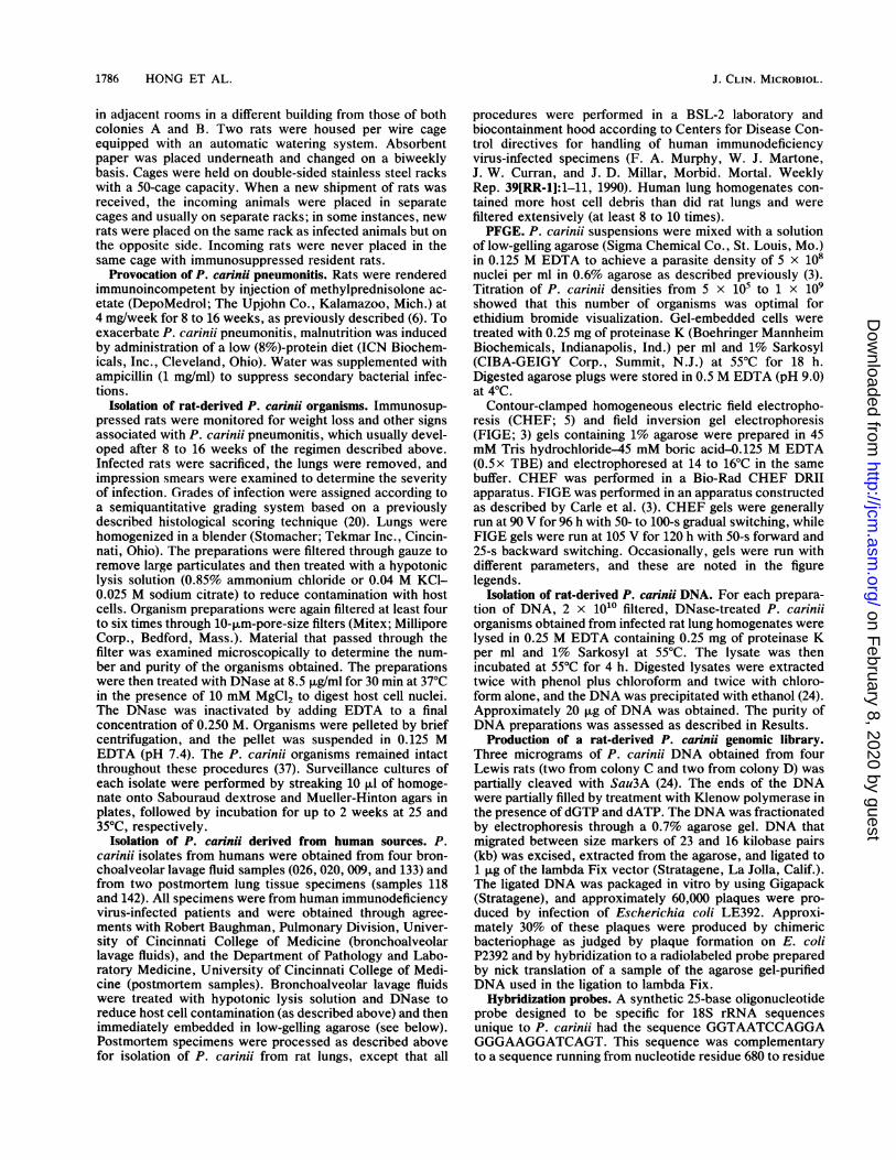
FIG. 1. P. carinIchromosomes resolved by CHEF. Samplesprepared from colonies B and D were subjected to CHEF at 90 V for96 h with an initial switching interval of 50 s. The switching intervalwas graduaiiy increased to 100 s. Lane i contained S. cerevisiae(Sc). Lanes 2 to 5 contained replicate piugs containing P. cariniipreparation SD(6-4) (Table 1). Lanes 6 to 9 contained replicatesamples of preparation L(8-1).
cases, animals grouped for P. carinil isolation were from thesame colony. Later preparations of P. carinii were usuaiiyfrom individual animals.
Figure i shows the results of CHEF analysis of twosamples of P. carinii DNA, one from colony B [SD(6-4)] andone from colony D [L(8-1)]. Both P. carinii samples pro-duced a series of bands ranging from approximately 300 to700 kb, but the band patterns for the two samples weredifferent. [The DNA that migrated in the megabase range insample L(8-1) was later shown to be rat DNA (see beiow).]To verify that the CHEF bands were from P. carinii, gels
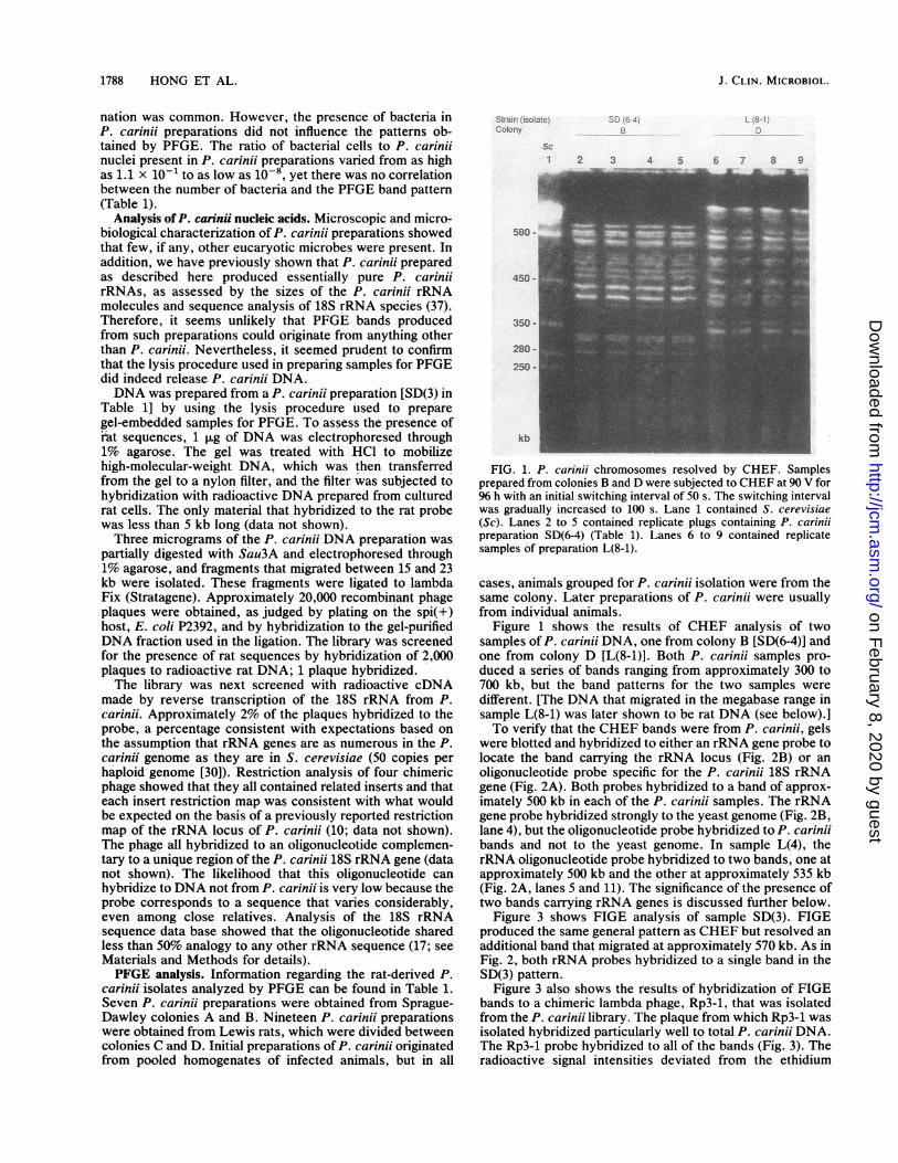
were blotted and hybridized to either an rRNA gene probe tolocate the band carrying the rRNA locus (Fig. 2B) or anoligonucleotide probe specific for the P. carinii 18S rRNAgene (Fig. 2A). Both probes hybridized to a band of approx-imateiy 500 kb in each of the P. carinii samples. The rRNAgene probe hybridized strongly to the yeast genome (Fig. 2B,lane 4), but the oligonucleotide probe hybridized toP. carinuibands and not to the yeast genome. In sample L(4), therRNA oligonucleotide probe hybridized to two bands, one atapproximately 500 kb and the other at approximately 535 kb(Fig. 2A, lanes 5 and 11). The significance of the presence oftwo bands carrying rRNA genes is discussed further beiow.
Figure 3 shows FIGE analysis of sample SD(3). FIGEproduced the same general pattern as CHEF but resolved anadditional band that migrated at approximately 570 kb. As inFig. 2, both rRNA probes hybridized to a single band in theSD(3) pattern.
Figure 3 also shows the results of hybridization of FIGEbands to a chimeric lambda phage, Rp3-1, that was isolatedfrom the P.cariniilibrary. The plaque from which Rp3-1 wasisolated hybridized particularly well to total P.carinii DNA.The Rp3-1 probe hybridized to ail of the bands (Fig. 3). Theradioactive signal intensities deviated from the ethidium
J. CLIN. MICROBIOL.
!. 0r-
on February 8, 2020 by guest
http://jcm.asm
.org/D
ownloaded from
P. CARINII KARYOTYPES 1789
Prcbe RNA o0QS. nr3SD L.3' ` 4A 3 C D
Sc
7 8 9 10 il 12
am ge
I 580 -
_ -d
OMM
450 -
B
Stra r: ico;a!e L.. L
IoobnV A CSc
Probe: rRNlA gene
SC:;I: L' 1
A C
Sc
2 3 4 5 6
580
450 -
kb
FIG. 2. Hybridization of P. carinfi chromosomes to 18S rRNA
gene probes. The gels in panels A and B were run under the
conditions stated in the legend to Fig. 1. Probes were prepared and
used as described in Materials and Methods. The lane labeled X
contained concatenated lambda genomes. Hybridization and wash
conditions were as follows. For pan-el A, the blot was hybridized in
6x SSPE-10 p.g of salmon sperm DNA per ml-0.1% SDS at 420C
and washed in 6x SSPE-0.5% SDS at 650C. For panel B, the blot
was hybridized in 50% formamide-6x SSPE-5x Denhardt solution-
100 kig of salmon sperm DNA per ml-0.4% SDS and washed in 0.2x
SSPE-0.5% SDS at 680C. Sc, S. cerevisiae.
bromide fluorescence intensities, suggesting that this chi-
meric phage carried a sequence that is repeated in the P.
carinhi genome and is present on ail chromosomes but that
the number of repeats varies among chromosomes. That the
hybridization signal was not due to the presence of lambda
sequences in the probe was verified by the experimentshown in Fig. 2B, in which a lambda phage carrying a
fragment from the P. carinii rRNA locus hybridized to a
single band. P. carinii DNAs from all four rat colonies
contained the repeated sequence present in the Rp3-1 cloned
DNA fragment, but rat and yeast DNAs showed no homol-
ogy to Rp3-1. Rp3-1 hybridized to bands that migrated
between 300 and 700 kb and, in some cases, to smaller DNAs
350 -
FIG. 3. P. carinii chromosomes resolved by FIGE hybridized toa repeated sequence. Multiple lanes of a 1% agarose gel were loadedwith sample SD(3) and subjected to FIGE at 105 V for 120 h withswitching forward for 50 s and backward for 25 s. The DNA was
transferred to a nylon membrane which was cut into strips whichwere hybridized to the probes indicated under the conditionsdescribed in Materials and Methods.
that migrated as a smear at the bottom of the gel (Fig. 4). TheRp3-1 probe did not hybridize to the DNA that migrated inthe megabase range. The data shown in Fig. 4C identifiedthis megabase DNA as originating from a rat, which explainswhy this large DNA was present in only some of the P.carinfi samples.Comparative analysis of P. carinii from rats. The experi-
ments described above showed that P. carinii derived fromdifferent colonies of rats produced distinct band patterns onCHEF gels. To better resolve these patterns, samples fromeach of the four colonies were analyzed by FIGE. Eachcolony produced a characteristic band pattern (Fig. 5). Thepatterns produced by Sprague-Dawley rats from colonies Aand B were very similar, the only differences being thatpattern B contained a prominent 560-kb band that was notpresent in the A pattern, and in pattern B the 560-kb bandand the 545-kb band stained less intensely than the 545-kbband in pattern A. This can be seen more clearly in Fig. 3.[Note that the bands in lane 2 are misaligned because thislane was from a different gel. The bands in sample SD(1)comigrated with samples from colony B (see Fig. 2A and 9).]Colony C, which housed Lewis rats, produced a bandpattern that was quite different from those of colonies A andB. However, the patterns from colonies A, B, and C allcontained 15 prominent bands that ranged between 300 and700 kb.
A Strain isolateColony
Probe_''-
x1 2
500
250 -
kb
SLD;3B
rRNAoligo
3 4Rp 3-1
1 2
rRNAgene
5 6
VOL. 28, 1990
kb
on February 8, 2020 by guest
http://jcm.asm
.org/D
ownloaded from

1790 HONG ET AL.
e',...- ..>4". _. ;~
._..
Sc1 2 3 4 5
+` ), :~B
S1I 2 3 4 5
p>core !;,e-z-- Di: 4: S' -. L.ii4 8-1. L :6
;; 3
Sc
3 4 52
e-._
1125 -
835 -
780 -
690 -580- ea_
-p
kb _'l.rn,, =
FIG. 4. Resolution of largest P. carinii chromosomes by CHEF and hybridization to rat DNA. CHEF was performed at 150 V for 45 h with100-s switching on a 1.5% agarose gel in 0.5 x TBE. Hybridization conditions were as described in Materials and Methods. Sc, S. cerevisiae.
The pattern of colony D, which also housed Lewis rats,differed from the other three colony patterns with respect tothe number of bands, showing 22; however, the size range ofthese bands conformed to the range observed in the other
Date Harvested 12J88 5189 68 89 589 6.89 58$- o three colonies (Fig. 6, lanes 1 through 6). The complexity ofStrain isolate> D(1/ 5DM)} 556-4 ;3 L 7 D7(9-5` z/E7 18the D band pattern suggested that the animals of this colonyColony A 6 B C G
sc harbored an infection caused by more than one type of P.1 2 3 4 5 6 7 8 9 carinii organism. A mixed infection was indicated by three
observations. (i) The D pattern contained a set of bands thatcomigrated with the bands in the C pattern in addition to
580~ ~ ~ ~ ~ ~
1 2 3 4 5 6 7 $ 9 10il112 13 14
0580- _ _ _ c:ra L~~~~~~~~~~~~~50
450-~~~~~~~~~~~~~~~~~~40
350,s.;_=_ 3
kb
FIG. 5. Variation of band patterns among P. carinii samples k
; * 4 _>;ku
trom danerent rat colonies and staboluty or the pattern rrom a gïvencolony over time. FIGE parameters were as described in the legendto Fig. 3. Note that lane 2 was from an earlier gel. The bands insample SD(1) comigrated with colony B bands in experiments inwhich both were run on the same gel (Fig. 2A). Sc, S. cerevisiae.
FIG. 6. FIGE analysis of multiple isolates from colonies C andD. The FIGE parameters used were as described in the legend toFig. 3. The P. carinii isolates are described in Table 1.
J. CLIN. MICROBIOL.
qwm
le
li;:.: -f
z. L.
on February 8, 2020 by guest
http://jcm.asm
.org/D
ownloaded from

P. CARINII KARYOTYPES 1791
several other bands. (ài) The rRNA oligonucleotide probehybridized to two bands in the D pattern, one of whichcomigrated with the rRNA gene-containing band in the Cpattern (Fig. 2A). (iii) All of the bands in the D patternhybridized to the P. carinii repeat probe (Fig. 4B), whichimplies that all of these bands were from P. carinii and werenot due to contaminants. The same pattern was producedwhether pooled homogenates or isolates from individual ratswere used, indicating that each animal in the D colony wasinfected by the same varieties of the organism in the samerelative amounts.
Analysis of P. carinui from humans. Samples were preparedfrom six individuals. Four samples were collected as bron-choalveolar lavage fluids (samples 026, 020, 009, and 133),and two were prepared from lung tissue (samples 118 and142). The CHEF band pattern produced by sample 142 isshown in Fig. 7A. The intense band that migrated near the250-kb marker was of unknown origin and was not present inthe other human-derived P. carinii sample that produced aband pattern (Fig. 7B).Sample 142 was also analyzed by FIGE, along with the
other five samples prepared from human sources (Fig. 7B).Only samples 133 and 142 produced FIGE band patterns.The patterns produced by samples 133 and 142 were differentfrom each other and from any of the patterns produced byrat-derived P. carinii, although the sizes of the bands presentin both human- and rat-derived specimens were within thesame 300- to 700-kb range. The number of human-P. cariniibands in these samples appeared to be 10 to 12. Visualizationof the P. carinii ethidium bromide-stained bands obtainedfrom human sources was more difficult than analysis ofrat-derived specimens because of problems inherent to hu-man specimens, including low organism number, decreasedorganism viability, and suboptimal sample preservation.Attempts to hybridize the bands seen in the human-
derived samples to total rat-derived P. carinii DNA, to a P.carinii 18S rRNA probe, or to an oligonucleotide specific forthe P. carinii 18S rRNA gene were unsuccessful. The failureof the 18S rRNA probe to hybridize to the human P. carinjibands suggests that the lack of hybridization was due totechnical factors, such as poor transfer of the bands to thenylon membrane or low organism numbers. The rRNA geneprobe would have been expected to hybridize to any eucary-otic 18S rRNA gene, and this probe did, in fact, hybridize toyeast DNA (Fig. 2B). When we performed reconstructionexperiments in which diminishing numbers of rat-derived P.carinii organisms were subjected to PFGE analysis, followedby blot hybridizations, bands as faint as those seen in thePFGE patterns produced from human-derived P. cariniiwere not detected by the rRNA gene probe and were barelyin evidence following hybridization to a radioactive probeprepared from total rat-derived P. carinii DNA. Furtherstudy of this issue will require isolation of more human-derived organisms, which have not yet become available.
Size estimates of P. carinii chromosomes. Figure 8 is aschematic representation of band patterns of P. cariniiorganisms from each of the four rat colonies resolved byCHEF. Although FIGE has superior resolving power, mo-lecular sizes of P. carinii bands were estimated by CHEFbecause sizing of FIGE-resolved bands is complicated byunpredictable migration of some chromosomes due to un-known factors (3). In our FIGE experiments, lambda mark-ers appeared to migrate according to size above 400 kb, butthe band pattern was aberrant in the small-molecular-sizerange. S. cerevisiae chromosomes also migrated aberrantlyon FIGE. By contrast, lambda and S. cerevisiae markers
A
xi
500 -
400 -
300 -
200 -
100 -
kb
B
Sc
SD(3Ï
2
L(1) 142
3 4Sc5
- 580
- 450
- 350
- 280- 250
kb
HUMAN
SDi3j 026 020 009 133 118 142x
2 3 4 5 6 7 8 9
580 -
450 - E7
350 -
kb
FIG. 7. Analysis of human-derived P. carinii. (A) CHEF through1% agarose at 90 V for 96 h with an initial switching interval of 50 sgradually increasing to 100 s. Lane 4 contained a sample derivedfrom human lung tissue. (B) The FIGE parameters used were asdescribed in the legend to Fig. 3. Lanes 3 to 6 contained broncho-alveolar lavage fluid samples. Lanes 7 and 8 contained samples fromhuman lung tissue. Sc, S. cerevisiae.
migrated according to size on CHEF gels. The CHEF runshown in Fig. 1 separated DNA molecules between 50 and600 kb in a manner that allowed the molecular sizes of all butthe largest bands in the P. carinii samples to be deduced byinterpolation. A second CHEF analysis was performed tobetter estimate the sizes of the largest three bands (Fig. 4).
VOL. 28, 1990
on February 8, 2020 by guest
http://jcm.asm
.org/D
ownloaded from

1792 HONG ET AL.
A
705
670655
575570
545525505485
445 -
425
B c SirainGoi;On:.is..f te
705
670635600575
ScI 'I 4 6-j, 5 7
690 -
580 - f550
545
515
490
440-m 430
410
335
320 325315
295 300FIG. 8. Schematic representation of CHEF band patterns prod'
duced by rat-derived P. carinfi. A, B, and C represent patterns fromcolonies A, B, and C. Bolder and fainter lines indicate bands thatstained more or less intensely, respectively, than would be expectedof an equimolar band.
450
If the simplest band pattern represents the karyotype of aP. carinii strain, patterns A, B, and C indicate that thegenome contains 15 to 19 chromosomes. Summing the sizesof the 15 discrete bands gives a haploid genome size of 7 x10⁶ base pairs. Inclusion of putative comigrating chromo-somes would increase the estimate of the genome size to 8 x10⁶ to 1 x 10⁷ base pairs.
Stability of band patterns. The band pattern produced fromP. carinii isolated from colony C remained essentially un-changed over a period of 6 months (Fig. 5), during whichtime new animals were received from the commercial vendor(Table 1), placed in the colony with infected animals, andrendered immunoincompetent. Organisms harvested at dif-ferent times from other colonies also usually produced thesame band pattern, the one exception being sample SD(8)(see below).Band patterns were not strictly rat strain specific. The
temporal constancy of band patterns allowed us to test thepossibility that animals introduced into a colony acquired theorganism from resident infected animals. The same experi-ment addressed the possibility that there might be Sprague-Dawley-specific P. carinii strains and Lewis-specific P.carinfi strains. Lewis rats were purchased from CharlesRiver Breeding Laboratories, Inc. (Wilmington, Mass.), andupon arrival they were divided into two groups. Lewis ratsfrom this vendor had been previously shown to produce P.carinii organisms with pattern C (Fig. 6, lanes 7 through 14).One group was placed with infected Lewis rats in colony C;the other group was housed with P. carinii-infected Sprague-Dawley rats in colony A. P. carinii band patterns from twoindividuals from each colony are shown in Fig. 9. Asexpected, the two Lewis rats placed in colony C yielded P.carinii organisms with pattern C (Fig. 9, lanes 6 and 7). Theresults obtained with Lewis rats placed in colony A weremore complex but showed that Lewis rats can acquire P.
350
kb
FIG. 9. Band patterns were not strictly rat strain specific. Organ-isms were prepared from Sprague-Dawley rats from colony A andfrom Lewis rats that had been housed in either colony C or colonyA. The FIGE parameters used were as described in the legend toFig. 3. Sc, S. cerevisiae.
carinii of the type characteristic of Sprague-Dawley ratsresiding in colony A. One animal produced P. carinii organ-isms with the characteristic A pattern (lane 4). The secondLewis rat from colony A produced a band pattern thatsuggested the presence of organisms typical of colony A, butthe pattern contained additional bands, perhaps due to amixed infection (lane 5). These results showed that Lewisrats can be infected by P. carinii formerly found only inSprague-Dawley rats.
Interestingly, a P. carinii isolate from a Sprague-Dawleyrat from the A colony obtained at the same time as the twoLewis rat-derived P. carinii samples were obtained (Fig. 9,lane 3) also produced a band pattern different from thatpreviously observed in P. carinii samples isolated fromcolony A (lane 2). This pattern appeared to contain all of thebands in the A pattern, as well as other bands, also suggest-ing a mixed infection. It is possible that this change in theband pattern was due to introduction of Lewis rats into thecolony, but further experiments are necessary to identify theexact origin of the infecting organism.
P. carinii organisms with different PFGE band patternsshow little restriction fragment length polymorphism in andaround a repeated DNA sequence. Variability in PFGE band
J. CLIN. MICROBIOL.
on February 8, 2020 by guest
http://jcm.asm
.org/D
ownloaded from

P. CARINII KARYOTYPES 1793
1 2 3 4
4-
1-
kb
FIG. 10. Analysis of restriction fragments containing a sequencerepeated in the P. carinii genome. Total DNAs from P. carinhisamples SD(3) and L(7) and from cultured rat and human cell lineswere digested to completion with EcoRI. Approximately 1 p.g ofeach digested DNA sample was electrophoresed through 1% agar-ose. The DNA was transferred to a nylon membrane and hybridizedto radioactive Rp3-1 DNA as described in Materials and Methods.Lanes: 1, SD(3); 2, L(7); 3, human; 4, rat.
patterns is common among members of the kingdom Pro-tista. While the functional ramifications of such variabilityare generally unclear, studies on S. cerevisiae have estab-lished that a single species can display considerable variabil-ity in PFGE band patterns (4, 8). The variability displayed byP. carinii isolates may be a similar phenomenon. Analysis atthe DNA sequence level should provide an independentindication of the genetic relatedness of P. carinii isolates thatproduce highly dissimilar PFGE band patterns.To examine genetic relatedness at the DNA sequence
level, we used the cloned P. carinii repeat Rp3-1 (see above;Fig. 3) to probe for restriction fragment length polymor-phisms between a P. carinii sample that exhibited the B bandpattern and a sample that exhibited the C band pattern. DNAwas prepared from each sample, digested to completion withEcoRI, electrophoresed through a 0.7% agarose gel whichwas blot transferred onto a nylon membrane, and hybridizedto the P. carinii repeat probe. The size distributions of theDNA fragments scanned by this repetitive probe were sim-ilar (Fig. 10).Although the function of the repeated sequence family in
question is not known, it seems likely that the P. cariniirepeat does not encode essential gene products. Consistentwith this supposition, preliminary experiments indicated thatthe repeat sequence is not represented in P. carinii RNA(data not shown). Since it is not highly expressed, the repeatmight be expected to be among the more variable sequences
in the P. carinii genome. However, the sizes of restrictionfragments carrying copies of the Rp3-1 repeat appeared to bemore similar than the PFGE band patterns. This suggeststhat the P. carinii isolates from colonies B and C areprobably less divergent than they appear to be on the basis ofkaryotype comparisons.
DISCUSSION
PFGE of P. carinii organisms prepared from infected lungtissue produced distinct band patterns. While PFGE is astandard technique that has been used to define the molec-ular karyotypes of numerous and diverse protists (2, 4, 12,19, 27, 34, 35, 39), the vagaries of working with P. cariniiengender circumspection in interpreting these data. The firstissue to resolve is the identities of the PFGE bands. Do theyoriginate from P. carinii? Several lines of evidence supportthe conclusion that the bands resolved by PFGE are P.carinii chromosomes. (i) The preparations subjected toPFGE contained no detectable eucaryotic microbes otherthan P. carinii. (ii) Previous sequence analysis of RNAisolated from P. carinii prepared by the methods used in thisstudy showed that this RNA was from P. carinii. (iii) Whentotal DNA prepared from P. carinii was inserted into alambda phage vector, the resulting library was essentiallyfree of rat sequences and four of four clones selected byhybridization to rRNA proved to contain P. carinii rRNAgenes. (iv) Oligonucleotide probes specifically complemen-tary toP. carinii 18S rRNA genes hybridized to a single bandin three of the four PFGE band patterns observed andhybridized to two bands in the pattern that appeared to bederived from more than one type of P. carinii. (v) All of thebands that migrated between 300 and 700 kb hybridized to acloned copy of a repeated sequence isolated from the P.carinii genomic library. This repeat did not hybridize toDNA from either rats or S. cerevisiae, indicating that therepeat was not something like a telomere that might not bespecific for P. carinii DNA. (vi) When P. carinii samples didcontain rat DNA, the contaminating host molecules migratedeither faster or slower than the P. carinii bands and werereadily distinguishable by hybridization to labeled rat DNA.
Another important consideration in discussing the resultsof PFGE analysis of P. carinii is that interpretation of suchdata must be qualified by recognition that the organismpopulations might not be composed of genetically identicalP. carinii strains. The potential for heterogeneity in P.carinii preparations cannot be eliminated because the organ-ism cannot be grown clonally in in vitro culture. Conse-quently, a PFGE band pattern produced from a given P.carinii preparation could represent either the chromosomalcomplement of a single genetic variety of the organism, i.e.,the karyotype, or a composite karyotype composed ofchromosomes from two or more genetic varieties of P.carinii. In addition, since the ploidy of the various morpho-logical forms of P. carinii is not known, PFGE band patternsmay represent a composite of the contents of haploid anddiploid forms of the organism.
In view of the potential for heterogeneity in P. cariniipreparations, the PFGE band patterns obtained were strik-ingly distinct and reproducible. Furthermore, with few ex-ceptions, the staining intensities of the bands within eachpattern were consistent with what would be expected from acollection of equimolar chromosomes. The distinctness,reproducibility, and stoichiometry of the PFGE band pat-terns from colonies A, B, and C support the notion that eachof these patterns is an accurate indication of each of three
VOL. 28, 1990
on February 8, 2020 by guest
http://jcm.asm
.org/D
ownloaded from

1794 HONG ET AL.
distinct P. carinii karyotypes. The alternative view, that theband patterns are each a composite formed by contributionsfrom more than one karyotype, seems highly unlikely,because for such a situation to occur, it would be necessaryfor these hypothetical mixed infections to be both obligatoryand invariant with respect to the relative amount of eachcontributing microbe.
If patterns A, B, and C are taken to be P. cariniikaryotypes, it appears that the genome of this organism iscomposed of 15 to 19 chromosomes which, in the aggregate,contain approximately 107 base pairs of DNA. The largestchromosome detected was approximately 700 kb. Othershave reported that P. carinii contains 20 chromosomes; 18between 300 and 700 kb and 2 between 1 and 1.5 megabases(46). We too observed megabase-pair bands in some P.carinii preparations, but hybridization experiments deter-mined that these bands were from the rat host. It is not clearwhether the megabase-pair bands reported by Yoganathanand Buck (46) were from P. carinfi or from the rat.The distinct and reproducible band patterns produced
from colonies A, B, and C suggest that in the immunosup-pressed rat model, pathological infections often arise from asingle dominant variety of P. carinii. By analogy with theresults obtained with rat-derived P. carinii, it appears thatthe two P. carinii populations isolated from humans wereeach composed largely of a single variety of the organism butthat each individual harbored a distinct type of P. carinii.When these findings are considered, two questions imme-
diately arise. What is the source of the dominant P. cariniivariety found in a given colony, and how is the dominantvariety maintained in a colony over time? While the data donot allow definitive answers, two factors probably contrib-ute. The rats that were used to establish a given colonyprobably already carried a dominant variety of P. carinii.Low-level infection of rodents appears to be commonplace(16). Rats used in this study were obtained exclusively fromspecific commercial vendor colonies. With the exception ofone experiment in which Lewis rats were housed withSprague-Dawley rats, colonies in this study were alwaysrestocked with rats from the vendor colony used to found thecolony. This practice is expected to minimize the possibilityof heterogeneity in the P. carinii isolated from a givencolony, provided that the animals residing in the vendorcolonies also harbor a particular variety of P. carinii. Sug-gestive evidence that this is the case comes from theobservation that the PFGE band patterns of P. carinjiisolated from colonies A and B, which both housed Sprague-Dawley rats obtained from a specific colony at HarlanIndustries, were nearly identical, although colony B wasestablished years before colony A.The second factor that may contribute to the maintenance
of a distinct P. carinii variety in a colony is transmission oforganisms from heavily infected animals to newcomers.Data supporting this idea include (i) previous reports that P.carinii pneumonia can be transmitted to uninfected rats kepteither in close (cagemates) or distant (in the same room)contact (15, 44), (ii) our previous observation that mainte-nance of a reliably infected rat colony requires the presenceof heavily infected animals in the colony at the time newanimals are introduced, and (iii) the observation reportedhere that Lewis rats kept with Sprague-Dawley rats acquiredP. carinii previously found only in a Sprague-Dawley colony(colony A). In the same experiment, a colony A Sprague-Dawley animal sampled after the introduction of Lewisanimals was infected by what appeared to be a combinationof two P. carinii varieties, the variety previously isolated
from colony A and the variety previously isolated fromLewis colony C. It is possible that this mixed infection wasdue to the presence of two varieties of P. carinii in thecolony. Experiments are under way to test this possibility.As mentioned above, the ploidy of P. carinii in its various
forms is unknown. While our data do not resolve thisquestion, it is interesting to consider the implications of theslight difference observed in the band patterns producedfrom P. carinii isolates from colonies A and B. Pattern Bcontained a band between 520 and 550 kb that was notpresent in the A pattern, and in the B pattern the new bandand the 520-kb band stained less intensely than the 520-kbband in pattern A. The less intense staining of the two bandsin question in the B pattern is what would be expected if theB pattern were produced by splitting of the 520-kb band inpattern A into two bands. Diploidy would provide an obvi-ous mechanism for such a split, since chromosome homologscan be different sizes. Of course, the difference betweenpatterns A and B could also be due to a mixed infection inwhich animals in colony B harbored equal numbers of twovarieties of P. carinii which differed only with respect to thelength of a single chromosome. While this seems unlikely,this possibility cannot be completely ruled out until P. cariniican be cultured in vitro to the extent that geneticallyhomogeneous populations can be prepared by clonal expan-sion from a single cell.The fact that PFGE can be used to distinguish varieties of
P. carinii presents a new opportunity to refine our under-standing of the pathogen and the disease it causes. Sincespecific varieties of P. carinii can be reproducibly isolatedfrom infected rat colonies over extended periods of time, itshould be possible to distinguish between transmission oftheorganism and activation of a persistent resident strain.PFGE analysis of P. carinii isolated from humans holdssimilar promise as an epidemiological tool. Immunoblottingstudies of humans with P. carinii pneumonia indicate thatrecurrent episodes of the disease are accompanied bychanges in P. carinii antigens (29). A number of mechanismscould lead to these changes, including selective pressureexerted by chemotherapy and infection by more than onevariety of P. carinii. It should be possible to use molecularkaryotyping to examine possible correlations betweenpathogenicity and response to therapy, to distinguish re-lapses from reinfection, and to investigate outbreaks orclusters of cases.
ACKNOWLEDGMENTS
We thank Maria Blase for technical assistance.This work was supported in part by Public Health Service grant
AI-25897 and contract N01-AI-72646 from the National Institutes ofHealth and by the Medical Research Service, Department of Vet-erans Affairs.
LITERATURE CITED1. Barton, E. G., and W. G. Campbell, Jr. 1969. Pneumocystis
carinii in the lungs of rats treated with cortisone acetate. Am. J.Pathol. 54:209-236.
2. Bernards, A., J. M. Kooter, P. A. M. Moberts, and P. Borst.1986. Pulsed field gradient electrophoresis of DNA digested inagarose allows the sizing of the large duplication unit of asurface antigen gene in trypanosomes. Gene 42:313-322.
3. Care, G. F., M. Frank, and M. V. Olsen. 1986. Electrophoreticseparation of large DNA molecules by periodic inversion of theelectric fields. Science 232:65-68.
4. Care, G. F., and M. V. Olsen. 1985. An electrophoretic karyo-type for yeast. Proc. Natl. Acad. Sci. USA 82:3756-3760.
5. Chu, G., D. Vollrath, and R. W. Davis. 1986. Separation of large
J. CLIN. MICROBIOL.
on February 8, 2020 by guest
http://jcm.asm
.org/D
ownloaded from

P. CARINII KARYOTYPES 1795
DNA molecules by contour-clamped homogeneous electricfields. Science 234:1582-1585.
6. Cushion, M. T., J. A. DeStefano, and P. D. Walzer. 1989.Pneumocystis carinii: surface reactive carbohydrates detectedby lectin probes. Exp. Parasitol. 67:137-147.
7. Cushion, M. T., J. J. Ruffolo, and P. D. Walzer. 1988. Analysisof the developmental stages of Pneumocystis carinii in vitro.Lab. Invest. 58:324-331.
8. De Jonge, P., F. C. M. De Jongh, R. Meijers, H. Y. Steensma,and A. Scheffers. 1986. Orthogonal-field-alternation gel electro-phoresis banding patterns of DNA from yeasts. Yeast 2:193-204.
9. Edman, J. C., J. A. Kovacs, H. Masur, D. V. Santi, H. J.Elwood, and M. L. Sogin. 1988. Ribosomal RNA sequenceshows Pneumocystis carinii to be a member of the fungi. Nature(London) 334:519-522.
10. Edman, J. C., J. A. Kovacs, H. Masur, D. V. Santi, H. J.Elwood, and M. L. Sogin. 1989. Ribosomal RNA genes ofPneumocystis carinii. J. Protozool. 36:18S-20S.
11. Frenkel, J. K. 1976. Symposium on Pneumocystis carinii infec-tion. Natl. Cancer Inst. Monogr. 43:13-30.
12. Giannini, S. H., M. Schittini, J. S. Keithly, P. W. Warburton,C. R. Cantor, and L. H. T. Van der Ploeg. 1986. Karyotypeanalysis of Leishmania species and its use in classification andclinical diagnosis. Science 232:762-765.
13. Gigliotti, F., L. R. Ballou, W. T. Hughes, and B. D. Mosley.1988. Purification and initial characterization of a ferret Pneu-mocystis carinii surface antigen. J. Infect. Dis. 158:848-854.
14. Graves, D. C., S. J. McNabb, M. A. Whorley, J. D. Downs, andM. H. Ivey. 1986. Analysis of rat Pneumocystis carinii antigensrecognized using Western immunoblotting. Infect. Immun. 54:96-103.
15. Hughes, W. T. 1982. Natural mode of acquisition for de novoinfection with Pneumocystis carinii. J. Infect. Dis. 145:842-848.
16. Hughes, W. T. 1989. Pneumocystis carinii pneumonitis, p.97-104. CRC Press, Inc., Boca Raton, Fla.
17. Huysmans, E., and R. De Wachter. 1987. Compilation of smallribosomal subunit RNA sequences. Nucleic Acids Res. 14:r73-rll8.
18. Kagan, I. G., and L. G. Norman. 1976. Serology of pneumocys-tosis. Natl. Cancer Inst. Monogr. 43:121-125.
19. Kemp, D. J., L. M. Corcoran, R. L. Coppel, H. D. Stahl, A. E.Bianco, G. V. Brown, and R. F. Anders. 1985. Size variation inchromosomes from independent cultured isolates of Plasmo-dium falciparum. Nature (London) 315:347-350.
20. Kim, C. K., J. M. Foy, M. T. Cushion, D. Stanforth, M. J.Linke, H. L. Hendrix, and P. D. Walzer. 1987. A comparison ofhistologic and quantitative techniques in the evaluation ofexperimental Pneumocystis carinii pneumonia. Antimicrob.Agents Chemother. 31:197-201.
21. Kovacs, J. A., J. W. Hiemenz, A. M. Macher, D. Stover, H. W.Murray, J. Shelhamer, H. C. Lane, C. Urmacher, C. Honig,D. L. Longo, M. M. Parker, C. Natanson, J. E. Parrillo, A. S.Fauci, P. A. Pizzo, and H. Masur. 1984. Pneumocystis cariniipneumonia: a comparison between patients with the acquiredimmunodeficiency syndrome and patients with other immuno-deficiencies. Ann. Intern. Med. 100:663-671.
22. Linke, M. J., M. T. Cushion, and P. D. Walzer. 1989. Propertiesof the major rat and human Pneumocystis carinii antigens.Infect. Immun. 57:1547-1555.
23. Long, E. C., J. S. Smith, and J. L. Meier. 1986. Attachment ofPneumocystis carinii to rat pneumocytes. Lab. Invest. 54:609-614.
24. Maniatis, T., E. F. Fritsch, and J. Sambrook. 1982. Molecularcloning: a laboratory manual, p. 194-195. Cold Spring HarborLaboratory, Cold Spring Harbor, N.Y.
25. Matsumoto, Y., and Y. Yoshida. 1984. Sporogony in Pneumo-cystis carinii: synaptonemal complexes and meiotic nucleardivisions observed in precysts. J. Protozool. 31:420-428.
26. Meade, J. C., J. Shaw, S. Lemaster, G. Gallagher, and J. R.
Stringer. 1987. Structure and expression of a tandem gene pairin Leishmania donovani that encodes a protein structurallyhomologous to eucaryotic cation-transporting ATPases. Mol.Cell. Biol. 7:3937-3946.
27. Merz, W. G., C. Connelly, and P. Hieter. 1988. Variation ofelectrophoretic karyotypes among clinical isolates of Candidaalbicans. J. Clin. Microbiol. 26:842-845.
28. Mills, J. 1986. Pneumocystis carinii and Toxoplasma gondiiinfections in patients with AIDS. Rev. Infect. Dis. 8:1001-1011.
29. Peglow, S. L., A. G. Smulian, M. J. Linke, J. Crisler, J. W. M.Phair, J. Gold, D. Armstrong, and P. D. Walzer. 1990. Serologicresponses to specific Pneumocystis carinii antigens in healthand disease. J. Infect. Dis. 161:296-306.
30. Petes, T. D. 1979. Meiotic mapping of yeast ribosomal DNA onchromosome XII. J. Bacteriol. 138:185-192.
31. Pifer, L. L., W. T. Hughes, S. Stagno, and D. Woods. 1978.Pneumocystis carinii infection: evidence for high prevalence innormal and immunosuppressed children. Pediatrics 61:35-41.
32. Ruffolo, J. J., M. T. Cushion, and P. D. Walzer. 1989. Ultra-structural observations of life cycle stages of Pneumocystiscarinii. J. Protozool. 36:53S-54S.
33. Schwartz, D. C., and C. R. Cantor. 1984. Separation of yeastchromosome-sized DNAs by pulsed field gradient gel electro-phoresis. Cell 37:67-75.
34. Sor, D. R., and H. Fukuhara. 1989. Analysis of chromosomalDNA patterns of the genus Kluyveromyces. Yeast 5:1-10.
35. Steele, P. E., G. F. Care, G. S. Kobayashi, and G. Medoff. 1989.Electrophoretic analysis of Histoplasma capsulatum chromo-somal DNA. Mol. Cell. Biol. 9:983-987.
36. Stringer, J. R., R. M. Kuhn, J. L. Newman, and J. C. Meade.1985. Unequal homologous recombination between tandemlyarranged sequences stably incorporated into cultured rat cells.Mol. Cell. Biol. 5:2613-2622.
37. Stringer, S. L., J. R. Stringer, M. A. Blase, P. D. Walzer, andM. T. Cushion. 1989. Pneumocystis carinii: sequence fromribosomal RNA implies a close relationship with fungi. Exp.Parasitol. 68:450-461.
38. Topp, W. C. 1981. Normal rat cell lines deficient in nuclearthymidine kinase. Virology 113:408-411.
39. Van der Ploeg, L. H. T., M. Smits, T. Ponnudurai, A. Ver-meulen, J. H. E. T. Meuwissen, and G. Langsley. 1985. Chro-mosome-sized DNA molecules of Plasmodiumfalciparum. Sci-ence 229:658-661.
40. Vavra, J., and K. Kucera. 1970. Pneumocystis carinii Delanoe,its ultrastructure and ultrastructural affinities. J. Protozool.17:463-483.
41. Vossen, M. E. M. H., P. J. A. Beckers, J. E. H. T. Meuwissen,and A. M. Stadhouders. 1978. Developmental biology of Pneu-mocystis carinii, an alternative view on the life cycle of theparasite. Z. Parasitenkd. 55:101-118.
42. Walzer, P. D., and M. J. Linke. 1987. A comparison of theantigenic characteristics of rat and human Pneumocystis cariniiby immunoblotting. J. Immunol. 138:2257-2265.
43. Walzer, P. D., and M. E. Rutledge. 1980. Comparison of rat,mouse, and human Pneumocystis carinii by immunofluores-cence. J. Infect. Dis. 142:449.
44. Walzer, P. D., V. Schnelle, D. Armstrong, and P. P. Rosen. 1977.The nude mouse: a new experimental model for Pneumocystiscarinii infection. Science 197:177-179.
45. Watanabe, J., H. Hori, K. Tanabe, and Y. Nakamura. 1989.Phylogenetic association of Pneumocystis carinii with the'Rhizopoda/Myxomycota/Zygomycota' indicated by compari-son of 5s ribosomal RNA sequences. Mol. Biochem. Parasitol.32:163-168.
46. Yoganathan, T., H. Lin, and G. A. Buck. 1989. An electropho-retic karyotype and assignment of ribosomal genes to resolvedchromosomes of Pneumocystis carinii. Mol. Microbiol. 3:1473-1480.
47. Yoneda, K., and P. D. Walzer. 1983. Attachment of Pneumo-cystis carinii to type I alveolar cells studied by freeze-fractureelectron microscopy. Infect. Immun. 40:812-815.
VOL. 28, 1990
on February 8, 2020 by guest
http://jcm.asm
.org/D
ownloaded from