pneumaticity and soft-tissue reconstructions in the neck ... · pneumaticity and soft−tissue...
TRANSCRIPT

Pneumaticity and soft−tissue reconstructions in theneck of diplodocid and dicraeosaurid sauropods
DANIELA SCHWARZ, EBERHARD FREY, and CHRISTIAN A. MEYER
Schwarz, D., Frey, E., and Meyer, C.A. 2007. Pneumaticity and soft−tissue reconstructions in the neck of diplodocid anddicraeosaurid sauropods. Acta Palaeontologica Polonica 52 (1): 167–188.
The axial soft−tissue system in the neck of Dicraeosauridae and Diplodocidae, including pneumatic diverticula, liga−ments, and muscles, is reconstructed on the basis of phylogenetic and functional morphological comparisons with extantcrocodylians and birds and compared with other soft−tissue reconstructions for sauropods. Bifurcation of the neuralspines separated the paired supraspinal ligament into two sheets. A paired interspinal septum was attached to the cranialand caudal margins of the neural spines. The dorsal and the lateral portions of the cervical musculature must have beenstrongly segmented, whereas the laterocostal portion was divided with one myoseptum per vertebral segment. Thehypaxial cervical muscle was most probably small and only poorly segmented. In Diplodocidae and Dicraeosauridae, thedistribution of external pneumatic structures is similar, whereas only Diplodocidae possess intraosseous pneumatic struc−tures. Supravertebral pneumatic diverticula are reconstructed for both groups, which, together with dorsal ligamentsfilled the gap between the metapophyses of bifurcate neural spines. Comparisons between the vertebrae of juvenile andadult diplodocids strongly indicate that pneumatisation proceeded from the supramedullary diverticula into the neuralarch and the neural spine. The regular branching pattern of the pneumatic cavities as well as the vertical I−beam construc−tion of the vertebral corpora is interpreted as a consequence of the biomechanical constraints of the vertebral corpora indiplodocids. These reconstructions form the ground for functional morphological considerations in Diplodocidae andDicraeosauridae while addressing the possible mechanical consequences of pneumatic structures for the integrity of thesupport system of the neck.
Key words: Diplodocidae; Dicraeosauridae, vertebral pneumaticity; cervical ligaments; cervical musculature; func−tional morphology; ontogeny; tomography.
Daniela Schwarz [[email protected]] and Christian A. Meyer [[email protected]], Naturhistorisches Mu−seum Basel, Augustinergasse 2, CH−4001 Basel, Switzerland;Eberhard Frey [[email protected]], Staatliches Museum für Naturkunde Karlsruhe, Abteilung Geologie, Erbprinzenstr.13, D−76133 Karlsruhe, Germany.
Introduction
With body lengths exceeding 30 m and masses presumablyreaching more than 26 tons (Henderson 2004), sauropodswere the largest terrestrial vertebrates that ever existed (Up−church et al. 2004a). Their presacral vertebrae bear externallaminae and are in most cases hollowed out by a complex pat−tern of cavities. These structures have been explained as theosteological traces of a pneumatic system similar to that ofextant birds (Seeley 1870; Janensch 1947; Britt 1997; Britt etal. 1998; Wedel et al. 2000a; O’Connor 2006). Pneumaticityof the sauropod axial skeleton in sauropods was recognizedearly (Seeley 1870; Cope 1877; Marsh 1877), signalled bypresence of large pneumatic foramina and fossae in the pre−sacral vertebrae. The most comprehensive description ofpneumatic structures and a first detailed outline nomenclatureof laminae in sauropods was published by Janensch (1950).Since non−invasive techniques such as computed tomographybecame available for analyzing the detailed internal morphol−ogy of sauropod vertebrae in detail, research on the pneu−maticity of sauropod bones has come under renewed scrutiny
(Britt 1993, 1997; Wedel et al. 2000a, b; Wedel 2003a, b).The distribution and general pattern of pneumatic structuresin sauropod presacral vertebrae ranges from simple externalpneumatic fossae (e.g., Haplocanthosaurus, Dicraeosaurus,Amargasaurus; Janensch 1947; Salgado and Bonaparte 1991;Britt 1993; Wedel 2003a) to large internal pneumatic cham−bers (e.g., Diplodocus, Camarasaurus, Apatosaurus; Britt1993; Wedel et al. 2000a; Wedel 2003a) or a densely spacedmeshwork of many small camellae (e.g., Austrosaurus, Sau−roposeidon, Saltasaurus; Mamenchisaurus; Longman 1933;Young and Zhao 1972; Wilson and Sereno 1998; Wedel et al.2000b). Pneumatic structures in sauropods are highly variablewithin species and individuals (Britt 1993; Curtice 1998;Wedel et al. 2000a; Wedel 2003a), but some of their osteolo−gical traces, such as the laminae (Wilson 1999) or the struc−ture and position of pneumatic fossae, can be used for taxon−omy (see characters in Upchurch 1995; Salgado et al. 1997;Upchurch 1998; Wilson and Sereno 1998; Wilson 2002;Upchurch et al. 2004a).
Hypotheses on the possible functions of vertebral pneu−maticity in sauropods have included mostly weight reduction,
http://app.pan.pl/acta52/app52−167.pdfActa Palaeontol. Pol. 52 (1): 167–188, 2007

but also other possibilities such as body cooling (Janensch1947; Coombs 1978; Britt 1993, 1997; Wedel et al. 2000a;Wedel 2005; O’Connor 2006). Vertebral pneumaticity in sau−ropods indicates the presence of lung dilatations or air sacs inthe trunk, which could have had an impact on mechanisms oftheir respiratory apparatus (Perry and Reuter 1999; Wedel2003b; O’Connor 2006). A possible biomechanical role ofpneumatic structures in the sauropod axial skeleton as stiffen−ing device was suggested by Akersten and Trost (2000, 2001,2004). However, there is general agreement on the fact that themain function of vertebral pneumaticity in sauropods was toreduce the density of the skeleton, making the attainment ofsuch large body sizes possible at all (Britt 1993, 1997; Wedel2003b, a, 2004, 2005; O’Connor 2006).
A rationale for the distribution, amount, and possible bio−logical roles of pneumatic diverticula in the axial skeletonsof eusauropods is difficult to explain without detailed recon−structions of the distribution of the pneumatic diverticula, incontext with the tendinomuscular bracing system. Recon−structions of the distribution of pneumatic diverticula in theneck of sauropods are provided by Wedel (2005) and Wedelet al. (2000a), but overall soft−part reconstructions are re−stricted to isolated muscle portions and ligaments (see e.g.,Tsuihiji 2004) or muscles and ligaments depicted only bytheir assumed force vectors (Wedel et al. 2000a; Wedel andSanders 2002). An integrative functional morphological ana−lysis of the cervical vertebral column of different sauropods,including both soft−tissue reconstructions and evaluations ofneck mobility, could be used to understand better how thelong sauropod necks were supported. The results of such ananalysis could then be taken as basis for the understanding offeeding ranges and locomotor options of sauropods, there−fore for niche partitioning amongst sauropods or their settle−ment in different ecosystems.
Because birds are the phylogenetically closest extant rel−atives of sauropods, the reconstruction of sauropod soft−tis−sues hitherto was mainly based on the anatomy of ratites likeStruthio or Rhea (Wedel et al. 2000a; Wedel and Sanders2002; Tsuihiji 2004). Extant crocodylians form the otherpole of the extant phylogenetic bracket of sauropods (Wit−mer 1995, 1997), but topological similarities of crocodylianand sauropod cervical vertebrae have been generally ne−glected. Here, we present soft−tissue reconstructions for thenecks of diplodocid and dicraeosaurid sauropods by usingboth crocodylians and birds, and discuss differences betweenour and other reconstructions. We have chosen diplodocidand dicraeosaurid sauropods for different reasons. The diplo−docids Diplodocus Marsh, 1878 and Apatosaurus Marsh,1877 and the dicraeosaurids Dicraeosaurus Janensch, 1914and Amargasaurus Salgado and Bonaparte, 1991 are knownfrom well−preserved, near−complete skeletons and thereforeprovide a good data basis for soft−tissue reconstructions. Ex−isting CT scans of diplodocids (Britt 1993; Wedel et al.2000a; Wedel 2003a, 2005) could be supplemented by fur−ther CT scans of Diplodocus and Dicraeosaurus material,providing again a good data basis for soft−tissue recon−
structions. From a phylogenetic viewpoint, diplodocid anddicraeosaurid sauropods comprise well−defined and mostprobably monophyletic sauropod clades (Upchurch 1995,1998; Wilson 2002, 2005; Rauhut et al. 2005). Both cladeshave a rather unique morphology of their cervical vertebralcolumn in terms of the bifurcation of neural spines, vertebralpneumaticity and cervical ribs in comparison with othersauropods, and they have been studied in terms of their neckmobility (Stevens and Parrish 1999, 2005b, a). Therefore, re−constructions of soft−tissues in the neck of diplodocids anddicraeosaurids will provide a basis for investigating possiblebiological roles of pneumatic structures in the neck, for de−vising models of the bracing system of extremely long necksin sauropods, and with some restrictions also for reconstruc−tions of soft−tissues in other eusauropods.
Institutional abbreviations.—AMNH, American Museumof Natural History, New York, USA; CM, Carnegie Mu−seum of Natural History, Pittsburgh, USA; MACN, MuseoArgentino de Ciencias Naturales, Buenos Aires, Argentina;NMB, Naturhistorisches Museum Basel, Switzerland;SMNK, Staatliches Museum für Naturkunde Karlsruhe,Germany; SMA, Saurier−Museum Aathal, Switzerland;SNM, Naturmuseum Senckenberg, Frankfurt, Germany;USNM, National Museum of Natural History, SmithsonianInstitution, Washington, USA; YPM, Yale Peabody Mu−seum, New Haven, USA; ZMB, Museum für Naturkundeder Humboldt−Universität Berlin, Berlin, Germany.
Other abbreviations.—ASP, Airspace Proportion; CT, com−puted tomography; m., musculus (muscle); sprl, spinopre−zygapophyseal lamina; spol, spinopostzygapophyseal lamina.
Materials and methodsMaterial and techniques.—Postcranial material of Amarga−saurus, Apatosaurus, Barosaurus, Dicraeosaurus, Diplodo−cus, and Tornieria was examined (DS) at AMNH, CM,MACN, NMB, SMA, SNM, USNM, YPM, and ZMB. Onlytaxa with well−preserved cervical vertebrae were used, in par−ticular the diplodocids Apatosaurus (Gilmore 1936; Upchurchet al. 2004b), Barosaurus lentus (Marsh 1890; Lull 1919;McIntosh 2005), Diplodocus (Hatcher 1901, 1903; Holland1906), and Suuwassea (Harris and Dodson 2004; Harris2006a, b), and the dicraeosaurids Amargasaurus (Salgado andBonaparte 1991; Salgado 1999) and Dicraeosaurus hanse−manni (Janensch 1914, 1929).
Four cervical vertebrae and a cervical rib of juvenilespecimens of an undetermined diplodocid from the HoweStephens Quarry of the Late Jurassic (Kimmeridgian) Morri−son Formation (SMA D15−2, 4th cervical; SMA H25−1, axis;SMA H25−2, 3rd cervical; SMA I34−1, 5th cervical; SMAD15−6, cervical rib) as well as the 8th cervical vertebra of anadult specimen of Diplodocus (SMA L25−3, 8th cervical),were scanned with X−ray computed tomography at the De−partment of Medical Radiology of the University Hospital
168 ACTA PALAEONTOLOGICA POLONICA 52 (1), 2007

Basel. The scans were performed with a Multidetector CT−scanner (Sensation 16, Siemens, Erlangen; Germany). Thevertebrae were scanned along their long axis with a parame−ter setting of 140 keV and 350 mA and a primary collimationof 16 × 0.75 mm. The raw data were reconstructed applying astandard algorithm for human osseous structures using thestandard CT imaging processor with the imaging softwareversion VA 70C. The data were reconstructed in all orthogo−nal planes at 3 mm thickness and additionally along desig−nated planes along anatomical structures (Schwarz et al.2005).
Two cervical vertebrae of Dicraeosaurus sp. (ZMB E14;ZMB E27) were scanned in the Clinic for Small Pets of theFree University of Berlin, using a high−resolution Multi−slice−CT scanner (GE Healthcare Light Speed advantageQXi). A spiral scan with a 0.6 mm interval was made, leadingto a 383 images DICOM stack. The settings were 140 kV and220 mA (Schwarz and Fritsch 2006).
The skeletons of Crocodylus porosus (FUB OS 13), Tomi−stoma schlegeli (NMB, no collection number), Rhea ameri−cana (NMB 2670), Struthio camelus (NMB 8180), Dromaeusnovaehollandiae (NMB 2978), Casuarius casuarius (NMB1829), Sarcorhamphus gryphus (NMB 3295), Cygnus cygnus(NMB 10588), Varanus salvator (NMB 2139), Equus ca−ballus (NMB 7175) and Camelus dromedarius (NMB 1022)were examined for comparison. The necks of Palaeosuchuspalpebrosus, Meleagris gallopavo, Columba livia, Ardea ci−nerea, and Uromastyx acanthinurus were dissected. For the
comparison of internal structures, CT scans of a crane (Grusgrus) and a white−tailed eagle (Haliaeetus albicilla) were ex−amined. Measurements were taken with a metric slide gauge,tape measure, and angle ruler.
Reconstruction method.—For soft−tissue reconstructionsin extinct vertebrates, topographical similarities of tissue−at−tachment sites can be used in the context of an Extant−Phylo−genetic−Bracket (EPB) approach (Bryant and Russell 1992;Witmer 1995, 1997; Carrano and Hutchinson 2002). In thecase of sauropods, extant Crocodylia and Aves provide theanatomical framework for soft−tissue reconstructions accor−ding to extant phylogenetic bracketing (see for exampleGauthier et al. 1988; Witmer 1997; Benton 2004). Axialmuscles and ligaments often leave characteristic traces on thesurface of the cervical vertebrae of avians and crocodylians,such as rugosities, crests or striations. If such osteologicalcorrelates are present at vertebrae of sauropods, similar mus−cles and ligaments are reconstructed. However, the derivedosteology of sauropod necks and the presence of osteologicalcorrelates for vertebral pneumaticity only in extant avianshindered inference of many specific structures by extantphylogenetic bracketing. In these cases, soft structures werereconstructed by one−way phylogenetic comparison (e.g., forthe reconstructions of pneumatic diverticula, or many as−pects of the cervical musculature), extrapolatory inferenceby structural similarities (e.g., in case of the extremely highcervical neural spines of Amargasaurus), and mechanicalconsiderations. Indeed, an integrated reconstruction can be
http://app.pan.pl/acta52/app52−167.pdf
SCHWARZ ET AL.—NECK RECONSTRUCTIONS IN SAUROPODS 169
C
ducts leading into cranial
peduncular foramina
Fig. 1. Photograph of 8th cervical vertebra (SMA L25−3) of subadult Diplodocus sp., Howe Stephens Quarry, Wyoming, USA, Morrison Formation,Kimmeridgian, Late Jurassic (A) showing location of transverse sections (B–H) obtained from X−ray computed tomography.

tested as morphologically feasible when it leads to a bio−mechanical functioning model of the organism (Weishampel1995; Carrano and Hutchinson 2002; Gudo et al. 2002;Hutchinson 2004; Perry and Sander 2004), in concert withphylogenetic inferences. Such mechanical aspects of thesauropod neck construction can best be realistically recon−structed, if the soft−tissues and hard−parts are seen as a func−tionally coherent and operating entity (Frey et al. 1993;Herkner 1999; Salisbury 2001; Salisbury and Frey 2001), al−lowing the reconstruction and interpretation of mechanicalaspects of organismic form and operation, in the context ofthe anatomy of a coherent bracing system.
Anatomical nomenclature.—Criteria for recognizing osteo−logical correlates of pneumatic structures in sauropod verte−brae followed the work of Britt (1993), O’Connor (2003,2004, 2006), Wedel (2000a, 2005), and Witmer (1990, 1997).The reconstructions of vertebral pneumaticity in the sauropodnecks by a one−way phylogenetic comparison with extantbirds induces the usage of a standard nomenclature of pneu−matic structures for birds (Müller 1908; Duncker 1971;O’Connor 2003, 2006), which is supplemented by topographi−cal descriptors for additional pneumatic structures. For theintraosseous pneumatic structures of the vertebrae, the termsalready applied to sauropods (Britt 1993; Wedel et al. 2000a;Wedel 2003a, 2005) were used. No statements on a possiblehomology of the reconstructed muscles of sauropods with ex−tant archosaurs are made in this study, therefore the used no−menclature for these soft−tissues is purely topographical. Thenomenclature of the external laminae in sauropod vertebraefollows that of Wilson (1999).
Soft−tissue reconstructions inDiplodocidae and Dicraeosauridae
Pneumatic diverticula in the neck
Lateral vertebral diverticula.—In the cranial cervicalvertebrae of Diplodocidae, approximately three fourths ofthe lateral surfaces of the vertebral corpora, are occupied bysubdivided communicating pneumatic fossae (Appendix1). In the caudal cervical vertebrae, the communicatingpneumatic fossae are restricted to the cranial two thirds ofthe lateral surface (Figs. 1–3). In subadult to adult Diplo−docus and Apatosaurus, the middle and caudal cervical ver−tebrae possess several pneumatic camerae inside the verte−bral condyle, separated from each other by thin bony lami−nae (Figs 1, 2). The parapophyses and cervical ribs bearpneumatic foramina (Fig. 1), and are hollowed out by pneu−matic camellae (Appendix 1). The vertebral body of the cra−nial cervical vertebrae of juvenile diplodocids includingApatosaurus is excavated by a left and right pneumaticcamera, which is connected with the pneumatic fossa (Figs.2, 3; Appendix 1).
In Diplodocidae, the presence of deep pneumatic fossaeand large foramina is similar to the pneumatised vertebrae ofextant birds (O’Connor 2004, 2006). This indicates unam−biguously that the lateral surface of the cervical vertebralcorpora was in contact with lateral vertebral diverticula,which deeply excavated their lateral surface (Fig. 4). The lat−eral vertebral diverticulum system probably contacted theinfradiapophyseal diverticulum dorsally and the intracostaldiverticulum ventrally (Fig. 4). The parapophyseal pneu−matic fossa and the pneumatised cervical ribs suggest thepresence of an intracostal pneumatic diverticulum filling thecapitulotubercular incision (Fig. 4). Whether these diverti−cula expanded continuously over the entire lateral vertebralsurface of the cervical vertebral corpora as in extant birds(Müller 1908; Cover 1953; O’Connor 2003, 2004), or whe−ther they were segmented, as is indicated by the interfossallaminae at least in the adult specimens (Fig. 4), cannot be re−constructed with certainty. In any case, the extension of thecommunicating pneumatic fossae, the connection betweenthem, and the adjacent ventral and dorsal pneumatic struc−tures suggest a lateral vertebral pneumatic diverticula systemsimilar to the canalis intertransversarius in birds (Müller1908; Landolt and Zweers 1985). If this canal was present,then the lumen of the intertransversal foramina was mostlikely completely occupied by pneumatic diverticula in Dip−lodocidae. This compares well with the situation in birds,where the pneumatic intertransversal canal occupies at leastone fourth of the intertransversal space (Müller 1908; Lan−dolt and Zweers 1985).
In Dicraeosauridae, fossae of the vertebral corpora arenoncommunicating (blind) and divided only into a cranialand a caudal part (Appendix 1, Fig. 3F). The fossae extendinto the vertebral body, leaving a median bone strut, and theparapophysis bears a pneumatic foramen and is hollowed out(Appendix 1). The noncommunicating fossae of the vertebralcorpora of Dicraeosauridae provide no unambiguous evi−dence for vertebral pneumaticity as such fossae can be asso−ciated with other tissues (e.g., muscles or adipose tissue) inextant vertebrates (O’Connor 2003, 2006). The occurrenceof these fossae in similar places as at the vertebrae of diplo−docids, and the presence of a pneumatic foramen at theparapophysis makes it probable that dicraeosaurids posses−sed indeed lateral vertebral diverticula.
Pneumatic diverticula around the prezygapophyses andthe diapophyses.—An infraprezygapophyseal communicat−ing pneumatic fossa, roofed by the prezygodiapophyseal la−mina, and an infradiapophyseal communicating pneumaticfossa is present in Diplodocidae (Appendix 1, Fig. 3). Pneu−matic foramina within the prezgyapophyseal pneumatic fossagive way into pneumatic camellae hollowing out the prezy−gapophyses. The cranial part of the neural arch is hollowed outinternally to the infradiapophyseal pneumatic fossa (Appen−dix 1, Fig. 2). In the cervical vertebrae of the juvenile diplo−docids, around the pneumatic cavity in the neural arch smallspongiose areas of bone indicate a progressive resorption of
170 ACTA PALAEONTOLOGICA POLONICA 52 (1), 2007
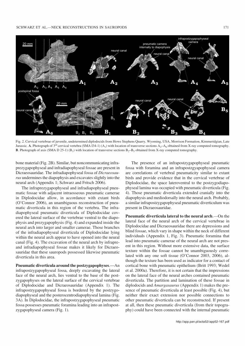
bone material (Fig. 2B). Similar, but noncommunicating infra−prezygapophyseal and infradiapophyseal fossae are present inDicraeosauridae. The infradiapophyseal fossa of Dicraeosau−rus undermines the diapophysis and excavates slightly into theneural arch (Appendix 1; Schwarz and Fritsch 2006).
The infraprezygapophyseal and infradiapophyseal pneu−matic fossae with adjacent intraosseous pneumatic cameraein Diplodocidae allow, in accordance with extant birds(O’Connor 2006), an unambiguous reconstruction of pneu−matic diverticula in this region of the vertebra. The infra−diapophyseal pneumatic diverticula of Diplodocidae cov−ered the lateral surface of the vertebrae ventral to the diapo−physis and prezygapophysis (Fig. 4) and expanded inside theneural arch into larger and smaller camerae. Those branchesof the infradiapophyseal diverticula of Diplodocidae lyingwithin the neural arch appear to have opened into the neuralcanal (Fig. 4). The excavation of the neural arch by infrapre−and infradiapophyseal fossae makes it likely for Dicraeo−sauridae that these sauropods possessed likewise pneumaticdiverticula in this area.
Pneumatic diverticula around the postzygapophyses.—Aninfrapostzygapophyseal fossa, deeply excavating the lateralface of the neural arch, lies ventral to the base of the post−zygapophyses on the lateral surface of the cervical vertebraeof Diplodocidae and Dicraeosauridae (Appendix 1). Theinfrapostzygapophyseal fossa is bordered by the postzygo−diapophyseal and the posterocentrodiapophyseal lamina (Fig.3A). In Diplodocidae, the infrapostzygapophyseal pneumaticfossa possesses pneumatic foramina leading into an infrapost−zygapophyseal camera (Fig. 1).
The presence of an infrapostzygapophyseal pneumaticfossa with foramina and an infrapostzgyapophyseal cameraare correlations of vertebral pneumaticity similar to extantbirds and provide evidence that in the cervical vertebrae ofDiplodocidae, the space lateroventral to the postzygodiapo−physeal lamina was occupied with pneumatic diverticula (Fig.4). Those pneumatic diverticula extended cranially into thediapophysis and mediodorsally into the neural arch. Probably,a similar infrapostzygapophyseal pneumatic diverticulum waspresent in Dicraeosauridae.
Pneumatic diverticula lateral to the neural arch.—On thelateral face of the neural arch of the cervical vertebrae inDiplodocidae and Dicraeosauridae there are depressions andblind fossae, which vary in shape within the neck of differentindividuals (Appendix 1, Fig. 3). Pneumatic foramina thatlead into pneumatic camerae of the neural arch are not pres−ent in this region. Without more extensive data, the surfacetexture within the fossae cannot be unambiguously corre−lated with any one soft tissue (O’Connor 2003, 2006), al−though the texture has been used as indicator for a contact ofcortical bone with pneumatic epithelium (Britt 1993; Wedelet al. 2000a). Therefore, it is not certain that the impressionson the lateral face of the neural arches contained pneumaticdiverticula. The partition and lamination of these fossae indiplodocids and Amargasaurus (Appendix 1) makes the pre−sence of pneumatic diverticula at least possible (Fig. 4), butneither their exact extension nor possible connections toother pneumatic diverticula can be reconstructed. If presentat all, then these pneumatic diverticula (from their topogra−phy) could have been connected with the internal pneumatic
http://app.pan.pl/acta52/app52−167.pdf
SCHWARZ ET AL.—NECK RECONSTRUCTIONS IN SAUROPODS 171
20 mm
20 mm
Fig. 2. Cervical vertebrae of juvenile, undetermined diplodocids from Howe Stephens Quarry, Wyoming, USA, Morrison Formation, Kimmeridgian, LateJurassic. A. Photograph of 3rd cervical vertebra (SMA I34−1) (A1) with location of transverse sections A2–A6 obtained from X−ray computed tomography.B. Photograph of axis (SMA D 25−1) (B1) with location of transverse sections B2–B5 obtained from X−ray computed tomography.

diverticula of the diapophysis, or the supravertebral pneu−matic diverticula or both (Fig. 4).
Supravertebral pneumatic diverticula.—In the neck ofadult Diplodocidae and Dicraeosauridae, the neural spines ofmost cervical vertebrae are bifurcate in their dorsal half orcompletely divided (Appendix 1). If the neural spines are notbifurcate, they possess a deep postspinal cavity. If they arepartially bifurcate, the postspinal cavity laterally widens into ashallow fossa on the medial surface on each ramus of the neu−ral spine (Fig. 4A–C). CT scans reveal internal pneumatic
ducts running from the metapophyses or the postspinal cavityinto the caudal camera of the neural arch of Diplodocidae (Fig.1A, B). Single neural spines and the metapophyses of bifur−cate neural spines of Diplodocidae contain small pneumaticcavities (Appendix 1). In Diplodocus, the completely bifur−cate neural spines can bear a large, longitudinally oval pneu−matic foramen penetrating the medial wall of either the left orthe right metapophysis (Appendix 2). In the axis of a juvenilediplodocid (SMA H25−1), the spongiose structure inside theneural spine (Fig. 2B) marks the beginning of intraosseouspneumatisation of this single neural spine.
172 ACTA PALAEONTOLOGICA POLONICA 52 (1), 2007
Fig. 3. Diplodocid and dicraeosaurid cervical vertebrae. A. 8th cervical vertebra (SMA L25−3) of subadult Diplodocus sp., Howe Stephens Quarry, Wyo−ming, USA, Morrison Formation, Kimmeridgian, Late Jurassic, in left lateral aspect (A1) and as schematic drawing indicating external pneumatic structures(A2). B. 7th and 8th cervical vertebra (SMA M34−1and M34−2) of juvenile undetermined diplodocid, Howe Stephens Quarry, Wyoming, USA, MorrisonFormation, Kimmeridgian, Late Jurassic, in left lateral aspect. C. Series of six cervical vertebrae of immature juvenile Apatosaurus louisae (CM 3390),Carnegie Museum Quarry at Dinosaur National Monument, Utah, USA, Morrison Formation, Late Jurassic, in right lateral aspect (C1) and with single ver−tebra in a larger scale (C2). D. Midcervical vertebrae of Barosaurus lentus (CM 1198), Carnegie Museum Quarry at Dinosaur National Monument, Utah,USA, Morrison Formation, Late Jurassic, in left lateral aspect, with broken distal half of cervical rib below. E. 8th cervical vertebra of Dicraeosaurushansemanni (ZMB “skelet m”), Middle Saurian Bed, Tanzania, Tendaguru Beds, Late Jurassic, in right lateral aspect (E1) and with schematic drawing ofexternal pneumatic structures (E2). Scale bars 50 mm, except D, for which is 300 mm.

The presence of pneumatic foramina and depressions onthe medial surface of the metapophyses of the cervical ver−tebrae indicate that in Diplodocidae supravertebral pneu−matic diverticula must have filled the gap between thesemetapophyses and the postspinal cavity, respectively (Fig.4). Pneumatic ducts connecting the intraosseous pneumaticstructures of the neural arch with the dorsal face of the neu−ral spine support this reconstruction (Figs. 1, 2). These
supravertebral pneumatic diverticula were most probablyextensions of the supramedullary diverticula, as in extantbirds (Müller 1908; Wedel 2003a: fig. 4; O’Connor 2006).
Although there is no osteological correlate for supra−vertebral pneumatic diverticula in Dicraeosauridae, thepossible reconstruction of supramedullary diverticula (seebelow) implies that they also possessed supravertebral di−verticula, protruding from the supramedullary diverticula.
http://app.pan.pl/acta52/app52−167.pdf
SCHWARZ ET AL.—NECK RECONSTRUCTIONS IN SAUROPODS 173
Fig. 4. Reconstruction of the distribution of pneumatic diverticula in diplodocids and dicraeosaurids. A. Schematic drawing of midcervical vertebra ofDiplodocus in left lateral aspect (A1), in dorsal aspect with single neural spine (A2) and in dorsal aspect with bifurcate neural spine (A3). The partitioning ofpneumatic diverticula at the lateral surface of the vertebral corpus is hypothetical, based on the strongly divided pneumatic fossae. B. 10th cervical vertebraof Amargasaurus cazaui in left lateral aspect. C. 8th cervical vertebra of Dicraeosaurus hansemanni in dorsal (C1) and in left lateral (C2) aspects. Not toscale.

Because the neural spines in Dicraeosaurus are tall, and themedial faces of the metapophyses are smooth and their tipsare medially inclined, the supravertebral pneumatic diver−ticula would have occupied the entire space between therami and thus had more volume than in Diplodocidae (Fig.4B).
In Amargasaurus, there is no structure that allows a reli−able reconstruction of a distinct supraspinal ligament ex−tending along the dorsal margins of consecutive cervicalneural spines (see also below in “Supraspinal ligament”). Incontrast, the ripple−and−striation pattern in the dorsal part ofthe cervical metapophyses of Amargasaurus (Table 2) issimilar to that found at the surface of the bony cores ofbovid horns (Bubenik and Bubenik 1990). Because it is alsoconsistent with the shape of the dorsally tapering andslightly caudally curving metapophyses, the striation pat−tern in the dorsal two thirds of the neural spines thereforemight indicate an integumental cover such as a keratinizedhorn sheath (see also Salgado 1999: fig. 9) or skin. In thiscase, the supravertebral pneumatic diverticula of Amarga−saurus would have occupied the ventral third in the medialcervical vertebrae, and in the cranial and caudal cervicalvertebrae the ventral half of the gap between the meta−pophyses (Fig. 4C).
It is probable that supravertebral pneumatic diverticulawere also present cranial to the neural spines of Diplodocidaeand Dicraeosauridae (Fig. 4A) as in extant birds.
Supramedullary diverticula.—A pneumatic meshworkconnects the neural canal of the cervical vertebrae of Diplo−docus, Apatosaurus and the undetermined juvenile diplo−docids with adjacent pneumatic cavities, e.g., the infradiapo−physeal or the infrapostzygapophyseal camerae (Appendix1, Figs. 1, 2). Apparently, this system reaches maximumcomplexity level with the diapophysis and even more pneu−matic cavities proliferate. From this osteological evidenceand the comparison with extant birds, it appears evident thatat least one supramedullary pneumatic diverticulum waspresent inside the neural canal of the cervical vertebrae ofDiplodocidae (Figs. 3, 7; Britt 1993; Wedel 2003a). It is im−possible to say, if these supramedullary diverticula formedsupramedullary pneumatic canals, which ran continuouslythrough the neural canal of the entire cervical column as inbirds (Müller 1908; Cover 1953; Duncker 1971; O’Connor2006) or if they were restricted to their respective vertebrae.
In contrast to diplodocids, the neural canal of the cervicalvertebrae of Dicraeosaurus exhibits no pneumatic structuresand the whole neural arch is unpneumatised (Schwarz andFritsch 2006). In extant birds with pneumatised cervicalvertebrae, the supramedullary diverticula system originatesfrom the cervical air sacs and enters the vertebral canal at thecervico−thoracic junction (Müller 1908; O’Connor 2006).The supramedullary diverticula and the lateral vertebral di−verticula communicate with each other by pneumatic ductsand small pneumatic diverticula (Müller 1908; Duncker
174 ACTA PALAEONTOLOGICA POLONICA 52 (1), 2007
Fig. 5. Cervical vertebrae of extant crocodylians and birds exposing osteological correlates for soft−tissue. A. Neural spine of 7th cervical vertebra ofCrocodylus porosus (FUB OS 13) in cranial (A1), caudal (A2) and lateral (A3) aspects. Note subdivision of the rugosity for the interlaminar elastic ligamentin A1 and A2. B. Neural spines of cervical vertebrae of Casuarius casuarius (NHM 1829) in craniodorsal (B1) and caudal (B2) aspects. Note bifurcate neuralspine and distinct rugosity for the interlaminar elastic ligament in B2. C. Neural spine of 14th cervical vertebra of Rhea americana (NHM 3534) in caudal(C1) and dorsal (C2) aspect, with rugosity for interspinal elastic ligament virtually not being distinguishable from rugosity for interlaminar elastic ligaments,as in B and D. D. Neural spines of Sarcorhamphus gryphus (NMB 3295) in craniolateral (D1) and caudal (D2) aspect. Scale bars 10 mm.

1971; Wedel 2003a; O’Connor 2006). If pneumatic diverti−cula are reconstructed for the neck of Dicraeosauridae ingeneral, they would have extended from a cervical air sac,making the presence of supramedullary diverticula likely aswell (Figs. 8, 9).
Reconstruction of major neck ligamentsand musculature
Supraspinal ligament.—The rugosities at the dorsal marginsof the neural spines of Diplodocidae and Dicraeosaurus (Ap−pendix 2, Figs. 5, 6) are similar to attachment areas of supra−spinal ligament elements in extant crocodylians (Frey 1988)and a homologous “ligamentum nuchae” system of ligamentsin birds (Tsuihiji 2004), thus indicating a rope−shaped supra−spinal ligament in Diplodocidae and Dicraeosaurus (Figs. 7,8) As in extant Crocodylia (Frey 1988), the dorsal margin
of the supraoccipital of Diplodocidae and Dicraeosaurus isrugose and bears a median nuchal crest (Holland 1924; Ja−nensch 1936; Berman and McIntosh 1978; Harris and Dodson2004; Harris 2006a), most likely representing the occipital in−sertion area for the supraspinal ligament.
As in extant crocodylians (Frey 1988; Salisbury 2001),the heights of the neural spines in the neck of Diplodocidaegradually increase from cranial to caudal (Appendix 2, Fig.7). In contrast, the cervical neural spines of extant birds arehigh in the cranial and caudal neck region, but very low inthe middle cervical region (Appendix 2). For the neck ofDiplodocidae, we therefore infer a configuration of thesupraspinal ligament similar to that of extant crocodylians,connecting the apices of each neural spine with each other(Fig. 7, 8). In contrast, the shorter 9th and 10th cervical neu−ral spine of Dicraeosaurus (Appendix 2, Fig. 8) make itpossible that single fibres of the supraspinal ligament ex−
http://app.pan.pl/acta52/app52−167.pdf
SCHWARZ ET AL.—NECK RECONSTRUCTIONS IN SAUROPODS 175
postspinal
cavity
Fig. 6. Cervical vertebrae of diplodocids exposing osteological correlates for soft−tissue. A. Cervical vertebra of Diplodocus (SMA L25−3), Howe StephensQuarry, Wyoming, USA, Morrison Formation, Kimmeridgian, Late Jurassic, in left lateral aspect (A1) and as schematic drawing with insertion areas fortendinomuscular apparatus (A2). B. 4th cervical vertebra (SMA D25−2) of undetermined juvenile diplodocid, Howe Stephens Quarry, Wyoming, USA, Morri−son Formation, Kimmeridgian, Late Jurassic, in cranial (B1) and caudal (B2) aspects. C. Isolated neural spine of a cervical vertebra of Apatosaurus excelsus(CM 555), Quarry D (Sheep Creek), Wyoming, USA, Morrison Formation, Late Jurassic, in caudal aspect showing postspinal fossa. D. Cervical vertebra ofBarosaurus lentus (CM 1198), Carnegie Museum Quarry at Dinosaur National Monument, Utah, USA, Morrison Formation, Late Jurassic, in caudal aspectshowing postspinal fossa containing pneumatic foramina. E. Cervical vertebra of Diplodocus (SMA L25−3), Howe Stephens Quarry, Wyoming, USA, Morri−son Formation, Kimmeridgian, Late Jurassic, in dorsolateral aspect showing large pneumatic foramen. F. Cervical vertebra of Diplodocus sp. (SMA, no collec−tion number), Howe Stephens Quarry, Wyoming, USA, Morrison Formation, Kimmeridgian, Late Jurassic, in cranial aspect with close−up showing pedunclefor interspinal elastic ligament (F1) and in caudal aspect (F2). Scale bars 50 mm.

tended over more than one vertebral segment in the middlecervical region.
According to the size of the insertion areas along the tipsof the neural spines, the diameter of the supraspinal ligamentof Diplodocidae and Dicraeosaurus did not exceed one tenthof the height of a neural spine, and was smaller when the liga−ment was stretched (McGowan 1999). In bifurcate neuralspines, the supraspinal ligament was split into two strings,each of them connecting the tips of one metapophysis (Figs.7A, 8A). A rugosity continues from the dorsal margin to themedial surface of the metapophyses in bifurcate neuralspines (Fig. 6C, E), indicating that the ligament was also in−serted medioventrally.
The height and shape of the neural spines of the presacralvertebrae of Amargasaurus is unique among eusauropods(Appendix 2, Fig. 9A, B, E). Because in Amargasaurus thereis no structure that allows a reliable reconstruction of a dis−tinct supraspinal ligament, this ligament was either com−pletely reduced or it was integrated in the interspinal elasticligament system.
Elastic ligament system.—In Diplodocidae, cranial andcaudal rugosities of single neural spines (Appendix 2, Fig. 6)are indicative for the presence of an elastic ligament systemas in extant crocodylians and birds (Boas 1929; Frey 1988).Because the medial tuberosity of bifurcate neural spines (Ap−pendix 2, Fig. 5) is positioned medially between the metapo−physes, and thus is equivalent to the cranial and caudalrugosities of the single neural spines, this medial tuberosityat the base of bifurcate neural spines (Appendix 2, Fig. 6C,E) probably represents the mineralised attachment area forthe elastic ligament in bifurcate neural spines.
Extant crocodylians and most extant birds possess aninterlaminar elastic ligament (Boas 1929; Landolt andZweers 1985; Frey 1988; Tsuihiji 2004), which connects thebases of the neural spines of adjacent vertebrae. In Rhea, asecond, interspinal elastic ligament is present, branching outfrom a dorsal string to insert at the caudal aspect of each neu−ral spines (Fig. 5C; Boas 1929; Tsuihiji 2004). The rugositiesfor the interspinal elastic ligament of Rhea and those for theinterlaminar elastic ligament of other birds, such as Sarco−rhamphus or Casuarius, are similar (Appendix 2, Fig. 5B,D). In extant crocodylians, the rugosity for the elastic liga−ment can be distinctly divided, although the ligament itself issimple and not partitioned (Appendix 2, Fig. 5A). Applyingthe situation of the elastic ligament system and its osteo−logical correlates in extant birds and crocodylians, we con−clude that it is not possible to specify the kind of elastic liga−ment as a specific interspinal or interlaminar elastic ligamentin Diplodocidae and Dicraeosauridae.
The differences between the height increase of the neu−ral spines in extant birds and Diplodocidae and Dicraeo−sauridae (Appendix 2) make a similar configuration of theelastic ligament in those sauropods and extant birds un−likely. In contrast, the similarities in neural spine presenceand height increase between extant Crocodylia (Frey 1988;
Salisbury 2001) and diplodocid and dicraeosaurid sauro−pods suggest that the elastic ligament of Diplodocidae andDicraeosauridae ran, as in extant crocodylians, craniocau−dally connecting the bases of successive neural spines. Theheight of the insertion scars for the elastic ligament in singleneural spines (see above) makes it probable that the elasticligament extended here at least along the ventral two−thirdsof the neural spines (Figs. 7–9). The tuberosities betweenthe metapophyses of bifurcate neural spines are restricted inheight to the ventral fourth of the neural spines. Most likelythese tuberosities coincide with the diameter of the elasticligament.
Interspinal septum.—In Alligator and most likely all otherextant crocodylians, the paired crests in the ventral two thirdsof the cranial and caudal neural spine margins are the inser−tion areas for a pair of interspinal septa, which enclose theinterlaminar elastic ligament (Appendix 2; Frey 1988). Dor−sally, the interspinal septa merge with the supraspinal liga−ment. Applying the configuration of these ligaments to thetopographically identical structures in Diplodocidae andDicraeosauridae (Appendix 2, Fig. 6) a paired interspinalseptum is inferable as present, which attached cranially andcaudally to the lateral crests of the spine margins. Betweensingle neural spines, the left and right sheet of the interspinalseptum embedded the elastic ligament as well as supra−vertebral pneumatic diverticula within the postspinal fossa(Fig. 7). In bifurcate neural spines, the interspinal septum ad−joined the supravertebral pneumatic diverticula around theelastic ligaments and dorsally contacted the supraspinal liga−ment (Fig. 7A, B).
Zygapophyseal articulation, interarticular ligament.—The articular surfaces of the pre− and postzygapophyses ofthe cervical vertebrae of Diplodocidae and Dicraeosauridaeare surrounded by an annular rugosity (Fig. 3A), which indi−cates similar to extant birds and crocodylians the presence ofa zygapophyseal articular capsule of fibrous connective tis−sue enclosing a synovial joint between the zygapophyses.
In the cervical vertebrae of Diplodocidae and Dicraeo−sauridae, a rugosity lies on the ventral part of the lateral sur−face of the prezygapophyseal peduncle adjacent to its articu−lar surface. From its position, this rugosity represents mostlikely the insertion area of a lateral interarticular ligament, asin Aves and Crocodylia (Frey 1988; Baumel and Raikow1993), which ran from the prezygapophysis cranioventrallyto the caudoventral margin of the cranially following post−zygapophysis and inserted on the slightly rugose lateral mar−gin of the vertebral foramen. The fibres of the lateral inter−articular ligament were probably connected with the articularcapsule of the zygapophyses (Fig. 7).
Intercostal ligaments.—In Diplodocidae and Dicraeosauri−dae, only the caudalmost part of the costal corpus ventro−laterally overlaps the cranial process or cranial portion of thecaudally following rib (Appendix 2). The morphology of thecervical ribs, the ventrolateral overlap of craniocaudally fol−
176 ACTA PALAEONTOLOGICA POLONICA 52 (1), 2007

lowing cervical ribs, and the presence of a striation pattern atthe cranial and caudal tips of the costal corpora in Diplo−docidae and Dicraeosauridae (Fig. 9D) is more similar to thecervical ribs of extant crocodylians (Appendix 2; Frey 1988)than to those of extant birds. This indicates the presence of anintercostal ligament connecting two adjacent ribs with eachother, similar to extant Crocodylia. If so, the intercostal liga−ment of Diplodocidae and Dicraeosauridae extended fromthe ventral and lateral surface of the costal corpora caudo−dorsally to the cranioventral and craniolateral surface of thecranial process of the caudally following cervical rib (Fig. 7).In the non−overlapping cervical ribs (i.e., in Apatosaurus),
the ligament fibres must have bridged the intercostal gap as alongitudinally oriented string, which ran in line with thecostal corpora.
Intervertebral articulation.—The cervical vertebrae of Di−plodocidae and Dicraeosauridae are opisthocoelous, with acranial hemispherical vertebral condyle and a caudal bowl−shaped cotyla (Upchurch 1995, 1998; Wilson and Sereno1998; Wilson 2002; Upchurch et al. 2004a). The cranial con−dyle and the caudal cotyla are both surrounded by a thinrugose rim (Fig. 6B, E). At its cranial and caudal margins ad−jacent to the articular surfaces of the vertebral corpora, the
http://app.pan.pl/acta52/app52−167.pdf
SCHWARZ ET AL.—NECK RECONSTRUCTIONS IN SAUROPODS 177
Fig. 7. Reconstruction of soft−tissues in the neck of Diplodocus. A. Transverse cross−sections through cervical vertebra with bifurcate neural spine in thediapophysis region (A1) and in caudal third of vertebra (A2). B. Transverse cross−sections through cervical vertebra with single neural spine in diapophysisregion (B1) and in caudal third of vertebra (B2), dashed outlines representing possible craniocervical extensor muscle analogous to m. biventer cervicis ofextant birds or m. transversospinalis capitis of extant crocodylians. C. Reconstruction of cervical ligaments in left lateral aspect. D. Reconstruction of cervi−cal axial musculature in left lateral aspect. Vertebrae and skull for C and D from Diplodocus carnegii (Hatcher, 1901). Not to scale.

lateral surface is covered with strong, longitudinally orientedstriae. The vertebral condyle bears a central high−oval fovea.The overall morphology of these intervertebral articular sur−faces is different to the heterocoelous articular condyles ofextant avians (Landolt and Zweers 1985; Baumel and Wit−mer 1993), but strikingly similar to the intervertebral articu−lations of the procoelous vertebrae of recent crocodilians.Additionally, the intervertebral articulation is not perfectlymatching as seen for example in the procoelous vertebrae ofmonitor lizards (Varanus; Goette 1897; Salisbury and Frey2001), but shows an intervertebral gap as in extant Croco−dylia (Goette 1897; Higgins 2005; Wettstein 1937; Frey1988; Salisbury 2001; Salisbury and Frey 2001). Therefore,it appears most likely that the intervertebral articulation ofDiplodocidae and Dicraeosauridae must have been similar tothat of extant Crocodylia.
The striae on the lateral surface cranially and caudally ofthe articular rims of the vertebral corpora represent parts ofthe annular rugosity, where the articular capsule inserted.The flat, sub−horizontal circumference around the articularsurfaces would then be the medial part of the annular rugo−sity and most likely housed the annulus fibrosus, from whicha cranial and caudal sheet of a fibrous intercorporal septumemerged that lined the articular cavity. The cranial sheet ofthe intercorporal septum was probably medially attached tothe vertebral condylus with a binding ligament. As in extantcrocodylians (Salisbury 2001; Salisbury and Frey 2001), thisligament inserted in the central fovea on the condylus ofDiplodocidae and Dicraeosauridae. The gap between theintercorporal septae was most likely filled with a synovialliquid, which was pumped around with the movement of thejoints and kept the intercorporal septae under tension. There−fore we assume that the intervertebral joint of Diplodocidaeand Dicraeosauridae was a synovial joint.
Dorsal epaxial muscle group.—In extant crocodylians, thecervical neural spines are plate−like in lateral view and bear afew vertical striae (Fig. 5A, Appendix 2). The main dorsalepaxial muscle of Crocodylia (m. transversospinalis cervicisI–III) is poorly segmented and inserts by fleshy fibres on thelateral face of the cervical neural spines (Appendix 2; Frey1988; Salisbury 2001). In extant avians, the neural spines canbe reduced to a slender median crest in the middle of the neck,whereas they increase in height cranially and caudally andbear in their dorsal half a lateral rugosity (Appendix 2). Themain dorsal epaxial muscle of birds (m. longus colli dorsalis)inserts with a tripartite system of muscle slips laterally andlaterodorsally at the cervical neural spines (Zusi and Storer1969; Landolt and Zweers 1985; Tsuihiji 2005). The neuralspines in the neck of Diplodocidae and Dicraeosauridae are,concerning their morphology, neither similar to extant birdsnor to extant crocodylians (Appendix 2). It is therefore diffi−cult to reconstruct the detailed configuration of the dorsalepaxial muscle group. However, for Diplodocidae and Dicra−eosaurus, the rugosity at the lateral face of the cervical neuralspines (Appendix 2, Fig. 6) suggests the presence of Sharpey's
fibres, indicating that a dorsal part of the epaxial muscle masswas present, inserting tendinous on the rugose lateral surfaceof the neural spines and fleshy on the interspinal septum be−tween the neural spines (Fig. 7). If so, a strongly segmenteddorsal epaxial muscle mass, such as suggested by Wedel andSanders (2002), would have inserted along the dorsal half ofthe lateral surface of the neural spines in Diplodocidae, and atleast along their dorsal fifth in Dicraeosaurus. The striationpattern at the lateral face of the cervical neural spines ofDicraeosaurus makes it possible, that the dorsal epaxial mus−cle contained a poorly segmented ventral portion, similar to m.transversospinalis cervicis II and III of extant crocodylians.The diameter of the dorsal epaxial muscle mass must havegradually increased caudally together with the height of theneural arches (Figs. 7, 8). Due to the higher cervical neuralspines, the dorsal epaxial muscle mass in Dicraeosaurus wasmost probably about three times as large as in Diplodocidae.In both Diplodocidae and Dicraeosauridae, the supraoccipitalat the skull shows a pair of bowl−shaped depressions lateral toits median vertical crest (Holland 1924; Janensch 1936; Ber−man and McIntosh 1978; Harris and Dodson 2004; Harris2006a). Probably these depressions served as an attachmentarea of the dorsal epaxial muscle group.
The lack of any rugosities at the lateral face of the cervicalmetapophyses in Amargasaurus (Appendix 2) argues againstthe presence of a strongly segmented dorsal epaxial musclegroup. According to the reconstruction of a keratinized hornsheath covering the dorsal two thirds of the cervical neuralspines and the presence of only a fine vertical striation ventrallyto the sheath, the dorsal epaxial muscle group in Amargasauruswas most probably poorly segmented and did not exceed theventral third of the height of the neural spine (Fig. 9).
Lateral epaxial muscle group.—The morphological changeat the bases of the lateral face of the neural spine from stria−tions towards pneumatic fossae, which occurs level with theepipophyses, might indicate a separation of the dorsal and lat−eral epaxial muscle mass by a border fascia in Diplodocidaeand Dicraeosauridae. The lateral epaxial muscle mass wouldhave extended ventrally adjacent to the latter (Fig. 8). If therewere pneumatic diverticula laterally at the base of the neuralarch, the lateral epaxial muscle portion must have been dor−sally interweaved by the latter (Fig. 7). The lateral epaxialmuscle mass would have inserted in the shallowly concave,rugose surface of the exoccipital dorsally and laterally to theforamen magnum. The muscle extended from the exoccipitalcaudally and inserted along the lateral faces of the neuralarches and the diapophyses. Its diameter must have graduallyincreased caudally together with the expansion of the cervicaldiapophyses (Figs. 7, 8).
The presence of structures as epipophyses, rugosities andcrests at the lateral face of the neural arches, diapophyses andzygapophyses at the cervical vertebrae of Diplodocidae andDicraeosauridae corresponds to extant birds (Appendix 2;Landolt and Zweers 1985, Tsuihiji 2005), but differs fromthe cervical neural arches of extant Crocodylia (Appendix 2).
178 ACTA PALAEONTOLOGICA POLONICA 52 (1), 2007

Apparently, the lateral epaxial muscle group of Diplodocidaeand Dicraeosauridae was, as in extant birds, a complex ten−dinomuscular system with segmental arranged insertion ten−dons and aponeuroses, as suggested for sauropods by Wedeland Sanders (2002). However, the lack of a complex systemof crests laterally at the lateral face of the diapophyses (thearea is mainly rugose here) and a less complex pattern ofcrests in Diplodocidae and Dicraeosauridae indicates that thearrangement of the tendinomuscular system was not identicalto that of birds, but could have been arranged as follows: The
postzygapophyseal epipophyses and ventral tuberosities atthe prezygapophyses were probably the insertion areas forcraniolaterally directed subcircular tendons. The longitudi−nally oriented crests at the lateral surface of the neural arches(Fig. 3A) could be explained by mineralised insertions of asystem of interstitial aponeuroses from which muscle fibresemerge. The caudal, roughened flanges or knobs at the dia−pophyses appear to represent an insertion area of a caudallydirected aponeurosis and with the distal margin of the flangesinto the postzygodiapophyseal lamina probably formed a
http://app.pan.pl/acta52/app52−167.pdf
SCHWARZ ET AL.—NECK RECONSTRUCTIONS IN SAUROPODS 179
elastic ligament
supraspinal ligament
dorsal epaxial muscle
dorsal epaxial muscle
lateral epaxial muscle
lateral epaxial muscle
diverticulum
supramedullary diverticulum
laterocostal muscle
laterocostal muscle
diverticula
hypaxial muscle
hypaxial muscle
carotid arteries
supravertebral
diverticulum
supraspinal ligament
elastic ligament
Fig. 8. Reconstruction of soft−tissues in the neck of Dicraeosaurus. A. Transversecross−sections through cervical vertebra in diapophysis region. B. Reconstruction ofcervical ligaments (B1) and distribution of cervical axial musculature (B2) in left lateralview, craniocervical extensor muscle (as depicted for diplodocids in Fig. 7) is implausi−ble in dicraeosaurids. Vertebrae and skull for B from Dicraeosaurus hansemanni(Janensch 1929). Not to scale.

180 ACTA PALAEONTOLOGICA POLONICA 52 (1), 2007
Fig. 9. Photographs and reconstructions of soft−tissues in the neck of Amargasaurus cazaui (MACN−N−15)), La Amarga, Neuquén, Argentina, La AmargaFormation, Hauterivian, Early Cretaceous. A. 7th and 8th cervical vertebra in left lateral (A1) and in cranial aspects (A2) and with close−up cranial view show−ing crests at the cranial face of neural spines (A3). B. Isolated cervical rib in dorsal (B1) and ventral (B2) aspects. C. Vertebral corpus of 5th cervical vertebrain ventral aspect. D. 10th cervical vertebral in left lateral aspect. E. Transverse cross−sections through cervical vertebra in the diapophysis region, with inter−nal extension of pneumatic cavities basing on Dicraeosaurus hansemanni (see also Fig. 8). Scale bars 60 mm, E is not to scale.

horizontally extended system of caudolaterally directed apo−neuroses. As the rugose dorsal surface of the diapophyses in−dicates, deep parts of the lateral epaxial muscle mass insertedfleshy on the diapophyses. The interdigitating tendinoapo−neurotical meshwork of the lateral epaxial muscle group inDiplodocidae and Dicraeosauridae was probably interwovenby a system of fascicular muscle fibres.
Laterocostal cervical muscle.—The morphological changebetween the diapophysis and the rib tuberculum of Diplo−docidae and Dicraeosauridae indicates a separation of thetendinomuscular lateral epaxial muscle group from a latero−costal muscle by a border fascia. The topographical similari−ties between the cervical ribs of extant crocodylians andthese of Diplodocidae and Dicraeosauridae (Appendix 2), es−pecially the slender and pillar−like costal tuberculum, make itlikely that the laterocostal cervical muscle was segmented asin Crocodylia (Frey 1988) with a myoseptum per vertebralsegment, the myoseptum inserting at the laterocaudal surfaceof the costal tuberculum. In contrast, there is no morphologi−cal change between the diapophysis and the costal process ofmodern birds, both being fused with each other, longitudi−nally expanded and bearing longitudinal crests serving as in−sertion points for muscle slips of m. intertransversarius (Ap−pendix 2; Boas 1929; Zusi and Storer 1969; Landolt andZweers 1985).
The occiput of Diplodocidae and Dicraeosauridae bears asharp crest separating the dorsal and ventral half of theparoccipital process (Holland 1924; Janensch 1936; Bermanand McIntosh 1978; Harris and Dodson 2004; Harris 2006a).It is likely that the laterocostal cervical muscle of these sauro−pods originated as in extant crocodylians (Frey 1988), fromthe ventral half of the paroccipital process, lateral to the oc−cipital condyle. From there, the muscle ran caudally, insert−ing on the cervical ribs and on the lateral face of the vertebralcorpora of the cervical vertebrae (Figs. 7, 8).
Hypaxial cervical muscle portion.—In extant crocodylians,m. longus colli is unsegmented and inserts by fleshy fibres atthe lateral faces of the hypapophyses, the ventral face of thecervical vertebral corpora and the medial face of the cervicalribs (Appendix 2; Frey 1988). In extant birds, the hypapo−physes and carotid processes serve as attachment points for thecomplex tendon system of m. longus colli ventralis (e.g., Boas1929; Landolt and Zweers 1985), which inserts also tendinousat the roughened spina processus costalis (Appendix 2). Incontrast to the situation in extant crocodylians and birds, thereare neither distinct hypapophyses nor carotid processes at theventral surface of the vertebral corpora of Diplodocidae andDicraeosauridae (Appendix 2; Wedel et al. 2000b), and thecervical ribs do not possess a spina processus costalis. It istherefore unlikely, that the hypaxial muscle of Diplodocidaeand Dicraeosauridae was as strongly segmented as in extantbirds. Similarly, the osteological correlates for a crocodylian−like hypaxial muscle (i.e., distinct hypapophyses) are not de−veloped in Diplodocidae and Dicraeosauridae, making a cro−
codylian−like hypaxial muscle in these sauropods implausible.Instead, the hypaxial muscle mass of Diplodocidae and Di−craeosauridae could have been an impair medial muscle masswith long external fibres and a poor segmentation, the seg−ments of which inserted at the ridges and crests of the ventralface of the vertebral bodies (Figs. 7–9). If so, then the basi−occipital tubera of Diplodocidae and Dicraeosauridae formedthe area of origin for the hypaxial cervical muscle mass, fromwhich it ran caudally along the ventral surface of the cervicalvertebral corpora (Fig. 9B).
Position of the carotid arteries.—The strongly concaveventral face of the cervical vertebral corpora of Diplodocidaeand Dicraeosauridae (Fig. 9D) resembles the sulcus or cana−lis caroticus of extant birds (Boas 1929; Baumel and Witmer1993). In birds, the paired carotid artery lies within this de−pression between the hypaxial cervical muscles m. inclususand m. longus colli ventralis (Crowe and Crowe 1979; Lan−dolt and Zweers 1985; Baumel 1993). However, in extantcrocodylians, the vertebral corpus is ventrally not concaveand the carotid arteries lie ventrally to the hypaxial muscle m.longus colli and the neck muscle m. capitisternalis (Wettstein1937). Due to the similarities in vertebral morphology, it isprobable that the position of the carotid arteries in Diplo−docidae and Dicraeosauridae was similar to extant birdswithin the ventral concavity formed by the vertebral corpusand cervical ribs (Figs. 7–9).
DiscussionComparison with other soft−tissuereconstructions for sauropods
Dorsal ligament system.—A strong dorsal ligament system,either developed as a ligamentum nuchae like in artiodactylmammals, or as a large, multisegmental interspinal elasticligament has been reconstructed for sauropods (Janensch1929; Alexander 1985, 1989; Frey and Martin 1997; Martinet al. 1998; Wilson and Sereno 1998; Stevens and Parrish1999; Tsuihiji 2004). In artiodactyls, the cervical neuralspines are low, but increase rapidly in height in the cranialthoracic region, and a strong nuchal ligament arises from thehigh neural spines of the cranialmost dorsal vertebrae to at−tach with several branches to the low neural spines of the cer−vical vertebrae (Dimery et al. 1985; Gellman et al. 2002). Incontrast to artiodactyls, the gradual height increase of thecervical neural spines of Diplodocidae and Dicraeosauridae(Appendix 2) makes a nuchal ligament unlikely.
The ligament topography in the sauropod neck with bifur−cate neural spines was previously reconstructed based on thatof Rhea, where the interspinal elastic ligament inserts in bran−ches dorsal to the interlaminar elastic ligament along the cau−dal margins of the neural spines (Tsuihiji 2004). We foundsimilar osteological correlates for an elastic ligament systemas described by Tsuihiji (2004) in diplodocids and dicraeo−
http://app.pan.pl/acta52/app52−167.pdf
SCHWARZ ET AL.—NECK RECONSTRUCTIONS IN SAUROPODS 181

saurids (Appendix 2), but we disagree in the reconstruction ofthe configuration of this ligament. First, we found it difficult todistinguish between an interlaminar and an interspinal elasticligament on the basis of ligament insertion scars in extantcrocodylians and birds (see Appendix 2 and above). Second,in all birds, neural spines are only present in the cranialmostand caudalmost cervical vertebrae (Boas 1929), whereas Di−plodocidae and Dicraeosauridae possess distinct neural spinesin all cervical vertebrae (Appendix 2). Third, Dodson and Har−ris (2001) did a biomechanical analysis of such an arrange−ment and pointed out that a multibranching ligament insertingmedially between the metapophyses and/or at the summits ofthe neural spines would have a very low mechanical advan−tage. These differences make a dorsally expanded interspinalelastic ligament, as in Rhea, unlikely for Diplodocidae andDicraeosauridae. In contrast to Rhea, modern crocodylianscan, due to the similarities in heights of the cervical neuralspines, give valuable hints for reconstructing the configurationof supraspinal and elastic ligaments in diplodocids and dicra−eosaurid sauropods.
Tsuihiji (2004) discussed an interesting aspect for soft−tissue reconstructions. He described that in Rhea, the inter−spinal elastic ligament is enclosed by a “… ligamentous orfascial sheath, which attaches to the tip of the bifurcated neu−ral spine as well as to the lamina connecting the neural spineand the postzygapophysis…” (Tsuihiji 2004: 166), andwhich is interpreted as a derivate of the supraspinal ligament.The described topography of the “ligamentous sheath”, inparticular its insertion at the cranial and caudal laminae of theneural spines makes it likely, that it represents the equivalentto the interspinal septum in extant crocodylians (Frey 1988).Therefore, the reconstruction of such an interspinal septumin the neck of Diplodocidae and Dicraeosauridae would bejustified by equivalent structures in both modern croco−dylians and birds.
Axial neck musculature.—A profound system of interseg−mental muscle slips identical to that of extant birds has beenreconstructed for sauropods on the base of partially similarinsertion scars (Appendix 2; Wedel et al. 2000a; Wedel andSanders 2002). In extant birds, who hold their neck in anS−shaped loop, the cervical muscles form three segment sys−tems (from cranial to caudal), interconnecting multiple verte−brae by muscular slips (Zweers et al. 1987; Bout 1997). Theoverall construction of the neck of a bird functions for intra−and intersegmental mobility of a strongly S−curved structure,which is evidenced by this multisegmental muscle and liga−ment system, allowing the movement of neck segmentsagainst each other (Elshoud and Zweers 1987). This is effec−tive in birds because acute curvatures, dorsally concave cau−dally and convex cranially, allow individual slips to spanseveral vertebrae and flex the curvature between them (mm.splenii colli and mm. cervicales ascendentes: Vanden Bergeand Zweers 1993), or even coalesce into larger muscle bel−lies, as with slips from caudal cervical vertebrae runningcraniodorsally to join the belly of m. longus colli dorsalis
cranialis (Vanden Berge and Zweers 1993). Only some of theosteological correlates of a bird−like cervical musculaturecan be found in diplodocids and dicraeosaurids, whereasother structures are more similar to extant crocodylians (seeAppendix 2 and above). Without a strong caudal neck curva−ture, multiple long slips coursing to join with an m. longuscolli dorsalis analogue could not extend the caudal part of thesauropod neck to the same degree as in birds, although theneck extensor muscles may have been of similar complexityto those of birds. Furthermore, a strictly bird−like neck mobil−ity is contraindicated for sauropods (Stevens and Parrish1999; Christian 2002; Stevens and Parrish 2005a, b). Wetherefore conclude that for sauropods like Diplodocidae andDicraeosauridae, the reconstruction of a longitudinally tri−partite cervical muscular system identical to that of birds isnot justified. Proportional and topological comparisons withmodern birds and crocodylians reveal that, although a strongsegmentation of the dorsal and lateral epaxial muscle mass islikely for Diplodocidae and Dicraeosauridae, a bird−like to−pology of the laterocostal and hypaxial cervical muscle massin these sauropods seems unlikely.
Soft−tissues between bifurcate neural spines in the neck.—Most hypotheses about the soft−part anatomy between the bi−furcate neural spines favour nuchal or interspinal ligaments(Janensch 1929; Alexander 1985, 1989; Wilson and Sereno1998), a medial axial muscle string (Osborn and Mook 1921;McIntosh 1990; Wedel et al. 2000a), or a combination of both(Tsuihiji 2004: fig. 5A). There is no osteological correlate fora cervical muscle portion dorsal or dorsomedially to the dorsalepaxial muscle group in Diplodocidae and Dicraeosauridae assuggested by Tsuihiji (2004) and Wedel and Sanders (2002),although such a muscle is present in both modern crocodylians(m. transversospinalis capitis, Frey 1988) and birds (Landoltand Zweers 1985; Vanden Berge and Zweers 1993). From ananatomical point of view (i.e., applying Extant PhylogeneticBracketing), the presence of a craniocervical muscle, runningdorsally above the neural spines can therefore not be excludedfor Diplodocidae (Fig. 7) and Dicraeosauridae. However, it isunlikely that the evolution of neural spines probably protrud−ing through the dermis like reconstructed for Amargasaurus(see above) could have evolved out of a preconstruction, inwhich a supraspinal tendinomuscular system used the neuralspines as a lever or was included into the bracing system.
The diameter of the notch between bifurcate neural spinesin Diplodocidae and Dicraeosauridae allows together withthe reconstructed cervical muscles (Figs. 7–9) a rough esti−mation of the cross−sectional area that would have been oc−cupied by a muscle filling the notch between bifurcate neuralspines completely. According to these reconstructions, sucha muscle would take at least 15% of the total cross−section ofthe neck vertebrae, thereby increasing the weight of the neckby approximately 15%. Although this assumed increase inneck weight makes it implausible that a muscle filled thespace between the bifurcate neural spines, it might have beencompensated by the weight−saving effect of vertebral pneu−
182 ACTA PALAEONTOLOGICA POLONICA 52 (1), 2007

maticity: The magnitude of bone reduction by pneumaticstructures can be quantified by calculations from the CT sec−tions of the bones (Airspace Proportion, or ASP, of Wedel2004, 2005) to have reached a maximum of 69% in some re−gions of the vertebral corpus of Apatosaurus (Wedel 2005).According to the CT sections presented in this work, and ifmuscles and ligaments throughout the whole neck are takeninto account, the neck of Dicraeosaurus (ca. 2.4 m in length)would have been lightened for at least 6% (Schwarz andFritsch 2006), that of a sub−adult diplodocid (ca. 4 m inlength) for at least 14% and that of an adult Diplodocus (ca. 7m in length) for at least 25% by their total of pneumatic struc−tures. A compensation of the additional neck weight by sucha dorsal epaxial muscle filling the gap between bifurcate neu−ral spines therefore would only be likely in diplodocids, butnot in dicraeosaurids.
The presence of ligaments in the area between bifurcateneural spines is supported by osteological correlates in Di−plodocidae and Dicraeosauridae (Tsuihiji 2004), as is, atleast for Diplodocidae, the presence of supravertebral pneu−matic diverticula between bifurcate neural spines and in thepostspinal cavity (see “Supravertebral pneumatic diverti−cula”). The hypothesis of pneumatic diverticula surroundingthese ligaments provides a reasonable model for the filling ofthis gap, as it is based on osteological correlates and does notlead to weight increase of the neck. Since supravertebraldiverticula in extant birds fill the interarticular foramen be−tween following cervical vertebrae (Müller 1908; O’Connor2003; Wedel 2003a; O’Connor 2006), such supravertebralpneumatic diverticula in Diplodocidae and Dicraeosauridaecould have risen from the supramedullary diverticula, andextended between the sheets of the interspinal septum.
Development, distribution and biomechanicalimplications of pneumatic structures
The extent of vertebral pneumatic structures in the neck ofDiplodocidae and Dicraeosauridae reaches a maximum in thecaudalmost cervical vertebrae. This corresponds to extantbirds (Hogg 1984a, b; O’Connor 2003, 2004) and supports thesuggestion of Wedel (2003b) for a possible position of a cervi−cal air sac at the base of the neck. From there, pneumaticdiverticula invaded the cervical vertebrae progressively fromcaudally to cranially during ontogeny (Müller 1908; King1966; Hogg 1984a; O’Connor 2003; Wedel 2003a; O’Connor2006).
The distribution of structures associated with vertebralpneumaticity around the cervical vertebrae of Diplodocidaeand Dicraeosauridae is similar to that generally found in othersauropods, differing in the amount of subdivisions (Appendix1, Fig. 3; Britt 1993; Carpenter and McIntosh 1994; Wedel etal. 2000a, b; Wedel 2003a, b; Schwarz and Fritsch 2006). Thisindicates a common pattern of pneumatic structures insauropod cervical vertebrae. Judging by the osteology of thecervical column in juveniles and adult diplodocids (Fig. 3;Carpenter and McIntosh 1994; Wedel 2003a), these systems
must have developed at early ontogenetic stages. During on−togeny, pneumatic diverticula progressively expanded to pro−duce the camerate and polycamerate pneumaticity patterncharacteristic for adult diplodocids (Figs. 1, 2; Britt 1993;Wedel et al. 2000a; Wedel 2003a).
Dicraeosauridae have no intraosseous pneumatisation, andexternal structures indicating vertebral pneumaticity are re−stricted mostly to fossae, which can extend deeply into the ver−tebrae. From a strict, comparative anatomical viewpoint, thepresence of vertebral pneumaticity in the neck of Dicraeo−sauridae remains uncertain. The position of pneumatic fossaein similar places as in Diplodocidae, their depth and obviouslyinvasive nature, the presence of at least a parapophyseal pneu−matic foramen, and the bracketing of Dicraeosauridae withsauropod taxa that possess pneumatic diverticula make thepresence of pneumatic diverticula in the neck of Dicraeo−sauridae highly probable. Dicraeosauridae possess short neckswith not more than 12 cervical vertebrae (Bonaparte 1999;Rauhut et al. 2005) and a relatively small overall size. Histo−logical studies (Sander 1999, 2000) show that Dicraeosaurusgrew fast and continuously, but reached sexual maturity with80% of its maximum size at a relatively larger size than othersauropods. Taken this together, there seems to be an earliergrowth arrest in Dicraeosauridae, which may have been ac−companied with a shortened pneumatisation of the postcranialskeleton (Schwarz and Fritsch 2006).
If the reconstruction of supramedullary pneumaticdiverticula in Diplodocidae and Dicraeosauridae holds, apneumatisation centre inside the neural arch can be postu−lated to have extended from the cervical air sacs as in modernbirds (Müller 1908; O’Connor 2006). In the cervical verte−brae of the juvenile diplodocids, the early presence of pneu−matic structures connected with the neural canal makes itplausible that the supramedullary diverticula formed prior toor parallel with the beginning of internal pneumatisation ofthe neural arch. A connection between the neural canal andthe surrounding pneumatic structures by pneumatic ductswas found in the cervical vertebrae of juvenile diplodocidsand other sauropods (Britt 1993; Wedel 2003a; Schwarz andFritsch 2006). Without the inclusion of the neural canal, nointernal pneumatisation of the neural arch has been docu−mented in sauropods. Internal pneumatisation of the neuralarch was therefore most likely triggered by supramedullarydiverticula, the latter being a major factor for pneumatisingthe neural arch. On the other hand, the case of Dicraeo−sauridae shows that supramedullary diverticula might havepersisted without further internal pneumatisation of the neu−ral arch. The function of the supramedullary pneumaticdiverticula within the neural canal of recent birds is notknown yet, and the same holds true for sauropods.
Although it has not been tested yet by mechanical ap−proaches, the distribution of intraosseous pneumatic struc−tures in juvenile and adult diplodocids might suggest thefollowing biomechanical explanations: Reducing the cervi−cal vertebral corpora and the ventral half of the neuralarches to a vertical I−beam construction could indicate a
http://app.pan.pl/acta52/app52−167.pdf
SCHWARZ ET AL.—NECK RECONSTRUCTIONS IN SAUROPODS 183

main stabilisation of the cervical vertebral column againstload in the vertical plane. As visible from the comparisonbetween the CT sections of juvenile and adult diplodocids,additional bony struts appeared on the lateral surface of theI−beam together with the increase of the absolute size of thevertebrae. The bone struts might have prevented local buck−ling of the median vertical lamella. If so, then the pro−gressive structural complexity of the cervical vertebrae inDiplodocidae might be explained as a consequence of in−creased mechanical demands in the context of a mass in−crease. Pneumatic cavities in the neural arch in Diplo−docidae are distributed irregularly, with secondary pneu−matic cavities randomly arranged around existing cavities,which indicates load differences between the neural archand the vertebral corpus.
The maximum size of the camerae could also be subjectto physiological constraints, probably in context with the re−spiratory apparatus. A paired cervical air sac, which can bereconstructed by the presence of vertebral pneumaticity indiplodocids and dicraeosaurids, would have been part of therespiratory systems as in birds (Duncker 1971; Schmidt−Nielsen 1971). In sauropods, cervical air sacs could haveacted as air reservoirs and could have contributed to ventilat−ing the gas exchanged tissue in the lungs (Perry and Reuter1999; Perry and Sander 2004). Surely, the ongoing research(Codd and Perry 2005; Perry 2006) on the functional mor−phology of the respiratory apparatus in sauropods will givemuch more valuable information on this issue. It cannot beexcluded that pneumatic diverticula in the neck of diplodo−cids and dicraeosaurids did also contribute to respiration byacting as additional air reservoirs. However, the uncertaintiesin reconstructing the extension of the cervical pneumaticdiverticula, and the multiple and small connections betweenindividual pneumatic diverticula probably hindering rapidexchange of air, make this assumption highly speculative.Possible biological roles of vertebral pneumaticity in theneck of sauropods that can be better evaluated at the momentare a weight reduction and possibly a contribution to necksupport (see below).
Biomechanical implications for neck support bythe reconstructed soft−tissue apparatus
The sauropod neck can be modelled as a segmented beam an−chored at the trunk and loaded by the weight of the skull(Martin et al. 1998). The neutral position of the necks ofDiplodocidae and Dicraeosauridae is reconstructed to haveformed a slightly ventrally sloping curve, with the short cer−vical ribs overlapping only in a short area or not at all(Salgado 1999; Stevens and Parrish 1999; Christian 2002).
In Diplodocidae and Dicraeosauridae, the double supra−spinal and the large elastic ligaments were well developed,yet they were not extraordinarily large. Bifurcation of theneural spines improved the dorsal ligament support systemof the neck, since it forced the supraspinal ligament to divideinto two parts, therefore to form a dorsal double−string. Dor−
sal ligaments surely were one important support mechanismin the neck of Diplodocidae and Dicraeosauridae: Duringflexion and extension, elastic energy was stored in the dorsalligament apparatus and launched an elastic recoil, as it hasbeen described for extant vertebrates (Alexander andBennet−Clark 1977; Dimery et al. 1985; Bennett and Alexan−der 1987). The intercostal ligament apparatus around the cer−vical ribs probably provided elastic recoil when the neck wasextended. Another factor in bracing the neck must have beenthe strongly segmented dorsal and lateral epaxial muscles,which could have stiffened the neck as a whole during simul−taneous contraction. Axial neck muscles did also achieveneck mobility. In contrast to these dorsal bracing mecha−nisms, cervical ribs as ventral bracing elements were reducedand did most probably not contributed to bracing of the neckof Diplodocidae and Dicraeosauridae.
The reconstructed arrangement of pneumatic diverticulaaround the cervical vertebrae and in the notch between bifur−cate neural spines suggests that the cervical pneumatic sys−tem in sauropods might have contributed to neck support aspassive support structures (Akersten and Trost 2000, 2001,2004; Schwarz and Frey 2006; Schwarz et al. 2006). There isno indication that vertebral pneumaticity in Diplodocidaeand Dicraeosauridae was a separate system from the respira−tory apparatus, so that most likely, pneumatic diverticula inthese sauropods were linked as dead space to cervical airsacs. If it is assumed, that these pneumatic diverticula, like inbirds, were brimming with air, the large pneumatic systemsin the neck could theoretically have passively supported theneck, but only together with its ligaments and muscles. How−ever, this would also require the presence of regulationmechanisms for the volume and pressure of the pneumaticdiverticula system of the neck of sauropods, which cannot bereconstructed by osteological evidence. Pressure and volumeof air sacs of birds is connected with a complicate array ofregulating mechanisms for respiration, such as control unitsfor the bronchial diameters (Brown et al. 1995) or the regula−tion of muscle activity for driving air sac ventilation (Beckerset al. 2003). In chickens, the ostia width can be controlled toregulate the amount of air passing through the lungs to a cer−tain degree (Cook et al. 1986). The presence of vertebralpneumaticity in the neck of Diplodocidae and Dicraeosauri−dae as in extant birds makes it likely, that air sacs could pos−sibly be regulated in the same way as in birds.
In the case that the pneumatic diverticula systems in theneck of diplodocid and dicraeosaurid sauropods did contrib−ute to neck support, the combination of pneumatic and liga−mental support of the neck would have made a large physio−logical cross−section of the axial muscle obsolete, thereforereducing the weight of the neck. It would be consistent withthe reconstruction of non−multibranching and not outstand−ingly large dorsal ligaments in the neck of Diplodocidae andDicraeosauridae, and might explain the lack of effective ven−tral bracing structures such as long, overlapping cervicalribs, in their necks. In Dicraeosauridae, the presence of largesupravertebral pneumatic diverticula systems would be an
184 ACTA PALAEONTOLOGICA POLONICA 52 (1), 2007

explanation for the enormous height increase in neural spinesand the possibility of a reduction of the supraspinal ligamentin Amargasaurus.
ConclusionsThe reconstructions of the configuration of the elastic liga−ments and the axial cervical muscles in the neck of Diplo−docidae and Dicraeosauridae differ partly from those postu−lated by other authors for sauropods. Differences in the soft−tissue reconstructions cannot be taxonomically biased, asmany of the osteological structures observed in Diplodocidaeand Dicraeosauridae and discussed in this paper are also foundin other sauropods. They are interpreted here as the result of areconstruction method acting purely on the basis of birds,which have very specialized necks. In contrast, inclusion ofextant Crocodylia into soft−tissue reconstructions of sauro−pods helps to understand their soft−part anatomy better and isconsistent with functional morphological models for the sau−ropod neck support. The special neck morphology of diplo−docid and dicraeosaurid sauropods does often allow only a oneway phylogenetic comparison with either birds or croco−dylians (Level II inference, Witmer 1995, 1997), which leadsto uncertainties in the reliability of these reconstructions.
Taking into account different ontogenetic stages showsdetails of the development of pneumatic structures in sauro−pod necks and allows some conclusions for the developmentand biomechanical importance of these pneumatic struc−tures. The presence of supramedullary pneumatic diverticulawas important for further pneumatisation of the neural arch.The pattern of internal pneumatic structures and their onto−genetic change in Diplodocidae might be explained havingbeen induced by the increasing mass during growth. In con−trast, the lack of internal pneumatisation and the simple pat−tern of pneumatic fossae in Dicraeosauridae prohibit an un−ambiguous reconstruction of vertebral pneumaticity in theneck of this sauropod group in a strict comparative anatomi−cal approach. Nevertheless, the similarly distributed pneu−matic fossae in Dicraeosauridae and in Diplodocidae, and thebracketing of Dicraeosauridae with sauropod taxa possess−ing pneumatised necks make the presence of vertebral pneu−maticity in the neck of Dicraeosauridae highly probable.
With the inclusion of all osteological evidence, a modelfor the filling of the gap between the metapophyses of bifur−cate neural spines by ligaments and supravertebral pneu−matic diverticula is represented. If so, then it is even possiblethat growth and interseptal segmentation of supravertebraldiverticula in sauropods influenced or triggered bifurcationof neural spines during ontogeny. Bifurcation of the neuralspines improved the dorsal ligamentous support system ofthe neck by forcing the supraspinal ligament to divide intotwo parts. Our soft−tissue reconstructions, in particular thereconstructed array of pneumatic diverticula around the cer−vical vertebrae lead to a biomechanical consistent model forthe bracing of the neck of Diplodocidae and Dicraeosauri−
dae. It is plausible that dorsal ligaments and epaxial musclescontributed to neck bracing, whereas ventral cervical ribs asventral bracing elements were reduced. Within this dorsallydominated bracing systems, it is possible that the pneumaticsystem was an additional, passively stabilising and shape−controlling mechanism in the neck.
AcknowledgementsWe are very grateful to Hans−Jakob “Kirby” and Yolanda Siber (bothSaurier−Museum Aathal, Switzerland), who lent us several vertebraefor the tomographies and provided at any time access to their collec−tions. Many thanks also to Eberhard Lehmann and Peter Vontobel(both Paul−Scherrer−Institute, Villigen, Switzerland) for the NeutronTomography and the discussion of the results. We are indebted toGeorg Bongartz, Georgia Ralli (both Kantonsspital Basel, Switzer−land), and Guido Fritsch (Clinic and Policlinic for Small Pets, Berlin,Germany) for the CT scans. For sharing many of their data and for dis−cussions on the problems of pneumaticity we thank Mathew J. Wedeland Patrick M. O’Connor. Two anonymous reviewers are thanked fortheir thoughtful comments and advice for improvement of an earlierversion of this manuscript. This manuscript benefited considerablyfrom the useful comments and corrections of the referees, Kristina A.Curry Rogers, Eric Snively, Jeffrey A. Wilson and an anonymous ref−eree. This study is part of a project on the role of pneumatisation insauropods funded by the Swiss National Science Foundation (SNFNo. 200021−101494/1 and 200020−109131/1).
ReferencesAkersten, W. and Trost, C.H. 2000. Function of avian air sac diverticula, im−
plications for sauropod cervical biomechanics. Journal of VertebratePaleontology 20 (Supplement to No. 3): 25A.
Akersten, W. and Trost, C.H. 2001. Studies on the function of avian air sacdiverticula and possible comparisons with saurischian vertebral bio−mechanics II. Journal of Vertebrate Paleontology 21 (Supplement toNo. 3): 27A.
Akersten, W. and Trost, C.H. 2004. Air sac diverticula as passive support de−vices in birds and saurischian dinosaurs: an overlooked biomechanicalsystem. Journal of Morphology 260: 275.
Alexander, R.M. 1985. Mechanics of posture and gait of some large dino−saurs. Zoological Journal of the Linnean Society of London 83: 1–25.
Alexander, R.M. 1989. Dynamics of Dinosaurs and Other Extinct Giants.167 pp. Columbia University Press, New York.
Alexander, R.M. and Bennet−Clark, H.C. 1977. Storage of elastic strain en−ergy in muscle and other tissues. Nature 265: 114–117.
Baumel, J.J. 1993. Systema Cardiovasculare. In: J.J. Baumel, A.S. King,J.E. Breazile, H.E. Evans, and J.C. Vanden Berge (eds.), Handbook ofAvian Anatomy: Nomina Anatomica Avium, 407–475. Nuttal Ornitho−logical Club, Cambridge, Massachusetts.
Baumel, J.J. and Raikow, R.J. 1993. Arthrologia. In: J.J. Baumel, A.S. King,J.E. Breazile, H.E. Evans, and J.C. Vanden Berge (eds.), Handbook ofAvian Anatomy: Nomina Anatomica Avium, 133–187. Nuttal Ornitho−logical Club, Cambridge, Massachusetts.
Baumel, J.J. and Witmer, L.M. 1993. Osteologia. In: J.J. Baumel, A.S. King,J.E. Breazile, H.E. Evans, and J.C. Vanden Berge (eds.), Handbook ofAvian Anatomy: Nomina Anatomica Avium, 45–132. Nuttal Ornitho−logical Club, Cambridge, Massachusetts.
Beckers, G.J.L., Suthers, R.A., and ten Cate, C. 2003. Mechanisms of fre−quency and amplitude modulation in ring dove song. The Journal of Ex−perimental Biology 206: 1833–1843.
http://app.pan.pl/acta52/app52−167.pdf
SCHWARZ ET AL.—NECK RECONSTRUCTIONS IN SAUROPODS 185

Bennett, M.B. and Alexander, R.M. 1987. Properties and function of exten−sible ligaments in the necks of turkeys (Meleagris gallopavo) and otherbirds. Journal of Zoology, London 212: 275–281.
Benton, M.J. 2004. Origin and relationships of Dinosauria. In: D.B. Weis−hampel, P. Dodson, and H. Osmólska (eds), The Dinosauria, 7–19. Uni−versity of California Press, Berkeley.
Berman, D.S. and McIntosh, J.S. 1978. Skull and relationship of the UpperJurassic sauropod Apatosaurus (Reptilia, Saurischia). Bulletin of theCarnegie Museum of Natural History 8: 1–35.
Boas, J.E.V. 1929. Biologisch−Anatomische Studien über den Hals derVögel. Det Kongelige Danske Videnskabernes Selskabs Skrifter, Natur−videnskabelig og Mathematisk Afdeling 9 (1): 10–222.
Bonaparte, J.F. 1999. Evolución de las vértebras presacras en Sauropodo−morpha. Ameghiniana 36: 115–187.
Bout, R.G. 1997. Postures of the avian craniocervical column. Journal ofMorphology 231: 287–295.
Britt, B.B. 1993. Pneumatic Postcranial Bones in Dinosaurs and OtherArchosaurs. 383 pp. PhD thesis, University of Calgary.
Britt, B.B. 1997. Postcranial pneumaticity. In: P.J. Currie, and K. Padian(eds.), Encyclopedia of Dinosaurs, 590–593. Academic Press, SanDiego.
Britt, B.B., Makovicky, P.J., Gauthier, J., and Bonde, N. 1998. Postcranialpneumatization in Archaeopteryx. Nature 395: 374–376.
Brown, R.E., Kovacs, C.E., Butler, J.P., Wang, N., Lehr, J., and Banzetti,R.B. 1995. The avian lung: is there an aerodynamic expiratory valve?Journal of Experimental Biology 198: 2349–2357.
Bryant, H.N. and Russell, A.P. 1992. The role of phylogenetic analysis inthe inference of unpreserved attributes of extinct taxa. PhilosophicalTransactions of the Royal Society of London B 337: 405–418.
Bubenik, G.A. and Bubenik, A.B. 1990. Horns, pronghorns, and antlers:evolution, morphology, physiology, and social significance. 562 pp.Springer, New York.
Carpenter, K. and McIntosh, J.S. 1994. Upper Jurassic sauropod babiesfrom the Morrison Formation. In: K. Carpenter, K.F. Hirsch, and J.R.Horner (eds.), Dinosaur Eggs and Babies, 265–278. Cambridge Uni−versity Press, Cambridge.
Carrano, M.T. and Hutchinson, J.R. 2002. Pelvic and hindlimb musculatureof Tyrannosaurus rex (Dinosauria: Theropoda). Journal of Morphology253: 207–228.
Christian, A. 2002. Neck posture and overall body design in sauropods.Mitteilungen aus dem Museum für Naturkunde Berlin, Geowissen−schaftliche Reihe 5: 271–281.
Codd, J.R. and Perry, S.F. 2005. Bird lungs: The key to getting really big:The allometry of respiratory parameters in sauropods. Journal of Verte−brate Paleontology 25 (Supplement to No. 3): 46A.
Cook, R.D., Vaillant, C.R., and King, A.S. 1986. The abdominal air sacostium of the domestic fowl: a sphincter regulated by neuro−epithelialcells? Journal of Anatomy 149: 101–111.
Coombs, W.P.J. 1978. Theoretical aspects of cursorial adaptations in dino−saurs. The Quarterly Review of Biology 53: 393–418.
Cope, E.D. 1877. On a gigantic saurian from the Dakota Epoch of Colorado.Palaeontological Bulletin 25: 5–10.
Cover, M.S. 1953. Gross and microscopic anatomy of the respiratory systemof the turkey. III. The air sacs. American Journal of Veterinary Re−search 14: 239–245.
Crowe, T.M. and Crowe, A.A. 1979. Anatomy of the vascular system of thehead and neck of the helmeted guineafowl Numida meleagris. Journalof Zoology, London 188: 221–233.
Curtice, B.D. 1998. Sauropod vertebral variation and its phylogenetic impact.Journal of Vertebrate Paleontology 18 (Supplement to No. 3): 37A.
Dimery, N.J., Alexander, R.M., and Deyst, K.A. 1985. Mechanics of theligamentum nuchae of some artiodactyls. Journal of Zoology, London206: 341–351.
Dodson, P. and Harris, J.D. 2001. Necks of sauropod dinosaurs: support of anuchal ligament? Journal of Morphology 248: 224.
Duncker, H.−R. 1971. The lung air sac system of birds. Advances in Anat−omy, Embryology, and Cell Biology 45: 1–171.
Elshoud, G.C.A. and Zweers, G.A. 1987. Avian cranio−cervical systemsPart III: Robot Kinematics for Cervical Systems. Acta MorphologicaNeerlando−Scandinavica 25: 235–260.
Frey, E. 1988. Anatomie des Körperstammes von Alligator mississippiensisDaudin. Stuttgarter Beiträge zur Naturkunde A 24: 1–106.
Frey, E., Herkner, B., Schrenk, F., and Seiffert, C. 1993. Reconstructingorganismal constructions and the problem of Leptictidium‘s locomo−tion. Kaupia 3: 89–95.
Frey, E. and Martin, J. 1997. Long necks in sauropods. In: P.D. Currie and K.Padian (eds.), Encyclopedia of Dinosaurs, 406–409. Academic Press, SanDiego.
Gauthier, J.A., Kluge, A.G., and Rowe, T. 1988. Amniote phylogeny andthe importance of fossils. Cladistics 4: 105–209.
Gellman, K.S., Bertram, J.E.A., and Hermanson, J.W. 2002. Morphology,histochemistry, and function of epaxial cervical musculature in thehorse (Equus caballus). Journal of Morphology 251: 182–194.
Gilmore, C.W. 1936. Osteology of Apatosaurus with special reference tospecimens in the Carnegie Museum. Memoirs of the Carnegie Museum11 (4): 175–300.
Goette, A. 1897. Über den Wirbelbau bei den Reptilien und einigen anderenWirbelthieren. Zeitschrift für wissenschaftliche Zoologie 62: 343–394.
Gudo, M., Gutmann, M., and Scholz, J. 2002. Concepts of functional engineer−ing and constructional morphology: introductory remarks. Senckenber−giana lethaea 82: 7–10.
Harris, J.D. 2006a. Cranial osteology of Suuwassea emilieae (Sauropoda:Diplodocoidea: Flagellicaudata) from the Upper Jurassic Morrison For−mation of Montana, USA. Journal of Vertebrate Paleontology 26:88–102.
Harris, J.D. 2006b. The axial skeleton of the dinosaur Suuwassea emilieae(Sauropoda: Flagellicaudata) from the Upper Jurassic Morrison Forma−tion of Montana, USA. Palaeontology 49: 1091–1121.
Harris, J.D. and Dodson, P. 2004. A new diplodocoid sauropod dinosaurfrom the Upper Jurassic Morrison Formation of Montana, USA. ActaPalaeontologica Polonica 49: 197–210.
Hatcher, J.B. 1901. Diplodocus (Marsh): its osteolology, taxonomy, andprobable habits, with a restoration of the skeleton. Memoirs of the Car−negie Museum 1: 347–355.
Hatcher, J.B. 1903. Additional remarks on Diplodocus. Memoirs of the Car−negie Museum 2: 72–75.
Henderson, D.M. 2004. Tipsy punters: sauropod dinosaur pneumaticity,buoyancy and aquatic habits. Proceedings of the Royal Society of Lon−don B 271 (Supplement): 180–183.
Herkner, B. 1999. Über die evolutionäre Entstehung des tetrapoden Loko−motionsapparates der Landwirbeltiere. Carolinea, Beihefte 13: 1–353.
Higgins, G.M. 2005. Development of the primitive reptilian vertebral col−umn, as shown by a study of Alligator mississippiensis. American Jour−nal of Anatomy 31 (4): 373–407.
Hogg, D.A. 1984a. The delevopment of pneumatization in the postcranialskeleton of the domestic fowl. Journal of Anatomy 139: 105–113.
Hogg, D.A. 1984b. The distribution of pneumatization in the skeleton of theadult domestic fowl. Journal of Anatomy 138: 617–629.
Holland, W.J. 1906. The osteology of Diplodocus Marsh. Memoirs of theCarnegie Museum 2 (6): 225–278.
Holland, W.J. 1924. The skull of Diplodocus. Memoirs of the Carnegie Mu−seum 9 (3–4): 379–403.
Hutchinson, J.R. 2004. Biomechanical modelling of musculoskeletal func−tion in extinct taxa: methods, assumptions, and challenges. Journal ofMorphology 260: 301.
Janensch, W. 1914. Übersicht über die Wirbeltierfauna der Tendaguru−Schichten, nebst einer kurzen Charakterisierung der neu aufgeführtenArten von Sauropoden. Archiv für Biontologie 3: 81–110.
Janensch, W. 1929. Die Wirbelsäule der Gattung Dicraeosaurus. Palae−ontographica Supplement 7 (2): 37–133.
Janensch, W. 1936. Die Schädel der Sauropoden Brachiosaurus, Barosaurusund Dicraeosaurus aus den Tendaguru−Schichten Deutsch−Ostafrikas.Palaeontographica Supplement 7 (2): 147–298.
186 ACTA PALAEONTOLOGICA POLONICA 52 (1), 2007

Janensch, W. 1947. Pneumatizität bei Wirbeln von Sauropoden und anderenSaurischiern. Palaeontographica Supplement 7 (3): 1–25.
Janensch, W. 1950. Die Wirbelsäule von Brachiosaurus brancai. Palae−ontographica Supplement 7 (3): 27–92.
King, A.S. 1966. Structural and functional aspects of the avian lungs and airsacs. International Review of General and Experimental Zoology 2:171–267.
Landolt, R. and Zweers, G. 1985. Anatomy of the muscle−bone apparatus ofthe cervical system in the mallard (Anas platyrhynchos L.). NetherlandsJournal of Zoology 35: 611–670.
Longman, H.A. 1933. A new dinosaur from the Queensland Cretaceous.Memoirs of the Queensland Museum 10: 131–144.
Lull, R.S. 1919. The sauropod dinosaur Barosaurus Marsh. Memoirs of theConneticut Academy of Arts and Sciences 6: 1–42.
Marsh, O.C. 1877. Notice of new dinosaurian reptiles from the Jurassic For−mation. American Journal of Science 3: 514–516.
Marsh, O.C. 1890. Description of new dinosaurian reptiles. American Jour−nal of Science, Serie 3 39: 81–86.
Martin, J., Martin−Rolland, V., and Frey, E. 1998. Not cranes or masts, butbeams: The biomechanics of sauropod necks. Oryctos 1: 113–120.
McGowan, C. 1999. A Practical Guide to Vertebrate Mechanics. 301 pp.Cambridge University Press, Cambridge.
McIntosh, J.S. 1990. Sauropoda. In: D.B. Weishampel, P. Dodson, and H.Osmólska (eds.), The Dinosauria, 345–401. University of CaliforniaPress, Berkeley.
McIntosh, J.S. 2005. The Genus Barosaurus Marsh (Sauropoda, Diplodoci−dae). In: V. Tidwell and K. Carpenter (eds.), Thunder−lizards. TheSauropodomorph Dinosaurs, 38–77. Indiana University Press, Bloo−mington.
Müller, B. 1908. The air sacs of the pigeon. Smithsonian Miscellaneous Col−lections 50: 365–414.
O’Connor, M.P. 2003. Pulmonary Pneumaticity in Extant Birds and ExtinctArchosaurs. 304 pp. PhD thesis, Stony Brook University.
O’Connor, M.P. 2004. Pulmonary pneumaticity in the postcranial skeletonof extant aves: a case study examining Anseriformes. Journal of Mor−phology 261: 141–161.
O’Connor, M.P. 2006. Postcranial pneumaticity: An evaluation of soft−tissueinfluences on the postcranial skeleton and the reconstruction of pulmonaryanatomy in archosaurs. Journal of Morphology 267: 1199–1226.
Osborn, H.F. and Mook, C.C. 1921. Camarasaurus, Amphicoelias, andother sauropods of Cope. Memoirs of the American Museum of NaturalHistory 3: 247–387.
Perry, S.F. 2006. Method for reconstructing the respiratory system of extinctanimals. Sauropod dinosaurs as a case in point. Comparative Biochem−istry and Physiology A 143: 61–64.
Perry, S.F. and Reuter, C. 1999. Hypothetical lung structure of Brachio−saurus (Dinosauria: Sauropoda) based on functional constraints. Mittei−lungen aus dem Museum für Naturkunde Berlin, GeowissenschaftlicheReihe 2: 75–79.
Perry, S.F. and Sander, P.M. 2004. Reconstruction of the evolution of the re−spiratory apparatus in tetrapods. Respiration Physiology & Neuro−biology 144: 125–139.
Rauhut, O.W.M., Remes, K., Fechner, R., Cladera, G., and Puerta, P. 2005.Discovery of a short−necked sauropod dinosaur from the Late Jurassicperiod of Patagonia. Nature 435: 670–672.
Salgado, L. 1999. The macroevolution of the Diplodocimorpha (Dinosauria;Sauropoda): a developmental model. Ameghiniana 36: 203–216.
Salgado, L. and Bonaparte, J.F. 1991. Un nuevo saurópodo Dicraeosauridae,Amargasaurus cazaui gen. et sp. nov. de la Formación La Amarga,Neocomiano de la Provincia Neuquén, Argentina. Ameghiniana 28:222–236.
Salgado, L., Coria, R.A., and Calvo, J.O. 1997. Evolution of titanosauridsauropods. I: phylogenetic analysis based on the postcranial evidence.Ameghiniana 34: 3–32.
Salisbury, S.W. 2001. A biomechanical transformation model for the evolu−tion of the eusuchian−type bracing system. 554 pp. PhD thesis, Univer−sity of New South Wales, Sydney.
Salisbury, S.W. and Frey, E. 2001. A biomechanical transformation modelfor the evolution of semi−spheroidal articulations between adjoiningvertebral bodies in crocodilians. In: G.C. Grigg, F. Seebacher, and C.E.Franklin (eds.), Crocodilian Biology and Evolution, 85–134. SurryBeatty & Sons, Chipping Norton, Australia.
Sander, P.M. 1999. Life history of Tendaguru sauropods as inferred fromlong bone histology. Mitteilungen aus dem Museum für NaturkundeBerlin, Geowissenschaftliche Reihe 2: 103–112.
Sander, P.M. 2000. Longbone histology of the Tendaguru sauropods: impli−cations for growth and biology. Paleobiology 26 (3): 466–488.
Schmidt−Nielsen, K. 1971. How birds breathe. Scientific American 225 (6):72–79.
Schwarz, D. and Frey, E. 2006. Blow 'em up! An experimental approach forsauropod necks. Hantkeniana 5: 107.
Schwarz, D. and Fritsch, G. 2006. Pneumatic structures in the cervical verte−brae of the Late Jurassic (Kimmerigian–Tithonian) Tendaguru sauro−pods Brachiosaurus brancai and Dicraeosaurus. Eclogae geologicaeHelvetiae 99: 65–78.
Schwarz, D., Meyer, C.A., and Frey, E. 2006. Pneumaticity in the neck andtrunk of sauropods and implications for locomotion and ecology. In:P.M. Barrett and S.E. Evans (eds.), Ninth International Symposium onMesozoic Terrestrial Ecosystems and Biota, Abstracts and Proceed−ings, 114–117. The Natural History Museum, London.
Schwarz, D., Meyer, C.A., Lehmann, E.H., Vontobel, P., and Bongartz, G.2005. Neutron tomography of internal structures of vertebrate remains: Acomparison with X−ray tomography. Palaeontologia Electronica 8 (2);30A: 1–11.
Seeley, H.G. 1870. On Ornithopsis, a gigantic animal of the pterodactylekind from the Wealden. Annals and Magazine of Natural History,Series 4 5: 279–283.
Stevens, K.A. and Parrish, M.J. 1999. The posture and feeding habits of twoJurassic sauropod dinosaurs. Science 284: 798–800.
Stevens, K.A. and Parrish, M.J. 2005a. Digital reconstructions of sauropoddinosaurs and implications for feeding. In: K.A. Curry Rogers and J.A.Wilson (eds.), The Sauropods: Evolution and Paleobiology, 178–200.California University Press, Berkeley.
Stevens, K.A. and Parrish, M.J. 2005b. Neck posture, dentition, and feedingstrategies in Jurassic sauropod dinosaurs. In: V. Tidwell and K. Carpen−ter (eds.), Thunder−lizards. The Sauropodomorph Dinosaurs, 212–232.Indiana University Press, Bloomington and Indianapolis.
Tsuihiji, T. 2004. The ligament system in the neck of Rhea americana andist implications for the bifurcated neural spines of sauropod dinosaurs.Journal of Vertebrate Paleontology 24: 165–172.
Tsuihiji, T. 2005. Homologies of the transversospinalis muscles in the ante−rior presacral region of Sauria (Crown Diapsida). Journal of Morphol−ogy 263: 151–178.
Upchurch, P. 1995. The evolutionary history of sauropod dinosaurs. Philo−sophical Transactions of the Royal Society of London B 349: 365–390.
Upchurch, P. 1998. The phylogenetic relationships of sauropod dinosaurs.Zoological Journal of the Linnean Society of London 124: 43–103.
Upchurch, P., Barrett, P.M., and Dodson, P. 2004a. Sauropoda. In: D.B.Weishampel, P. Dodson, and H. Osmólska (eds.), The Dinosauria,259–322. University of California Press, Berkeley.
Upchurch, P., Tomida, Y., and Barrett, P.M. 2004b. A new specimen ofApatosaurus ajax (Sauropoda: Diplodocidae) from the Morrison For−mation (Upper Jurassic) of Wyoming, USA. National Science MuseumMonographs 26: 1–107.
Vanden Berge, J.C. and Zweers, G.A. 1993. Myologia. In: J.J. Baumel, A.S.King, J.E. Breazile, H.E. Evans, and J.C. Vanden Berge (eds.), Hand−book of Avian Anatomy: Nomina anatomica avium, 189–247. NuttalOrnithological Club, Cambridge.
Wedel, M.J. 2003a. The evolution of vertebral pneumaticity in sauropod di−nosaurs. Journal of Vertebrate Paleontology 23: 344–357.
Wedel, M.J. 2003b. Vertebral pneumaticity, air sacs, and the physiology ofsauropod dinosaurs. Paleobiology 29: 243–255.
Wedel, M.J. 2004. Skeletal pneumaticity in saurischian dinosaurs and its im−
http://app.pan.pl/acta52/app52−167.pdf
SCHWARZ ET AL.—NECK RECONSTRUCTIONS IN SAUROPODS 187

plications for mass estimates. Journal of Vertebrate Paleontology 24(Supplement to No. 3): 127A.
Wedel, M.J. 2005. Postcranial skeletal pneumaticity in sauropods and its im−plications for mass estimates. In: K.A. Curry Rogers and J.A. Wilson(eds.), The Sauropods: Evolution and Paleobiology, 201–228. Univer−sity of California Press, Berkeley.
Wedel, M.J., Cifelli, R.I., and Sanders, R.K. 2000a. Osteology, paleo−biology, and relationships of the sauropod dinosaur Sauroposeidon.Acta Palaeontologica Polonica 45: 343–388.
Wedel, M.J., Cifelli, R.I., and Sanders, R.K. 2000b. Sauroposeidon proteles,a new sauropod from the Early Cretaceous of Oklahoma. Journal of Verte−brate Paleontology 20: 109–114.
Wedel, M.J. and Sanders, R.K. 2002. Osteological correlates of cervicalmusculature in Aves and Sauropoda (Dinosauria: Saurischia), withcomments on the cervical ribs of Apatosaurus. PaleoBios 22 (3): 1–6.
Weishampel, D.B. 1995. Fossils, function, and phylogeny. In: J. Thomason(ed.), Functional Morphology in Vertebrate Paleontology, 34–54. Cam−bridge University Press, Cambridge.
Wettstein, O.v. 1937. Crocodilia. In: W. Kükenthal (ed.), Handbuch derZoologie, 236–424. Fischer−Verlag, Jena.
Wilson, J.A. 1999. A nomenclature for vertebral laminae in sauropods andother saurischian dinosaurs. Journal of Vertebrate Paleontology 19 (4):639–653.
Wilson, J.A. 2002. Sauropod dinosaur phylogeny: critique and cladisticanalysis. Zoological Journal of the Linnean Society 136: 217–276.
Wilson, J.A. 2005. Overview of sauropod phylogeny and evolution. In:
K.A. Curry Rogers and J.A. Wilson (eds.), The Sauropods. Evolutionand Paleobiology, 15–49. University of California Press, Berkeley.
Wilson, J.A. and Sereno, P.S. 1998. Early evolution and higher−level phy−logeny of sauropod dinosaurs. Journal of Vertebrate Paleontology,Memoir 5, 18: 1–68.
Witmer, L.M. 1990. The craniofacial air sac system of mesozoic birds(Aves). Zoological Journal of the Linnean Society 100: 327–378.
Witmer, L.M. 1995. The Extant Phylogenetic Bracket and the importance ofreconstructing soft tissues in fossils. In: J. Thomason (ed.), FunctionalMorphology in Vertebrate Paleontology, 19–33. Cambridge UniversityPress, Cambridge.
Witmer, L.M. 1997. The evolution of the antorbital cavity in archosaurs: astudy in soft−tissue reconstruction in the fossil record with analysis ofthe function of pneumaticity. Journal of Vertebrate Paleontology,Memoir 3 17: 1–73.
Young, C.C. and Zhao, X.−J. 1972. Mamenchisaurus hochuanesis sp. nov.[in Chinese with English abstract]. Institute of Vertebrate Paleontologyand Paleoanthropology Monographs, A 8: 1–30.
Zusi, R.L. and Storer, R.W. 1969. Osteology and myology of the head andneck of the pied−billed grebes (Podilymbus). Miscellaneous Publica−tions, Museum of Zoology, University of Michigan 139: 1–49.
Zweers, G.A., Vanden Berge, J.C., and Koppendraier, R. 1987. Aviancranio−cervical systems Part I: anatomy of the cervical column in thechicken (Gallus gallus L.). Acta Morphologica Neerlando−Scandina−vica 25: 131–155.
188 ACTA PALAEONTOLOGICA POLONICA 52 (1), 2007