pnas the rat behavior
TRANSCRIPT
8/3/2019 PNAS the rat behavior
http://slidepdf.com/reader/full/pnas-the-rat-behavior 1/6
Changes in brain testosterone and allopregnanolonebiosynthesis elicit aggressive behaviorGraziano Pinna*, Erminio Costa, and Alessandro Guidotti
Psychiatric Institute, Department of Psychiatry, College of Medicine, University of Illinois, Chicago, IL 60612
Contributed by Erminio Costa, December 23, 2004
In addition to an action on metabolism, anabolicandrogenic ste-
roids also increase sex drive and mental acuity. If abused, suchsteroids can cause irritability, impulsive aggression, and signs of
major depression [Pearson, H. (2004) Nature 431, 500–501], but the
mechanisms that produce these symptoms are unknown. Thepresent study investigates behavioral and neurochemical alter-
ations occurring in association with protracted (3-week) adminis-tration of testosterone propionate (TP) to socially isolated (SI) and
group-housed male and female mice. Male but not female SI mice
exhibit aggression that correlates with the down-regulation ofbrain neurosteroid biosynthesis. However, in female mice, long-
term TP administration induces aggression associated with a de-
crease of brain allopregnanolone (Allo) content and a decrease(40%) of 5-reductase type I mRNA expression. In spayed mice
treated with TP, restitution experiments with progesterone andestrogen normalize brain Allo content and prevent aggression.
Submicromolar doses of S-norfluoxetine (S-NFLX) that are insuffi-
cient to inhibit serotonin reuptake selectively increase brain Allocontent and abolish TP-induced aggression. Our results support the
view that TP-induced aggressive behavior is the result of a TP-mediated neurosteroid biosynthesis down-regulation that can be
reversed by the S-NFLX-induced increase of brain Allo content.
5-reductase anabolic steroids neurosteroids social
isolation castration
The athletic performance-enhancing anabolicandrogenicsteroids (AAS) are becoming a major public health concern
worldwide (1, 2). Contrary to expectations, AAS abuse is not aproblem restricted to elite athletes, but, because of their abilityto enhance mental acuity, these agents are becoming part of thegeneral recreational drug armamentarium. In fact, not onlynon-athlete adolescents of both sexes but also elderly subjectsabuse them (1–5). AAS abuse may increase the risk of cardio-
vascular and hepatic disorders and prostate cancer, may upsetmenstrual cycles (3, 6), and may elicit a w ide range of psychiatricsymptoms (e.g., episodic mania, impulsive aggression, and majordepression) in both males and females (reviewed in refs. 7 and8). If administered in large doses to female rodents, AASpromote the onset of male behavior by remodeling synaptic linksin specific CNS circuits (reviewed in ref. 9). Episodes of irrita-bility and aggressive behavior, commonly referred to as steroidrage (colloquially called ‘‘roid rage’’), are among the side effects
frequently expressed by AAS users (10, 11). These episodes canescalate to extreme violence, including murder attempts (7).
AAS abuse may lead to dependence, and drug discontinuationcan cause severe withdrawal symptoms that may lead to suicideattempts (12, 13).
Despite the social impact caused by the high incidence of AASabuse, the neuronal mechanisms underlying this drug-inducedsyndrome are not understood.
The symptoms of such behavioral disorders strongly suggest amodification of neuronal events mediated by GA BAergic trans-mission (8, 13–17). Although M c oncentrations of testosteronecan directly exert a positive allosteric modulation of the actionof GABA at different recombinant GABA A receptors (16), ithas also been shown that protracted daily exposure to largedoses
of testosterone results in the development of GA BAergic trans-mission down-regulation (8, 16). Hence, GABAergic transmis-sion may be an important mechanism underlying the actionof AAS.
Several studies in mice unrelated to AAS have unequivocallyshown that a GABAergic transmission impairment plays apivotal role in the expression of aggressive behavior (18 –20). Forexample, the down-regulation of GABA A receptor signal trans-duction (18–23) in socially isolated (SI) aggressive male mice isassociated with a down-regulation of 3-hydroxysteroid-5-pregnan-20-one (allopregnanolone, Allo) biosynthesis (19–21,24). Allo acts as a potent (nM concentrations) positive allostericmodulator of signal transduction at several GABA A receptor
subtypes (25–27).Unlike SI male mice, SI female mice fail to develop aggressionor to express Allo down-regulation (19). Hence, these datastrongly implicate a lack of positive GABA A receptor functionmodulation by 3,5-pregnane steroids and invite speculationon the pivotal mechanism underlying mouse aggressive behavior.
The experiments to be reported were designed to answer thebroad question: Is testosterone propionate (TP)-elicited aggres-sive behavior mediated by brain Allo content down-regulation?
To answer this question, the following brain functions werestudied: (i) Allo levels; (ii) theexpression of themRNAencodingthe rate-limiting step enzyme of pregnane steroid biosynthesis,i.e., 5-reductase type I (5-RI) (28); and (iii) aggressivebehavior in normal or spayed female mice treated or untreatedfor 3 weeks with a daily TP dose of 0.5 mgkg s.c.
Methyltestosterone, oxandrolone, stanozolol, nandrolone,and a number of congeners of testosterone are among the mostcommonly abused A AS (7). However, the most extensivelyabused AAS is testosterone (2). Therefore, the present investi-gation is focused on the behavioral and neurochemical correlatesof long-term TP administration.
Materials and Methods
Animals and Drug Treatment. Adult male and female Swiss–Webster mice (Harlan Breeders, Indianapolis), 22–25 g of body
weight, maintained under a 12-h darklight cycle, and food and water ad libitum, were used for all experiments. Animals weregroup housed (GH) (five to six per 24 17 12-cm cage) or SIin a cage of the same size for a period of 3 weeks, during which
they were subjected to daily s.c. injections of TP, estradiol,progesterone, or vehicle. Treatment was discontinued 24 hpreceding our behavioral and biochemical studies (19). Neuro-steroids were measured in the olfactory bulbs (OB) because of the importance of this brain region in aggressive behavior (29),
Abbreviations: S-NFLX, S-norfluoxetine; SI , social isolation; SI, socially isolated; GH, group
housed; ORX, orchiectomized; Allo, allopregnanolone; TP, testosterone propionate; AAS,
anabolicandrogenic steroids; 5-RI, 5-reductase type I; OB, olfactory bulbs; HFBA, hep-
tafluorobutyric acid anhydride.
*To whom correspondence should be addressed at: Psychiatric Institute, Department of
Psychiatry, University of Illinois, 1601 West Taylor Street, Chicago, IL 60612. E-mail:
© 2005 by The National Academy of Sciences of the USA
www.pnas.orgcgidoi10.1073pnas.0409643102 PNAS February 8, 2005 vol. 102 no. 6 2135–2140
8/3/2019 PNAS the rat behavior
http://slidepdf.com/reader/full/pnas-the-rat-behavior 2/6
and because this area expresses the highest levels of neuro-steroids in the rodent brain (23, 28).
Ovariectomy (dorsal route), orchiectomy, or olfactory bulbec-tomy were performed under nembutal anesthesia. Vehicle ortested drugs were prepared in sesame oil and given s.c., as 0.1 mlper 10 g of body weight. S-norfluoxetine (S-NFLX) was admin-istered to SI mice i.p., 30 min before behavioral test.
Because estrous cycle stage was deemed an important variablein most of the behavioral and biochemical tests, vaginal smears
were conducted in mice at the time of testing.S-NFLX was a generous gift from Eli Lilly. Testosterone,estradiol, and progesterone were from Sigma. T P was fromSteraloids (Newport, RI).
Resident–Intruder Test. Aggressive behavior in SI mice. To test theaggressive behavior of SI mice, an intruder mouse of the samegender as the SI resident male (malemale resident–intruder)(19, 20) or male bulbectomized intruder for SI resident female(femalemale resident–intruder) (30) was placed in the residenthome cage, and resident–intruder interactions were videotapedfor 10 min. Olfactory bulbectomized males were used as intrud-ers because they elicit aggressive behavior in SI female mice thatreceived 3 weeks of TP treatment (30). The aggressive behaviorof resident SI mice was characterized by an initial pattern of
exploratory activity around the intruder, which was followed byrearing and tail rattle and, a few seconds later, by wrestlingandor a violent biting attack. The total duration of these attacksandor wrestling during the 10-min observation period wasevaluated as described (19).Social interaction test. Because GH female mice fail to develop anovert territorial-dependent aggression, to test whether TP in-duces social interaction changes in GH female mice, female mice
were placed individually in a novel cage for 60 min beforeintroducing a bulbectomized male intruder. In TP-treated fe-male mice, the resident initiates social interactions to a bulbec-tomized intruder that were characterized by exploratory activity,genitalia sniffing, and licking the male intruder, followed byoccasional rough grooming and bites. These forms of residentintruder interactions were expressed as duration of ‘‘aggressive
grooming’’ during 10 min of the resident exposure to theintruder.
Measurement of Locomotor Activity. A computerized AccuScan 12 Animal Activity Monitoring System (Columbus Instruments,Columbus, OH) assisted by VERSAMAX software (AccuScanInstruments, Columbus, OH) was used to quantitatively monitorlocomotor activity in mice, as described (31). Each activity cageconsisted of a Perspex box (20 20 20 cm) surrounded byhorizontal and vertical infrared sensor beams. The interruptionsof the horizontal sensors during 15 min were taken as a measureof horizontal activity, whereas those of vertical sensors measuredrearing activity. Between 1:00 and 3:00 p.m., activity was re-corded from GH and SI mice for 15 min 24 h after drugdiscontinuation.
Measurement of Brain Neurosteroid Content. Extraction, derivati-zation, and GC-MS analyses of neurosteroids were performed
with minor modifications as described (19). (i) Olfactory bulbs were homogenized in 10 vol of distilled water containing 2–5fmolml [3H]progesterone, [3H]Allo, and [3H]testosterone (NewEngland Nuclear) to monitor the HPLC retention profile and 1pmol of deuterium-labeled progesterone (progesterone-1,2,6,7-D4), deuterium-labeled Allo (Allo-17,21,21,21-D4) and deute-rium-labeled testosterone (17-hydroxyandrost-4-en-3-one-16,16,17-D3) (Sigma) were used as internal standards. Thesupernatants were extracted with ethyl acetate and after lyoph-ilization were purified with HPLC, as described (23). (ii) TheHPLC fractions containing progesterone, Allo, or testosterone
were derivatized with heptafluorobutyric acid anhydride(HFBA) and subjected to GC-mass fragmentography analysis.
Mass fragmentography analysis of derivatized progesteroneand Allo was performed in the standard electron impact (EI)mode. Testosterone was analyzed in the negative ion chemicalionization mode (NICI) by using methane as the reaction gas.
The detection limit for progesterone, Allo, and testosterone was 10 fmol; the standard curve was linear between 5 and 105
fmol. For progesterone, Allo, or testosterone quantification, the
m z ion-monitoring mode was 510 for HFBA-progesterone, 518for HFBA-D-progsterone, 496 for HFBA-Allo, 500 for HFBA-D-Allo, 660 for HFBA-testosterone, and 663 for HFBA-D-testosterone.
Quantitative RT-PCR Analyses of 5-RI mRNAs. The 5-RI mRNAs were quantified with competitive RT-PCR as described byDong et al., (28). Primers for 5-RI mRNA quantification wereas follows: reverse 308–331 (5-ACCATGACTCATTGCTC-CCTGCTT-3) and forward 1–24 (5-CATCATCAGTGG-TACCTCGAGAAG-3). Templates for 5-RI internal stan-dards contained a restriction endonucleases site XbaI, whichon digestion generated fragments of 135 and 182 bp (28). Eachprimer pair annealed to a single RNA template on multipleBLAST comparisons and yielded a single band of the correct
molecular size after amplification of the RNA isolated fromthe mouse brain.
Statistical Analyses. Data are given as means SEMs unlessotherwise indicated. Comparisons between the control groupand each of the treatment groups were performed by one-way
ANOVA followed by Dunnett’s test.
Results
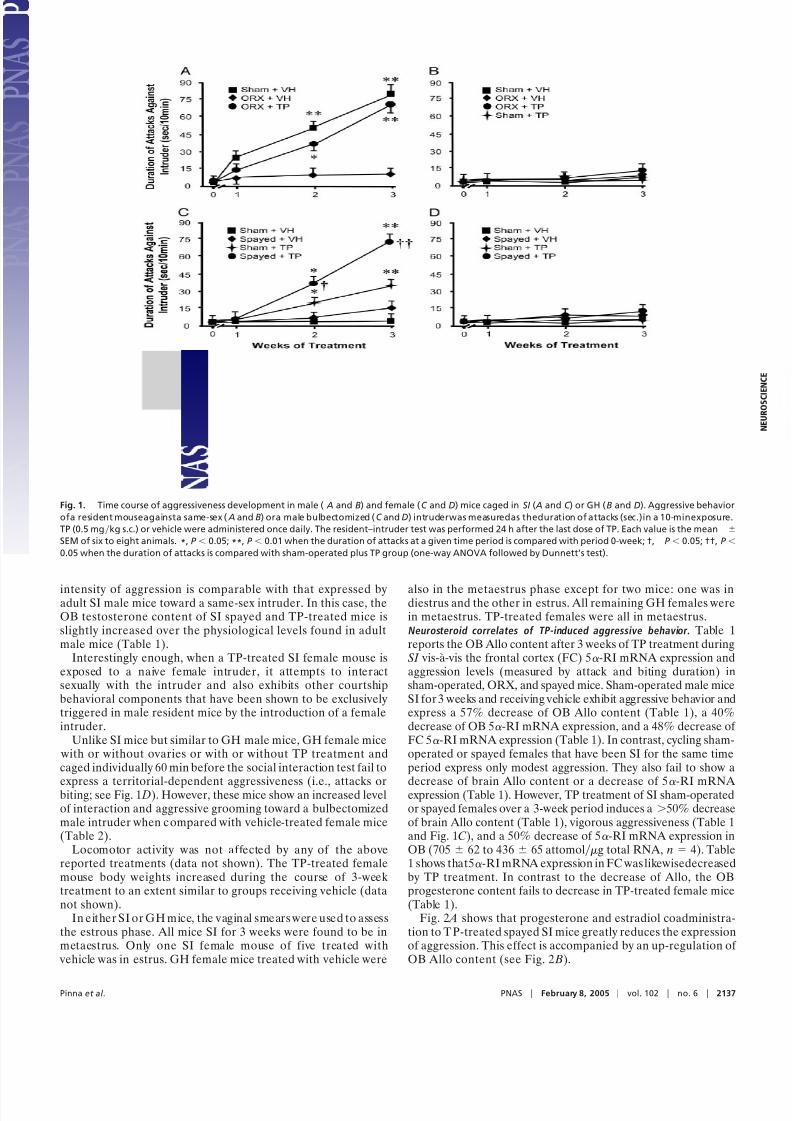
Testosterone-Induced Aggression in Orchiectomized (ORX) Mice. Inmost species, orchiectomy inhibits aggression (32, 33). Fig. 1 Ashows that when sham-operated adult (2.5-month-old) malemice are SI, there is a time-related increase of aggressionintensity toward a same-sex intruder. After 3 weeks of socialisolation (SI ), the OB Allo content and the OB and frontal
cortex 5-RI mRNA ex pression are reduced by 50% (Table 1).In contrast, SI ORX mice not only exhibit a lower level of aggressive behavior (Fig. 1 A), but also a normal OB Allo level(Table 1) and a 10-fold reduction in OB testosterone levels(Table 1). To underscore the important interaction betweentestosterone brain levels and the expression of aggressive be-havior, ORX mice were administered TP (0.5 mgkg) once dailyfor 3 weeks of SI . Resident–intruder tests performed in thesemice 24 h after the last TP dose showed an aggressive behaviorlevel comparable with that of sham-operated SI mice (Table 1).This TP dose normalizes the OB testosterone level and reducesthe OB Allo content to a level similar to that expressed by
vehicle-treated sham-operated mice after an identical isolationperiod (Table 1).
Aggressive Behavior and Brain Neurosteroid Content in TP-TreatedFemale Mice. Behavioral correlates of TP administration. Unlike malemice, females fail to develop a territorially defined aggressive-ness (see Materials and Methods) even after 3 weeks of SI (Fig.1C). When female mice are SI for a period of 3 weeks andadministered TP daily (0.5 mgkg s.c.), they develop a time-related increase of aggression measured 24 h after the last TPinjection (Fig. 1C). This dose of TP increases the female OBtestosterone content to values similar to that of male mice(Table 1).
SI induces only modest levels of aggression in spayed mice(Fig. 1C). However, when SI spayed mice are administered TPdaily for 3 weeks, they evince a time-dependent high level of aggression (Fig. 1C) against a bulbectomized male intruder. The
2136 www.pnas.orgcgidoi10.1073pnas.0409643102 Pinna et al .
8/3/2019 PNAS the rat behavior
http://slidepdf.com/reader/full/pnas-the-rat-behavior 3/6
intensity of aggression is comparable with that expressed byadult SI male mice toward a same-sex intruder. In this case, theOB testosterone content of SI spayed and TP-treated mice isslightly increased over the physiological levels found in adultmale mice (Table 1).
Interestingly enough, when a TP-treated SI female mouse isexposed to a naive female intruder, it attempts to interactsexually with the intruder and also exhibits other courtshipbehavioral components that have been shown to be exclusivelytriggered in male resident mice by the introduction of a femaleintruder.
Unlike SI mice but similar to GH male mice, GH female mice with or without ovaries or with or without TP treatment andcaged individually 60 min before the social interaction test fail to
express a territorial-dependent aggressiveness (i.e., attacks orbiting; see Fig. 1 D). However, these mice show an increased levelof interaction and aggressive grooming toward a bulbectomizedmale intruder when c ompared with vehicle-treated female mice(Table 2).
Locomotor activity was not affected by any of the abovereported treatments (data not shown). The TP-treated femalemouse body weights increased during the course of 3-weektreatment to an extent similar to groups receiving vehicle (datanot shown).
In either SI or GH mice, the vaginal smears were used to assessthe estrous phase. All mice SI for 3 weeks were found to be inmetaestrus. Only one SI female mouse of five treated with
vehicle was in estrus. GH female mice treated with vehicle were
also in the metaestrus phase except for two mice: one was indiestrus and the other in estrus. All remaining GH females werein metaestrus. TP-treated females were all in metaestrus.Neurosteroid correlates of TP-induced aggressive behavior. Table 1reports the OB Allo content after 3 weeks of TP treatment duringSI vis-a-vis the frontal cortex (FC) 5-RI mRNA expression andaggression levels (measured by attack and biting duration) insham-operated, ORX, and spayed mice. Sham-operated male miceSI for 3 weeks and receiving vehicle exhibit aggressive behavior andexpress a 57% decrease of OB Allo content (Table 1), a 40%decrease of OB 5-RI mRNA expression, and a 48% decrease of FC 5-RI mRNA expression (Table 1). In contrast, cycling sham-operated or spayed females that have been SI for the same timeperiod express only modest aggression. They also fail to show a
decrease of brain Allo content or a decrease of 5-RI mRNAexpression (Table 1). However, TP treatment of SI sham-operatedor spayed females over a 3-week period induces a 50% decreaseof brain Allo content (Table 1), vigorous aggressiveness (Table 1and Fig. 1C), and a 50% decrease of 5-RI mRNA expression inOB (705 62 to 436 65 attomolg total RNA, n 4). Table1 shows that5-RI mRNA expression in FC was likewisedecreasedby TP treatment. In contrast to the decrease of Allo, the OBprogesterone content fails to decrease in TP-treated female mice(Table 1).
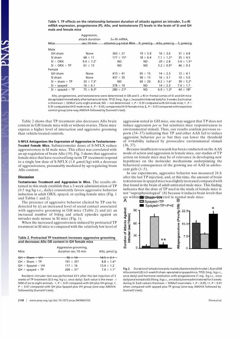
Fig. 2 A shows that progesterone and estradiol coadministra-tion to T P-treated spayed SI mice greatly reduces the expressionof aggression. This effect is accompanied by an up-regulation of OB Allo content (see Fig. 2 B).
Fig. 1. Time course of aggressiveness development in male ( A and B) and female (C and D) mice caged in SI ( A and C ) or GH (B and D). Aggressive behavior
ofa resident mouseagainsta same-sex ( A and B) ora male bulbectomized (C and D) intruderwas measuredas theduration of attacks (sec.)in a 10-minexposure.
TP (0.5 mgkg s.c.) or vehicle were administered once daily. The resident–intruder test was performed 24 h after the last dose of TP. Each value is the mean
SEM of six to eight animals. *, P 0.05; **, P 0.01 when the duration of attacks at a given time period is compared with period 0-week; †, P 0.05; ††, P
0.05 when the duration of attacks is compared with sham-operated plus TP group (one-way ANOVA followed by Dunnett’s test).
Pinna et al . PNAS February 8, 2005 vol. 102 no. 6 2137
8/3/2019 PNAS the rat behavior
http://slidepdf.com/reader/full/pnas-the-rat-behavior 4/6
8/3/2019 PNAS the rat behavior
http://slidepdf.com/reader/full/pnas-the-rat-behavior 5/6
Modulation of GABAA Receptors by Testosterone. Alterations of GABAergic transmission in the mammalian forebrain includingthe OB probably play a pivotal role in lowering the threshold for
aggressive behavior (19, 30, 38–40). Furthermore, several linesof evidence recently reviewed by Clark and Henderson (8)suggest that down-regulation of the GABAergic signal trans-duction is prominent in the regulation of aggressive behavior by
AAS (8).For example, it has been reported that protracted exposure to
high (7.5 mgkg per day) doses of 17-methyltestosteronesignificantly decreases the expression of GABA A receptor sub-units (1, 2, 5, 1, and 2) mRNAs preferentially in peripu-bertal females (41). In contrast, moderate (0.75 mgkg per day)doses of 17-methyltestosterone fail to induce significantchanges of GABA A receptor subunit expression in adult mice of either sex (41).
We have shown (Table 1) that, after the TP schedule (0.5mgkg per day) used in our experiments, brain testosteronelevels in SI ORX or spayed mice reach values slightly higher thanthose found in brain tissue of intact male mice. These concen-trations are in the nM range, i.e., three orders of magnitudelower than the concentrations causing a direct positive allostericmodulatory action at GABA A receptors (8). It is also unlikelythat these brain testosterone concentrations are sufficient toinduce significant changes of GABA A receptor subunit expres-sion in adult male or female mice (41).
Down-Regulation of Neurosteroid Biosynthesis. Because accumulat-ing evidence suggests that a decreased signal transduction of GABA A receptors to GABA or GA BAmimetic drugs in SI micemay be related to a down-regulation of Allo biosynthesis (19–23), we were prompted to study whether TP action on aggressive
behavior is associated with a down-regulation of Allo braincontent.
In this study, we provide evidence that TP reduces the brain Allo content by 50% in female and male mice that are eitherGH or SI, and sham-operated or spayed.
The rationale for measuring neurosteroids in the OB issupported by the crucial role that this brain region plays inaggressive behavior (29), and also by the finding that this brainstructure expresses the highest levels of neurosteroids andpresumably the highest rate of steroid biosynthesis (23, 28).Moreover, previous studies could correlate the biosynthesis ratesof Allo in OB with the intensity of aggressive behavior (19).
To test the hypothesis that a decrease of Allo biosynthesis maymodulate the threshold and duration of the aggressive behavior
accompanying TP treatment, we injected mice with proges-terone plus estradiol in doses that increase OB Allo levels (Fig.2). We found that coadministration of progesterone and estra-diol reduces aggression induced by SI and protracted treatment
with TP.S-NFLX is a pregnane steroid-enhancing drug that in submi-
cromolar doses increases brain Allo content and attenuatesaggressive behavior in SI male mice (19) in a manner that isindependent of 5HT reuptake (22). S-NFLX administered in
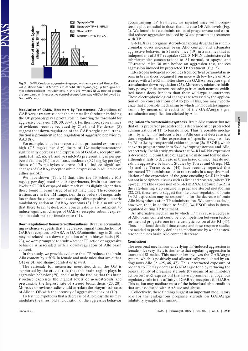
submicromolar concentrations to SI normal, or spayed andTP-treated mice 30 min before an aggression test, reducesaggression induced by protracted TP treatment (Fig. 3).
Electrophysiological recordings from cortical pyramidal neu-rons in brain slices obtained from mice with low levels of Allotreated with a 5-RI inhibitor showed a GABA A receptor signaltransduction down-regulation (25). Moreover, miniature inhib-itory postsynaptic current recordings from such neurons exhib-ited faster decay kinetics than their wild-type counterparts.These electrophysiological changes are reversed by the applica-tion of low concentrations of Allo (25). Thus, one may hypoth-esize that a possible mechanism by which TP modulates aggres-sion is by means of a reduction of the GABAergic signaltransduction amplification elicited by Allo.
Regulation of Neurosteroid Biosynthesis. Brain Allo content but notthat of its precursor progesterone is decreased after protractedadministration of TP to female mice. Thus, a possible mecha-nism by which TP induces a brain Allo content decrease is adown-regulation of the expression of specific enzymes, i.e.,5-RI or 3-hydroxysteroid-oxidoreductase (3-HSOR), whichconverts progesterone into 5-dihydroprogesterone and Allo,respectively. In this study, we show that 5-RI mRNA expressionis decreased in brain tissue of aggressive male and female micealthough it fails to decrease in brain tissue of mice that do notexhibit aggressive behavior. Studies by Torres and Ortega (42,43) and by Torres et al. (44) have recently reported thatprotracted TP administration to rats results in a negative mod-ulation of the expression of the gene encoding 5-RI in brain,but not in peripheral tissues such as liver and prostate, where TP
up-regulates the expression of 5-RI mRNA. Because 5-RI isthe rate-limiting step enzyme in pregnane steroid metabolism(22, 28), these results suggest that the down-regulation of brain5-RI expression may be responsible for the decrease of brain
Allo biosynthesis after TP administration. We cannot exclude,however, that, in addition to 5-RI, 3-HSOR also is down-regulated during TP treatment.
An alternative mechanism by which TP may cause a decreaseof Allo brain content could be a competition between testos-terone and progesterone in the catalytic action of 5-RI (45).Thus, additional detailed time-course and dose-response studiesare needed to precisely define the mechanisms by which testos-terone induces brain Allo content decrease.
Conclusions
The neuronal mechanism underlying TP-induced aggression infemale mice very likely is similar to that regulating aggression inuntreated SI males. This mechanism involves the GABAergicsystem, which is positively and allosterically modulated by en-dogenous Allo (21–25, 46, 47). Thus, protracted exposure of rodents to TP may decrease GABAergic tone by reducing thebioavailability of pregnane steroids (by means of an inhibitoryaction on 5-RI expression) that have a prominent endogenousregulatory role in the affinity of GABA A receptors for GABA.This action may mediate most of the behavioral abnormalitiesthat are associated with AAS use and abuse.
Collectively, these findings suggest an important modulatoryrole for the endogenous pregnane steroids on GABAergicinhibitory synaptic transmission.
Fig. 3. S-NFLX reduces aggression in spayed or sham-operated SI mice. Each
value is themean SEMof four mice. S-NFLX(1.8molkg,i.p.)was given 30
min before resident–intruder tests. *, P 0.01 when S-NFLX-treated groups
are compared with respective control groups (one-way ANOVA followed by
Dunnett’s test).
Pinna et al . PNAS February 8, 2005 vol. 102 no. 6 2139
8/3/2019 PNAS the rat behavior
http://slidepdf.com/reader/full/pnas-the-rat-behavior 6/6
It is noteworthy that the pregnane-enhancing drug S-NFLX insubmicromolar doses increases brain Allo content (19, 20, 22)and attenuates aggressive behavior in TP-treated mice. Likely,this observation may have important implications for designingfuture treatments for androgen abuse.
We thank Drs. Maria Luisa Barbaccia (Department of Neuroscience,School of Medicine, University of Rome ‘‘Tor Vergata,’’ Rome), Vas-
silios Papadopoulos (Departments of Cell Biology and Pharmacologyand of Neuroscience, Georgetown University Medical Center, Wash-ington, DC), and David H. Farb (Department of Pharmacology, Schoolof Medicine, Boston University, Boston) for constructive criticism andsuggestions in the preparation of the manuscript, and Ulana Liskevychfor excellent technical assistance. This study was supported by NationalInstitute of Mental Health Grants MH 56890 (to A.G.) and MH071667-01A1 (to E.C.) and by a Campus Research Board Award(to G.P.).
1. National Institute on Drug Abuse (2000) NIDA Research Report: Steroid Abuse and Addiction (National Clearinghouse on Alcohol and Drug Information,Rockville, MD), NIH Publication No. 00-3721 (http:165.112.78.61ResearchReportsSteroids Anabolicsteroids.html).
2. Pearson, H. (2004) Nature 431, 500–501.3. Lukas, S. E. (1996) Annu. Rev. Pharmacol. Toxicol. 36, 333–357.4. Faigenbaum, A. D., Zaichkowshy, L. D., Gardner, D. E. & Micheli, L. J. (1998)
Pediatrics 101, 1–6.5. Irving, L. M., Wall, M., Neumark-Sztainer, D. & Story, M. (2002) J . Adolesc.
Health 30, 243–252.6. Urhausen, A., Albers, T. & Kindermann, W. (2004) Heart 90, 496–501.7. Pope, H. G., Jr., & Brower, K. J. (2000) in Comprehensive Textbook of
Psychiatry, eds. Sadock, B. J. & Sadock, B. A. (Lippincott William & Wilkins,Philadelphia), 7th Ed., pp. 1085–1095.
8. Clark, A. S. & Henderson, L. P. (2003) Neurosci. Biobehav. Rev. 27, 413–436.9. Morris, J. A., Jordan, C. L. & Breedlove, S. M. (2004) Nat. Neurosci. 7,
1034–1039.
10. Corrigan, B. (1996) Med. J . Aust. 165, 222–226.11. Hartgens, F. & Kuipers, H. (2004) Sport Med. 34, 513–554.12. Brower, K. J., Blow, F. C., Beresford, T. P. & Fuelling, C. (1989) J . Clin.
Psychiatry. 50, 31–33.13. Thiblin, I., Lindquist, O. & Rajs, J. (2000) J . Forensic Sci. 45, 16–23.14. Bitran, D., Kellogg, C. K. & Hilvers, R. J. (1993) Horm. Behav. 27, 568–583.15. Wilson, M. A. (1996) Crit. Rev. Neurobiol. 10, 1–37.16. Clark, A. S., Jones, B. L., Yang, P. & Henderson, L. P. (2004) in Neurosteroid
Effects in the Central Nervous System: The Role of the GABA A Receptor , ed.Smith, S. S. (CRC, Boca Raton, FL), pp. 119–141.
17. Jorge-Rivera, J. C., McIntyre, K. L. & Henderson, L. P. (2000) J . Neurophysiol.83, 3299–3309.
18. Miczek, K. A., Fish, E. W. & De Bold, J. F. (2003) Horm. Behav. 44, 242–257.19. Pinna, G., Dong, E., Matsumoto, K., Costa, E. & Guidotti, A. (2003) Proc. Natl.
Acad. Sci. USA 100, 2035–2040.20. Guidotti, A., Dong, E., Matsumoto, K., Pinna, G., Rasmusson, A. M. & Costa,
E. (2001) Brain Res. Rev. 37, 110–115.21. Matsumoto, K., Uzunova, V., Pinna, G., Taki, K., Uzunov, D. P., Watanabe,
H., Mienvielle, J. M., Guidotti, A. & Costa, E. (1999) Neuropharmacology 38,
955–963.22. Pinna, G., Costa, E. & Guidotti, A. (2004) Proc. Natl. Acad. Sci. USA 101,
6222–6225.23. Pinna, G., Uzunova, V., Matsumoto, K., Puia, G., Mienville, J.-M., Costa, E.
& Guidotti, A. (2000) Neuropharmacology 39, 440–448.24. Serra, M., Pisu, M. G., Littera, M., Papi, G., Sanna, E., Tuveri, F., Usala, L.,
Purdy, R. H. & Biggio, G. (2000) J . Neurochem. 75, 732–740.
25. Puia, G., Mienville, J.-M., Matsumoto, K., Takahata, H., Watanabe, H., Costa,E. & Guidotti, A. (2002) Neuropharmacology 44, 49–55.
26. Puia, G., Santi, M. R., Vicini, S., Pritchett, D. B., Purdy, R. H., Paul, S. M.,Seeburg, P. H. & Costa, E. (1990) Neuron 4, 759–765.
27. Lambert, J. J., Belelli, D., Peden, D. R., Vardy, A. W. & Peters, J. A. (2003) Prog. Neurobiol. 71, 67–80.
28. Dong, E., Matsumoto, K., Uzunova, V., Sugaya, I., Costa, E. & Guidotti, A.(2001) Proc. Natl. Acad. Sci. USA 98, 2849–2854.
29. Siegel, A., Roeling, T. A., Gregg, T. R. & Kruk, M. R. (1999) Neurosci. Biobehav. Rev. 23, 359–389.
30. Denenberg, V. H., Gaulin-Kremer, E., Gandelman, R. & Zarrow, M. X. (1973) Anim. Behav. 21, 590–598.
31. Pinna, G., Galici, R., Schneider, H. H., Stephens, D. N. & Turski, L. (1997) Proc. Natl. Acad. Sci. USA 94, 2719–2723.
32. Knol, B. W. & Egberink-Alink, S. T. (1989) Vet. Q. 11, 102–107.33. Schneider, J. S., Stone, M. K., Wynne-Edwards K. E., Horton, T. H., Lydon, J.,
O’Malley, B. & Levine, J. E. (2003) Proc. Natl. Acad. Sci. USA 100, 2951–2956.
34. Breuer, M. E., McGinnis, M. Y., Lumia, A. R. & Possidente B. P. (2001) Horm. Behav. 40, 409–418.
35. McGinnis, M. Y., Lumia, A. R., Breuer, M. E. & Possidente, B. (2002) Horm. Behav. 41, 101–110.
36. Bronson, F. H. (1996) Pharmacol. Biochem. Behav. 53, 329–334.37. Bronson, F. H., Nguyen, K. Q. & De La Rosa, J. (1996) Physiol. Behav. 59,
49–55.38. Mandel, P., Haug, M., Puglisi-Allegra, S., Kempf, E. & Mack, G. (1981) inThe
Biology of Aggression, eds. Brian, P. & Benton, D. (Sijthoff & Noordhoff, Alphen aan der Rijn, The Netherlands), pp. 169–173.
39. Puglisi-Allegra, S. & Mandel, P. (1980) Psychopharmacology (Berlin) 70,
287–290.40. Molina, V., Ciesielski, L., Gobaille, S. & Mandel, P. (1986) Pharmacol.
Biochem. Behav. 24, 657–664.41. McIntyre, K. L., Porter, D. M. & Henderson, L. P. (2002) Neuropharmacology
43, 634–645.42. Torres, J. M. & Ortega, E. (2003) FASEB J . 17, 1428–1433.43. Torres, J. M. & Ortega, E. (2003) Biochem. Biophys. Res. Commun. 308,
469–473.44. Torres, J. M., Ruiz, E. & Ortega, E. (2003) Prostate 56, 74–79.45. Sturenburg, H. J., Fries, U. & Kunze, K. (1997) Neuropsychobiology 35,
143–146.46. Barbaccia, M. L., Serra, M., Purdy, R. H. & Biggio, G. (2001) Int. Rev.
Neurobiol. 46, 243–272.47. Park-Chung, M., Malayev, A., Purdy, R. H., Gibbs, T. T. & Farb, D. H. (1999)
Brain Res. 830, 72–87.
2140 www.pnas.orgcgidoi10.1073pnas.0409643102 Pinna et al .