plug-and-play pairing via defined divalent streptavidins
TRANSCRIPT

Article
Michael Fairhe
0022-2836/$ - see front m
Plug-and-Play Pairing via DefinedDivalent Streptavidins
ad, Denis Krndija1, Ed D.
Lowe and Mark HowarthDepartment of Biochemistry, University of Oxford, South Parks Road, Oxford OX1 3QU, UK
Correspondence to Mark Howarth: [email protected]://dx.doi.org/10.1016/j.jmb.2013.09.016Edited by T. Yeates
Abstract
Streptavidin is one of the most important hubs for molecular biology, either multimerizing biomolecules,bridging one molecule to another, or anchoring to a biotinylated surface/nanoparticle. Streptavidin has theadvantage of rapid ultra-stable binding to biotin. However, the ability of streptavidin to bind four biotinylatedmolecules in a heterogeneous manner is often limiting. Here, we present an efficient approach to isolatestreptavidin tetramers with two biotin-binding sites in a precise arrangement, cis or trans. We geneticallymodified specific subunits with negatively charged tags, refolded a mixture of monomers, and usedion-exchange chromatography to resolve tetramers according to the number and orientation of tags. Wesolved the crystal structures of cis-divalent streptavidin to 1.4 Å resolution and trans-divalent streptavidin to1.6 Å resolution, validating the isolation strategy and explaining the behavior of the Dead streptavidin variant.cis- and trans-divalent streptavidins retained tetravalent streptavidin's high thermostability and low off-rate.These defined divalent streptavidins enabled us to uncover how streptavidin binding depends on the nature ofthe biotin ligand. Biotinylated DNA showed strong negative cooperativity of binding to cis-divalent but nottrans-divalent streptavidin. A small biotinylated protein bound readily to cis and trans binding sites. We alsosolved the structure of trans-divalent streptavidin bound to biotin-4-fluorescein, showing how one ligandobstructs binding to an adjacent biotin-binding site. Using a hexaglutamate tag proved a more powerful way toisolate monovalent streptavidin, for ultra-stable labeling without undesired clustering. These forms ofstreptavidin allow this key hub to be used with a new level of precision, for homogeneous molecular assembly.
© 2013 The Authors. Published by Elsevier Ltd. All rights reserved.
Introduction
Streptavidin isa tetramericprotein fromStreptomycesavidinii [1] that, along with the structurally similar proteinfrom chicken egg white, avidin, exhibits extraordinarilyhigh affinity for the vitamin biotin [2,3]. The femtomolaraffinity of streptavidin/avidin for biotinylated targets,combined with resilience to harsh conditions and anon-rate of ~107 M−1 s−1, is thebasis for biotin's centralrole as a specific biological “glue” in diverse researchmethods and biotechnologies [1–3].Although streptavidin has a long history of use for
capture of biotinylated ligands from complexmixtures,it has gainedmany recent applications for precise andstable assembly in the emerging field of bionanotech-nology [4–6]. Such assemblies can be centered on asingle streptavidin, for example, streptavidin:MHC(major histocompatibility complex) tetramers, to ana-
atter © 2013 The Authors. Published by El
lyze immune function [7]. Assemblies can also dependon multiple streptavidin molecules holding togethernetworks, such as for amplification in histochemistry [8]or to bridge biomolecules to facilitate structure deter-mination by electron microscopy, atomic force micros-copy, and X-ray crystallography [9–11].A central issue that needs addressing is to control
the nature of the four binding sites in the streptavidintetramer. While the binding of four ligands may bedesirable in some applications, in other applications,it may be necessary to limit binding to only one, two,or three subunits to achieve the desired assembliesand geometries [11–13]. Splitting up the streptavidintetramer into monomers or dimers is possible, butthis has always been accompanied by orders ofmagnitude lower affinity for biotin [14–17], becauseof the contribution of the neighboring subunit to thebiotin-binding site [1,18]. Such monomers or dimers
sevier Ltd. All rights reserved. J. Mol. Biol. (2014) 426, 199–214

Fig. 1. Principle of defined divalent streptavidins.Numbering of subunits in the streptavidin tetramer, witheach monomer colored separately and biotin in space-fill(from PDB ID: 3RY2). Below are arrangements of 1,2 or1,3 biotin-binding sites (biotin-bound monomers, green;non-binding monomers, blue), showing relative orienta-tions and distances between the biotin carboxyl groupcarbons.
200 Crystal Structures of Divalent Streptavidins
give desirable reversibility for purification but forfeitmuch of the advantage of streptavidin's stability forassembly applications.We previously generated streptavidin heterotetra-
mers with exactly one, two, or three functional biotin-binding sites, through purifying tetramers containingdefined numbers of non-biotin-binding “Dead” andHis6-tagged wild-type subunits [19]. However, tetra-mers with two functional and two Dead subunits canassemble in three different arrangements. Thesearrangements are the 1,2, 1,3, and 1,4 divalent formsof streptavidin (Fig. 1). In the 1,2 arrangement thebiotin-binding sites of streptavidin are in a cis config-uration as the binding sites both point in the samedirection (Fig. 1). However, in the 1,3 arrangement,both sites are in a trans configuration, pointing awayfrom each other (Fig. 1). Taking a vector fromN1 to theterminal carbon atom of biotin, in 1,2, both biotins point23° from the two-fold axis in Fig. 1, while in 1,3, onebiotin is 23° from this axis and the other is 160°. In 1,2,the terminal carbon atoms of the two biotins are 20 Åapart, and in 1,3, they are 35Åapart (Fig. 1). Therefore,any biotinylated ligands to divalent streptavidin willadopt different distances and angles with respect toeach other. Such mixed divalent species, containing
all three arrangements, would not be suitable forapplications where defined spatial control is neces-sary. For example, a cis arrangement may be suitablefor capping biotinylated protein oligomers [20], result-ing in molecules with a uniform length, while the transarrangement may serve as a linker in the middle ofsuch an assembly [20,21].Control over subunit orientation could be achieved
by generating single-chain dimers or tetramers [22],but the symmetry and folding characteristics ofstreptavidin mean that connection of subunits (usingcircular permutation) has been accompanied byimpaired folding and affinity [16,23].To generate a controlled and stable bridge for
bionanotechnology, we demonstrate here a methodfor the simple isolation of streptavidin variants withprecise cis-divalent or 1,3 trans-divalent arrange-ments. We confirmed the variant structures bycrystallography and investigated the binding char-acteristics of these defined divalents, which helps toresolve decades of data about interaction betweenbinding sites on streptavidin/avidin [24–28].
Results
Streptavidins with defined valencies and subunitorientation by engineering of charge
To prepare streptavidin tetramers with controlledpairs of biotin-binding sites (Fig. 1), we added chargedtags at specific sites in the subunits to enable resolutionof the tetramers by ion-exchange chromatography(Figs. 2 and 3). We refolded from inclusion bodies themixture of charged and non-charged streptavidinvariants and then separated the tetramers on aMonoQ column (anion exchange), since the tetramerswith increasing negative charge required higher ionicstrength for elution. Geometrically distinct speciesbearing the same number of charged tags can beseparated from each other. On that basis, two differentengineering strategies made it possible to isolate thecis-divalent and 1,3 trans-divalent forms (Fig. 1).
C-terminal polyglutamate tag allowed 1,3trans-divalent isolation
In the first instance, we placed a hexaglutamatetag at the C-terminus of core streptavidin monomer(termed SAe) (Fig. 2a). First, these charges ought toallow easy isolation of forms containing 0–4biotin-binding sites by ion-exchange chromatogra-phy, giving clearer separation than the previousmethod of separation using a C-terminal His6 tagand Ni-NTA, where peaks from different tetramerforms largely overlapped [19,29]. Second theC-termini are close together between subunits 1and 3 (27 Å) but far apart between subunits 1 and 2

Fig. 2. Generation of the 1,3 trans-divalent streptavidin. (a) Amino acid sequence of streptavidin-E6 (SAe) with E6 tag red andunderlined, along with Dead (D) streptavidin showing residues impairing biotin binding in blue. (b) Generation of the differentvalency formsofSAe/Dbymixed refoldand ionexchange,with predictedpIofmonomerand tetramer indicated. (c) Ion-exchangechromatogramof amixtureof tetramers containingdifferent proportions of SAeandD. (d)Analysis of peaks from ionexchangebySDS-PAGE with Coomassie staining, unboiled (left) to show tetramer mobility, or boiled (right) to show subunit composition.
201Crystal Structures of Divalent Streptavidins
(57 Å) or subunits 1 and 4 (50 Å), so that elution fromthe ion-exchange resin would be predicted to occurlater for the 1,3 divalent (Fig. 1).SAe was refolded together with Dead (D) strepta-
vidin (N23A, S27D, S45A) [19] (Fig. 2a). Sevendifferent tetramer forms are possible: one nullivalent,one monovalent, one trivalent, one tetravalent, andthree divalent (Fig. 2b). The ion-exchange chro-matogram showed six peaks (Fig. 2c), consistentwith our hypothesis that 1,2 and 1,4 divalents wouldco-elute, while the 1,3 divalent would elute later.We analyzed the eluted peaks by SDS-PAGE,
where streptavidin will remain a tetramer without
boiling (Fig. 2d) [30]. The tetramers do not runaccording to their molecular weight: the increasingnegative charge increases migration of tetramerscontaining a higher proportion of SAe. Upon boiling inSDS, the tetramer breaks up into monomers and therelative proportion of SAe and D subunits is shown(Fig. 2d). This gel analysis confirmed that peak 1 wasD4, peak 2 was the monovalent SAe1D3, peak 5 wasthe trivalent SAe3D1, peak 6 was the tetravalentSAe4, and peaks 3 and 4 were both divalent. Atitration with biotin-4-fluorescein (whose fluorescenceis quenched dramatically upon streptavidin binding)[31,32] confirmed the expected number of biotin-

Fig. 3. Generation of cis-divalent streptavidin. (a) Amino acid sequence of core streptavidin (SA), along with Dead(Dd) containing a polyaspartate insertion in the 3/4 loop (red and underlined) and residues impairing biotin binding inblue. (b) Generation of the different valency forms of SA/Dd by mixed refold and ion exchange, with predicted pI ofmonomer and tetramer indicated. (c) Ion-exchange chromatogram of a mixture of tetramers containing differentproportions of SA and Dd. (d) Analysis of peaks from ion exchange by SDS-PAGE with Coomassie staining, unboiled(left) to show tetramer mobility, or boiled (right) to show subunit composition.
202 Crystal Structures of Divalent Streptavidins
binding sites for D4, SAe4, SAe1D3, and the putative1,3 trans-divalent (Supplementary Data Fig. S1a).
Multiple aspartates in a surface loop allowedcis-divalent isolation
To isolate cis-divalent streptavidin (1,2 arrangementof biotin-binding subunits) (Fig. 1), we introduced aseries of aspartates into the loop between β-strands 3and 4 (L3,4) of the Dead subunit (Fig. 3a). Afterexploring a number of different insertions for optimalexpression and separation, we chose the sequence
termed Dd (Fig. 3a). Introduction of the charges in thisposition means that in the 1,3 and 1,4 arrangements,these charged regions should be far apart, while in the1,2 arrangement, the charged loops should beapposed (Fig. 3b). Since L3,4 is important for biotinbinding affinity [1,18,33], it was necessary to insertthese residues on the Dead subunit, so that thecis-divalent streptavidin could maintain high biotinbinding affinity. Ion-exchange chromatography of amixed refold of SA/Dd again allowed efficient separa-tion of tetramers, giving six peaks, two of whichcorresponded to divalent species (Fig. 3b and c).
203Crystal Structures of Divalent Streptavidins
We analyzed peaks by SDS-PAGE with andwithout boiling. For unboiled samples, the negativecharge on Dd accelerated the mobility of thetetramers (Fig. 3d). Upon boiling, the varyingintensities of SA and Dd bands clearly showed thatthe tetramers contained the expected ratios of eachsubunit (Fig. 3d). This gel analysis confirmed thatpeak 1 was tetravalent SA4, peak 2 was the trivalentSA3Dd1, peak 5 was the monovalent SA1Dd3, peak6 was the non-binding Dd4, and peaks 3 and 4 wereboth divalent. A titration of the different tetramerforms with biotin-4-fluorescein also confirmed theexpected number of biotin-binding sites (Supple-mentary Data Fig. S1b). Overall, the introduction of aseries of negative charges into the surface-exposed3,4-loop of streptavidin allowed the isolation of asecond divalent species, cis-divalent.
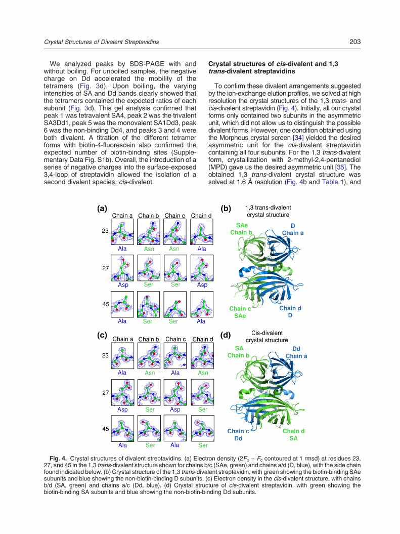
Fig. 4. Crystal structures of divalent streptavidins. (a) Elect27, and 45 in the 1,3 trans-divalent structure shown for chains bfound indicated below. (b) Crystal structure of the 1,3 trans-divasubunits and blue showing the non-biotin-binding D subunits. (b/d (SA, green) and chains a/c (Dd, blue). (d) Crystal strucbiotin-binding SA subunits and blue showing the non-biotin-bi
Crystal structures of cis-divalent and 1,3trans-divalent streptavidins
To confirm these divalent arrangements suggestedby the ion-exchange elution profiles, we solved at highresolution the crystal structures of the 1,3 trans- andcis-divalent streptavidin (Fig. 4). Initially, all our crystalforms only contained two subunits in the asymmetricunit, which did not allow us to distinguish the possibledivalent forms. However, one condition obtained usingthe Morpheus crystal screen [34] yielded the desiredasymmetric unit for the cis-divalent streptavidincontaining all four subunits. For the 1,3 trans-divalentform, crystallization with 2-methyl-2,4-pentanediol(MPD) gave us the desired asymmetric unit [35]. Theobtained 1,3 trans-divalent crystal structure wassolved at 1.6 Å resolution (Fig. 4b and Table 1), and
ron density (2Fo − Fc contoured at 1 rmsd) at residues 23,/c (SAe, green) and chains a/d (D, blue), with the side chainlent streptavidin, with green showing the biotin-binding SAec) Electron density in the cis-divalent structure, with chainsture of cis-divalent streptavidin, with green showing thending Dd subunits.

Table 1. Data collection, refinement statistics, and structure validation for the 1,3 trans-divalent and cis-divalentstreptavidin structures.
1,3 trans-divalent(4BX6)
cis-divalent(4BX5)
1,3 trans-divalent withbiotin-4-fluorescein (4BX7)
Data collectionSpace group P1211 P1211 P3121Asymmetric unit Tetramer Tetramer DimerUnit cell size (Å) 47.04 × 81.59 × 65.12 46.22 × 84.24 × 58.25 64.41 × 64.41 × 103.13Unit cell angles (°) 90 × 95.94 × 90 90 × 98.81 × 90 90 × 90 × 120Completeness (%) 98.00 92.59 99.77RefinementResolution (Å) 1.59–24.98 1.43–42.12 2.26–49.06No. of reflections 64,074 75,108 12,059Rwork/Rfree 0.1640/0.1866 0.1601/0.1873 0.1839/0.2369No. of atomsProtein 3756 3739 1768Heteroatoms 24 42 54Water 466 517 78B-factors (mean of all atoms, Å2)Wilson plot 20.98 14.65 42.43Mean B value 28.40 22.00 45.10rmsdBond length (Å) 0.011 0.01 0.01Bond angle (°) 1.28 1.14 0.94Structure validationMolProbity clash score 3.65 (97th percentile) 2.71 (98th percentile) 2.29 (100th percentile)Poor rotamers (%) 2.01 0.57 2.33Ramachandran outliers (%) 0.43 0.00 0.00Ramachandran favored (%) 97.02 97.65 97.85MolProbity score 1.55 (85th percentile) 1.13 (98th percentile) 1.32 (100th percentile)Residues with bad bonds (%) 0.00 0.00 0.00Residues with bad angles (%) 0.00 0.00 0.00
204 Crystal Structures of Divalent Streptavidins
cis-divalent streptavidin was solved at 1.4 Å resolution(Fig. 4d and Table 1), with the phases in each casesolved using molecular replacement.We identified each subunit (whether SAe/D or SA/
Dd) unambiguously by examiningwhether the electrondensity around residues 23, 27, and 45 correspondedto the residues of the wild-type or Dead subunit (N23A,S27D, S45A). For the putative 1,3 trans-divalent, theelectron density closely fitted residues 23, 27, and 45only if chains b/c were SAe and chains a/d were D(Fig. 4a), confirming that the biotin-binding subunitsindeed had a 1,3 trans orientation (Fig. 4b). For theputative cis-divalent sample, the electron densityclosely fits if chains a/c are Dd and chains b/d are SA(Fig. 4c), also confirming that the biotin-bindingsubunits indeed had a cis orientation (Fig. 4d).Furthermore, while the electron density around
L3,4 could be clearly observed in the SA chains band d of the cis-divalent form, the electron density ofL3/4 was poorly defined in Dd chains a and c(Supplementary Data Fig. S2). The engineered L3,4of Dd (chains a/c) is expected to be highly flexible,which would lead to weak diffraction in this region.The rmsd of any of the chains in 1,3 trans-divalent
and cis-divalent streptavidin is less than 0.5 Å to chaina of the 0.95-Å structure of streptavidin [Protein DataBank (PDB) ID: 3RY2] [35]. The Dead streptavidinsubunit has not been previously analyzed by crystal-
lography, and the changes to the structure arelocalized to the mutated residues, consistent withthe comparable thermostability of tetramers contain-ing wild-type or Dead subunits [19,33]. The overlay ofD and a wild-type subunit bound to biotin illustrateshow the three mutations in D greatly reduced thenumber of polar contacts to biotin (Fig. 5a). In theoverlay, Asp27 has van der Waals clash with biotinand there would be repulsion between the Asp27carboxylate and biotin's carbonyl oxygen lone pair(Fig. 5a).
Crystal structure of biotin-4-fluorescein boundto streptavidin
To understand interaction between ligand bindingsites, we solved 1,3 trans-divalent streptavidin'scrystal structure bound to biotin-4-fluorescein. Bio-tin-4-fluorescein represents the largest ligand wherethere is a crystal structure with streptavidin or avidin,as well as a widely used probe of the binding kineticsand capacity of biotin-binding proteins [19,31,32,36–38]. The fluorescence of biotin-4-fluorescein isquenched ~90% on binding to streptavidin/avidin,allowing a clear and sensitive test of ligandbinding without any separation step [31,32]. Our2.3-Å-resolution structure shows the 1,4 dimer in theasymmetric unit (one copy of SAe and one copy of

Fig. 5. Structures of streptavidin interaction with ligands. (a) Structure of the biotin-binding pocket in the Dead subunit(N23A, S27D, and S45A), showing chain a of the 1,3 trans-divalent streptavidin (carbon atoms in blue) overlaid withbiotin-bound wild-type streptavidin (PDB ID: 3RY2, carbon atoms in yellow). Hydrogen bonds from residues 23, 27, and 45to biotin in wild-type streptavidin are shown as broken lines. (b) Crystal structure of the 1,3 trans-divalent streptavidinbound to biotin-4-fluorescein (space-fill, carbons in cyan), with green for SAe and blue for D subunits. (c) Residuessurrounding the fluorescein tail (carbon atoms in cyan) in the 1,3 trans-divalent:biotin-4-fluorescein structure, labeledaccording to chain (b, carbon atoms in green; d, carbon atoms in blue), and with putative interactions as broken lines.Electron density for biotin-4-fluorescein is shown as blue mesh and contoured at 1 rmsd. (d) Clash for cis-boundbiotin-4-fluorescein, generated from the 1,3 trans-divalent biotin-4-fluorescein structure, if two biotin-4-fluoresceinmolecules (carbon atoms of one in yellow and the other in cyan) were to bind in the same conformation in cis as they do in1,3 trans-divalent.
205Crystal Structures of Divalent Streptavidins
D). Building the native streptavidin tetramer from thisdimer shows that the two biotin-4-fluorescein mole-cules are in the expected trans (1,3) arrangement(Fig. 5b). All the contacts of the 1,3 trans-divalentstreptavidin with the biotinmoiety are as expected [18].Fluorescein and the ethylene diamine spacer formseveral non-polar contacts to streptavidin not observedwith biotin (Fig. 5c). Features that have been proposedto quench fluorescein are photoinduced electrontransfer to nearby tryptophans or tyrosines [39],co-planarity of the xanthenone and benzoate rings[40], and hydrogen bonding to O3 of fluorescein [41].Trp120 is 5 Å from fluorescein (Fig. 5c) and mayexplain part of the quenching of fluorescence.Biotin-4-fluorescein bound in chain b reaches over
and interactswith residues of chain d. However, wedonot believe this conformation of biotin-4-fluorescein isthe same as when four ligands are bound to the same
tetramer: if the SAe:biotin-4-fluorescein complex werein a cis arrangement, there would be an obvious clashbetween the fluorescein moieties of neighboringligands (Fig. 5d).
Low off-rate and high thermostability ofdefined divalents
Having confirmed the identity of the defined diva-lents, we initially assessed these proteins by measur-ing the off-rate for a biotin conjugate (Fig. 6a). Thestrong biotin-binding stability of wild-type streptavidinwas retained by the defined divalents. Both divalentshad dissociation rates for biotin-4-fluorescein compa-rable to the original SA4 or SAe4 tetramers. In fact, thebinding stability for the 1,3 trans-divalent streptavidinwas slightly superior to that of the cis-divalent one(P ≤ 0.001, n = 3) and that of the wild-type SA4

Fig. 6. Off-rate and thermostability of divalent streptavidins. (a) Biotin-4-fluorescein dissociation over time at 37 °C inthe presence of competing biotin, for a tetramer of wild-type streptavidin subunits (SA4), a tetramer of SAe subunits(SAe4), and cis-divalent or 1,3 trans-divalent streptavidin (mean of triplicate + 1 SD). (b) Tetramer thermostability forstreptavidin variants determined by SDS-PAGE with Coomassie staining, following incubation in PBS for 3 min at theindicated temperatures. In B, the protein was boiled in the presence of SDS to achieve complete conversion to monomers.The mobilities of the starting tetramer and the resulting monomers are marked.
206 Crystal Structures of Divalent Streptavidins
tetramer (P ≤ 0.001, n = 3, one-way ANOVA withTukey's multiple comparison) (Fig. 6a).We next compared the thermostability of the two
divalent forms with tetramers containing only theconstituent monomers. We incubated at varyingtemperatures and then analyzed the resulting proteinforms by SDS-PAGE (Fig. 6b). Both cis-divalent and1,3 trans-divalent forms started to denature to their
constituent monomers above 70 °C, in a similarfashion to SA4, Dd4, SAe4, and D4. Some mixing ofthe divalent tetramers into new species could beobserved at 70 °C before full denaturation into themonomer species (Fig. 6b). To check whether thismixing would be an issue at 37 °C or room temper-ature, we incubated cis-divalent and 1,3 trans-divalentstreptavidins for 1–8 days, but we observed no
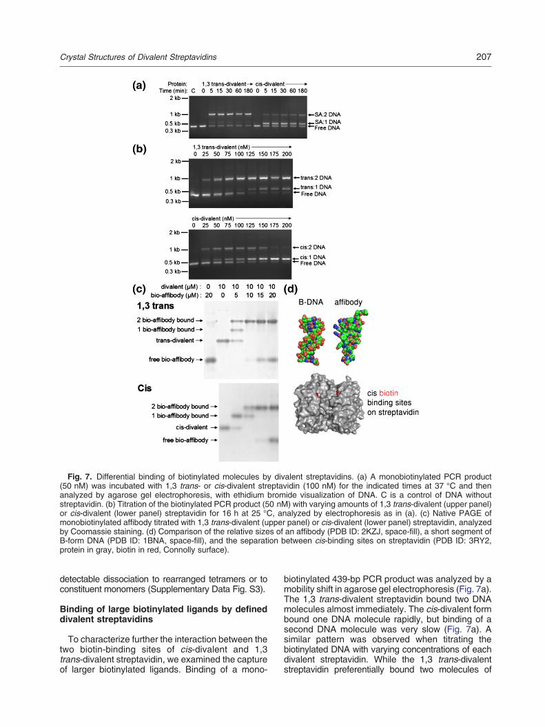
Fig. 7. Differential binding of biotinylated molecules by divalent streptavidins. (a) A monobiotinylated PCR product(50 nM) was incubated with 1,3 trans- or cis-divalent streptavidin (100 nM) for the indicated times at 37 °C and thenanalyzed by agarose gel electrophoresis, with ethidium bromide visualization of DNA. C is a control of DNA withoutstreptavidin. (b) Titration of the biotinylated PCR product (50 nM) with varying amounts of 1,3 trans-divalent (upper panel)or cis-divalent (lower panel) streptavidin for 16 h at 25 °C, analyzed by electrophoresis as in (a). (c) Native PAGE ofmonobiotinylated affibody titrated with 1,3 trans-divalent (upper panel) or cis-divalent (lower panel) streptavidin, analyzedby Coomassie staining. (d) Comparison of the relative sizes of an affibody (PDB ID: 2KZJ, space-fill), a short segment ofB-form DNA (PDB ID: 1BNA, space-fill), and the separation between cis-binding sites on streptavidin (PDB ID: 3RY2,protein in gray, biotin in red, Connolly surface).
207Crystal Structures of Divalent Streptavidins
detectable dissociation to rearranged tetramers or toconstituent monomers (Supplementary Data Fig. S3).
Binding of large biotinylated ligands by defineddivalent streptavidins
To characterize further the interaction between thetwo biotin-binding sites of cis-divalent and 1,3trans-divalent streptavidin, we examined the captureof larger biotinylated ligands. Binding of a mono-
biotinylated 439-bp PCR product was analyzed by amobility shift in agarose gel electrophoresis (Fig. 7a).The 1,3 trans-divalent streptavidin bound two DNAmolecules almost immediately. The cis-divalent formbound one DNA molecule rapidly, but binding of asecond DNA molecule was very slow (Fig. 7a). Asimilar pattern was observed when titrating thebiotinylated DNA with varying concentrations of eachdivalent streptavidin. While the 1,3 trans-divalentstreptavidin preferentially bound two molecules of

208 Crystal Structures of Divalent Streptavidins
DNA, the cis-divalent formboundoneor twomolecules(Fig. 7b).We also analyzed binding of a biotinylated protein,
avoiding the high charge of polynucleotides. Titration ofthedivalentswith asite-specifically biotinylatedaffibody[42], analyzed by the mobility shift in native PAGE,yielded a similar pattern (Fig. 7c). With streptavidinbinding sites in excess, one or two molecules of themonobiotinylated affibody could bind to the 1,3trans-divalent streptavidin, while for the cis-divalentone, only a single affibody bound. However, at higherconcentrations of affibody, two molecules of affibodybound to the cis-divalent form (Fig. 7c). The sizes of theaffibody and DNA double helix relative to streptavidinare illustrated in Fig. 7d.
Monovalent streptavidin isolatedby ion-exchangechromatography allowed specific cellular imaging
Monovalent streptavidin has found diverse uses,such as a highlighter in electron microscopy [43],calibrating energies in protein folding [44], enhancingantibody detection sensitivity [45], and DNA origami-mediated nano-assembly [46]. The clean resolution ofmonovalent streptavidin SAe1D3 from other valencyforms by ion-exchange chromatography (Fig. 2c) issuperior to our previous Ni-NTA method, whereelution of His6-tagged monovalent and divalentforms substantially overlapped [19,29]. Therefore,we validated that the new SAe1D3 form was stilleffective for cellular imaging and the altered chargedid not lead to high non-specific cellular binding. HeLacells were transfected with ER-resident biotin ligaseand acceptor peptide-tagged low-density lipoproteinreceptor (LDLR) [29] and then imagedwith AlexaFluor555-labeled SAe1D3. SAe1D3 clearly labeled cellsexpressing AP-GFP-LDLR but did not label nearbynon-expressing cells or cells that were not co-trans-fected with biotin ligase (Supplementary Data Fig.S4), consistent with specific biotin detection.
Discussion
Herein, we describe the generation and analysis oftetramers of streptavidin with a defined orientation oftwin biotin-binding sites. We were able to engineercis-divalent streptavidin with comparably low biotinoff-rate to wild-type streptavidin but 1,3 trans-divalent streptavidin showed even better bindingstability. Defined divalent streptavidins should act asan efficient and simple bridge for diverse applica-tions in bionanotechnology.Although new in its application to streptavidin, the
separation of multimers by ion-exchange chromatog-raphy according to the position of charged tagshas been applied previously [47–49]. A similarorientation-dependent separation was observed forwild-type streptavidin tetramers containing two bound
biotins because of biotin's charge. However, the biotinoccupancy of such streptavidin tetramers changedover hours [27], whereas our defined divalents werestable for at least a week at 37 °C and should bestable indefinitely at −80 °C. We found high thermo-stability of both divalent tetramers, with negligibledissociation into monomers until above 60 °C.The introduced mutations have also not reducedbiotin-conjugate binding affinity, a problem frequentlyencountered when engineering streptavidin [14–17].Despite testing several strategies to isolate 1,4
divalent streptavidins, we could never attain a cleanseparation. However, we anticipate that the mostimportant thing is having one defined trans-divalentstreptavidin, since the biotin–biotin separation distancefor 1,3 and 1,4 is very close.The work here also establishes a substantially
improved route to monovalent streptavidin. Monova-lent streptavidin with an E6 tag can be purified on alarger scale compared to the His6-tagged monovalentstreptavidin, because its elution is well separated fromdivalent streptavidin. SAe1D3 is alsoeasier to analyzefor purity, because it is much better resolved onSDS-PAGE. The E6 tag did not impair the use ofmonovalent streptavidin for specific cellular imaging.Divalent streptavidins without defined orientation
have been used for generating monovalent quantumdots [50] and for studying mechanical interactionsbetween MHC class I and T-cell receptors [51],illustrating areas where our defined divalents wouldimprove control in nano-assembly.Streptavidin and avidin are the most widely used
biotin-binding proteins, but recently, several othershave been found, including the dimers rhizavidin [52]and shwanavidin [53]. These relativesmay find a rangeof applications, but the natural dimers contain disulfidesand provide only one divalent orientation (equivalent to1,4), and streptavidin appears to have stronger bindingto biotin conjugates [53,54]. A further advantage ofretaining the whole streptavidin tetramer is the possi-bility in futurework to functionalizeDead subunits, suchas by fusion to an antibody fragment [55] or via anexposed cysteine [56], to create a multifunctional hub.The cooperativity that can exist for biotinylated ligand
binding is not well appreciated by the wider communityusing streptavidin/avidin. For biotin itself, even thoughstreptavidin has a conformational change and in-creased tetramer stability when biotin binds [18],there is no cooperativity of biotin binding [27]. However,for large biotinylated ligands, there can be substantialnegative cooperativity in binding to streptavidin/avidin,as shown for biotin metal–ligand complexes [24].Similarly, the binding of more than two biotinylatedDNA molecules to wild-type streptavidin was notreadily observed [5,57]. Our defined divalents alloweddirect analysis of these ligand–ligand repulsions at cisor 1,3 trans binding sites. Binding of two largebiotinylated ligands was efficient for the 1,3 trans-divalent streptavidin. Even with a ligand as small as

209Crystal Structures of Divalent Streptavidins
biotin-4-fluorescein, our structure showed the interac-tions that would inhibit cis binding. For moderate-sizedligands, such as a biotinylated affibody, we found adegree of negative cooperativity for cis binding. For alarger and densely charged ligand such as biotinylatedDNA, binding in cis was both slow and requiredsubstantial ligand excess. The two binding sites inthe cis-divalent streptavidin are 20 Å apart, and theDNAdouble helix is 20 Å in diameter; hence, binding ofthe first DNA molecule provides substantial steric andelectrostatic hindrance to binding of a second mole-cule. In particular, the 1,3 trans-divalent form should bepreferable in generating precise DNA–streptavidinassemblies, as only fully loaded complexes (twoDNA molecules per tetramer) were observed, ratherthan heterogeneous mixtures seen with wild-typestreptavidin [5,57].Numerous chemical (e.g., click chemistry) [58] and
biological (e.g., SNAP-tag [59], HaloTag [60,61],SpyTag [62], and coiled coils [63]) approaches havebeen developed in recent years for stable proteinassembly. However, streptavidin is still ubiquitousbecause of the wide-ranging biotinylated resourcesavailable and streptavidin's simple, nearly diffusion-limited, and high-yield binding, enabling “plug andplay” construction. Our defined divalent streptavidinsshould be valuable modules for researchers tocontrive novel nano-assemblies using nucleicacids, proteins, sugars, and non-biological buildingblocks [64].
2www.addgene.org
Materials and methods
Constructs
pET21-Core streptavidin (SA) [65] and pET21-Deadstreptavidin (D) [19] were used as templates for PCR withKODHot Start polymerase (EMDMillipore). For theadditionof a C-terminal hexaglutamate tag to core streptavidin to giveSAe (GenBank accession number KF378616), 5′-ATACA-TATGGCTGAAGCTGGTATCACCGGCACCTGG and5′-CGCAAGCTTTTATTACTCTTCCTCTTCCTCTTCG-GAAGCAGCGGACGGTTTAACTTTGG were used. Theresulting PCR product was digested with NdeI and HindIIIand subcloned into pET21. We inserted a polyaspartateloop into the Dead variant to give Dd (GenBank accessionnumber KF378617) using the Site-directed Ligase-Inde-pendent Mutagenesis (SLIM) protocol [66] with 5′-GAT-GACGATGGTGACGATGACGGTGATGACGATG-G A G C T G A A T C T A G A T A C G T T C T G A C C ,5′-GCTGAATCTAGATACGTTCTGACC, 5′-TCCATCGT-CATCACCGTCATCGTCACCATCGTCATCACCAA-CAGCGGCTTCGTAGGT , and 5 ′ - ACCAACAGCGGCTTCGTAGGT. pET21b-AP-Affibody was madeby inverse PCR [67] from pET21b-ZSPA [68] with anN-terminal acceptor peptide (AP tag, GLNDIFEAQ-KIEWHE) for BirA-mediated biotinylation [69]. To intro-duce mutations to bind IGF1R (type 1 insulin-like growthfactor receptor) [42], we generated AP-AffibodyIGF1R (Gen-Bank accession number KF378618) using the forward primer
5′-AATCGAAAACAGTCTACCGCATTTATTTCTAGCCTT-GAAGATGACCCAAGCCAAAGCGCT and the reverseprimer 5′-TAGATTCGGTAATGCCAGGATTTCGATTG-CAGCATAGAAACCTTCTTTGTTGAATTTGTTGTC.Biotin ligase with a signal sequence and ER retention
sequence (pDisplay-BirA-ER) and pEGFP-AP-GFP-LDLR(human LDLR containing an N-terminal acceptor peptideand GFP) were previously described [70].All constructs were verified by sequencing. Plasmids for
streptavidin constructs are available from Addgene:2 DPlasmid 20859, SAe 46367, and Dd 46368.
Protein expression and purification
SA, SAe, D, and Dd streptavidin variants were expressedin Escherichia coli and refolded from inclusion bodies bydilution into phosphate-buffered saline (PBS) as previouslydescribed [29]. After refolding, the protein mixtures of SA/Ddor SAe/Dwere first purified on a 5-mL iminobiotin-Sepharoseaffinity column (Affiland, S.A.) using 50 mM sodium borateand300 mMNaCl, pH 11.0, as thebindingbuffer and20 mMKH2PO4, pH 2.2, as the elution buffer, with a 5-mL/min flowrate. The eluate was then exchanged into 20 mM Tris–HCl,pH 8.0, by dialysis and loaded onto a 1-mL Mono-Q column(GE Healthcare). The different tetramers were then isolatedusing a 100-column-volume (i.e., 100 mL) linear gradient of0–1 M NaCl, collecting 1-mL fractions with a 1-mL/min flowrate. For larger-scale preparations, a 30-mL Q-SepharoseHigh-Performance column (GE Healthcare) and a30-column-volume gradient from 0.15 to 0.4 M NaCl wasrun. Eluted fractionswere concentrated to 5–10 mg/mL usinga Vivaspin cutoff 30-kDa centrifugal concentrator (GEHealthcare), dialyzed thrice into PBS, and stored at −80 °C.All purification steps were performed using an ÄKTA purifier10 (GE Healthcare). Yield from refolding 1 L of biotin-bindingsubunit mixed with 1 L of non-binding subunit was approx-imately 9 mg of the relevant divalent and 12 mg of themonovalent streptavidin.Monovalent streptavidin (SAe1D3) can also be purified, if
preferred, in a gravity-flow column. The mixed refold of SAeand D, prepared in 20 mM Tris–HCl, pH 8.0, as above, isloadedonto 1 mLQ-SepharoseHigh-Performance resin (GEHealthcare) in a Poly-Prep chromatography column(Bio-Rad) pre-equilibrated with binding buffer of 20 mMTris–HCl, pH 8.0. The column is washed with 10 mL 20 mMTris–HCl, pH 8.0, plus 0.15 M NaCl (to wash out D4 andimpurities), followed by elution of SAe1D3 with 5 mL 20 mMTris–HCl, pH 8.0, plus 0.25 M NaCl, collecting 1-mLfractions. The resin is regenerated by washing with 10 mL20 mM Tris–HCl, pH 8.0, plus 2 M NaCl.Glutathione S-transferase-BirA (the plasmid was a kind
gift from Chris O'Callaghan, University of Oxford) wasexpressed in E. coli and purified using glutathione-Sepharose as described previously [71]. AP-Affibodywas expressed in E. coli and purified using Ni-NTA(Qiagen) [68]. Enzyme-mediated biotinylation was per-formed as described previously [19].Protein concentrations were determined from A280 via
ProtParam. Concentrations of all streptavidin forms refer tothe concentration of the monomer. pI was predicted usingProtParam.

210 Crystal Structures of Divalent Streptavidins
SDS-PAGE
SDS-PAGE was performed on 10% or 14% polyacryl-amide gels, using an XCell SureLock system (Life Technol-ogies). Protein samples (10 μM) for denaturation weremixedwith an equal volumeof 2×SDS loading buffer (20%glycerol,100 mM Tris–HCl, 4% SDS, and 0.2% bromophenol blue,pH 6.8) and heated for 3 min at 95 °C. Non-denaturedsamples were loaded without boiling, in SDS-free loadingbuffer. Gels were typically run for 1 h at room temperature at200 V in running buffer (25 mM Tris–HCl, 192 mM glycine,and 0.1% SDS, pH 8.2).Biotinylated affibody and streptavidin were incubated at
the indicated concentrations in PBS for 16 h at 25 °C,before mixing with an equal volume of 2× loading buffer(20% glycerol, 100 mM Tris–HCl, and 0.2% bromophenolblue, pH 6.8). Native PAGE was performed on 10%polyacrylamide gels using standard running and loadingbuffers but without SDS. Gels were stained with Instant-Blue (Expedeon) and imaged using a ChemiDoc XRSimager and QuantityOne (version 4.6) software(Bio-Rad).
Thermostability testing
Samples at 5 μM in PBS were heated at the indicatedtemperature for 3 min in a DNA Engine® Peltier ThermalCycler (Bio-Rad). After cooling to room temperature,samples were mixed with an equal volume of 2× loadingbuffer (20% glycerol, 100 mM Tris–HCl, and 0.2%bromophenol blue, pH 6.8). SDS-PAGE to discriminatemonomers from tetramers was performed on 14% poly-acrylamide gels as previously described [36]. Wild-typestreptavidin samples heated for 3 min at 95 °C in thepresence of SDS were included as controls.
Divalent streptavidin crystallization and data collection
Apo 1,3 trans-divalent streptavidin crystals (space groupP1211; a = 47.04 Å, b = 81.59 Å, c = 65.12 Å, with twoSAe and two D monomers in the asymmetric unit) wereobtained from a 1-μL drop of a solution containing 0.5 μL of740 μM 1,3 trans-divalent streptavidin in PBS mixed with0.5 μL of reservoir solution, 65% v/v MPD, and 0.1 M2-(N-morpholino)ethanesulfonic acid (Mes), pH 6.0. Crys-tals were obtained by the sitting-drop vapor-diffusionmethod at 291 K, reached a maximum size after~10 days, and were harvested soon after.For crystallization of 1,3 trans-divalent streptavidin with
biotin-4-fluorescein, we added biotin-4-fluorescein to afinal concentration of 100 μM along with 75 μM 1,3trans-divalent streptavidin in PBS (0.5 mL final volume).This sample was then concentrated 10-fold using aVivaspin cutoff 30-kDa centrifugal concentrator. Crystals(space group P3121; a =64.41 Å, b = 64.41 Å, c =103.13 Å, with one SAe and one D monomer in theasymmetric unit) were obtained from a 1-μL drop of asolution containing 0.5 μL 750 μM 1,3 trans-divalentstreptavidin mixed with 0.5 μL reservoir solution: 65% v/v MPD and 0.1 MMes, pH 4.0. Crystals were obtained bythe sitting-drop vapor-diffusion method at 277 K, reacheda maximum size after ~14 days, and were harvestedsoon after.
Apo cis-divalent streptavidin crystals (space groupP1211; a = 46.22 Å, b = 84.24 Å, c = 58.25 Å, with twoSA and two Dd monomers in the asymmetric unit) wereobtained from a 0.1-μL drop of a solution containing113 μM cis-divalent streptavidin and 28 μM ALAD-Streptag [11] and a reservoir solution of 2.5% w/vpolyethylene glycol 1000, 12.5% w/v polyethylene glycol3350, 12.5% v/v MPD, 30 mM of ethylene glycol mix(di-ethyleneglycol, tri-ethyleneglycol, tetra-ethyleneglycol,penta-ethyleneglycol), and 0.1 M Mes/imidazole, pH 6.5,corresponding to condition E4 of the Morpheus screen [34].Crystals appeared after 2 days and were harvested soonafter.Prior to data collection, crystals were mounted and
immersed into liquid nitrogen. Crystallographic data werecollected at 100 K, using an Oxford Cryosystems 700series Cryostream on an ADSC Quantum 315 charge-coupled device detector with an oscillation range of 0.5° atbeamline iO4 at the Diamond Light Source, Didcot, UK.
Structure solution and refinement
Data were auto-indexed and integrated by the xia2program upon collection [72]. The cis- and 1,3 trans-divalent streptavidin structures were phased by molecularreplacement using PDB 3RY1 (apo streptavidin) [35] andfurther refined using the Phenix [73] suite of programsimplemented through a Python graphical user interface[74]. The models were altered to better fit the electrondensity using Coot [75]. Throughout the refinement of apo1,3 trans-divalent, all data were included from 24.98 Åresolution to the highest limit (1.59 Å), and anisotropictemperature factors were refined. For the 1,3 trans-divalent streptavidin with bound biotin-4-fluorescein, alldata from 49.06 Å resolution to the highest limit (2.26 Å)and isotropic temperature factors were refined. Similarly,for the cis-divalent structure, all data were included from42.12 Å resolution to the highest limit (1.43 Å), andanisotropic temperature factors were refined. All modelswere continually evaluated with MolProbity [76].The refinement statistics and other structure factors for
the two structures are shown in Table 1.Structures were visualized, and images for figures were
prepared using PyMOL (Schrödinger, LLC). Electrondensity maps were visualized and images were prepared(contoured at 1 rmsd) with the CCP4mg program [77].Predicting the clash between cis-bound biotin-4-fluoresceinmolecules was done by overlaying the biotin-4-fluorescein-bound streptavidin monomer with the adjacent ligand-freemonomer using PyMOL.
Biotin-4-fluorescein off-rate assay
The off-rate of biotin-4-fluorescein (Life Technologies) wasmeasured from the fluorescence increase of biotin-4-fluorescein upon unbinding from streptavidin at 37 °C withcompeting free biotin (Sigma-Aldrich), as previously de-scribed [36]. Samples (180 μL) in a sealed clear 96-wellmicrotiter plate containing 1 μM streptavidin variant in PBSwere incubated with 12 nM biotin-4-fluorescein at 37 °C for1 h. We then added 1 mM biotin (20 μL) and biotin-4-fluorescein release was monitored from the increase influorescence (λex = 480 nm, λem = 520 nm) using a

211Crystal Structures of Divalent Streptavidins
SpectraFluor Plus plate reader (Tecan) over time at 37 °C.The percentage of dissociationwas calculated as (signal withbiotin − signal without biotin)/(signal without quenching −signal without biotin) × 100. The signal without quenchingwas taken as the biotin-4-fluorescein fluorescence in theabsenceof streptavidin. One-wayANOVAstatistical analysiswith Tukey'smultiple comparison test for the dissociation ratecurves was performed using the Prism program (GraphPadSoftware).
Biotin-4-fluorescein titration
To 20 nM streptavidin variant in PBS was added 0 to15 nM biotin-4-fluorescein. Samples were incubated for1 h at 37 °C in PBS. Subsequently, the fluorescence wasmeasured (λex = 485 nm, λem = 523 nm) using aSpectraMax M5 plate reader (Molecular Devices).
Biotinylated DNA gel shift
A monobiotinylated 439-bp PCR product was preparedusing Taq polymerase with primer Fts1 and terminallybiotinylated primer bioFts3 (5′ biotin triethylene glycollinker), as previously described [36]. Streptavidin variant(100 nM) was incubated with 50 nM monobiotinylatedPCR product in electrophoretic mobility shift assay buffer(20 mM Hepes, pH 7.9, 1 mM reduced dithiothreitol,0.1 mM ethylenediaminetetraacetic acid, 50 mM KCl, 5%glycerol, and 0.2 mg/mL bovine serum albumin) for theindicated time at 37 °C, mixed with 2× loading buffer [10%glycerol, 2 mg/mL Orange G (Sigma-Aldrich)], and run on a1.5% agarose gel containing ethidium bromide with TAE(40 mM Tris–HCl, 20 mM acetic acid, and 1 mM ethyle-nediaminetetraacetic acid, pH 8.3) for 1 h at 130 V. Gelswere imaged under UV using a ChemiDoc XRS imagerand QuantityOne (version 4.6) software.For Fig. 7b, the indicated concentration of the strepta-
vidin variant was incubated with 50 nM of the monobioti-nylated PCR product in electrophoretic mobility shift assaybuffer for 16 h at 25 °C, before mixing with loading bufferand agarose electrophoresis as above.
Dye labeling
SAe1D3 was labeled with AlexaFluor 555 succinimidylester (Life Technologies) according to the manufacturer'sinstructions. After conjugation (10:1 dye:streptavidin mono-mer), excess dye was first removed by gel filtration on aPD10 column (GEHealthcare) and subsequently by dialysisthree times against excess PBS. The number of dyemolecules per streptavidin monomer was calculated,following manufacturer's instructions, from A280 and A555to be 1.2.
Cell culture and receptor labeling
HeLa cells (American Type Culture Collection) weregrown in high-glucose Dulbecco's modified Eagle'smedium with 10% fetal bovine serum (GE Healthcare),50 U/mL penicillin, and 50 μg/mL streptomycin (growthmedium). HeLa cells were either co-transfected with
AP-GFP-LDLR and BirA-ER plasmids or transfectedwith AP-GFP-LDLR plasmid alone using Lipofectamine2000 and incubated overnight in growth medium supple-mented with 10 μM biotin. Cells were then placed on ice,rinsed with cold PBS-Mg (PBS + 5 mM MgCl2), andlabeled with 200 nM SAe1D3-AlexaFluor 555 in PBS-Mgwith 1% dialyzed bovine serum albumin for 15 min on ice.After three washes with cold PBS-Mg, cells were imagedlive.
Microscopy
Cells were imaged using a wide-field DeltaVision fluores-cence microscope (Applied Precision) with a 40× oilimmersion objective and Optovar lens (1.6×), using soft-WoRx 5.0.0 software (Applied Precision). AlexaFluor 555was imaged with 540DF40 excitation, 600DF50 emission,and a Chroma 84100bs polychroic filter set. Typicalexposure times were 0.1–0.5 s. Samples shown in thesame figure were imaged, analyzed, and displayed underidentical conditions.
Accession numbers
Coordinates and structure factors have been depositedwith PDB ID: 4BX5 for cis-divalent streptavidin, PDB ID:4BX6 for 1,3 trans-divalent ligand-free streptavidin, andPDB ID: 4BX7 for 1,3 trans-divalent streptavidin bound tobiotin-4-fluorescein.Supplementary data to this article can be found online at
http://dx.doi.org/10.1016/j.jmb.2013.09.016.
Acknowledgements
M.F. and M.H. were funded by the Biotechnologyand Biological Sciences Research Council (BBSRC).D.K. and M.H. were funded by the Wellcome Trust.E.D.L.was funded by theDepartment of Biochemistry,University of Oxford. We would like to thank theMicron Oxford Advanced Bioimaging Unit and theDepartment of Biochemistry Biophysical Facility at theUniversity of Oxford.Conflict of interest: M.H. is an author on a patent
application in 2006 for monovalent streptavidincompositions (USPTO 20070099248).
Received 9 July 2013;Received in revised form 7 September 2013;
Accepted 12 September 2013Available online 19 September 2013
Keywords:avidin;
protein design;bivalent;
supramolecular;nanotechnology

This is an open-access article distributed under the termsof the Creative Commons Attribution License, whichpermits unrestricted use, distribution, and reproduction inany medium, provided the original author and source arecredited.
Present address: D. Krndija, Institut Curie, 12 rue Lhomond,
212 Crystal Structures of Divalent Streptavidins
Abbreviations used:LDLR, low-density lipoprotein receptor; MPD, 2-methyl-2,4-
pentanediol; PDB, Protein Data Bank; PBS, phosphate-buffered saline.
75005 Paris, France.
References
[1] Sano T, Vajda S, Reznik GO, Smith CL, Cantor CR.Molecular engineering of streptavidin. Ann N Y Acad Sci1996;799:383–90.
[2] Green NM. Avidin and streptavidin. Methods Enzymol1990;184:51–67.
[3] Laitinen OH, Hytonen VP, Nordlund HR, Kulomaa MS.Genetically engineered avidins and streptavidins. Cell MolLife Sci 2006;63:2992–3017.
[4] Kim M, Wang CC, Benedetti F, Rabbi M, Bennett V,Marszalek PE. Nanomechanics of streptavidin hubs formolecular materials. Adv Mater 2011;23:5684–8.
[5] Niemeyer CM, Adler M, Pignataro B, Lenhert S, Gao S, Chi L,et al. Self-assembly of DNA-streptavidin nanostructures andtheir use as reagents in immuno-PCR. Nucleic Acids Res1999;27:4553–61.
[6] Huber C, Liu J, Egelseer EM, Moll D, Knoll W, Sleytr UB, et al.Heterotetramers formed by an S-layer-streptavidin fusionprotein and core-streptavidin as a nanoarrayed template forbiochip development. Small 2006;2:142–50.
[7] Sims S, Willberg C, Klenerman P. MHC-peptide tetramers forthe analysis of antigen-specific T cells. Expert Rev Vaccines2010;9:765–74.
[8] Grumbach IM, Veh RW. The SA/rABC technique: a new ABCprocedure for detection of antigens at increased sensitivity. JHistochem Cytochem 1995;43:31–7.
[9] Yamamoto D, Nagura N, Omote S, Taniguchi M, Ando T.Streptavidin 2D crystal substrates for visualizing biomolecularprocesses by atomic force microscopy. Biophys J2009;97:2358–67.
[10] Wang L, Sigworth FJ. Liposomes on a streptavidin crystal: asystem to study membrane proteins by cryo-EM. MethodsEnzymol 2010;481:147–64.
[11] Sinclair JC, Davies KM, Venien-Bryan C, Noble ME.Generation of protein lattices by fusing proteins withmatching rotat ional symmetry. Nat Nanotechnol2011;6:558–62.
[12] Yeates TO, Padilla JE. Designing supramolecular proteinassemblies. Curr Opin Struct Biol 2002;12:464–70.
[13] Padilla JE, Colovos C, Yeates TO. Nanohedra: usingsymmetry to design self assembling protein cages, layers,crystals, and filaments. Proc Natl Acad Sci U S A2001;98:2217–21.
[14] Sano T, Vajda S, Smith CL, Cantor CR. Engineering subunitassociation of multisubunit proteins: a dimeric streptavidin.Proc Natl Acad Sci U S A 1997;94:6153–8.
[15] Lim KH, Huang H, Pralle A, Park S. Stable, high-affinitystreptavidin monomer for protein labeling and monovalentbiotin detection. Biotechnol Bioeng 2013;110:57–67.
[16] Aslan FM, Yu Y, Vajda S, Mohr SC, Cantor CR. Engineeringa novel, stable dimeric streptavidin with lower isoelectricpoint. J Biotechnol 2007;128:213–25.
[17] Wu SC, Wong SL. Engineering soluble monomeric strepta-vidin with reversible biotin binding capability. J Biol Chem2005;280:23225–31.
[18] Stayton PS, Freitag S, Klumb LA, Chilkoti A, Chu V, PenzottiJE, et al. Streptavidin–biotin binding energetics. Biomol Eng1999;16:39–44.
[19] Howarth M, Chinnapen DJ, Gerrow K, Dorrestein PC, GrandyMR, Kelleher NL, et al. A monovalent streptavidin with asingle femtomolar biotin binding site. Nat Methods2006;3:267–73.
[20] Mori Y, Wakabayashi R, Goto M, Kamiya N. Proteinsupramolecular complex formation by site-specific avidin–biotin interactions. Org Biomol Chem 2013;11:914–22.
[21] Oohora K, Burazerovic S, Onoda A, Wilson YM, Ward TR,Hayashi T. Chemically programmed supramolecular assem-bly of hemoprotein and streptavidin with alternating align-ment. Angew Chem Int Ed Engl 2012;51:3818–21.
[22] Nordlund HR, Hytonen VP, Horha J, Maatta JA, White DJ,Halling K, et al. Tetravalent single-chain avidin: from subunitsto protein domains via circularly permuted avidins. Biochem J2005;392:485–91.
[23] Lim KH, Huang H, Pralle A, Park S. Engineered streptavidinmonomer and dimer with improved stability and function.Biochemistry 2011;50:8682–91.
[24] Loosli A, Rusbandi UE, Gradinaru J, Bernauer K, SchlaepferCW, Meyer M, et al. (Strept)avidin as host for biotinylatedcoordination complexes: stability, chiral discrimination, andcooperativity. Inorg Chem 2006;45:660–8.
[25] Sano T, Cantor CR. Cooperative biotin binding by streptavidin.Electrophoretic behavior and subunit association of streptavidinin the presence of 6 M urea. J Biol Chem 1990;265:3369–73.
[26] Green NM. Avidin. 5. Quenching of fluorescence bydinitrophenyl groups. Biochem J 1964;90:564–8.
[27] Jones ML, Kurzban GP. Noncooperativity of biotin binding totetrameric streptavidin. Biochemistry 1995;34:11750–6.
[28] Green NM. Avidin. Adv Protein Chem 1975;29:85–133.[29] Howarth M, Ting AY. Imaging proteins in live mammalian
cells with biotin ligase and monovalent streptavidin. NatProtoc 2008;3:534–45.
[30] Bayer EA, Ehrlich-Rogozinski S, Wilchek M. Sodiumdodecyl sulfate-polyacrylamide gel electrophoretic methodfor assessing the quaternary state and comparative ther-mostability of avidin and streptavidin. Electrophoresis1996;17:1319–24.
[31] Kada G, Kaiser K, Falk H, Gruber HJ. Rapid estimation ofavidin and streptavidin by fluorescence quenching orfluorescence polarizat ion. Biochim Biophys Acta1999;1427:44–8.
[32] Kada G, Falk H, Gruber HJ. Accurate measurement of avidinand streptavidin in crude biofluids with a new, optimizedbiotin-fluorescein conjugate. Biochim Biophys Acta1999;1427:33–43.
[33] Chivers CE, Koner AL, Lowe ED, Howarth M. How the biotin–streptavidin interaction was made even stronger: investiga-tion via crystallography and a chimaeric tetramer. Biochem J2011;435:55–63.
[34] Gorrec F. The MORPHEUS protein crystallization screen.J Appl Crystallogr 2009;42:1035–42.

213Crystal Structures of Divalent Streptavidins
[35] Le Trong I, Wang Z, Hyre DE, Lybrand TP, Stayton PS,Stenkamp RE. Streptavidin and its biotin complex at atomicresolution. Acta Crystallogr D 2011;67:813–21.
[36] Chivers CE, Crozat E, Chu C, Moy VT, Sherratt DJ,Howarth M. A streptavidin variant with slower biotindissociation and increased mechanostability. Nat Methods2010;7:391–3.
[37] Mittal R, Bruchez MP. Biotin-4-fluorescein based fluores-cence quenching assay for determination of biotin bindingcapacity of streptavidin conjugated quantum dots. BioconjugChem 2011;22:362–8.
[38] TakakuraY, TsunashimaM,Suzuki J, UsamiS, KakutaY,OkinoN, et al. Tamavidins—novel avidin-like biotin-binding proteinsfrom the Tamogitake mushroom. FEBS J 2009;276:1383–97.
[39] Gotz M, Hess S, Beste G, Skerra A, Michel-Beyerle ME.Ultrafast electron transfer in the complex between fluoresceinand a cognate engineered lipocalin protein, a so-calledanticalin. Biochemistry 2002;41:4156–64.
[40] Urano Y, Kamiya M, Kanda K, Ueno T, Hirose K, Nagano T.Evolution of fluorescein as a platform for finely tunablefluorescence probes. J Am Chem Soc 2005;127:4888–94.
[41] Whitlow M, Howard AJ, Wood JF, Voss EW, Hardman KD.1.85-Angstrom structure of antifluorescein 4-4-20-Fab. Pro-tein Eng 1995;8:749–61.
[42] Li J, Lundberg E, Vernet E, Larsson B, Hoiden-Guthenberg I,Graslund T. Selection of affibody molecules to the ligand-binding site of the insulin-like growth factor-1 receptor.Biotechnol Appl Biochem 2010;55:99–109.
[43] Lau PW, Potter CS, Carragher B, MacRae IJ. DOLORS:versatile strategy for internal labeling and domain localizationin electron microscopy. Structure 2012;20:1995–2002.
[44] Blois TM, Hong H, Kim TH, Bowie JU. Protein unfolding witha steric trap. J Am Chem Soc 2009;131:13914–5.
[45] Kattah MG, Coller J, Cheung RK, Oshidary N, Utz PJ. HIT: aversatile proteomics platform for multianalyte phenotyping ofcytokines, intracellular proteins and surface molecules. NatMed 2008;14:1284–9.
[46] Sacca B, Meyer R, Erkelenz M, Kiko K, Arndt A, Schroeder H,et al. Orthogonal protein decoration of DNA origami. AngewChem Int Ed Engl 2010;49:9378–83.
[47] Sakash JB, Kantrowitz ER. The contribution of individualinterchain interactions to the stabilization of the T and Rstates of Escherichia coli aspartate transcarbamoylase. JBiol Chem 2000;275:28701–7.
[48] Miles G, Bayley H, Cheley S. Properties of Bacillus cereushemolysin II: a heptameric transmembrane pore. Protein Sci2002;11:1813–24.
[49] HammarstromP, Schneider F, Kelly JW. Trans-suppression ofmisfolding in an amyloid disease. Science 2001;293:2459–62.
[50] You C, Wilmes S, Beutel O, Lochte S, Podoplelowa Y, RoderF, et al. Self-controlled monofunctionalization of quantumdots for multiplexed protein tracking in live cells. AngewChem Int Ed Engl 2010;49:4108–12.
[51] Huang J, Zarnitsyna VI, Liu B, Edwards LJ, Jiang N, EvavoldBD, et al. The kinetics of two-dimensional TCR and pMHCinteractions determine T-cell responsiveness. Nature2010;464:932–6.
[52] Helppolainen SH, Nurminen KP, Maatta JA, Halling KK,Slotte JP, Huhtala T, et al. Rhizavidin fromRhizobium etli: thefirst natural dimer in the avidin protein family. Biochem J2007;405:397–405.
[53] Meir A, Bayer EA, Livnah O. Structural adaptation of athermostable biotin-binding protein in a psychrophilic envi-ronment. J Biol Chem 2012;287:17951–62.
[54] Nordlund HR, Hytonen VP, Laitinen OH, Kulomaa MS. Novelavidin-like protein from a root nodule symbiotic bacterium,Bradyrhizobium japonicum. J Biol Chem 2005;280:13250–5.
[55] Park SI, Shenoi J, Frayo SM, Hamlin DK, Lin Y, Wilbur DS,et al. Pretargeted radioimmunotherapy using geneticallyengineered antibody-streptavidin fusion proteins for treat-ment of non-Hodgkin lymphoma. Clin Cancer Res2011;17:7373–82.
[56] Stayton PS, Ding Z, Hoffman AS. Smart polymer–streptavidinconjugates. Methods Mol Biol 2004;283:37–43.
[57] Funabashi H, Ubukata M, Ebihara T, Aizawa M, Mie M,Kobatake E. Assessment of small ligand–protein interac-tions by electrophoretic mobility shift assay using DNA-modified ligand as a sensing probe. Biotechnol Lett2007;29:785–9.
[58] Sletten EM, Bertozzi CR. Bioorthogonal chemistry: fishing forselectivity in a sea of functionality. Angew Chem Int Ed Engl2009;48:6974–98.
[59] Hinner MJ, Johnsson K. How to obtain labeled proteins andwhat to do with them. Curr Opin Biotechnol 2010;21:766–76.
[60] Los GV, Encell LP, McDougall MG, Hartzell DD, Karassina N,Zimprich C, et al. HaloTag: a novel protein labeling technologyfor cell imaging and protein analysis. ACS Chem Biol2008;3:373–82.
[61] Liu DS, Phipps WS, Loh KH, Howarth M, Ting AY. Quantumdot targeting with lipoic acid ligase and HaloTag for single-molecule imaging on living cells. ACS Nano 2012;6:11080–7.
[62] Zakeri B, Fierer JO, Celik E, Chittock EC, Schwarz-Linek U,Moy VT, et al. Peptide tag forming a rapid covalent bond to aprotein, through engineering a bacterial adhesin. Proc NatlAcad Sci U S A 2012;109:E690–7.
[63] Woolfson DN, Bartlett GJ, Bruning M, Thomson AR. Newcurrency for old rope: from coiled-coil assemblies to alpha-helical barrels. Curr Opin Struct Biol 2012;22:432–41.
[64] Keren K, Berman RS, Buchstab E, Sivan U, Braun E. DNA-templated carbon nanotube field-effect transistor. Science2003;302:1380–2.
[65] Sano T, Pandori MW, Chen X, Smith CL, Cantor CR.Recombinant core streptavidins. A minimum-sized corestreptavidin has enhanced structural stability and higheraccessibility to biotinylated macromolecules. J Biol Chem1995;270:28204–9.
[66] Chiu J, March PE, Lee R, Tillett D. Site-directed, Ligase-Independent Mutagenesis (SLIM): a single-tube methodolo-gy approaching 100% efficiency in 4 h. Nucleic Acids Res2004;32:e174.
[67] Gama L, Breitwieser GE. Generation of epitope-taggedproteins by inverse PCR mutagenesis. Biotechniques1999;26:814–6.
[68] Holm L, Moody P, Howarth M. Electrophilic affibodies formingcovalent bonds to protein targets. J Biol Chem2009;284:32906–13.
[69] Beckett D, Kovaleva E, Schatz PJ. A minimal peptidesubstrate in biotin holoenzyme synthetase-catalyzed biotiny-lation. Protein Sci 1999;8:921–9.
[70] Howarth M, Liu W, Puthenveetil S, Zheng Y, Marshall LF,Schmidt MM, et al. Monovalent, reduced-size quantum dotsfor imaging receptors on living cells. Nat Methods2008;5:397–9.
[71] O'Callaghan CA, Byford MF, Wyer JR, Willcox BE, JakobsenBK. BirA enzyme: production and application in the study ofmembrane receptor–ligand interactions by site-specific bio-tinylation. In: McMichael AJ, Bell JI, editors. Anal Biochem1999;266:9–15.

214 Crystal Structures of Divalent Streptavidins
[72] Winter G. xia2: an expert system for macromolecular crystal-lography data reduction. J Appl Crystallogr 2010;43:186–90.
[73] Afonine PV, Grosse-Kunstleve RW, Echols N, Headd JJ,Moriarty NW, Mustyakimov M, et al. Towards automatedcrystallographic structure refinement with phenix.refine. ActaCrystallogr D 2012;68:352–67.
[74] Echols N, Grosse-Kunstleve RW, Afonine PV, Bunkoczi G,Chen VB, Headd JJ, et al. Graphical tools for macromolecularcrystallography in PHENIX. J Appl Crystallogr 2012;45:581–6.
[75] Emsley P, Cowtan K. Coot: model-building tools formolecular graphics. Acta Crystallogr D 2004;60:2126–32.
[76] Chen VB, Arendall III WB, Headd JJ, Keedy DA, ImmorminoRM, Kapral GJ, et al. MolProbity: all-atom structure validationfor macromolecular crystallography. Acta Crystallogr D2010;66:12–21.
[77] McNicholas S, Potterton E,Wilson KS, Noble ME. Presentingyour structures: the CCP4mg molecular-graphics software.Acta Crystallogr D 2011;67:386–94.