pleuroperitonealleakcomplicatingperitoneal dialysis...
TRANSCRIPT
SAGE-Hindawi Access to ResearchInternational Journal of NephrologyVolume 2011, Article ID 526753, 4 pagesdoi:10.4061/2011/526753
Case Report
Pleuroperitoneal Leak Complicating PeritonealDialysis: A Case Series
C. Kennedy,1 C. McCarthy,2 S. Alken,1 J. McWilliams,1 R. k. Morgan,2
M. Denton,1 P. J. Conlon,1 and C. Magee1
1 Department of Nephrology, Beaumont Hospital, Dublin 9, Ireland2 Department of Respiratory Medicine, Beaumont Hospital, Dublin 9, Ireland
Correspondence should be addressed to C. Kennedy, [email protected]
Received 6 March 2011; Revised 6 June 2011; Accepted 14 June 2011
Academic Editor: Hulya Taskapan
Copyright © 2011 C. Kennedy et al. This is an open access article distributed under the Creative Commons Attribution License,which permits unrestricted use, distribution, and reproduction in any medium, provided the original work is properly cited.
Pressure related complications such as abdominal wall hernias occur with relative frequency in patients on peritoneal dialysis.Less frequently, a transudative pleural effusion containing dialysate can develop. This phenomenon appears to be due to increasedintra-abdominal pressure in the setting of congenital or acquired diaphragmatic defects. We report three cases of pleuroperitonealleak that occurred within a nine-month period at our institution. We review the literature on this topic, and discuss managementoptions. The pleural effusion resolved in one patient following drainage of the peritoneum and a switch to haemodialysis. Onepatient required emergency thoracocentesis. The third patient developed a complex effusion requiring surgical intervention.The three cases highlight the variability of this condition in terms of timing, symptoms and management. The diagnosis of apleuroperitoneal leak is an important one as it is managed very differently to most transudative pleural effusions seen in thispatient population. Surgical repair may be necessary in those patients who wish to resume peritoneal dialysis, or in those patientswith complex effusions. Pleuroperitoneal leak should be considered in the differential diagnosis of a pleural effusion, particularlya right-sided effusion, in a patient on peritoneal dialysis.
1. Introduction
Peritoneal dialysis (PD) is a well-established means ofrenal replacement therapy. A Tenckhoff catheter is electivelyinserted into the peritoneal cavity before PD starts. After ahealing period of at least two weeks, PD training begins. Inour centre, 500 ml volumes are used for the first two days oftraining and titrated upwards, based on body surface area,over a two-week period.
In our centre, there are 35–40 patients on PD at any onetime. Approximately 2 patients join our program per month,and a similar number exit the program due to transplan-tation, switch to haemodialysis, or death. Patients startingPD in our unit use standard dextrose-based, lactate-buffereddialysate. If infusion pain is a problem, bicarbonate/lactatebuffered dialysate is used instead. A daytime icodextrin-based dialysate dwell is often required in patients with littleresidual renal function for extra ultrafiltration. Icodextrin isa hyperosmolar glucose polymer.
The main complications of PD are either infectious, suchas peritonitis and exit site infections, or pressure related,such as abdominal wall hernias and gastrooesophageal reflux.Less frequently, a pleural effusion containing dialysate candevelop. This phenomenon appears to be due to increasedintraabdominal pressure in the setting of congenital oracquired diaphragmatic defects. The incidence rate of pleu-roperitoneal leak development is thought to be less than2% in newcomers to peritoneal dialysis [1]. We report threecases of pleuroperitoneal leak that occurred within a nine-month period at our institution. This corresponded to a 12%incidence rate amongst newcomers to PD in that calendaryear (25 newcomers). Prior to that, there had been no cases inour department for over 10 years [2]. We review the literatureon this topic and discuss management options.
2. Case 1A 35-year-old Philipino female presented to our unit withadvanced chronic kidney disease, secondary to medullary
2 International Journal of Nephrology
Inspiration
L
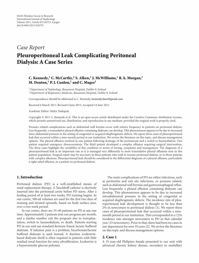
Figure 1: Chest radiograph at presentation.
Table 1: Biochemical results.
Pleural fluid Serum
pH 7.64 7.38
Albumin (g/dL) <15 32
HCO3—(mmol/L) 30.1 28
LDH (iu/L) <50 474
Glucose (mmol/L) 12.1 5.2
Amylase (iu/L) <10 40
cystic kidney disease, which was diagnosed many years earlierin the Philipines. She had no significant heart disease. Oneyear later, she was approaching end-stage kidney disease anda Tenckhoff catheter was inserted. Training for PD began fourweeks later, using standard dextrose-based solutions.
She presented after four days of PD training withdyspnoea. She was otherwise well and was afebrile. Bloodtests and ECG were unchanged. She had clinical evidenceof a large, rightsided pleural effusion. This was confirmedon a chest radiograph (Figure 1). A diagnostic aspiratewas performed and yielded serous fluid. The pleural fluidbiochemistry was consistent with a transudative process(Table 1). The high-pleural-fluid-serum-glucose ratio con-firmed the clinical suspicion of a pleuroperitoneal leak.
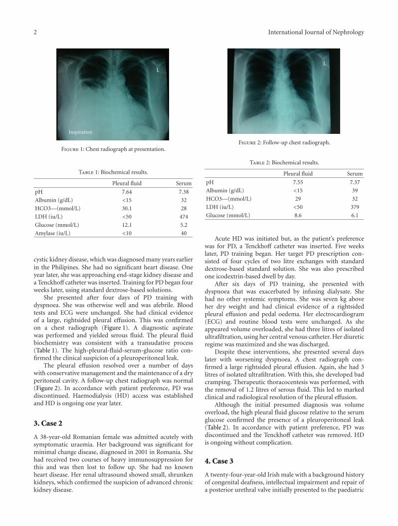
The pleural effusion resolved over a number of dayswith conservative management and the maintenance of a dryperitoneal cavity. A follow-up chest radiograph was normal(Figure 2). In accordance with patient preference, PD wasdiscontinued. Haemodialysis (HD) access was establishedand HD is ongoing one year later.
3. Case 2
A 38-year-old Romanian female was admitted acutely withsymptomatic uraemia. Her background was significant forminimal change disease, diagnosed in 2001 in Romania. Shehad received two courses of heavy immunosuppression forthis and was then lost to follow up. She had no knownheart disease. Her renal ultrasound showed small, shrunkenkidneys, which confirmed the suspicion of advanced chronickidney disease.
L
Figure 2: Follow-up chest radiograph.
Table 2: Biochemical results.
Pleural fluid Serum
pH 7.55 7.37
Albumin (g/dL) <15 39
HCO3—(mmol/L) 29 32
LDH (iu/L) <50 379
Glucose (mmol/L) 8.6 6.1
Acute HD was initiated but, as the patient’s preferencewas for PD, a Tenckhoff catheter was inserted. Five weekslater, PD training began. Her target PD prescription con-sisted of four cycles of two litre exchanges with standarddextrose-based standard solution. She was also prescribedone icodextrin-based dwell by day.
After six days of PD training, she presented withdyspnoea that was exacerbated by infusing dialysate. Shehad no other systemic symptoms. She was seven kg aboveher dry weight and had clinical evidence of a rightsidedpleural effusion and pedal oedema. Her electrocardiogram(ECG) and routine blood tests were unchanged. As sheappeared volume overloaded, she had three litres of isolatedultrafiltration, using her central venous catheter. Her diureticregime was maximized and she was discharged.
Despite these interventions, she presented several dayslater with worsening dyspnoea. A chest radiograph con-firmed a large rightsided pleural effusion. Again, she had 3litres of isolated ultrafiltration. With this, she developed badcramping. Therapeutic thoracocentesis was performed, withthe removal of 1.2 litres of serous fluid. This led to markedclinical and radiological resolution of the pleural effusion.
Although the initial presumed diagnosis was volumeoverload, the high pleural fluid glucose relative to the serumglucose confirmed the presence of a pleuroperitoneal leak(Table 2). In accordance with patient preference, PD wasdiscontinued and the Tenckhoff catheter was removed. HDis ongoing without complication.
4. Case 3
A twenty-four-year-old Irish male with a background historyof congenital deafness, intellectual impairment and repair ofa posterior urethral valve initially presented to the paediatric
International Journal of Nephrology 3
Table 3: Biochemical results.
Pleural fluid Serum
pH 7.23 7.36
Albumin (g/dL) 28 29
HCO3—(mmol/L) 20 26
LDH (iu/L) 2816 614
Glucose (mmol/L) <0.6 4
Amylase (iu/L) <10 32
nephrology services with nephrotic syndrome. A renal biopsyrevealed secondary focal segmental glomerulosclerosis andsignificant tubuleinterstitial fibrosis. Following introductionof Renin-Angiotensin-Aldosterone system blockade, his pro-teinuria was controlled.
Despite this, his chronic kidney disease progressed. Tenyears later, he was approaching end-stage kidney disease. ATenckhoff catheter was inserted without complication. Sixweeks later, PD training was initiated. A bicarbonate/lactatebuffered solution was used to avoid infusion pain, which hewould be unable to verbalise. The following months werecomplicated by dialysis-associated pericarditis that resolvedwith intensive haemodialysis for a number of weeks. Hisbaseline PD prescription consisted of seven cycles of twolitre exchanges with 1.36% dextrose solution. He was alsoprescribed an icodextrin-based daytime dwell.
Five months later, he was admitted with clinical andradiological evidence of a large, rightsided pleural effusionwith pleural thickening. The exact duration of the effusionwas unclear as he was unable to verbalise symptoms. It wasthought to have developed over weeks given the apparentabsence of symptoms and the degree of pleural thickening.A chest radiograph four months earlier showed normal lungfields.
He was afebrile and systemically well. His serum bio-chemistry and haematology were unchanged and his inflam-matory markers were not raised. ECG and echo were nor-mal. A diagnostic pleural aspirate was performed underultrasound guidance. This yielded serous fluid and alsoidentified numerous loculations within the collection.
The pleural fluid biochemistry was atypical for thatseen with pleuroperitoneal leak (Table 3). However, as thepatient was so well clinically, and numerous pleural fluidcultures were sterile, it was felt that the effusion was notrelated to infection. It was also felt that the effusion wasnot due to volume overload given the degree of loculationwithin the effusion and the absence of other clinical signsof volume overload. The low pleural fluid glucose and highlactate dehydrogenase (LDH) were difficult to interpret as thesampled fluid was walled off within a loculation, probably formany weeks. A clinical diagnosis of pleuroperitoneal leak wasmade and PD was discontinued.
The effusion did not resolve with drainage of theperitoneal cavity, and a chest drain was inserted. This drainedminimal serous fluid due to the highly loculated nature ofthe effusion. A second drain was inserted and intrapleuralalteplase administered. This drain became dislodged and wasreplaced by a third drain with further intrapleural alteplase
administration. At this point, however, a chest radiographconfirmed persistence of the large, loculated effusion with asmall pneumothorax and a thick pleural rind.
Video-assisted thorascopic surgery with decorticationwas performed. A rapid postoperative recovery ensued andthe postoperative chest drains were removed without event.A chest radiograph six weeks later showed a well-expandedright lung, without effusion or pleural abnormality. HD isongoing without complication.
5. Discussion
The first description of a pleuroperitoneal leak causing apleural effusion in a PD patient was in 1967 [3]. Sincethen, several reports have estimated the incidence of thiscomplication; the largest report estimated an incidence of1.6% [1].
It is thought that congenital or acquired communicationsbetween the pleura and peritoneum underpin this problem.This had been demonstrated both in scintigraphy [4] and inautopsy specimens with localized absence of diaphragmaticmuscle fibres [5]. The raised intraabdominal pressure withdialysate infusion, in a patient with such a communication,promotes the translocation of dialysate into the pleural space.
An increased incidence of pleuroperitoneal leak is seenin patients with polycystic kidney disease [6]. This isprobably related to the increased intraabdominal pressurein these patients and, therefore, higher pleuroperitonealpressure gradients [6]. There is also a generalized connectivetissue weakness in this condition that may contribute toinherent diaphragmatic weakness [6]. Patients with previousperitonitis are also at higher risk of developing this condition.This is probably due to a weakening of diaphragmatic tissueduring peritonitis [7].
The timing of this complication varies from days toyears after the initiation of PD. Half of cases occur withinone month of starting PD [3]; these probably representpatients with congenital diaphragmatic defects. Those casesthat occur later probably represent those with acquireddiaphragmatic defects. Patients typically present with dys-pnoea, although a significant proportion are asymptomatic[3]. Clinical examination reveals a pleural effusion that isusually rightsided [3]. Presumably the preponderance ofrightsided cases is due to diaphragmatic protection by theheart on the left.
Pleural effusions are broadly divided into transudativeand exudative effusions. Transudative effusions developwhen systemic factors affect the pleural Starling forces, asseen, for example, in congestive cardiac failure. Exudativeeffusions develop when local factors influence the formationof pleural fluid, as seen in malignant effusions. Light’s criteriaare applied to the pleural and serum biochemistry to makethe distinction between a transudate and an exudate [8]. Aneffusion is an exudate if the pleural-fluid-to-serum-proteinratio is >0.5 or the pleural-fluid-to-serum-LDH ratio is>0.6 [8]. Pleuroperitoneal leaks typically cause transudativeeffusions with low LDH and low cell count [5].
The differential diagnosis of a pleural effusion in adialysis patient is extensive. Transudative pleural effusions
4 International Journal of Nephrology
due to volume overload or congestive cardiac failure arerelatively common in this population. In PD patients, sucheffusions would typically be managed by increasing thedialysate volume and tonicity to increase ultrafiltration.However, this can exacerbate the problem if the effusion isdue to a pleuroperitoneal leak.
This highlights the importance of the pleural fluidglucose measurement. In the absence of loculation, a highpleural fluid to serum glucose concentration gradient is avery sensitive and specific test for diagnosing a pleuroperi-toneal leak [9]. The ratio of pleural fluid to serum glucose isdynamic, and varies depending on the type of fluid instilled,the volume and the contact time. However, a positivegradient is highly suggestive of a pleuroperitoneal leak [9].This is due to the presence of hypertonic, usually dextrose-based, dialysate in the pleural space—a “sweet hydrothorax”[10]. Atypical biochemistry can be seen when the fluid hasbeen in the pleural cavity for a prolonged period and hasbeen partially reabsorbed by lymphatics, as seen in Case 3.
Other diagnostic tests are less practical and includeTechnetium-99m labelled peritoneal scintigraphy [4] andcomputed tomography (CT) with intraperitoneal dye [11].
In terms of management, emergency large volume thora-cocentesis is occasionally required [3]. However, most casesof pleuroperitoneal leak are initially managed by drainage ofthe peritoneal cavity [3]. Temporary HD may be required,especially for those with minimal residual renal function.Many patients choose to remain on HD in the long term and,therefore, formal repair of the diaphragm is not required.
For those that wish to return to PD, there are a number ofmanagement options. One group advocates early diaphrag-matic repair and continuation of peritoneal dialysis [12]. Inthis case, thorascopic diaphragmatic repair was performedusing absorbable polyglycolic acid felt, and fibrin glue [12].
Most advocate temporary discontinuation of PD. Thisallows resolution of the effusion and, in some cases, healingof the diaphragmatic defects. A trial of low-volume PD two–six weeks later is successful in a significant proportion ofcases [13].
Recurrent hydrothorax necessitates either a permanentswitch to haemodialysis or definitive management of thepleural-peritoneal communication. Chemical pleurodesisvia an intercostal drain has been used in several caseswith success [13]. Agents used include tetracycline, bloodand fibrin glue. This method had relatively high rates ofsuccess, with 48% of patients resuming long-term PD afterpleurodesis in one large systematic review [13].
Video-assisted thorascopic surgery, with direct visualiza-tion of the diaphragmatic defect and suture repair, has shownmuch promise in recent times for the management of thiscondition [13, 14]. Using this approach, 88% of patientsin one study successfully resumed long-term PD withoutrecurrence [13].
The three cases described above highlight the variabilityof this condition in terms of timing, symptoms, andmanagement. Pleuroperitoneal leak should be considered inthe differential diagnosis of a pleural effusion, particularly arightsided effusion, in a patient on peritoneal dialysis.
References
[1] Y. Nomoto, T. Suga, K. Nakajima et al., “Acute hydrothoraxin continuous ambulatory peritoneal dialysis—a collaborativestudy of 161 centers,” American Journal of Nephrology, vol. 9,no. 5, pp. 363–367, 1989.
[2] A. Green, M. Logan, W. Medawar et al., “The managementof hydrothorax in continuous ambulatory peritoneal dialsyis(CAPD),” Peritoneal Dialysis International, vol. 10, no. 4, pp.271–274, 1990.
[3] S. R. Edwards and A. M. Unger, “Acute hydrothorax—a newcomplication of peritoneal dialysis,” Journal of the AmericanMedical Association, vol. 199, no. 11, pp. 853–855, 1967.
[4] H. Tokmak, A. Mudun, C. Turkmen, Y. Sanli, S. Cantez,and S. Bozfakioglu, “The role of peritoneal scintigraphy inthe detection of continuous ambulatory peritoneal dialysiscomplications,” Renal Failure, vol. 28, no. 8, pp. 709–713,2006.
[5] R. Garcia Ramon and A. Miguel Carrasco, “Hydrothorax inperitoneal dialysis,” Peritoneal Dialysis International, vol. 18,no. 1, pp. 5–10, 1998.
[6] S. Fletcher, J. H. Turney, and A. M. Brownjohn, “Increasedincidence of hydrothorax complicating peritoneal dialysis inpatients with adult polycystic kidney disease,” NephrologyDialysis Transplantation, vol. 9, no. 7, pp. 832–833, 1994.
[7] C. C. Chow, Y. J. Sung, C. K. Cheung, C. Hamilton-Wood, andK. N. Lai, “Massive hydrothorax in continuous ambulatoryperitoneal dialysis: diagnosis, management and review of theliterature,” New Zealand Medical Journal, vol. 101, no. 850, pp.475–477, 1988.
[8] R. W. Light, M. I. Macgregor, P. C. Luchsinger, and W. C. Ball,“Pleural effusions: the diagnostic separation of transudatesand exudates,” Annals of Internal Medicine, vol. 77, no. 4, pp.507–513, 1972.
[9] K. M. Chow, C. C. Szeto, T. Y. H. Wong, and P. K. T.Li, “Hydrothorax complicating peritoneal dialysis: diagnosticvalue of glucose concentration in pleural fluid aspirate,”Peritoneal Dialysis International, vol. 22, no. 4, pp. 525–528,2002.
[10] P. Mangana, D. Arvanitis, D. Vlassopoulos, S. Tang, and K.N. Lai, “Acute hydrothorax in peritoneal dialysis patients:diagnosis and treatment options,” Nephrology Dialysis Trans-plantation, vol. 18, no. 11, pp. 2451–2452, 2003.
[11] T. W. Kang and C. K. Kim, “Pleuroperitoneal communicationof peritoneal dialysis demonstrated by multidetector-row CTperitoneography,” Abdominal Imaging, vol. 34, no. 6, pp. 780–782, 2009.
[12] H. Kumagai, M. Watari, and M. Kuratsune, “Simple surgi-cal treatment for pleuroperitoneal communication withoutinterruption of continuous ambulatory peritoneal dialysis,”General Thoracic and Cardiovascular Surgery, vol. 55, no. 12,pp. 508–511, 2007.
[13] K. M. Chow, C. C. Szeto, and P. K. T. Li, “Management optionsfor hydrothorax complicating peritoneal dialysis,” Seminars inDialysis, vol. 16, no. 5, pp. 389–394, 2003.
[14] S. Tang, W. H. Chui, A. W. C. Tang et al., “Video-assistedthoracoscopic talc pleurodesis is effective for maintenanceof peritoneal dialysis in acute hydrothorax complicatingperitoneal dialysis,” Nephrology Dialysis Transplantation, vol.18, no. 4, pp. 804–808, 2003.

Submit your manuscripts athttp://www.hindawi.com
Stem CellsInternational
Hindawi Publishing Corporationhttp://www.hindawi.com Volume 2014
Hindawi Publishing Corporationhttp://www.hindawi.com Volume 2014
MEDIATORSINFLAMMATION
of
Hindawi Publishing Corporationhttp://www.hindawi.com Volume 2014
Behavioural Neurology
EndocrinologyInternational Journal of
Hindawi Publishing Corporationhttp://www.hindawi.com Volume 2014
Hindawi Publishing Corporationhttp://www.hindawi.com Volume 2014
Disease Markers
Hindawi Publishing Corporationhttp://www.hindawi.com Volume 2014
BioMed Research International
OncologyJournal of
Hindawi Publishing Corporationhttp://www.hindawi.com Volume 2014
Hindawi Publishing Corporationhttp://www.hindawi.com Volume 2014
Oxidative Medicine and Cellular Longevity
Hindawi Publishing Corporationhttp://www.hindawi.com Volume 2014
PPAR Research
The Scientific World JournalHindawi Publishing Corporation http://www.hindawi.com Volume 2014
Immunology ResearchHindawi Publishing Corporationhttp://www.hindawi.com Volume 2014
Journal of
ObesityJournal of
Hindawi Publishing Corporationhttp://www.hindawi.com Volume 2014
Hindawi Publishing Corporationhttp://www.hindawi.com Volume 2014
Computational and Mathematical Methods in Medicine
OphthalmologyJournal of
Hindawi Publishing Corporationhttp://www.hindawi.com Volume 2014
Diabetes ResearchJournal of
Hindawi Publishing Corporationhttp://www.hindawi.com Volume 2014
Hindawi Publishing Corporationhttp://www.hindawi.com Volume 2014
Research and TreatmentAIDS
Hindawi Publishing Corporationhttp://www.hindawi.com Volume 2014
Gastroenterology Research and Practice
Hindawi Publishing Corporationhttp://www.hindawi.com Volume 2014
Parkinson’s Disease
Evidence-Based Complementary and Alternative Medicine
Volume 2014Hindawi Publishing Corporationhttp://www.hindawi.com