pleural pressure immediately after pulmonary lobectomy: single versus double chest tubes for suction
TRANSCRIPT
Brief Clinical Reports
DISCUSSIONMycotic aneurysms of the coronary arteries are exceed-
ingly uncommon, accounting for less than 3% of all coro-
nary aneurysms.2 Most are associated with infective
endocarditis, although coronary stents are increasingly im-
plicated in the formation of mycotic coronary aneurysms.2,3
We present a unique case of a mycotic aneurysm of the left
main coronary artery in a splenectomized patient after a pneu-
mococcal pneumonia complicated by purulent pericarditis.
The rapid growth of the pseudoaneurysm within 3 months
coupled with the septicemia and purulent pericarditis support
the mycotic origin of the pseudoaneurysm. Pre-existing coro-
nary calcifications may have led to an initial endarteritis fol-
lowed by a contained rupture and purulent pericarditis.
Although the presentation was ischemic in nature owing to
coronary artery stenosis, it was likely that the pseudoaneur-
ysm caused compression on the coronary arteries as sug-
gested by the positional feature of the patient’s symptoms.
Preoperative assessment is essential to define the relation-
ship of the pseudoaneurysm to the surrounding structures.
High-resolution multislice CT allows excellent visualization
of the proximal coronary arteries and relationships with
surrounding structures. Although covered stents have been
From the Division of Thoracic Surgery,a Ospedali Riuniti, Ancona, Italy, and the
Division of General Thoracic Surgery,b Mayo Clinic, Rochester, Minn.
Disclosures: Dr Brunelli is a member of the International Advisory Board of Medela
AG, Baar, Switzerland.
Dr Fibla is the recipient of the AATS Evarts Graham Traveling Fellowship 2009-2010.
Received for publication Feb 1, 2010; accepted for publication April 10, 2010;
available ahead of print May 14, 2010.
Address for reprints: Alessandro Brunelli, Division of Thoracic Surgery, Ospedali
Riuniti Ancona, Via Conca 71, 60020, Ancona, Italy (E-mail: brunellialex@
gmail.com).
J Thorac Cardiovasc Surg 2010;140:e52-3
0022-5223/$36.00
Copyright � 2010 by The American Association for Thoracic Surgery
doi:10.1016/j.jtcvs.2010.04.013
e52 The Journal of Thoracic and Cardiovascular Surg
reported as a treatment for mycotic coronary pseudoaneurysm,
open surgical management remains the gold standard for
patients without prohibitive risk.4,5 Access to a pseudoaneur-
ysm of the left main coronary artery requires transection of the
main pulmonary artery in most circumstances, especially in
the presence of dense adhesions, as in the present case. Un-
roofing of the aneurysm wall with proximal ligation of the cor-
onary artery is mandatory. Although technically challenging,
in situ revascularization of the proximal circumflex artery is
feasible, as performed in our case. The proximal LAD is often
difficult to revascularize and is probably best revascularized
with the left internal thoracic artery. Appropriate antibiotic
treatment is essential to minimize recurrence.
References1. Nichols L, Lagana S, Parwani A. Coronary artery aneurysm: a review and hypoth-
esis regarding etiology. Arch Pathol Lab Med. 2008;132:823-8.
2. Garg N, Garg R, Gordon C, Singh R, Singh A. Acute coronary syndrome caused by
coronary artery mycotic aneurysm due to late stent infection localized with radio-
labeled autologous leukocyte imaging. Clin Nucl Med. 2009;34:753-5.
3. Schoenkerman AB, Lundstrom RJ. Coronary stent infections: a case series. Cath-
eter Cardiovasc Interv. 2009;73:74-6.
4. Christiansen S, Klocke A, Hoffman A, Autschbach R. Surgical management of
a left main stem coronary artery aneurysm. J Card Surg. 2008;23:69-70.
5. Shariff N, Combs W, Roberts J. Large mycotic pseudoaneurysm of the left circum-
flex treated with antibiotics and covered stent. J Invasive Cardiol. 2009;21:E37-8.
Pleural pressure immediately after pulmonary lobectomy: Singleversus double chest tubes for suction
Alessandro Brunelli, MD,a Stephen D. Cassivi, MD,b Juan Fibla, MD,b and Luca Di Nunzio, MD,a
Ancona, Italy, and Rochester, Minn
Recent studies have shown that a single chest tube after
pulmonary lobectomy is at least as effective as 2 chest tubes
in evacuating air and fluid from the pleural space.1-3 In addi-
tion, a single tube may produce less pain and may be a more
suitable option for patients being treated with fast-tracking
approaches. Most surgeons still favor the use of �20 cm
H2O suction in the first postoperative hours, with the theo-
retic aim of promoting lung expansion. This level of suction
has persisted in most practices since being adapted from
early thoracic drainage devices designed by Emerson in
the 1940s. The benefits of chest tube suction, however, re-
main a topic of debate. This study investigated whether
the use of 1 or 2 chest tubes after pulmonary lobectomy
might influence the level of intrapleural pressure as mea-
sured in the first postoperative hours.
MATERIALS AND METHODSThe study was approved by the local hospital institutional review boards,
and patients gave informed consent for collection and use of their data in the
database for research purposes. This post hoc analysis of a prospective study
to assess the efficacy of a novel chest drain system included 145 patients un-
dergoing pulmonary lobectomy through a lateral muscle-sparing thoracot-
omy in 2 separate thoracic surgery centers (2007–2008). After completion
of the lobectomy, 2 chest tubes (1 at the apex and the other placed at the
base) or 1 chest tube (placed in a lateral position up to the apex, all tubes
ery c September 2010
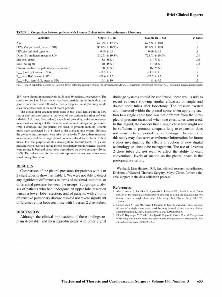
TABLE 1. Comparison between patients with 1 versus 2 chest tubes after pulmonary lobectomy
Variables Single (n ¼ 89) Double (n ¼ 56) P value
Age 67.3% � 12.5% 67.5% � 10.8 .9
FEV1 (% predicted, mean � SD) 83.8% � 18.7% 83.6% � 19.8 .9
FEV1/forced vital capacity 0.68 � 0.1 0.69 � 0.1 .8
DLCO (% predicted, mean � SD) 80.2% � 19.3% 72.8% � 19.8% .08
Site (no. upper) 52 (58%) 41 (73%) .06
Side (no. right) 40 (45%) 37 (66%) .02
Chronic obstructive pulmonary disease (no.) 28 (31%) 16 (29%) .8
Pmax (cm H2O, mean � SD) �11.5 � 8 �11.3 � 7 .8
Pmin (cm H2O, mean � SD) �21.6 � 7.2 �22.2 � 8.2 .7
Pmax�Pmin (cm H2O, mean � SD) 10.1 � 10 11 � 9.5 .3
FEV1, Forced expiratory volume in 1 second; DLCO, diffusing capacity of lung for carbon monoxide; Pmax, maximum intrapleural pressure; Pmin, minimum intrapleural pressure.
Brief Clinical Reports
28F) were placed intraoperatively in 56 and 89 patients, respectively. The
choice to use 1 or 2 chest tubes was based mainly on the individual sur-
geon’s preference and reflected in part a temporal trend (favoring single
chest tube placement in the most recent period).
The digital chest drainage units used in this study had a built-in flow
sensor and pressure sensor at the level of the canister featuring software
(Medela AG, Baar, Switzerland) capable of providing real-time measure-
ments and recordings of the maximal and minimal intrapleural pressures.
Only 1 drainage unit per patient was used, to promote mobility. Double
tubes were connected by a Y piece to the drainage unit system. Because
the pressure measurements were taken distal to the Y piece, these measure-
ments represented the average pleural pressure value detected by the 2 chest
tubes. For the purpose of this investigation, measurements of pleural
pressures were recorded during the 6th postoperative hour, when all patients
were resting in bed and chest tubes were placed on active suction (�20 cm
H2O). The values used for the analysis represent the average values mea-
sured during this period.
RESULTSComparison of the pleural pressures for patients with 1 or
2 chest tubes is shown in Table 1. We were not able to detect
any significant differences in terms of maximal, minimal, or
differential pressure between the groups. Subgroups analy-
ses of patients who had undergone an upper lobe resection
versus a lower lobe resection, and of patients with chronic
obstructive pulmonary disease also did not reveal significant
differences either between those with 1 versus 2 chest tubes.
DISCUSSIONAlthough the clinical implications of these findings re-
main debatable, and their reproducibility with other digital
The Journal of Thoracic and Ca
drainage systems should be confirmed, these results add to
recent evidence showing similar efficacies of single and
double chest tubes after lobectomy. The pressure exerted
and measured within the pleural space when applying suc-
tion to a single chest tube was not different from the intra-
pleural pressure measured when two chest tubes were used.
In this regard, the concern that a single chest tube might not
be sufficient to promote adequate lung re-expansion does
not seem to be supported by our findings. The results of
this study may also serve as reference information for future
studies investigating the effects of suction or new digital
technology on chest tube management. The use of 1 versus
2 chest tubes did not seem to affect the ability to exert
conventional levels of suction on the pleural space in the
postoperative setting.
We thank Lisa Halgren, RN, lead clinical research coordinator,
Division of General Thoracic Surgery, Mayo Clinic, for her valu-
able support in the data collection process.
References1. Alex J, Ansari J, Bahalkar P, Agarwala S, Rehman MU, Saleh A, et al. Com-
parison of the immediate postoperative outcome of using the conventional two
drains versus a single drain after lobectomy. Ann Thorac Surg. 2003;76:
1046-9.
2. Gomez-Caro A, Roca MJ, Torres J, Cascales P, Terol E, Castaner J, et al. Success-
ful use of a single chest drain postlobectomy instead of two classical drains:
a randomized study. Eur J Cardiothorac Surg. 2006;29:562-6.
3. Okur E, Baysungur V, Tezel C, Sevilgen G, Ergene G, Gokce M, et al. Comparison
of the single or double chest tube applications after pulmonary lobectomies. Eur
J Cardiothorac Surg. 2009;35:32-6.
rdiovascular Surgery c Volume 140, Number 3 e53