platelet-derived growth factor receptor- positive ... · tion with hoechst 33258 (invitrogen,...
TRANSCRIPT

Chronic critical limb ischemia (CLI) is defined as the end stage of lower limb ischemia due to peripheral arte-
rial disease (PAD). The prognosis of CLI patients is poor with an 1-year major amputation rate of 30%.1,2) Moreover, although revascularization of the ischemic limb with sur-gical bypass or endovascular approaches may be helpful for limb salvage, revascularization procedures may not be possible in the severe stage of CLI. Therefore, we suggest CLI be one of the main targets for cell therapy as stem cells and progenitor cells are believed to have an important function in tissue regeneration after damage.
Platelet-Derived Growth Factor Receptor-Positive Pericytic Cells of White Adipose Tissue
from Critical Limb Ischemia Patients Display Mesenchymal Stem Cell-Like Properties
Eo Jin Kim, MD, Sang Gyo Seo, MD*, Hyuk Soo Shin, MD†, Doo Jae Lee, MD†, Ji Hye Kim, BS†, Dong Yeon Lee, MD†
Department of Orthopedic Surgery, Hanil General Hospital, Seoul, *Department of Orthopedic Surgery, Asan Medical Center, Seoul, †Department of Orthopedic Surgery, Seoul National University Hospital, Seoul, Korea
Background: The pericytes in the blood vessel wall have recently been identified to be important in regulating vascular forma-tion, stabilization, remodeling, and function. We isolated and identified pericyte-like platelet-derived growth factor receptor beta-positive (PDGFRb+) cells from the stromal vascular fraction (SVF) of adipose tissue from critical limb ischemia (CLI) patients and investigated their potential as a reliable source of stem cells for cell-based therapy. Methods: De-identified subcutaneous fat tissues were harvested after amputation in CLI patients. Freshly isolated SVF cells and culture-expanded adipose-derived stem cells (ADSCs) were quantified using flow cytometry. A matrigel tube formation assay and multi-lineage differentiation were performed to assess pericytic and mesenchymal stem cell (MSC)-like characteristics of PDGFRb+ ADSCs. Results: PDGFRb+ cells were located in the pericytic area of various sizes of blood vessels and coexpressed mesenchymal stem cell markers. PDGFRb+ cells in freshly isolated SVF cells expressed a higher level of stem cell markers (CD34 and CXCR4) and mes-enchymal markers (CD13, CD44, CD54, and CD90) than PDGFRb– cells. In vitro expansion of PDGFRb+ cells resulted in enrichment of the perivascular mesenchymal stem-like (PDGFRb+/CD90+/CD45–/CD31–) cell fractions. The Matrigel tube formation assay revealed that PDGFRb+ cells were located in the peritubular area.Conclusions: PDGFRb+ ADSCs cells demonstrated a good multilineage differentiation potential. Pericyte-like PDGFRb+ cells from the SVF of adipose tissue from CLI patients had MSC-like characteristics and could be amplified by in vitro culture with preserva-tion of their cell characteristics. We believe PDGFRb+ cells in the SVF of adipose tissue can be used as a reliable source of stem cells even in CLI patients.Keywords: Platelet-derived growth factor, Adipose tissue, Adult stem cells, Ischemia
Original Article Clinics in Orthopedic Surgery 2017;9:239-248 • https://doi.org/10.4055/cios.2017.9.2.239
Copyright © 2017 by The Korean Orthopaedic AssociationThis is an Open Access article distributed under the terms of the Creative Commons Attribution Non-Commercial License (http://creativecommons.org/licenses/by-nc/4.0)
which permits unrestricted non-commercial use, distribution, and reproduction in any medium, provided the original work is properly cited.Clinics in Orthopedic Surgery • pISSN 2005-291X eISSN 2005-4408
Received January 20, 2017; Accepted February 24, 2017Correspondence to: Dong Yeon Lee, MD Department of Orthopedic Surgery, Seoul National University Hospital, 101 Daehak-ro, Jongno-gu, Seoul 03080, KoreaTel: +82-2-2072-1863, Fax: +82-2-764-2718E-mail: [email protected]

240
Kim et al. Platelet-Derived Growth Factor Receptor Beta-Positive Pericytic Cells for Critical Limb Ischemia Clinics in Orthopedic Surgery • Vol. 9, No. 2, 2017 • www.ecios.org
On the basis of numerous successful animal experi-ments, cell therapies for PAD using bone marrow-derived mononuclear cells (BM-MNCs),3-7) peripheral blood-derived mononuclear cells (PB-MNCs),8,9) and adipose-derived stem cells (ADSCs)1,10) have been reported to be promising, though more evidence needs to be accumulated before being used as a standard modality of treatment.
Among the various sources of stem or progeni-tor cells, adipose tissue represents an attractive source of autologous adult stem cells used in regenerative therapy owing to its abundance, surgical accessibility, and high content of multipotent mesenchymal stem cell (MSC)-like cells.11) It has been reported that the adipose tissue yields far more colony-forming units than the bone marrow when the weight of the tissues used is the same.12) How-ever, knowledge of the cellular origin of culture-expanded mesenchymal-like ADSCs remains elusive because of the complex organization of stromal cells surrounding the small blood vessels.13)
Recent studies have suggested that the perivascular niche may be an important reservoir of MSCs in multiple developed organs.14,15) While it is evident that not all peri-vascular cells are MSCs, the majority of MSCs are believed to reside in the perivascular area.16) Although there are no specific cell surface markers for perivascular cells or peri-cytes that regulate vessel stability and vascular survival, platelet-derived growth factor receptor beta (PDGFRβ) is thought to be a major cell surface marker defining the pericytes.17,18)
We hypothesized that PDGFRβ-positive (PDGFRβ+) perivascular cells of the white adipose tissue from CLI pa-tients would have characteristics of the MSCs and would be a good source for autologous cell transplantation as a treatment for CLI.
The purposes of our study were (1) to identify PDGFRβ+ perivascular cells in the white adipose tis-sue from CLI patients; (2) to isolate and characterize PDGFRβ+ cells from the stromal vascular fraction (SVF) of patient’s adipose tissue in vitro; and (3) to investigate the differentiation potential of PDGFRβ+ cells as a reliable source of MSCs that can be used for the treatment of CLI.
METHODS
Acquisition of the Fresh Subcutaneous Human Adipose Tissues and Preparation of the SVF This study was approved by Seoul National University Hospital Institutional Review Board (No. 1103-035-354) and informed consent was obtained from all patients. Among the 28 patients who had undergone lower limb
amputation surgery (below knee amputation or above knee amputation) due to CLI from March 2010 to Septem-ber 2011, 10 specimens from 10 patients were included in this study. The other 18 patients declined to participate and were excluded. Subcutaneous adipose tissue (1–3 g) was obtained aseptically from each amputated specimen and transported to our laboratory in aseptic bottles with phosphate buffered saline (PBS). Medical records were not profiled in accordance with IRB instructions as they could be used for the identification of tissue donors. Samples from three patients were allocated to immunofluorescence staining of fresh fat tissue and fluorescence-activated cell sorter (FACS) analysis of fresh SVF and remaining 7 sam-ples were used after expansion to obtain enough amount of cells for extensive FACS analysis (triple samples for each analysis).
The SVF of adipose tissue was isolated as described in previous studies.19-21) Fresh fat tissues were washed at least three times with PBS to remove blood, and then di-gested at 37°C for 1 hour with 0.075% collagenase type 1 (Sigma-Aldrich, St. Louis, MO, USA) in Dulbecco’s Modi-fied Eagle Medium (DMEM; Gibco, New York, NY, USA). Mature adipocyte fractions were separated from stromal fractions by centrifuging at 1,200 × g for 10 minutes. The remained fractions were treated with red blood cell lysis buffer for 10 minutes at room temperature (RT) and then filtered through 100-μm nylon mesh to exclude remaining erythrocyte debris, and then centrifuged at 1,200 × g for 10 minutes.
Immunofluorescence of the Fresh Fat Tissue Pieces of harvested adipose tissues were washed in PBS, 10% formalin (Sigma-Aldrich), and held for at least 24 hours at 4°C, before being embedded in paraffin. Sec-tions (6 to 8 μm) were cut on a rotary microtome (Leica RM2145, Leica Microsystems, Nussloch, Germany) fixed for 1 hour at 56°C, and then stored at RT. Before staining, sections were deparaffinized in xylenes. Tissue rehydration and all subsequent washes were performed by 25-minute incubations in a Zytomed wash buffer (Zytomed systems GmbH, Berlin, Germany). All incubations were completed at ambient temperature. For fluorescent immuno-staining, rehydrated tissue sections were pretreated with protein blocking in serum-free protein blocks (Dako, Glostrup, Denmark) and incubated with antibodies for 2 hours. Nuclear staining was attained through 10-minute incuba-tion with Hoechst 33258 (Invitrogen, Carlsbad, CA, USA). Slides were mounted in Histomount (National Diagnos-tics, Atlanta, GA, USA), and observed under a fluores-cence microscopy (BX61; Olympus, Tokyo, Japan) and a

241
Kim et al. Platelet-Derived Growth Factor Receptor Beta-Positive Pericytic Cells for Critical Limb Ischemia Clinics in Orthopedic Surgery • Vol. 9, No. 2, 2017 • www.ecios.org
digital imaging system (DCF 500; Leica Microsystems).Antibodies used in these studies were anti-CD140b
(PDGFRβ, 1:50; BD Biosciences, San Jose, CA, USA), anti-CD146 (1:50; R&D Systems, Minneapolis, MN, USA), anti-CD90 (1:100; BD Biosciences), and anti-CD31 (1:100; BD Biosciences). All antibodies were diluted in an an-tibody diluent with background reducing components (Dako).
Analysis of Cell Surface Antigen Profile of the Fresh SVF Cells and Culture Expansion of Fluorescence-Activated Cell Sorted PDGFR-Positive CellsCell surface antigen profiles of freshly isolated SVF cells were quantified by flow cytometry with a FACS.13,22,23) Fat tissue was thoroughly minced with scissors and digested for 30 minutes in DMEM and 0.075% collagenase type I (Sigma Aldrich) on a rotator at 37°C. Mature adipocytes were eliminated by centrifugation (1,200 × g, ambient tem-perature, 10 minutes) and cell pellets were resuspended in blood cell lysis buffer, incubated for 10 minutes at RT and washed in PBS.
Freshly isolated cells from the SVF were maintained on ice, and stained for analytical flow cytometry and cell sorting experiments as previously described.24) Cell sus-pensions were centrifuged (1,200 × g, 10 minutes) and the cells were simultaneously stained with monoclonal mouse anti-human fluorochrome-conjugated antibodies: CD31-fluorescein isothiocyanate (FITC), CD34-FITC, CD90-FITC, CD140b-phycoerythrin (PE), CD146-PE, CD54-allophycocyanin (APC) (BD Biosciences), CD146-FITC, neuron-glial antigen 2 (NG2)-FITC, α-smooth muscle actin (αSMA)-PE, vascular endothelial growth factor re-ceptor 2 (VEGFR2)-PE, C-X-C chemokine receptor type 4 (CXCR4)-PE, CD140b-APC (R&D systems), CD105-FITC, CD44-FITC (AbD Serotec, Kidlington, Oxford, UK), CD14-PE, CD45-PE (Dako), CD13-FITC (Novus Biological, Littleton, CO, USA), CD133-APC (Miltenyi Biotec, Auburn, CA, USA), and unconjugated antibod-ies (Desmin; Abcam, Cambridge, MA, USA). Analytical samples were fixed with 1% paraformaldehyde (PFA) for 10 minutes at RT. Four-color, 6-parameter data files were acquired on a 2-laser FACSCalibur flow cytometer (BD Biosciences) at a maximum of 10,000 events per second.
Cell sorting was performed using a 3-laser FAC-SAria II cell sorter (BD Biosciences).
Unfixed CD140b-PE stained samples were suspend-ed in PBS, and 0.5% bovine serum albumin (BSA). Sam-ples were continuously cooled to 4°C and single cell sort-ing was performed at 70,000 events per second. Samples were collected into BD Falcon round-bottom polypropyl-
ene tubes (BD Biosciences) containing 1 mL of DMEM/Nutrient Mixture F-12 (DMEM/F12; Gibco) and plated in tissue culture dish (TPP; Techno Plastic Products, Trasa-dingen, Switzerland) at a density of 10,000 to 20,000 cell/cm2. Adherent sorted and unsorted cells were expanded in equal volumes of DMEM/F12 and supplemented with 10% FBS, and 100 U/mL penicillin (Sigma-Aldrich).
Matrigel Tube Formation AssayWe evaluated the pericytic function of PDGFRβ+ cells that were collected from cultured ADSCs using FACS. We seeded human umbilical vein endothelial cells (HUVECs) and PDGFRβ+ ADSCs on the surface of a 100% Matrigel (BD Biosciences) in Endothelial Cell Growth Medium 2 (EGM2; Sigma-Aldrich) as previously reported.25) Briefly, Matrigel was added to a 24-well plate followed by incuba-tion for 30 minutes at 37°C. PDGFRβ+ cells (passage 2) and HUVEC were labeled with anti-von Willebrand factor (vWF; Abcam) and anti-CD140b (PDGFRβ, BD Biosci-ences). They were then detached from the culture plate by trypsin and seeded onto the Matrigel in DMEM/F12 culture medium at a ratio of 1:1. In total, 1 × 10⁵ cells were seeded in each well of 24-well plates. Capillary tube forma-tion was investigated after five hours by fluorescence micros-copy (IX51; Olympus), inverted microscopy (IX50; Olympus) and confocal laser scanning microscope (TCS SP8; Leica Microsystems) microscopy.
Multilineage Differentiation AssayFor osteogenesis, cells at 70% confluence were cultivated in DMEM, 10% FBS, 0.1 mM dexamethasone, 50 mg/mL L-ascorbic acid, and 10 mM b-glycerophosphate. After 14 days, cells were fixed in citrate fixative (citrate solution-acetone-37% formaldehyde) for 1 minute. For detection of alkaline phosphatase (ALP) activity, the fixed cells were incubated for 15 minutes in a mixture of Naphthol AS-BI alkaline solution (Sigma-Aldrich). Cells were then rinsed with deionized water and mounted in GVA aqueous mounting solution (Genemed, San Francisco, CA, USA).
For chondrogenesis, pellets were prepared by spin-ning down 3 × 10⁵ cultured cells and grown in serum-free DMEM containing an insulin-transferrin selenious acid mix (Sigma-Aldrich), 50 mg/mL L-ascorbic acid 2-phos-phate (Sigma-Aldrich), 100 mg/mL sodium pyruvate, 40 mg/mL L-proline (Sigma-Aldrich), 0.1 mM dexametha-sone (Sigma-Aldrich), and 10 ng/mL transforming growth factor beta 1 (TGF-β1; R&D Systems). After 21 days, pel-lets were fixed in 10% formalin, dehydrated in ethanol, and embedded in paraffin. For the Safranin O staining, specimen sections (thickness, 6 μm) were deparaffinized

242
Kim et al. Platelet-Derived Growth Factor Receptor Beta-Positive Pericytic Cells for Critical Limb Ischemia Clinics in Orthopedic Surgery • Vol. 9, No. 2, 2017 • www.ecios.org
with xylene and ethanol and Safranin O solution (Sigma-Aldrich) was applied for 5–10 minutes.
For adipogenic differentiation, cultured cells at 70% confluence were switched to DMEM, 10% FBS, 1 mM dexamethasone, 0.5 mM isobutylmethylxanthine, 60 mM indomethacin, and 170 mM insulin (Sigma-Aldrich). Af-ter 21 days, cells were fixed in 5% PFA at RT, washed in PBS, and incubated with oil red O for 10 minutes at RT for
the detection of lipids.
Statistical AnalysisAll data are expressed as means ± standard deviations. Mann-Whitney U-test was used to compare branch points and tube numbers in Matrigel assay. IBM SPSS ver. 22.0 (IBM Co., Armonk, NY, USA) was used for the statistical analysis, and probability values of less than 0.05 were con-
A
B
C D
Fig. 1. Immunofluorescence of endothelial and perivascular markers in fresh white adipose tissue of human subjects. (A) H&E stain shows abundant vessels in the fat tissue including a small artery (large square), an arteriole (medium square), and a capillary (small square). The small artery (B), arteriole (C), and capillary (D) stained with indicated markers showed that platelet-derived growth factor receptor beta (PDGFRb; CD140b)-positive (+) cells were located in the pericytic area while CD31 staining was exclusively visualized at the luminal surface for the small blood vessel. Costaining with a mesenchymal marker (CD90) showed that pericytic PDGFRb+ cells also expressed the mesenchymal marker in small arterioles (C and D), while CD90-positive cells and PDGFRb+ cells were mutually exclusive in the larger artery (B).

243
Kim et al. Platelet-Derived Growth Factor Receptor Beta-Positive Pericytic Cells for Critical Limb Ischemia Clinics in Orthopedic Surgery • Vol. 9, No. 2, 2017 • www.ecios.org
sidered statistically significant.
RESULTS
PDGFRβ+ Cells Located in the Perivascular Area Coexpressed MSC-Like Markers in the Small Arterioles of Fresh Human White Adipose Tissue White adipose tissue from the subcutaneous fat of the human lower leg is a richly vascularized tissue (Fig. 1A). Immunofluorescence staining using endothelial (CD31)
and pericyte markers (PDGFRβ and CD146) showed the exclusively perivascular location of PDGFRβ+ cells in var-ious sizes of the blood vessels (small artery, arteriole, and capillary) (Fig. 1B–D). Co-staining with a mesenchymal marker (CD90) showed that perivascular PDGFRβ+ cells also expressed mesenchymal markers in small arterioles (Fig. 1C and D), while CD90-positive cells and PDGFRβ+ cells were mutually exclusive in larger arteries (Fig. 1B).
104
CX
CR
4
104
CD31
Q117.9%
Q20.785%
Q470.3%
Q311.0%
103
102
101
100
103
102
101
100
104
VE
GF
R2
104
CD34
Q10.031%
Q20.382%
Q453.8%
Q345.8%
103
102
101
100
103
102
101
100
104
CD
45
104
CD44
Q10.545%
Q220.2%
Q427.4%
Q351.9%
103
102
101
100
103
102
101
100
104
CD
13
104
CD54
Q51.54%
Q695.6%
Q80.845%
Q72.04%
103
102
101
100
103
102
101
100
PDGFR +�
104
CX
CR
4
104
CD31
Q12.11%
Q20.041%
Q494.6%
103
102
101
100
103
102
101
100
104
VE
GF
R2
104
CD34
Q10.019%
Q20.00%
Q488.3%
Q311.7%
Q3%3.28
103
102
101
100
103
102
101
100
104
CD
45
104
CD44
Q15.37%
Q244.4%
Q438.4%
Q311.8%
103
102
101
100
103
102
101
100
104
CD
13
104
CD54
Q512.1%
Q658.7%
Q824.0%
Q75.19%
103
102
101
100
103
102
101
100
104
CD
144
104
CD90
Q14.85%
Q29.90%
Q419.8%
Q365.4%
103
102
101
100
103
102
101
100
104
CD
14
104
CD105
Q15.92%
Q20.570%
Q484.6%
Q38.86%
103
102
101
100
103
102
101
100
104
Desm
in
104
CD133
Q50.220%
Q60.022%
Q899.3%
Q70.506%
103
102
101
100
103
102
101
100
104
�SM
A
104
NG2
Q154.7%
Q20.982%
Q444.3%
Q30.039%
103
102
101
100
103
102
101
100
104
CD
144
104
CD90
Q12.16%
Q20.465%
Q479.3%
Q318.1%
103
102
101
100
103
102
101
100
104
CD
14
104
CD105
Q10.101%
Q20.00%
Q499.4%
Q30.537%
103
102
101
100
103
102
101
100
104
Desm
in
104
CD133
Q51.23%
Q60.00%
Q898.6%
Q70.184%
103
102
101
100
103
102
101
100
104
�SM
A
104
NG2
Q113.0%
Q25.39%
Q481.6%
Q30.00%
103
102
101
100
103
102
101
100
PDGFR�
PDGFR +�
PDGFR�
Fig. 2. Fluorescence-activated cell sorter analysis of fresh stromal vascular fraction. Platelet-derived growth factor receptor beta-positive (PDGFRb+) cells expressed a higher level of stem cell markers (CD34 and CXCR4) and mesenchymal markers (CD13, CD44, CD54, and CD90) than PDGFRb– cells. Cell populations with a positive pericyte marker (α-smooth muscle actin [αSMA]) or endothelial markers (CD31 and CD144) were more abundant in PDGFRb+ cells. VEGFR: vascular endothelial growth factor receptor, NG: neuron-glial antigen.

244
Kim et al. Platelet-Derived Growth Factor Receptor Beta-Positive Pericytic Cells for Critical Limb Ischemia Clinics in Orthopedic Surgery • Vol. 9, No. 2, 2017 • www.ecios.org
Characterization of the PDGFRβ+ Cells from Fresh SVF of Human Adipose Tissue Multilabel flow cytometric analysis revealed that PDGFRβ+ cells from fresh SVF of human adipose tissue expressed a higher level of a pericyte marker (αSMA), stem cell markers (CD34 and CXCR4), and mesenchymal markers (CD13, CD44, CD54, and CD90) than PDGFRβ– cells. PDGFRβ+ cells also expressed a slightly higher level of endothelial markers (CD31 and CD144) than PDGFRβ– cells (Fig. 2).24)
The CD45-positive cells are thought to include a mixture of leukocytes contained within the vasculature as well as cells resident in the adipose tissue.24) Staining of freshly isolated cells for PDGFRβ and other markers showed that there was a substantial number of perivascu-lar mesenchymal stem-like (PDGFRβ+/CD90+/CD44+/CD45–) cell fractions in fresh SVF of human adipose tissue.
Culturing Isolated Cells from SVF on an Uncoated Tissue Culture Plastic Resulted in Providing Enrichment to the Perivascular Mesenchymal Stem-Like (PDGFRβ+/CD90+/CD45–) Cell FractionsPlating of isolated cells from SVF on an uncoated tis-sue culture plastic resulted in selective adherence of cell
population. Multilabel flow cytometric analysis of culture-expanded ADSCs revealed that FACS analysis from fresh SVF of human adipose tissue showed that a population of endothelial (CD31 and CD144) and leukocytic (CD45) marker-positive cells was greatly reduced (Fig. 3). Cultured PDGFRβ+ cells maintained the properties of PDGFRβ+ cells from fresh SVF except that they had a lower level of stem cell marker (CD34 and CXCR4)-positive cells. Cultured PDGFRβ+ cells expressed high levels of mesen-chymal markers (CD13, CD44, CD54, and CD90), and a pericyte marker (αSMA). However, they expressed neither the endothelial markers (CD31 and CD144) nor the leu-kocytic marker (CD45).24)
PDGFRβ+ Cells Were Located in the Perivascular Niche during Endothelial Tube FormationWhen PDGFRβ+ ADSCs were cocultured with HUVEC, they demonstrated the formation of a tubular network (Fig. 4A). Tube number was higher (124.5 ± 13.2 per plate, p < 0.05) than that formed by HUVEC (97.5 ± 5.5) or ADSC (62.8 ± 5.6) only. At a higher magnification, they showed a pericytic location, where PDGFRβ+ ADSCs adhered to HUVEC (Fig. 4B). These results suggested that PDGFRβ+ ADSCs indeed possess a pericytic phenotype and stabilize
CD31
VE
GF
R2
CD34
CD
45
CD44
CD
13
CD54
104
CX
CR
4
104
Q11.02%
Q20.293%
Q498.6%
Q30.098%
103
102
101
100
103
102
101
100
104
104
Q10.010%
Q20.073%
Q496.0%
Q33.94%
103
102
101
100
103
102
101
100
104
104
Q10.00%
Q26.99%
Q40.041%
Q393.0%
103
102
101
100
103
102
101
100
104
104
Q10.010%
Q248.6%
Q40.493%
Q350.9%
103
102
101
100
103
102
101
100
CD
144
CD90
CD
14
CD105
Desm
in
CD133
�SM
A
NG2
104
104
Q10.011%
Q27.72%
Q40.475%
Q391.8%
103
102
101
100
103
102
101
100
104
104
Q10.00%
Q20.483%
Q42.36%
Q397.2%
103
102
101
100
103
102
101
100
104
104
Q10.095%
Q20.00%
Q499.9%
Q30.021%
103
102
101
100
103
102
101
100
104
104
Q129.8%
Q21.44%
Q467.5%
Q31.18%
103
102
101
100
103
102
101
100
Fig. 3. Fluorescence-activated cell sorter analysis of culture-expanded adipose-derived stem cell from fresh stromal vascular fraction (SVF) of human adipose tissue. A population with endothelial (CD31 and CD144) and leukocytic (CD45) markers was substantially reduced. Cultured platelet-derived growth factor receptor beta-positive (PDGFRb+) cells maintained the feature of PDGFRb+ cells from fresh SVF except that they had a lower level of stem cell marker (CD34 and CXCR4)-positive cells. Cultured PDGFRb+ cells expressed high mesenchymal markers (CD13, CD44, CD54, and CD90) and a pericyte marker (α-smooth muscle actin [αSMA]). However, they did not express endothelial (CD31 and CD144) nor leukocytic (CD45) marker. VEGFR: vascular endothelial growth factor receptor, NG: neuron-glial antigen.

245
Kim et al. Platelet-Derived Growth Factor Receptor Beta-Positive Pericytic Cells for Critical Limb Ischemia Clinics in Orthopedic Surgery • Vol. 9, No. 2, 2017 • www.ecios.org
the vascular tube-like network formed by HUVEC.
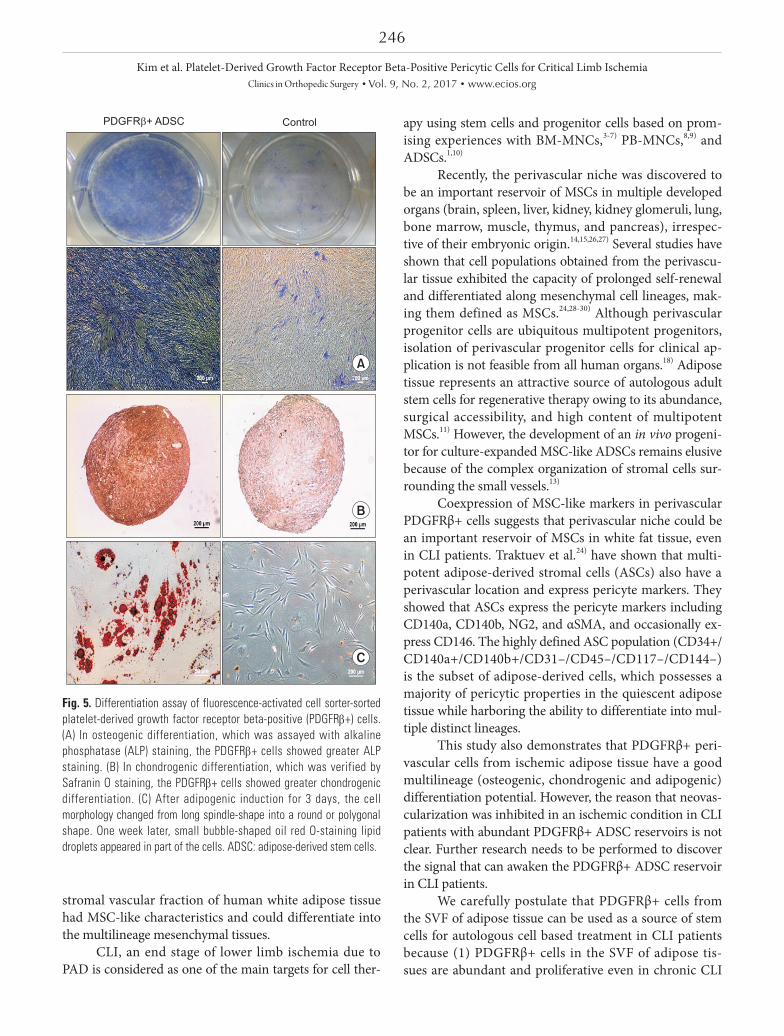
PDGFRβ+ Cells Displayed a Good Multilineage (Osteogenic, Chondrogenic, and Adipogenic) Differentiation PotentialTo examine whether these cells have a multilineage dif-ferentiation ability, PDGFRβ+ cells were induced to differ-entiate into the osteogenic, chondrogenic, and adipogenic lineages. During the analysis for osteogenic differentia-tion assayed by ALP staining, the PDGFRβ+ cells showed greater ALP staining (Fig. 5A). On the chondrogenic dif-ferentiation potential, verified by Safranin O staining, the PDGFRβ+ cells showed greater chondrogenic differentia-tion (Fig. 5B). After adipogenic induction for 3 days, the
cell morphology changed from long spindle-shaped into a round or polygonal shape (data not shown). One week later, small bubble-shaped oil red O-staining lipid droplets appeared in some parts of the cells. The size of lipid drop-lets increased after 2 weeks, and most of the differentiated cells showed red lipid droplets throughout the cytoplasm (Fig. 5C). These results demonstrate that PDGFRβ+ cells have a multi-lineage differentiation ability.
DISCUSSION
In this study, we demonstrated that PDGFRβ+ cells are located in the perivascular area in fresh white adipose tis-sues of CLI patients and they coexpressed the MSC-like markers. We also showed that PDGFRβ+ cells from the
Fig. 4. Matrigel tube formation of fluorescence-activated cell sorter-sorted platelet-derived growth factor receptor beta-positive (PDGFRb+) cells. Human umbilical vein endothelial cells (HUVECs) and CD140b (+) cells were labeled with von Willebrand factor (vWF; green) and CD140b (red), respectively. Nuclei were labeled by DAPI stain (blue). (A) Tubular network formation was more abundant when PDGFRb+ adipose-derived stem cells (ADSCs) were cocultured with HUVECs (c) than when HUVEC only (a) or ADSC only (b) were cultured. (B) When PDGFR beta-positive (PDGFRb+) ADSCs were cocultured with HUVECs, they showed the pericytic location of PDGFRb+ ADSCs (red) which adhered to HUVECs (green) when observed at higher magnification using a confocal microscope.
vWF CD140b vWF + CD140b MergeA
B
a
b
c
246
Kim et al. Platelet-Derived Growth Factor Receptor Beta-Positive Pericytic Cells for Critical Limb Ischemia Clinics in Orthopedic Surgery • Vol. 9, No. 2, 2017 • www.ecios.org
stromal vascular fraction of human white adipose tissue had MSC-like characteristics and could differentiate into the multilineage mesenchymal tissues.
CLI, an end stage of lower limb ischemia due to PAD is considered as one of the main targets for cell ther-
apy using stem cells and progenitor cells based on prom-ising experiences with BM-MNCs,3-7) PB-MNCs,8,9) and ADSCs.1,10)
Recently, the perivascular niche was discovered to be an important reservoir of MSCs in multiple developed organs (brain, spleen, liver, kidney, kidney glomeruli, lung, bone marrow, muscle, thymus, and pancreas), irrespec-tive of their embryonic origin.14,15,26,27) Several studies have shown that cell populations obtained from the perivascu-lar tissue exhibited the capacity of prolonged self-renewal and differentiated along mesenchymal cell lineages, mak-ing them defined as MSCs.24,28-30) Although perivascular progenitor cells are ubiquitous multipotent progenitors, isolation of perivascular progenitor cells for clinical ap-plication is not feasible from all human organs.18) Adipose tissue represents an attractive source of autologous adult stem cells for regenerative therapy owing to its abundance, surgical accessibility, and high content of multipotent MSCs.11) However, the development of an in vivo progeni-tor for culture-expanded MSC-like ADSCs remains elusive because of the complex organization of stromal cells sur-rounding the small vessels.13)
Coexpression of MSC-like markers in perivascular PDGFRβ+ cells suggests that perivascular niche could be an important reservoir of MSCs in white fat tissue, even in CLI patients. Traktuev et al.24) have shown that multi-potent adipose-derived stromal cells (ASCs) also have a perivascular location and express pericyte markers. They showed that ASCs express the pericyte markers including CD140a, CD140b, NG2, and αSMA, and occasionally ex-press CD146. The highly defined ASC population (CD34+/CD140a+/CD140b+/CD31–/CD45–/CD117–/CD144–) is the subset of adipose-derived cells, which possesses a majority of pericytic properties in the quiescent adipose tissue while harboring the ability to differentiate into mul-tiple distinct lineages.
This study also demonstrates that PDGFRβ+ peri-vascular cells from ischemic adipose tissue have a good multilineage (osteogenic, chondrogenic and adipogenic) differentiation potential. However, the reason that neovas-cularization was inhibited in an ischemic condition in CLI patients with abundant PDGFRβ+ ADSC reservoirs is not clear. Further research needs to be performed to discover the signal that can awaken the PDGFRβ+ ADSC reservoir in CLI patients.
We carefully postulate that PDGFRβ+ cells from the SVF of adipose tissue can be used as a source of stem cells for autologous cell based treatment in CLI patients because (1) PDGFRβ+ cells in the SVF of adipose tis-sues are abundant and proliferative even in chronic CLI
PDGFR + ADSC� Control
A
B
C
Fig. 5. Differentiation assay of fluorescence-activated cell sorter-sorted platelet-derived growth factor receptor beta-positive (PDGFRb+) cells. (A) In osteogenic differentiation, which was assayed with alkaline phosphatase (ALP) staining, the PDGFRb+ cells showed greater ALP staining. (B) In chondrogenic differentiation, which was verified by Safranin O staining, the PDGFRb+ cells showed greater chondrogenic differentiation. (C) After adipogenic induction for 3 days, the cell morphology changed from long spindle-shape into a round or polygonal shape. One week later, small bubble-shaped oil red O-staining lipid droplets appeared in part of the cells. ADSC: adipose-derived stem cells.

247
Kim et al. Platelet-Derived Growth Factor Receptor Beta-Positive Pericytic Cells for Critical Limb Ischemia Clinics in Orthopedic Surgery • Vol. 9, No. 2, 2017 • www.ecios.org
patients; (2) the pericytic localization of the PDGFRβ+ ADSCs and their direct contact with endothelial cells can be helpful to increase neovascularization in the ischemic limb; and (3) MSC-like characteristics of PDGFRβ+ AD-SCs can be maintained during subsequent cell expansion in vitro. However, collecting SVF from other fat sites such as abdominal fat tissue might be better in clinical situation of CLI patients for saving ischemic limbs before major am-putation.
In conclusion, we demonstrated that pericyte-like PDGFR-positive cells from the SVF of adipose tissue from CLI patients might have the potential to enhance vascu-larization. We also found that they could be amplified and cells characteristics could be preserved in in vitro cul-ture. Considering the pericytic localization of PDGFRβ+ ADSCs and their direct contact with endothelial cells,
PDGFRβ+ cells in the SVF of adipose tissue can be used as a source of stem cells for autologous cell-based treatment in CLI patients.
CONFLICT OF INTEREST
No potential conflict of interest relevant to this article was reported.
ACKNOWLEDGEMENTS
This study was supported by the Seoul National University Hospital Research Fund (No. 03-2012-0430) and by Basic Science Research Program through the National Research Foundation of Korea (NRF) funded by the Ministry of Science, ICT & Future Planning (No. NRF-2012R1A1A2044067).
REFERENCES
1. Bura A, Planat-Benard V, Bourin P, et al. Phase I trial: the use of autologous cultured adipose-derived stroma/stem cells to treat patients with non-revascularizable critical limb ischemia. Cytotherapy. 2014;16(2):245-57.
2. Dormandy JA, Rutherford RB. Management of peripheral arterial disease (PAD): TASC Working Group: TransAtlantic Inter-Society Consensus (TASC). J Vasc Surg. 2000;31(1 Pt 2):S1-296.
3. Esato K, Hamano K, Li TS, et al. Neovascularization in-duced by autologous bone marrow cell implantation in pe-ripheral arterial disease. Cell Transplant. 2002;11(8):747-52.
4. Higashi Y, Kimura M, Hara K, et al. Autologous bone-mar-row mononuclear cell implantation improves endothelium-dependent vasodilation in patients with limb ischemia. Circulation. 2004;109(10):1215-8.
5. Saigawa T, Kato K, Ozawa T, et al. Clinical application of bone marrow implantation in patients with arterio-sclerosis obliterans, and the association between efficacy and the number of implanted bone marrow cells. Circ J. 2004;68(12):1189-93.
6. Matoba S, Tatsumi T, Murohara T, et al. Long-term clinical outcome after intramuscular implantation of bone mar-row mononuclear cells (Therapeutic Angiogenesis by Cell Transplantation [TACT] trial) in patients with chronic limb ischemia. Am Heart J. 2008;156(5):1010-8.
7. Tateishi-Yuyama E, Matsubara H, Murohara T, et al. Therapeutic angiogenesis for patients with limb ischaemia by autologous transplantation of bone-marrow cells: a pilot study and a randomised controlled trial. Lancet.
2002;360(9331):427-35.
8. Kinoshita M, Fujita Y, Katayama M, et al. Long-term clini-cal outcome after intramuscular transplantation of granu-locyte colony stimulating factor-mobilized CD34 positive cells in patients with critical limb ischemia. Atherosclerosis. 2012;224(2):440-5.
9. Fujita Y, Kinoshita M, Furukawa Y, et al. Phase II clinical trial of CD34+ cell therapy to explore endpoint selection and timing in patients with critical limb ischemia. Circ J. 2014;78(2):490-501.
10. Lee HC, An SG, Lee HW, et al. Safety and effect of adipose tissue-derived stem cell implantation in patients with criti-cal limb ischemia: a pilot study. Circ J. 2012;76(7):1750-60.
11. Gimble JM, Grayson W, Guilak F, Lopez MJ, Vunjak-Nova-kovic G. Adipose tissue as a stem cell source for musculo-skeletal regeneration. Front Biosci (Schol Ed). 2011;3:69-81.
12. Strem BM, Hicok KC, Zhu M, et al. Multipotential differ-entiation of adipose tissue-derived stem cells. Keio J Med. 2005;54(3):132-41.
13. Zimmerlin L, Donnenberg VS, Pfeifer ME, et al. Stromal vascular progenitors in adult human adipose tissue. Cytom-etry A. 2010;77(1):22-30.
14. Crisan M, Yap S, Casteilla L, et al. A perivascular origin for mesenchymal stem cells in multiple human organs. Cell Stem Cell. 2008;3(3):301-13.
15. Zannettino AC, Paton S, Arthur A, et al. Multipotential human adipose-derived stromal stem cells exhibit a peri-vascular phenotype in vitro and in vivo. J Cell Physiol.

248
Kim et al. Platelet-Derived Growth Factor Receptor Beta-Positive Pericytic Cells for Critical Limb Ischemia Clinics in Orthopedic Surgery • Vol. 9, No. 2, 2017 • www.ecios.org
2008;214(2):413-21.
16. Caplan AI. All MSCs are pericytes? Cell Stem Cell. 2008;3(3):229-30.
17. Song S, Ewald AJ, Stallcup W, Werb Z, Bergers G. PDG-FRbeta+ perivascular progenitor cells in tumours regulate pericyte differentiation and vascular survival. Nat Cell Biol. 2005;7(9):870-9.
18. Crisan M, Corselli M, Chen WC, Peault B. Perivascular cells for regenerative medicine. J Cell Mol Med. 2012; 16(12):2851-60.
19. Bhang SH, Cho SW, Lim JM, et al. Locally delivered growth factor enhances the angiogenic efficacy of adipose-derived stromal cells transplanted to ischemic limbs. Stem Cells. 2009;27(8):1976-86.
20. Katz AJ, Tholpady A, Tholpady SS, Shang H, Ogle RC. Cell surface and transcriptional characterization of human adi-pose-derived adherent stromal (hADAS) cells. Stem Cells. 2005;23(3):412-23.
21. Lee RH, Kim B, Choi I, et al. Characterization and expres-sion analysis of mesenchymal stem cells from human bone marrow and adipose tissue. Cell Physiol Biochem. 2004;14(4-6):311-24.
22. Amos PJ, Shang H, Bailey AM, Taylor A, Katz AJ, Peirce SM. IFATS collection: the role of human adipose-derived stromal cells in inflammatory microvascular remodel-ing and evidence of a perivascular phenotype. Stem Cells. 2008;26(10):2682-90.
23. Bagley RG, Rouleau C, Morgenbesser SD, et al. Pericytes from human non-small cell lung carcinomas: an attrac-tive target for anti-angiogenic therapy. Microvasc Res. 2006;71(3):163-74.
24. Traktuev DO, Merfeld-Clauss S, Li J, et al. A population of multipotent CD34-positive adipose stromal cells share peri-cyte and mesenchymal surface markers, reside in a perien-dothelial location, and stabilize endothelial networks. Circ Res. 2008;102(1):77-85.
25. Ponce ML. Tube formation: an in vitro matrigel angiogen-esis assay. Methods Mol Biol. 2009;467:183-8.
26. Shi S, Gronthos S. Perivascular niche of postnatal mesen-chymal stem cells in human bone marrow and dental pulp. J Bone Miner Res. 2003;18(4):696-704.
27. da Silva Meirelles L, Chagastelles PC, Nardi NB. Mesenchy-mal stem cells reside in virtually all post-natal organs and tissues. J Cell Sci. 2006;119(Pt 11):2204-13.
28. Collett GD, Canfield AE. Angiogenesis and pericytes in the initiation of ectopic calcification. Circ Res. 2005;96(9):930-8.
29. Farrington-Rock C, Crofts NJ, Doherty MJ, Ashton BA, Griffin-Jones C, Canfield AE. Chondrogenic and adipo-genic potential of microvascular pericytes. Circulation. 2004;110(15):2226-32.
30. Dore-Duffy P, Katychev A, Wang X, Van Buren E. CNS mi-crovascular pericytes exhibit multipotential stem cell activ-ity. J Cereb Blood Flow Metab. 2006;26(5):613-24.