plasma contact factors as novel biomarkers for diagnosing
TRANSCRIPT

RESEARCH Open Access
Plasma contact factors as novel biomarkersfor diagnosing Alzheimer’s diseaseJung Eun Park1, Do Sung Lim1,2, Yeong Hee Cho1,2, Kyu Yeong Choi3, Jang Jae Lee3, Byeong C. Kim4,Kun Ho Lee1,3 and Jung Sup Lee1,2,3*
Abstract
Background: Alzheimer’s disease (AD) is the most common cause of dementia and most of AD patients sufferfrom vascular abnormalities and neuroinflammation. There is an urgent need to develop novel blood biomarkerscapable of diagnosing Alzheimer’s disease (AD) at very early stage. This study was performed to find out newaccurate plasma diagnostic biomarkers for AD by investigating a direct relationship between plasma contact systemand AD.
Methods: A total 101 of human CSF and plasma samples from normal and AD patients were analyzed. The contactfactor activities in plasma were measured with the corresponding specific peptide substrates.
Results: The activities of contact factors (FXIIa, FXIa, plasma kallikrein) and FXa clearly increased and statisticallycorrelated as AD progresses. We present here, for the first time, the FXIIa cut-off scores to as: > 26.3 U/ml for prodromalAD [area under the curve (AUC) = 0.783, p < 0.001] and > 27.2 U/ml for AD dementia (AUC = 0.906, p < 0.001). We alsodescribe the cut-off scores from the ratios of CSF Aβ1–42 versus the contact factors. Of these, the representative ratiocut-off scores of Aβ1–42/FXIIa were to be: < 33.8 for prodromal AD (AUC = 0.965, p < 0.001) and < 27.44 for ADdementia (AUC = 1.0, p < 0.001).
Conclusion: The activation of plasma contact system is closely associated with clinical stage of AD, and FXIIa activity aswell as the cut-off scores of CSF Aβ1–42/FXIIa can be used as novel accurate diagnostic AD biomarkers.
Keywords: Alzheimer’s disease, Biomarkers, Contact factor, FXIIa, Plasma
BackgroundAlzheimer’s disease (AD) is the most common cause ofdementia, accounting for 60–80% of all dementia patients.However, the cause of AD is poorly understood and thereare no treatments currently available. To date, the onlyway to take care of AD is to find out in advance the onsetand prevent the progression [1, 2]. Over the past decades,there have been many efforts to develop blood biomarkers
that can easily and reliably detect the onset of Alzheimer’s,but it has been delayed due to the lack of reproducibilityand other problems related to clinical use [3, 4]. Neverthe-less, the identification of novel blood biomarkers for pre-dicting and monitoring disease progression that canreliably detect the onset of AD at the early stage is crucial.AD is a neurodegenerative disease characterized by
amyloid-beta (Aβ) deposits in brain [5–7], and most ofAD patients suffer from vascular abnormalities [8, 9]and neuroinflammation [10]. Both vascular abnormal-ities and inflammation can trigger neuronal death; how-ever, the effect of Aβ on these pathologies has not beenelucidated. However, a few recent reports have suggestedthat FXII–initiated contact system can trigger both
© The Author(s). 2021 Open Access This article is licensed under a Creative Commons Attribution 4.0 International License,which permits use, sharing, adaptation, distribution and reproduction in any medium or format, as long as you giveappropriate credit to the original author(s) and the source, provide a link to the Creative Commons licence, and indicate ifchanges were made. The images or other third party material in this article are included in the article's Creative Commonslicence, unless indicated otherwise in a credit line to the material. If material is not included in the article's Creative Commonslicence and your intended use is not permitted by statutory regulation or exceeds the permitted use, you will need to obtainpermission directly from the copyright holder. To view a copy of this licence, visit http://creativecommons.org/licenses/by/4.0/.The Creative Commons Public Domain Dedication waiver (http://creativecommons.org/publicdomain/zero/1.0/) applies to thedata made available in this article, unless otherwise stated in a credit line to the data.
* Correspondence: [email protected] of Biomedical Science, College of Natural Sciences, ChosunUniversity, 309 Pilmun-Daero, Gwangju 61452, Republic of Korea2Department of Integrative Biological Sciences & BK21-Four EducationalResearch Group for Age-associated Disorder Control Technology, ChosunUniversity, Gwangju, Republic of KoreaFull list of author information is available at the end of the article
Park et al. Biomarker Research (2021) 9:5 https://doi.org/10.1186/s40364-020-00258-5

vascular pathology and inflammation [11], and depletionof coagulation FXII can ameliorate brain pathology andcognitive impairment in AD mice [12].The plasma contact system, which is composed of the
intrinsic pathway of coagulation and the kallikrein-kininsystem, plays an essential role in innate immunity [13–15]. Activation of the plasma contact system triggers sev-eral cascade systems that involve three serine protease zy-mogens [factor XII (FXII), factor XI (FXI), and pre-kallikrein (PKK)] and a non-enzymatic cofactor protein[high-molecular weight kininogen (HK)] [13]. The enzy-matically active FXIIa activates PKK to plasma kallikrein(PK; KLKB1) that cleaves HK to release the vasoactive andproinflammatory nanopeptide, bradykinin (BK).Several recent studies have shown a possible relation-
ship between plasma contact system and AD. However,they did not provide any solid evidence, because of theiruses of data from a very few human CSF samples ormurine models [11, 12, 15–19]. Accordingly, there arestill a variety of issues related to contact system and AD,including 1) Is there a close correlation between contactsystem and AD progression in fact?; and 2) Is it possibleto obtain various cut-off scores and certain ratios fordistinguishing accurately prodromal AD and AD demen-tia from normal subjects?In this study, we addressed these issues and present,
for the first time, the direct relationship between thecontact system activation and AD progress, and the cut-off scores of FXIIa and CSF Aβ1–42/FXIIa ratio, whichare capable of discriminating accurately prodromal ADand AD dementia from normal subjects.
MethodsStudy participantsThis study was conducted on participants in Gwangjuand Jeollanam-do, Republic of Korea, from August 2015to October 2017. All participants in this study providedtheir written consents, and the study protocol was ap-proved by Chosun University Hospital Institutional Re-view Board (IRB file numbers 2013–12–018-068 and2016–10–005-009). A total of 101 subjects included inthe study were classified into three groups (50 normalelderly people, 23 patients with prodromal AD, and 28patients with AD dementia) according to the clinical cri-teria proposed by the IWG-2 guidelines with amyloidPET [16]. However, PET positive group in normal sub-jects (preclinical stage of AD) and amyloid PET negativegroup in mild cognitive impairment (MCI) were ex-cluded. Patients with AD dementia met the clinical cri-teria for Alzheimer’s disease dementia potentialproposed by the NIA-AA or IWG-2 working group, andthe diagnosis of MCI was also made according to theMCI criteria suggested by the NIA-AA or IWG-2 group[20, 21].
Determination of the concentrations of Aβ1–42, t-Tau, p-Tau181, and bradykininThe collection and storage of CSF used in this study wasperformed in the same process as follows [22]. The con-centrations of Aβ1–42, t-Tau, and p-Tau181 in CSF weremeasured using INNOTEST ELISA kit (Fujirebio,Ghent, Belgium) and that of bradykinin (BK) was quanti-tated with Bradykinin ELISA kit (Enzo Life Sciences,Farmingdale, NY, USA) according to the protocols pro-vided by the manufacturers.
Measurement of the activities of FXIIa, FXIa, FXa, and PKin plasmaPlasma used in this study was collected and stored fromparticipants according to the Molecular MedicineIreland (MMI) guidelines for standardized biobanking[23]. To measure and make the standard curves forFXIIa, FXIa, FXa, and PK activities, various concentra-tions of FXIIa (0, 0.5, 1, 1.5, 2, 2.5, and 4 U/ml), FXIa (0,0.01, 0.02, 0.05, 0.1, 0.2, and 0.4 U/ml), FXa (0, 0.01,0.05, 0.1, 0.2, 0.5, and 1 U/ml), and PK (0, 0.02, 0.04,0.08, 0.1, 0.2, and 0.4 U/ml) were serially diluted in phos-phate buffered saline (PBS, pH 7.5). After the dilutions,90 μl each of samples was mixed with 10 μl of corre-sponding synthetic peptide substrates (S-2302 for FXIIa,S-2366 for FXIa, S-2765 for FXa, and H-D-Val-Leu-Arg-AFC for kallikrein) dissolved in PBS (pH 7.5) at a finalconcentration of 4 mM and incubated for 30 min at37 °C, during which the increase in absorbance at 405nm for chromogenic substrates or in fluorescence forfluorogenic substrate (excitation 400 nm/emission 505nm) was recorded with a 96-well plate reader (MolecularDevices, San Jose, CA, USA) as described previously[13]. All experiments were performed in triplicate. Usingthe data obtained, standard curves for the enzymes werethen made using a sigmoidal 4 parameter curve fitting(Supplementary Fig. 1). To validate the measurementmethod, total 6 plasma samples and 3 different concen-trations of contact factors (FXIIa, FXIa, and PK) andFXa were tested over different days.
Statistical analysisStatistical analysis was performed with SPSS version 24.0(IBM Corp., Armonk, NY, USA), and analysis of variance(ANOVA) was used to compare the three groups (nor-mal, prodromal AD, and AD dementia). Pearson’s cor-relation analysis was used to analyze the associationbetween the activity of plasma contact system and thetypical CSF AD biomarkers. Receiver operator character-istic (ROC) curves were generated to calculate areasunder the curves (AUCs) to determine the diagnosticabilities of the contact factors and the typical CSF ADbiomarkers for prodromal AD and AD dementia. Thestandard deviations from the mean value of the
Park et al. Biomarker Research (2021) 9:5 Page 2 of 8

quantitative analysis or activity of each protein in the co-hort used in this study was calculated as the z-score.
Results and discussionConcentration of CSF biomarkers in ADThe clinical characteristics of total 101 subjects (50 nor-mal subjects, 23 patients with prodromal AD, and 28 pa-tients with AD dementia) and the concentrations oftypical AD biomarkers, including Aβ1–42, t-Tau, and p-Tau181 in CSF and contact factors (FXIIa, FXIa, PK, andBK) and FXa in plasma samples are summarized inTable 1. As reported previously [2, 5, 24, 25], Aβ1–42level in CSF decreased, while t-Tau and p-Tau181 con-centrations increased with AD progression (Figs. 1a, c).The concentrations of CSF Aβ1–42 were estimated to be1059, 550 and 484 pg/ml in normal, prodromal AD, andAD dementia groups, respectively (Table 1). Contrary tothese, the levels of t-Tau and p-Tau181 increased as ADprogressed, in which the concentrations of t-Tau werefound to be 230, 389, and 536 pg/ml, and those of p-Tau181 were 44, 66, and 78 pg/ml in normal, prodromalAD, and AD dementia groups, respectively (Table 1).
These results were well in accordance with reported pre-vious studies [2, 24, 25].
Activation of contact system in ADTo measure the activation of the contact system inplasma, the assay validation standard curves were cre-ated using the recombinant contact factor (FXIIa, FXIa,and PK) and FXa enzymes. As a result, the R2 values ofstandard curves were higher than 0.95 (SupplementaryFig. 1). In addition, the coefficients of variation (CV%)were analyzed to confirm the precision and reproducibil-ity of the measurement method for contact factor activ-ity. As a result, the inter-assay and intra-assay CVs (%)ranged for FXIIa from 3.2 to 4.5% and from 7.2 to12.7%, which indicated the excellent reproducibility ofthis method (Table 2).The enzymatic activities of the plasma contact factors
(FXIIa, FXIa, and PK) and FXa clearly increased withAD progression. The enzymatic activities for the normal,prodromal AD, and AD dementia groups were as fol-lows: 23.9, 28.0, and 30.6 U/ml for FXIIa; 1.11, 1.18, and1.30 U/ml for FXIa; 0.77, 0.81, and 0.97 U/ml for FXa;1.20, 1.35, and 1.54 U/ml for PK, respectively (Table 1).
Table 1 Demographic and biochemical characteristics of normal subjects, prodromal AD, and AD dementia
Demographic data/molecules in CSF or plasma Total numbers of subjects Normal Prodromal AD AD dementia p value
Number of subjects 101 50 23 28
Age 101 72.0 (6.1) 71.1 (8.6) 69.4 (6.0) 0.262
Years of education 101 9.5 (5.0) 8.0 (4.7) 6.70 (3.8) a 0.033
Gender, female, n (%) 101 26.0 (52.0) 12 (52.2) 17.0 (60.7) 0.736
K-MMSE score 101 26.8 (2.3) 25.3 (3.9) 17.9 (5.4) a, b < 0.001
CDR 101 0.3 (0.2) 0.5 (0.0) a 0.9 (0.4) a, b < 0.001
CDR sum of boxes 101 0.5 (0.6) 1.3 (0.6) 5.1 (2.6) a, b < 0.001
GDS 101 1.6 (0.5) 3.0 (0.2) a 4.1 (0.9) a, b < 0.001
B-ADL 101 20.0 (0.0) 20.0 (0.2) 19.0 (1.8) a, b < 0.001
I-ADL 101 0.04 (0.08) 0.22 (0.14) a 0.63 (0.20) a, b < 0.001
CSF biomarkers
Aβ1–42, pg/ml 101 1,059 (177) 550 (223) a 484 (192) a < 0.001
t-Tau, pg/ml 101 230 (77) 389 (236) a 536 (209) a, b < 0.001
p-Tau181, pg/ml 101 44 (14) 66 (33) a 78 (28) a < 0.001
BK, pg/ml 72 83 (49) 55 (45) 39 (33) a 0.002
Plasma factors
FXIIa, U/ml 101 23.90 (3.70) 28.0 (3.4) a 30.6 (3.7) a < 0.001
FXIa, U/ml 101 1.11 (0.21) 1.18 (0.21) 1.30 (0.30) a 0.005
FXa, U/ml 101 0.77 (0.11) 0.81 (0.12) 0.97 (0.29) a, b < 0.001
Kallikrein, U/ml 101 1.20 (0.25) 1.35 (0.34) 1.54 (0.39) a < 0.001
BK, pg/ml 90 13,365 (9,305) 13,497 (7,945) 17,679 (21,186) 0.406
Data are presented as mean (± standard deviation) or number (%). Abbreviations: K-MMSE Korean Mini-Mental State Examination, CDR Clinical Dementia Rating,GDS Global Deterioration Scale, B-ADL Barthel Activities of Daily Living, I-ADL Instrumental Activities of Daily Living, CSF cerebrospinal fluid, Aβ amyloid beta-protein, t-Tau total Tau protein, p-Tau phosphorylated Tau protein, AD Alzheimer’s disease, BK bradykinin. astatistically significant difference between the indicatedgroup and the normal group; bstatically significant difference between prodromal AD and AD dementia groups
Park et al. Biomarker Research (2021) 9:5 Page 3 of 8

Taken as a whole, the activities of contact factors (FXIIa,FXIa, or PK) and FXa had a statically difference betweenthe three groups (normal, prodromal AD, and AD de-mentia groups) (Fig. 1e, h). As for BK, which is a finalproduct of the kallikrein/kinin system [26], its concen-tration increased in plasma as expected, but rather de-creased in CSF with the progression of AD (Table 1; Fig.1d). The concentrations of BK in plasma were 13,365,13,497, and 17,679 pg/ml and those in CSF were 83, 55,and 39 pg/ml in normal, prodromal AD, and AD demen-tia groups, respectively. These results are consistent withthe those of recent reports [27] and also seemed to berelated, in part, to the fact that BK can evoke blood–brain barrier (BBB) leakage and neuro-inflammation,
resulting in CSF BK efflux [15, 28]. All these results sug-gest that the activation of plasma contact system andAD progression are obviously related, and the activitiesof contact factors can be used for discriminating pro-dromal AD and AD from normal groups.
Analysis of correlation between AD biomarker in CSF andcontact factor activity in plasmaTo examine further the strength of a link between thetypical CSF AD biomarkers and the contact factors, weperformed correlation analyses (Fig. 1i - l; Supplemen-tary Table 1). As for the typical CSF AD biomarkers, thePearson’s correlation coefficients of Aβ1–42 versus t-Tauand p-Tau181 were to be - 0.445 and - 0.359, respectively,
Fig. 1 Activation of plasma contact factors with clinical stage of AD and correlation strengths between biomarkers. Concentrations of typical ADbiomarkers, including Aβ1–42 (a), t-Tau (b), p-Tau181 (c) and BK (d) in CSF were measured from normal (NR), prodromal AD (P-AD), and ADdementia (AD-D) groups by ELISA. The activities of plasma contact factors such as FXIIa (e), FXIa (f), FXa (g), and kallikrein (h) were measured inthe presence of 0.4 mM each of corresponding specific synthetic peptide substrates as described in Study design. Statistical analysis wasperformed using SPSS version 24.0 (IBM Corp., Armonk, NY, USA), and analysis of variance (ANOVA) was performed for comparisons between thethree groups (NR, P-AD, and AD-D). Data are presented as mean values for each group. *: statistically significant difference between the indicatedgroup and the NR; †: statistically significant difference between the P-AD and the AD-D groups. Correlation plots of FXIIa versus Aβ1–42 (i), FXIIaversus t-Tau (j), FXIIa versus p-Tau181 (k), and CSF BK versus Aβ1–42 (l). Pearson’s correlation analysis was used to analyze the correlations betweenthe activity of plasma contact factor and CSF AD biomarker as indicated, in which statistical significance was set at p < 0.05. In panels a to l, cyan,blue, and red colored circles represent NR, P-AD, and AD-D groups, respectively. Dynamics of biomarkers analyzed in AD pathological cascade(m). Lines show z-scores in mean values of normalized biomarker levels for each AD group
Park et al. Biomarker Research (2021) 9:5 Page 4 of 8
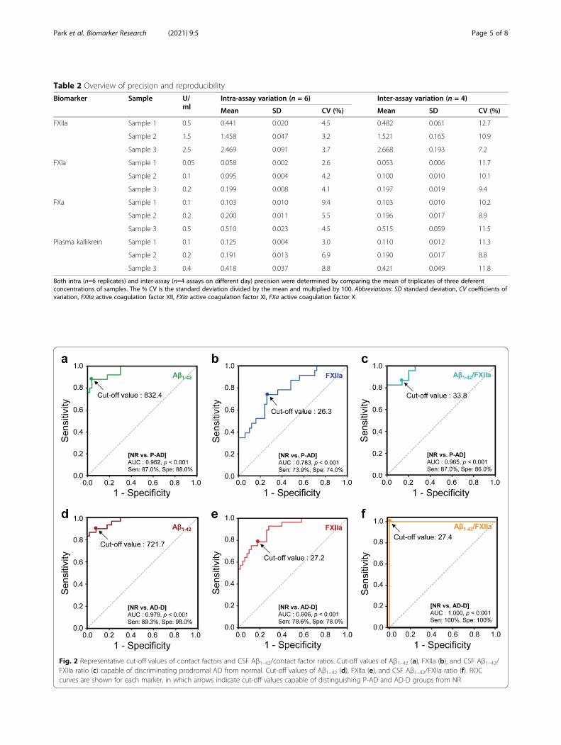
Table 2 Overview of precision and reproducibility
Biomarker Sample U/ml
Intra-assay variation (n = 6) Inter-assay variation (n = 4)
Mean SD CV (%) Mean SD CV (%)
FXIIa Sample 1 0.5 0.441 0.020 4.5 0.482 0.061 12.7
Sample 2 1.5 1.458 0.047 3.2 1.521 0.165 10.9
Sample 3 2.5 2.469 0.091 3.7 2.668 0.193 7.2
FXIa Sample 1 0.05 0.058 0.002 2.6 0.053 0.006 11.7
Sample 2 0.1 0.095 0.004 4.2 0.100 0.010 10.1
Sample 3 0.2 0.199 0.008 4.1 0.197 0.019 9.4
FXa Sample 1 0.1 0.103 0.010 9.4 0.103 0.010 10.2
Sample 2 0.2 0.200 0.011 5.5 0.196 0.017 8.9
Sample 3 0.5 0.510 0.023 4.5 0.515 0.059 11.5
Plasma kallikrein Sample 1 0.1 0.125 0.004 3.0 0.110 0.012 11.3
Sample 2 0.2 0.191 0.013 6.9 0.190 0.017 8.8
Sample 3 0.4 0.418 0.037 8.8 0.421 0.049 11.8
Both intra (n=6 replicates) and inter-assay (n=4 assays on different day) precision were determined by comparing the mean of triplicates of three deferentconcentrations of samples. The % CV is the standard deviation divided by the mean and multiplied by 100. Abbreviations: SD standard deviation, CV coefficients ofvariation, FXIIa active coagulation factor XII, FXIa active coagulation factor XI, FXa active coagulation factor X
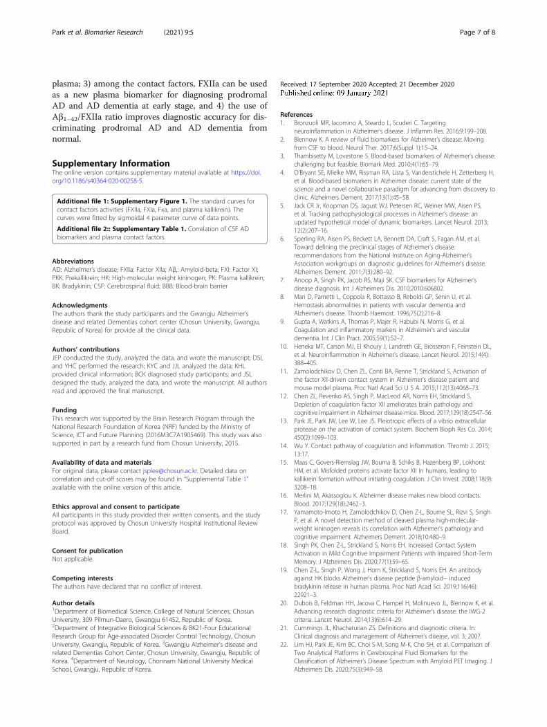
Fig. 2 Representative cut-off values of contact factors and CSF Aβ1–42/contact factor ratios. Cut-off values of Aβ1–42 (a), FXIIa (b), and CSF Aβ1–42/FXIIa ratio (c) capable of discriminating prodromal AD from normal. Cut-off values of Aβ1–42 (d), FXIIa (e), and CSF Aβ1–42/FXIIa ratio (f). ROCcurves are shown for each marker, in which arrows indicate cut-off values capable of distinguishing P-AD and AD-D groups from NR
Park et al. Biomarker Research (2021) 9:5 Page 5 of 8

at their r-values, indicating that these biomarkers are mod-erately correlated, under the guidelines of correlationstrength [29]. In cases of contact factors, the r-values ofFXIIa versus FXIa, FXa, and PK were to be 0.8, 0.692, and0.59, respectively, suggesting that these factors are corre-lated statistically in significant level (SupplementaryTable 1). Prominently, FXIIa was negatively correlated toCSF Aβ1–42 (r = − 0.463) and positively to both t-Tau (r =0.509) and p-Tau181 (r = 0.452) in all moderate relation-ships (Fig. 1i - l; Supplementary Table 1). All these resultssuggest that the activation of contact system is certainlycorrelated to AD progression as reported previously [17].The changes in the z-values of typical CSF AD bio-
markers and plasma contact factors as AD progresseswere also analyzed (Fig. 1m). As expected [5, 12, 16, 30],the z-score of Aβ1–42 decreased, whereas those of t-Tauand p-Tau181 increased in the order of normal, pro-dromal AD, and AD dementia groups (Fig. 1m). As forplasma contact factors, the scores of FXIIa, FXIa, FXa,and PK noticeably increased (Fig. 1m), whereas CSF BKdecreased (data not shown) in the AD pathological cas-cade. These results strongly suggest that plasma contactsystem is closely associated with AD progression, and itsdegree of activation can reflect the disease progression.
Analysis of potential as a plasma biomarker for ADdiagnosisWe generated receiver operating characteristic (ROC)curves to analyze the potential of each protein for use as
a new biomarker for AD diagnosis (Fig. 2 and Table 3).The ROC curves and the areas under the curves (AUCs)[28, 31] showed that Aβ1–42 can discriminable in highlyaccurate for both prodromal AD (AUC = 0.962; cut-offvalue = < 832.4 pg/ml) and AD dementia (AUC = 0.979;cut-off value = < 721.7 pg/ml) from normal (Fig. 2a, d;Table 3). Among the contact factors, only FXIIa seemedto have a capability able to discriminate both prodromalAD (AUC = 0.783; cut-off value = > 26.3 U/ml) and ADdementia (AUC = 0.906; cut-off value = > 27.2 U/ml)from normal (Fig. 2b,e; Table 3). These results indicatethat the contact factor FXIIa can be a new plasma bio-marker for diagnosing prodromal AD in acceptable andAD dementia in very accurate from normal.In particular, the CSF Aβ1–42/FXIIa ratio showed very
accurate for prodromal AD (AUC = 0.965; cut-off value= < 33.8) and perfect diagnostic abilities for AD demen-tia (AUC = 1.0; cut-off value = < 27.44) (Fig. 2c, f; Table3). Taken together, these results suggest that, 1) FXIIacan be used as a plasma biomarker for early diagnosis ofprodromal AD, and 2) use of the Aβ1–42/FXIIa ratio im-proves diagnostic accuracy.
ConclusionsBased on the results, we conclude that 1) activation ofplasma contact system is not only correlated with ADprogression, but also is available for AD diagnosis; 2) thedegree of AD progress can be quickly determined bymeasuring the activities of contact factors in blood
Table 3 Cut-off scores and sensitivity/specificity values of fluid biomarkers for discriminating the prodromal AD or AD dementiafrom normal subjects
Characteristics Normal (n = 50) vs. Prodromal AD (n = 23) groups Normal (n = 50) vs. AD dementia (n = 28) groups
Cut-off Sen (%) Spe (%) AUC p value Cut-off Sen (%) Spe (%) AUC p value
Plasma factors
FXIIa activity, U/ml > 26.3 73.9 74.0 0.783 < 0.001 > 27.2 78.6 78.0 0.906 < 0.001
FXIa activity, U/ml > 1.13 52.2 52.0 0.566 0.367 > 1.17 57.1 58.0 0.659 0.020
FXa activity, U/ml > 0.76 56.5 54.0 0.573 0.316 > 0.82 71.4 70.0 0.779 < 0.001
Kallikrein activity, U/ml > 1.21 56.5 56.0 0.609 0.138 > 1.29 64.3 64.0 0.772 < 0.001
CSF biomarkers
Aβ1–42 levels, pg/ml < 832.4 87.0 88.0 0.962 < 0.001 < 721.7 89.3 98.0 0.979 < 0.001
t-Tau levels, pg/ml > 252.6 65.2 66.0 0.701 0.006 > 315.3 85.7 86.0 0.954 < 0.001
p-Tau181 levels, pg/ml > 46.1 65.2 64.0 0.690 0.009 > 46.1 78.6 78.0 0.870 < 0.001
BK levels, pg/ml > 46.1 54.5 56.5 0.656 0.073 > 52.8 74.1 73.9 0.783 0.001
Ratios
Aβ1–42 / FXIIa < 33.8 87.0 86.0 0.965 < 0.001 < 27.4 100.0 100.0 1.000 < 0.001
Aβ1–42 / FXIa < 742.5 82.6 82.0 0.930 < 0.001 < 635.4 96.4 96.0 0.993 < 0.001
Aβ1–42 / FXa < 1,091 82.6 82.0 0.933 < 0.001 < 941.8 96.4 96.0 0.994 < 0.001
Aβ1–42 / Kallikrein < 680.9 87.0 86.0 0.943 < 0.001 < 584.8 96.4 96.0 0.994 < 0.001
Statistically-derived optimal cut-off value was determined with the best balance between sensitivity (Sen) and specificity (Spe) values. Discrimination of prodromalAD and AD dementia from normal group was evaluated by receiver operator characteristics (ROC) curve analysis and quantified by the area under the curve(AUC) using SPSS software version 24.0
Park et al. Biomarker Research (2021) 9:5 Page 6 of 8
plasma; 3) among the contact factors, FXIIa can be usedas a new plasma biomarker for diagnosing prodromalAD and AD dementia at early stage, and 4) the use ofAβ1–42/FXIIa ratio improves diagnostic accuracy for dis-criminating prodromal AD and AD dementia fromnormal.
Supplementary InformationThe online version contains supplementary material available at https://doi.org/10.1186/s40364-020-00258-5.
Additional file 1: Supplementary Figure 1. The standard curves forcontact factors activities (FXIIa, FXIa, Fxa, and plasma kallikrein). Thecurves were fitted by sigmoidal 4 parameter curve of data points.
Additional file 2:: Supplementary Table 1. Correlation of CSF ADbiomarkers and plasma contact factors.
AbbreviationsAD: Alzheimer’s disease; FXIIa: Factor XIIa; Aβ,: Amyloid-beta; FXI: Factor XI;PKK: Prekallikrein; HK: High-molecular weight kininogen; PK: Plasma kallikrein;BK: Bradykinin; CSF: Cerebrospinal fluid; BBB: Blood-brain barrier
AcknowledgmentsThe authors thank the study participants and the Gwangju Alzheimer’sdisease and related Dementias cohort center (Chosun University, Gwangju,Republic of Korea) for provide all the clinical data.
Authors’ contributionsJEP conducted the study, analyzed the data, and wrote the manuscript; DSLand YHC performed the research; KYC and JJL analyzed the data; KHLprovided clinical information; BCK diagnosed study participants; and JSLdesigned the study, analyzed the data, and wrote the manuscript. All authorsread and approved the final manuscript.
FundingThis research was supported by the Brain Research Program through theNational Research Foundation of Korea (NRF) funded by the Ministry ofScience, ICT and Future Planning (2016M3C7A1905469). This study was alsosupported in part by a research fund from Chosun University, 2015.
Availability of data and materialsFor original data, please contact [email protected]. Detailed data oncorrelation and cut-off scores may be found in “Supplemental Table 1”available with the online version of this article.
Ethics approval and consent to participateAll participants in this study provided their written consents, and the studyprotocol was approved by Chosun University Hospital Institutional ReviewBoard.
Consent for publicationNot applicable.
Competing interestsThe authors have declared that no conflict of interest.
Author details1Department of Biomedical Science, College of Natural Sciences, ChosunUniversity, 309 Pilmun-Daero, Gwangju 61452, Republic of Korea.2Department of Integrative Biological Sciences & BK21-Four EducationalResearch Group for Age-associated Disorder Control Technology, ChosunUniversity, Gwangju, Republic of Korea. 3Gwangju Alzheimer’s disease andrelated Dementias Cohort Center, Chosun University, Gwangju, Republic ofKorea. 4Department of Neurology, Chonnam National University MedicalSchool, Gwangju, Republic of Korea.
Received: 17 September 2020 Accepted: 21 December 2020
References1. Bronzuoli MR, Iacomino A, Steardo L, Scuderi C. Targeting
neuroinflammation in Alzheimer’s disease. J Inflamm Res. 2016;9:199–208.2. Blennow K. A review of fluid biomarkers for Alzheimer’s disease: Moving
from CSF to blood. Neurol Ther. 2017;6(Suppl 1):15–24.3. Thambisetty M, Lovestone S. Blood-based biomarkers of Alzheimer’s disease:
challenging but feasible. Biomark Med. 2010;4(1):65–79.4. O'Bryant SE, Mielke MM, Rissman RA, Lista S, Vanderstichele H, Zetterberg H,
et al. Blood-based biomarkers in Alzheimer disease: current state of thescience and a novel collaborative paradigm for advancing from discovery toclinic. Alzheimers Dement. 2017;13(1):45–58.
5. Jack CR Jr, Knopman DS, Jagust WJ, Petersen RC, Weiner MW, Aisen PS,et al. Tracking pathophysiological processes in Alzheimer’s disease: anupdated hypothetical model of dynamic biomarkers. Lancet Neurol. 2013;12(2):207–16.
6. Sperling RA, Aisen PS, Beckett LA, Bennett DA, Craft S, Fagan AM, et al.Toward defining the preclinical stages of Alzheimer’s disease:recommendations from the National Institute on Aging-Alzheimer’sAssociation workgroups on diagnostic guidelines for Alzheimer’s disease.Alzheimers Dement. 2011;7(3):280–92.
7. Anoop A, Singh PK, Jacob RS, Maji SK. CSF biomarkers for Alzheimer’sdisease diagnosis. Int J Alzheimers Dis. 2010;2010:606802.
8. Mari D, Parnetti L, Coppola R, Bottasso B, Reboldi GP, Senin U, et al.Hemostasis abnormalities in patients with vascular dementia andAlzheimer's disease. Thromb Haemost. 1996;75(2):216–8.
9. Gupta A, Watkins A, Thomas P, Majer R, Habubi N, Morris G, et al.Coagulation and inflammatory markers in Alzheimer’s and vasculardementia. Int J Clin Pract. 2005;59(1):52–7.
10. Heneka MT, Carson MJ, El Khoury J, Landreth GE, Brosseron F, Feinstein DL,et al. Neuroinflammation in Alzheimer’s disease. Lancet Neurol. 2015;14(4):388–405.
11. Zamolodchikov D, Chen ZL, Conti BA, Renne T, Strickland S. Activation ofthe factor XII-driven contact system in Alzheimer’s disease patient andmouse model plasma. Proc Natl Acad Sci U S A. 2015;112(13):4068–73.
12. Chen ZL, Revenko AS, Singh P, MacLeod AR, Norris EH, Strickland S.Depletion of coagulation factor XII ameliorates brain pathology andcognitive impairment in Alzheimer disease mice. Blood. 2017;129(18):2547–56.
13. Park JE, Park JW, Lee W, Lee JS. Pleiotropic effects of a vibrio extracellularprotease on the activation of contact system. Biochem Bioph Res Co. 2014;450(2):1099–103.
14. Wu Y. Contact pathway of coagulation and inflammation. Thromb J. 2015;13:17.
15. Maas C, Govers-Riemslag JW, Bouma B, Schiks B, Hazenberg BP, LokhorstHM, et al. Misfolded proteins activate factor XII in humans, leading tokallikrein formation without initiating coagulation. J Clin Invest. 2008;118(9):3208–18.
16. Merlini M, Akassoglou K. Alzheimer disease makes new blood contacts.Blood. 2017;129(18):2462–3.
17. Yamamoto-Imoto H, Zamolodchikov D, Chen Z-L, Bourne SL, Rizvi S, SinghP, et al. A novel detection method of cleaved plasma high-molecular-weight kininogen reveals its correlation with Alzheimer's pathology andcognitive impairment. Alzheimers Dement. 2018;10:480–9.
18. Singh PK, Chen Z-L, Strickland S, Norris EH. Increased Contact SystemActivation in Mild Cognitive Impairment Patients with Impaired Short-TermMemory. J Alzheimers Dis. 2020;77(1):59–65.
19. Chen Z-L, Singh P, Wong J, Horn K, Strickland S, Norris EH. An antibodyagainst HK blocks Alzheimer’s disease peptide β-amyloid− inducedbradykinin release in human plasma. Proc Natl Acad Sci. 2019;116(46):22921–3.
20. Dubois B, Feldman HH, Jacova C, Hampel H, Molinuevo JL, Blennow K, et al.Advancing research diagnostic criteria for Alzheimer's disease: the IWG-2criteria. Lancet Neurol. 2014;13(6):614–29.
21. Cummings JL, Khachaturian ZS. Definitions and diagnostic criteria. In:Clinical diagnosis and management of Alzheimer’s disease, vol. 3; 2007.
22. Lim HJ, Park JE, Kim BC, Choi S-M, Song M-K, Cho SH, et al. Comparison ofTwo Analytical Platforms in Cerebrospinal Fluid Biomarkers for theClassification of Alzheimer’s Disease Spectrum with Amyloid PET Imaging. JAlzheimers Dis. 2020;75(3):949–58.
Park et al. Biomarker Research (2021) 9:5 Page 7 of 8

23. Guerin JS, Murray DW, McGrath MM, Yuille MA, McPartlin JM, Doran PP.Molecular medicine Ireland guidelines for standardized biobanking.Biopreserv Biobank. 2010;8(1):3–63.
24. Slemmon JR, Meredith J, Guss V, Andreasson U, Andreasen N, Zetterberg H,et al. Measurement of Aβ1–42 in cerebrospinal fluid is influenced by matrixeffects. J Neurochem. 2012;120(2):325–33.
25. Parnetti L, Chiasserini D, Eusebi P, Giannandrea D, Bellomo G, De Carlo C,et al. Performance of abeta1-40, abeta1-42, total tau, and phosphorylatedtau as predictors of dementia in a cohort of patients with mild cognitiveimpairment. J Alzheimers Dis. 2012;29(1):229–38.
26. Wu J, Akaike T, Hayashida K, Miyamoto Y, Nakagawa T, Miyakawa K, et al.Identification of bradykinin receptors in clinical cancer specimens andmurine tumor tissues. Int J Cancer. 2002;98(1):29–35.
27. Singh PK, Chen Z-L, Ghosh D, Strickland S, Norris EH. Increased plasmabradykinin level is associated with cognitive impairment in Alzheimer’spatients. Neurobiol Dis. 2020;139:104833.
28. Marcos-Contreras OA, Martinez de Lizarrondo S, Bardou I, Orset C, PruvostM, Anfray A, et al. Hyperfibrinolysis increases blood-brain barrierpermeability by a plasmin- and bradykinin-dependent mechanism. Blood.2016;128(20):2423–34.
29. Mukaka MM. Statistics corner: A guide to appropriate use of correlationcoefficient in medical research. Malawi Med J. 2012;24(3):69–71.
30. Hu WT, Watts KD, Shaw LM, Howell JC, Trojanowski JQ, Basra S, et al. CSFbeta-amyloid 1-42 - what are we measuring in Alzheimer's disease? AnnClin Transl Neur. 2015;2(2):131–9.
31. Greiner M, Pfeiffer D, Smith RD. Principles and practical application of thereceiver-operating characteristic analysis for diagnostic tests. Prev Vet Med.2000;45(1–2):23–41.
Publisher’s NoteSpringer Nature remains neutral with regard to jurisdictional claims inpublished maps and institutional affiliations.
Park et al. Biomarker Research (2021) 9:5 Page 8 of 8