plasma coagulation in cardiac surgery - gupea: home · understood. fibrinogen is a final step in...
TRANSCRIPT
1
From the
Department of Medicine/Hematology and Coagulation Disorders
Sahlgrenska University Hospital
and
Department of Molecular and Clinical Medicine
Institute of Medicine, Sahlgrenska Academy
University of Gothenburg
Plasma coagulation in cardiac surgery
Vladimir Radulovic
Gothenburg 2013

2
Plasma coagulation in cardiac surgery © vladimir radulovic 2013
ISBN 978-91-628-8784-1
Printed in Gothenburg, Sweden 2013
Ineko Intellecta

3

4
5
Abstract
Background: Cardiac surgery using cardiopulmonary bypass is a complex
procedure, sometimes accompanied with excessive bleeding. The nature and
pathophysiology of this bleeding is multifactorial and not completely
understood. Fibrinogen is a final step in the coagulation cascade and its
substitution can alter postoperative hemorrhage.
Aims: To investigate different aspects of plasma coagulation in cardiac surgery.
Firstly, to assess potential changes in coagulation factor levels during surgery
and its relation to bleeding. Secondly, to study plasma’s potential to generate
thrombin after cardiac surgery. Thirdly, to compare two different protocols to
dose anticoagulant heparin in regard to thrombin building capacity. Fourthly, to
investigate effects of recombinant human fibrinogen ex vivo.
Material and methods: In study I, coagulation factor activities were measured
before and after surgery in a cohort of 57 patients undergoing first time
elective coronary artery bypass grafting (CABG). Study II comprises the same
cohort, now measuring thrombin generation potential using calibrated
automated thrombography (CAT). Study III is a prospective trial, randomizing
60 elective CABG or valve replacement surgery patients to either
anticoagulation with weight-based heparin dosing or using heparin and
protamine titration with a bedside device. In study IV, plasma of 10 cardiac
surgery patients was spiked with various concentrations of human plasma
derived fibrinogen or recombinant human fibrinogen. Ex vivo clot formation
was assessed by rotational thromboelastometry.
Results: There is pronounced variation in level of individual coagulation factors
after surgery. Concentration of fibrinogen and FXIII two hours after surgery
showed a weak correlation to total bleeding volume. Pronounced deterioration
of thrombin generation capacity after surgery was found, possibly caused by
persistent heparin effect and/or heparin rebound. Different heparin dosing
protocol had no effect on per- and postoperative plasma’s thrombin generation
capacity or on bleeding volume. Ex vivo, there is no difference between the
new recombinant human fibrinogen concentrate and plasma derived regarding
clot formation ability.

6
Conclusions: Postoperative decline (or rise) of individual coagulation factors
does not seem to affect the postoperative bleeding volume, with reservation
for fibrinogen and FXIII. Heparin effect is still present at 2 and 4 hours after
surgery, affecting thrombin generation. More precise heparin and protamine
dosing protocole does not influence this phenomenon. Recently manufactured
recombinant human fibrinogen concentrate is able to generate a clot of similar
viscoelastic properties as the one plasma derived.

7
Sammanfattning på svenska
Hjärtkirurgi med användning av hjärtlungmaskin är en tämligen vanlig procedur
idag. Drygt sex tusen ingrepp görs i Sverige varje år. Heparin används för att
motverka blodproppsbildning under själva kirurgin. Komplikationer under och
efter hjärtkirurgi är inte så vanliga men förekommer. En av de vanligaste är
ökad blödning, och omkring fem procent av patienterna måste opereras på nytt
på grund av blödningen. Blödningsorsakerna är mångfaktoriella och till en stor
del fortfarande oklara. Huvudsyftet med vårt projekt var att studera blodets
koagulation (levringsförmåga) efter att den initiala trombocytpluggen hade
bildats. Vi har funnit att aktiviteten av koagulationfaktorer varierar kraftigt
efter kirurgi och att låga nivåer av fibrinogen och faktor XIII är kopplade till
ökad blödningsbenägenhet. Vidare har vi noterat att efter operationerna har
patienterna sänkt förmåga att bilda aktiverad faktor II (trombin), en faktor av
särskild betydelse för fullgod koagulation. Detta var orsakat av kvarstående
heparineffekt. En annan metod att dosera heparin med hjälp av ett patientnära
instrument har också testats, men utan att kunna minska heparinets negativa
effekt på blodplasmas trombinbildande potential. Fibrinogen har också tidigare
visats vara särskilt viktigt för adekvat koagulation. En del patienter som drabbas
av stora blödningar behandlas därför idag med fibrinogenkoncentrat. Dessa
utvinns ur lagrad plasma, samlad från många blodgivare. Sådant förfarande
innebär en teoretisk smittorisk i fall att en ny, okänd infektiös agens uppstår.
Vår grupp har fått möjlighet att testa fibrinogen som är framställt med
rekombinant teknik (fritt från mänskliga proteiner i samtliga steg av
tillverkningsprocessen). I testförsök utanför kroppen har det rekombinanta
fibrinogenet visat sig kunna forma blodkoagler av lika bra kvalité som
fibrinogen framställt från human plasma.

8
Original papers
This thesis is based on the following papers, which will be referred to in the
text by their Roman numerals:
I) Ternström L, Radulovic V, Karlsson M, Baghaei F, Hyllner M, Bylock A,
Hansson KM, Jeppsson A
Plasma activity of individual coagulation factors, hemodilution and
blood loss after cardiac surgery
Thromb Res 2010; 126: e128-e133
II) Radulovic V, Hyllner M, Ternström L, Karlsson M, Bylock A, Hansson
KM, Baghaei F, Jeppsson A
Sustained heparin effect contributes to reduced plasma thrombin
generation capacity early after cardiac surgery
Thromb Res 2012; 130(5): 769-74
III) Radulovic V, Laffin A, Hansson KM, Backlund E, Baghaei F, Jeppsson A
Heparin and protamine titration does not improve thrombin
generation capacity after cardiac surgery: a prospective randomized
study
Manuscript
IV) Radulovic V, Baghaei F, Blixter I, Samuelsson S, Jeppsson A
Comparable effects of recombinant and plasma derived human
fibrinogen concentrate on ex vivo clot formation after cardiac
surgery
J Thromb Hemost 2012; 10(8): 1696-8

9
Content
Abstract ................................................................................................................................................... 5
Sammanfattning på svenska ................................................................................................................... 7
Original papers ........................................................................................................................................ 8
Abbrevations ......................................................................................................................................... 11
Introduction ........................................................................................................................................... 14
Cardiac surgery today ........................................................................................................................ 15
Cardiac surgery and bleeding ............................................................................................................ 16
Extracorporeal circulation ................................................................................................................. 16
Coagulopathy of surgery with cardiopulmonary bypass ................................................................... 17
Heparin .............................................................................................................................................. 19
Protamine .......................................................................................................................................... 21
Thrombin ........................................................................................................................................... 21
Fibrinogen .......................................................................................................................................... 23
Aims of the study ................................................................................................................................... 25
Material and methods ........................................................................................................................... 26
Patients .............................................................................................................................................. 26
Paper I and II .................................................................................................................................. 26
Paper III .......................................................................................................................................... 26
Paper IV ......................................................................................................................................... 27
Clinical management ......................................................................................................................... 27
Study design ...................................................................................................................................... 28
Analyses ............................................................................................................................................. 29
Thrombin generation capacity .......................................................................................................... 30
Rotational thromboelastometry ....................................................................................................... 31
Fibrinogen .......................................................................................................................................... 32
Statistics ............................................................................................................................................ 33
Results ................................................................................................................................................... 34
Paper I................................................................................................................................................ 34
Paper II............................................................................................................................................... 37
Paper III.............................................................................................................................................. 39
Paper IV ............................................................................................................................................. 42
Discussion .............................................................................................................................................. 44
Summary ............................................................................................................................................... 49

10
Acknowledgements ............................................................................ Fel! Bokmärket är inte definierat.
References ............................................................................................................................................. 52
11
Abbreviations
A10 Amplitude 10 min after clotting time
ACT Activated clotting time
Alpha Alpha-angle
ANOVA Analysis of variance
APC Activated protein C
APTT Activated partial thromboplastin time
ASD Atrial septum defect
AT Antithrombin
AUC Area under the curve
BMI Body mass index
CABG Coronary artery bypass grafting
CAT Calibrated automated thrombography
CFT Clot formation time
CMV Cytomegalovirus
CPB Cardiopulmonary bypass
CT Clotting time
Da Dalton
ECAT External quality control of diagnostic assays and tests
ECC Extracorporeal circuit
ELISA Enzyme-linked immunosorbent assay
EPCR Endothelial protein C receptor
ETP Endogenous thrombin generation
F1.2 Prothrombin fragment 1 and 2
FFP Fresh frozen plasma
FGN Fibrinogen

12
GAG Glycosaminoglycans
HCII Heparin cofactor II
HDR Heparin dose response
HLM Heart and lung machine
HMWK High molecular weight kininogen
HS Heparan sulfate
ICU Intensive care unit
IU International units
LI30 Lysis index 39 min after clotting time
MCF Maximal clot firmness
ML Maximum lysis
PBS Phosphate buffered saline
PC Protein C
PCI Protein C inhibitor
pdFGN Plasma derived fibrinogen
PPP Platelet poor plasma
PRBC Packed red blood cells
PS Protein S
PT INR Prothrombin time INR
SDS-PAGE Sodium dodecyl sulphate polyacrylamid gel electrophoresis
TAFI Thrombin-activatable fibrinolysis inhibitor
TAT Thrombin-antithrombin complex
TFPI Tissue factor pathway inhibitor
TM Thrombomodulin
tPA Tissue plasminogen activator
TT Thrombin time
TTP Time to peak thrombin activity
UFH Unfractionated heparin
USP United States Pharmacopeia Units
VTE Venous thromboembolism
VWF Von Willebrand factor

13

14
Introduction
The first successful surgical intervention of the heart recorded was suturing
a knife stab wound by Axel Cappelen at Rikshospitalet, Oslo, in September
1895. Nevertheless, the patient died eventually in mediastinitis. Many years
were to pass until the next surgical breakthrough.
Gross closed a patent ductus arteriosus in 1939, Blalock succeeded with the
first open heart surgery repairing atrial septum defect (ASD) at Johns
Hopkins in Baltimore in 1944, Crafoord and Nylin performed resection of
coarctation of the aorta in Stockholm 1945, while Bailey managed to carry
out mitral commissurotomy in 1949. Several groups were engaged in
intensive pursuit of the means for safe and convenient “surgeon friendly”
intra-cardiac surgery. Lillehei at Minnesota Medical School operated using
hypothermia and cross circulation from mother/father to a child (having
placenta as inspiration), ”the only operation with a potential 200% mortality
rate” (1). Others focused on constructing a machine that could temporarily
replace heart and lungs in the operation theatre. Thus, Mustard
experimented with isolated rhesus monkey lungs as oxygenator in Toronto,
Dodrill (Wayne State Medical School, Detroit) worked in collaboration with
General Motors on a device resembling an old Cadillac V12 engine, Gibbon
(Jefferson Medical College, Philadelphia) developed a film oxygenator with
help of his wife and Malmros, chief engineer at International Business
Machine Corp. (IBM), not so unlike today´s computers, while Dennis
(Minnesota Medical School) used rotating oxygenator based on design by
Viking Olov Björk and Clarence Crafoord. A couple of years later Gibbon´s
device was refined by John Kirklin at Mayo (Mayo-Gibbon machine).
The first successful open heart surgery using heart and lung machine (HLM)
was done by John Gibbon and took place in Philadelphia, 5th of May 1953.
He closed ASD in an 18-years old patient. Partial bypass time was 45
minutes, complete 26 min; fresh heparinized blood was used for priming,
with 10 mg heparin added per 500 ml blood. The clots started to develop in
the machine and surgery had to be finished in a rush, but all ended up well
eventually. Later that evening, Gibbon went to his office and made two
telephone calls – one to Blalock in Baltimore and the other one to Crafoord

15
in Stockholm. Thanks to Gibbon´s modesty as a person and a scientist, this
case was first presented on a meeting many months later and was
subsequently published in a journal called Minnesota Medicine.
This initial success was unfortunately followed by a series of three patients
who died peroperatively, and Gibbon stopped operating and quit further
developing of the machine (2).
In 1955-56 there were only two hospitals (surgeons) in the world
performing open heart surgery on a daily basis – Lillehei and, some 60 miles
away, Kirklin. Åke Senning from Stockholm, who would be the first one to
implant a pacemaker a couple of years later, was visiting these two, like
many other colleagues from around the world, learning the technique.
Cardiac surgery today
Open heart surgery is performed routinely today and is an established
treatment for cardiovascular diseases. There is a huge discrepancy between
countries and regions in clinical practice. Some 1222 open heart surgeries
per million populations are done in North America, 569 per million in
Europe and just 18 per million in Africa (3). Ninety three percent of world´s
cardiac surgery patients are living outside of USA, Europe and Australia (4).
In Sweden, 6178 open heart procedures were performed during 2011
(excluding the pediatric population with congenital heart defects), with
overall 30-day mortality 2.9% (5).
Despite advances in surgical technique and anesthesia, cardiac surgery is
still accompanied with a risk of serious complications, such as infection,
stroke, pulmonary or renal dysfunction or bleeding, especially in an aging
population (6).

16
Cardiac surgery and bleeding
In the US, surgical procedures account for transfusion of almost 15 million
units PRBC every year and cardiac operations consume 10-15 % of nation’s
blood supply (7). Fifty percent of patients undergoing cardiac procedures
(entered into The Society of Thoracic Surgeons adult cardiac surgery
database) require transfusion, which itself may worsen short and long
outcomes (8). One of three undergoing routine cardiac surgery in UK is
exposed to allogenic blood products (which is however an improvement
comparing with 100 % rate in 1950’s) and 10 % of patients utilize 90 % of all
blood products issued (9). In a previously mentioned Swedish Registry (5),
308 patients (5.3%) were re-operated because of excessive bleeding during
2011. In another Swedish study on a smaller population, with data from
Linköping, Uppsala and Örebro, comprising 4232 patients who underwent
primary isolated CABG 2005-2008, 148 patients (3.5%) were re-explored
because of bleeding. The mean incremental costs were 6290 Euros per re-
exploration per patient, mostly due to prolonged hospital stay, cost of
surgery, blood products and hemostatic drugs (10). A surgical cause of such
a hemorrhage is found in about half of all cases (in some series 67 % (11)),
the rest is due to impaired per- and postoperative hemostasis. In general,
bleeding etiology in this setting is complex and multifactorial.
Extracorporeal circulation
The good results of modern open heart surgery could not be possible
without stopping the heart temporarily, which requires the use of
cardiopulmonary bypass in order to maintain peripheral organ perfusion.
The machines have evolved during the last 60 years but are still a major
challenge to blood and its homeostasis (12).

17
In order for blood not to clot, massive doses of anticoagulants are
administered. The roller or centrifugal pumps are used to maintain flow of
blood, which is exposed to a huge artificial surface. Gas exchange takes
place in an oxygenator with a surface of 1.5-2 m2 (5% of that in human
lungs). This must be compensated for with non-physiological turbulent flow
and longer blood dwell time. Furthermore, wound blood from the operation
field is often suctioned back to the systemic circulation.
Regarding anticoagulation regime in the circuit, heparin has been used ab
ovo, mostly due to the fact that there was no other anticoagulant, but even
because this drug is old, proven, easily administered, inexpensive and is
readily neutralized by protamine. In 1960, thrombin time (TT) was
introduced as a measure of complete heparin neutralization by protamine,
but no intraoperative monitoring of anticoagulant effect was performed
until 1975. Intra-bypass heparin monitoring was introduced first by Bull et al
(13), who reported more than 30 different heparin dosing algorithms in US
at that time. Today so commonly accepted ”safety limit” for providing
sufficient anticoagulation while on CPB (activated clotting time (ACT) >400 s)
is based mainly on data from Bull and a study measuring soluble fibrin
monomer in nine rhesus monkeys (14).
Coagulopathy of surgery with cardiopulmonary bypass
Procedures in cardiac surgery using CPB are among the most extensive
iatrogenic trauma human body can encounter. Exposure to artificial flow
and surfaces, activation of blood cells and proteins, surgical damage on
endothelium and extravascular tissues, massive local and systemic
inflammatory response and concomitant medication makes it a very
complex and dynamic model to study, impossible to mimic and investigate
in vitro. There is no research modality today being able to cover all aspects
of its complexity. The markers of coagulation are being formed and cleared
simultaneously and have different half-lives. Measuring their levels in vivo
or in vitro gives just a cross-sectional picture at the time of blood sampling
and cannot capture the dynamics of the whole system (15).

18
Initiation of CPB causes immense activation of the contact system (16) and
formation of FXIIa after contact with anionic surfaces, which in turn
converts prekallikrein to kallikrein. Kallikrein exerts positive feedback on FXII
and produces bradykinin, which induces tissue-plasminogen activator (tPA)
mobilization from endothelium. At the same time, bradykinin clearance in
the lungs is decreased due to minimal pulmonary blood flow (17).
Fibrinogen is adsorbing on the CPB surface, recruiting and activating the
platelets, causing defect in platelet function early during the CPB (18).
Consequently, platelets stop reacting towards ECC and this ”passivisation”
may be due to FXIIa replacement with a high molecular weight kininogen
(HMWK)(9). The importance of this contact activation as main coagulation
stimulus in vivo has been questioned. Boisclair et al (19) found no
correlation between thrombin generation (F1.2) and FXIIa levels.
Furthermore, patients with severe FXII deficiency demonstrate coagulation
activation while on CPB (20).
The extrinsic pathway with tissue factor-FVIIa interaction is therefore
considered as a main trigger for thrombin generation in this setting.
Paparella et al (21) found no difference in tissue factor production in ”on
pump” vs. ”off-pump” surgery, underlining the importance of surgical
wound as a tissue factor source. Apart from thrombin formed at the site of
the wound, there is excessive systemic production of non-wound bound
thrombin, a product of dysregulation of the coagulation system (17).
In short, coagulopathy in this setting is attributed to traumatic procedure,
hemodilution, platelet and coagulation defects, inflammation and increased
fibrinolysis.
Regarding components of plasma coagulation – clotting factors, several
studies have shown decrease of factor levels immediately after initiation of
CPB (22-25). In earlier studies this finding was thought to be due to per-
operative consumption (22, 26), but some authors ascribed it completely to
dilution effects (18, 23). Because the concentration of coagulation factors
measured in cardiac surgery settings is still much higher than the one seen
in single factor deficiency, this per-operative reduction is thought to be of
minor importance for bleeding tendency (27). The studies vary however in
clotting factors determined, patient populations, sampling timepoints,
perioperative procedures and laboratory assessment methods. No trial
previously has examined all coagulation factors at the same time.

19
Heparin
In the beginning of previous century a physiologist William Henry Howell at
Johns Hopkins in Baltimore was engaged in studies concerning blood
clotting. His theory was based on the existence of balance between clotting
inhibitor (termed antiprothrombin) and procoagulant (termed
thromboplastin). A second year medical student Jay McLean was assigned of
Howell to examine chemical purity of different cephalin preparations
(thromboplastin named after the site of primary isolation – canine brain).
The preparation from canine liver seemed to demonstrate anticoagulant
properties instead and cause bleeding in laboratory animals. Howell
continued research on this phenomenon and finally isolated heparin (from
Greek hepar) and presented his method in 1922.
The real ”fathers of heparin” are however Best and Scott in Toronto, who
managed to produce it in a larger scale. Erik Jorpes was introduced to their
work in Canada and started later himself with heparin research (28). Among
the first users of a new drug in a clinical setting was Clarence Crafoord in
Stockholm, publishing results from 800 post hysterectomy cases given
heparin as thromboprophylaxis in 1947 (29). The first randomized study on
heparin for treatment of venous thromboembolism (VTE) was issued in
1960 (30) and the notion of APTT prolongation to 1.5-2.5 times baseline
value during heparin treatment was introduced in 1972 (31).
Heparin is a linear polysaccharide, a member of heparan sulfate (HS) family
of glycosaminoglycans (GAG). It is extremely complex in its structure and is
heterogeneous in both sequence and size. Sulphatation grade is high, but
there are even unsulphated domains and more complex structures. Heparin
has been localized to secretory granules in mast cells by Hjalmar Holtgren in
1936 (32). It has even been found in hematopoietic and endothelial cells,
but then with slightly different structure (33). Strong negative charge of
heparin molecule secondary to sulphatation is important for interaction
with other constituents in granules. Its primary function seems not to be
anticoagulation but storage and retention of structures mentioned. Heparan
sulfate (HS) shares the same basic structure but is less heavily sulphated and
is produced by virtually all the cells of the human body.

20
The complete chemical characterization of any naturally occurring heparin is
not possible with today’s physicochemical techniques. Its molecular weight
is an experimental estimation, since there are no calibrants for
determination (34). The estimated molecular weight of unfractionated
heparin (UFH) is thus 10000 – 20000 Da, but contains even molecules under
3000 and over 100 000 Da. Purity of preparations in clinical use is a matter
of convention because of mixture of mucosal glycosaminoglycans that is
used for industrial production.
In a process of establishing the 5th International Standard Unfractionated
Heparin, all previous standards have been compared with the current ones
(European Pharmacopoeia, United States Pharmacopoeia and Chinese) by
Mulloy et al (35). During the 50 years there has been a shift from bovine to
porcine mucosa sources and increase in heparin specific activity, from 140
IU/mg in 40s and 50s to today’s 180 IU/mg or more. Current heparins have
also less material of low molecular weight. United States Pharmacopeia
calibrated its heparin potency reference standard with the international
standard issued by World Health Organization by decreasing the potency 10
%, as announced on August 21, 2009 (36).
In 1939, it was speculated by two independent authors that heparin needed
yet unknown substance for its effect (37, 38). Antithrombin was discovered
by Abildgaard first in 1968. Heparin’s most significant anticoagulant function
in vivo is potentiating the endogenous antithrombin action. The heparin-
binding site of antithrombin (AT) binds to a specific pentasaccharide
sequence in the heparin molecule, inducing conformational changes in AT,
and exposing its reactive centre loop (39). This makes the interaction
between AT and a certain active protease (thrombin (FIIa) for example)
easier, the loop is cleaved and thrombin trapped in a covalent bound. It is
essential that thrombin connect with the AT-bound heparin as well, and that
is why the heparin molecule must be at least 13 polysaccharides long (which
makes it 18 with a pentasaccharide sequence added) to bridge the gap
between AT and FIIa molecules. AT mediated inhibition of other proteases
(FXa, IXa and XIa) can occur even in presence of shorter heparin molecules,
with a molecular mass < 5400. This explanation model is probably
oversimplified regarding the fact that there is actually heparin
octasaccharide needed for AT connection (40), antithrombin occurs in two
variants, with different heparin affinity (41) and only one third of heparin
molecules bind to AT (42). In that sense, Jaques (43) was right talking about
21
”…misgivings of the pioneers in accepting heparin as a simple anticoagulant
substance”.
Moreover, heparin presence potentiates activity of heparin cofactor II
(HCII), protein C inhibitor (PCI) and tissue factor pathway inhibitor (TFPI).
Protamine
Protamines are highly basic, polycationic, arginine-rich molecules, that act in
a nucleus as DNA stabilizers during spermatogenesis (44). The protamine
sulphate is a purified mixture of proteins obtained from the sperm of
salmon species fished off the coast of Japan (previously Honshu but
nowadays Hokkaido, after tsunami 2011)(45).
Its initial use was in delaying insulin absorption (46). Experiments were then
made in attempt to prolong the heparin effect, but showed surprisingly
heparin neutralization instead (47). Heparin and protamine react
electrostatically, building an ion complex with no anticoagulant activity. The
higher sulphate content in a heparin preparation the better neutralization
by protamine. According to Schulman et al (48), 1 mg protamine sulphate
neutralizes 90 United States Pharmacoiea units (USP) of bovine UFH and 115
USP units of porcine origin UFH. Exact ratio is impossible to determine
however, and depends on protamine and heparin preparations and the
coagulation system used for testing (43). The substances have also different
half-lives (T½) - protamine 7 minutes and UFH 1-2 hours. Protamine is able
to activate platelets and complement system, to cause histamine release
from mast cells and is potentially antigenic (44).
Thrombin
Blood flow in vivo and preventing exsanguination in case of eventual
endothelial damage are tightly regulated by a fine-tuned hemostatic system.
One of its components is a complex network of (mostly) serine proteases,

22
acting in an amplification cascade, sometimes called ”secondary” or
”plasma” coagulation. The final product is factor IIa (thrombin), which
cleaves fibrinogen to its active form fibrin, which together with a platelet
plug forms a clot.
Thrombin is a serine protease with structural similarity to trypsin and
chymotrypsin and is by many considered the central player in the
coagulation cascade. Its precursor is prothrombin (FII), synthesized in the
liver. The half-life of thrombin in plasma is physiologically 10-15 s because of
a rapid inhibition by antithrombin (49). It has one active- and two exosites (I
and II), and is Na+-dependant. Thrombin’s role in the system of blood
coagulation is dual because it can act both as pro- and anticoagulant. In
procoagulant regard, its primary role is in conversion of fibrinogen to fibrin,
but it also activates FV, FVIII, FXI and FXIII; stimulates platelets via protease-
activated receptors (PARs) and glycoprotein V (GPV), binds even to GPIbα,
activates (in complex with thrombomodulin (TM)) thrombin-activatable
fibrinolysis inhibitor (TAFI) and finally, protects von Willebrand factor (VWF)
from ADAMTS-13 action by proteolysis. Thrombin’s anticoagulant function is
primarily activation of protein C (PC), reaction that is greatly enhanced by
thrombin binding to thrombomodulin (TM) on the intact endothelium and
PC being linked to its endothelial receptor (EPCR). Activated protein C (APC)
can then, in complex with its cofactor protein S (PS), inhibit FVa and FVIIIa
by protelytic cleavage. Thrombin propagation is being controlled and limited
to the endothelial injury site by antithrombin action, potentiated by heparin
or heparan sulfate presence. Apart from blood coagulation, thrombin seems
to be involved in inflammation, angiogenesis and tissue repair, but this has
been only investigated in vitro (in cell culture) or in animal models so far
(50). There is certain experimental and clinical evidence suggesting that
thrombin also is a mediator of myocardial ischemia-reperfusion injury (for
review see (51)).
Giving the central role of thrombin in hemostasis, the ability of blood to
generate adequate thrombin burst is crucial for timely forming of sufficient
blood clot, especially in surgical setting. Early methods for determining
thrombin generation (52) were laboursome, involving repeated manual
subsamplings. As soon as small amounts of thrombin are formed (upon
sample activation in a test tube) blood clotting occurs and the time needed
therein comprises the result of most of routine coagulation tests (53).
Further thrombin production remains thus undetected. Recently, a new
23
method has been described (54), allowing continuous automated detection
of thrombin generation in vitro. It has been tested in assessing venous
thromboembolism and bleeding risk in clotting factors deficiencies, but is
still relatively unproven in surgical settings (55).
Fibrinogen
Denis de Commercy suggested existence of a fibrin precursor he termed
fibrinogen in 1859. It was purified from horse plasma by Olof Hammarsten
in Uppsala twenty years later (56). Fibrinogen is a glycoprotein synthesized
by hepatocytes and secreted as assembled hexamer (AαBβγ)2, with a half-
life in plasma of approximately 120 hours (57). It is the only clotting factor
with concentration measured in grams and next to immunoglobulins and
albumin the most abundant protein in plasma.
Apart from its role in coagulation, fibrinogen seems to be linked to
inflammation, cancer, host defense and neuropathology (58). Under
controlled conditions in vitro, increasing concentrations of fibrinogen,
thrombin or both makes blood clot more densely-woven and homogeneous
in architecture.
Lately, several experimental, animal and human studies on fibrinogen
concentrate have been published (59). Current European trauma treatment
guidelines recommend treatment with fibrinogen concentrate in case of
bleeding accompanied with functional fibrinogen defect or low levels (60).
In small randomized clinical trials it was demonstrated that prophylactic use
of fibrinogen decreased transfusion rate and postoperative bleeding in
urologic (61) and cardiac surgery population (62). In some countries
fibrinogen products are already granted not only for hereditary a- and
hypofibrinogenemia but even for acquired deficiency (63). However, all
fibrinogen concentrates available today are plasma derived and, in spite of
several viral elimination steps during production, cannot be considered
completely safe regarding eventual infectious risk, especially concerning
non-lipid-enveloped viruses (entero-, circo-, parvo- or polyomavirus) and
prions (64, 65). In a hypothetical model of occurrence of a new pathogen
resistant to inactivation, there is 1000-fold greater risk for exposure using

24
fibrinogen concentrate in comparison to cryoprecipitate (66). That is why
manufacturers, physicians, regulatory agencies and public are showing a
great interest in developing products completely free from human or animal
proteins, in any part of manufacturing process (67).
25
Aims of the study
(1) To investigate activity of individual coagulation factors in cardiac
surgery with cardiopulmonary bypass (CPB) in relation to
hemodilution and bleeding volume after surgery (Paper I)
(2) To investigate the thrombin generation capacity after cardiac
surgery with CPB in correlation to reduction in activity of individual
coagulation factors (Paper II)
(3) To investigate standard weight-based heparin dosing compared to
dosing using heparin protamine titration regarding thrombin
generation capacity in cardiac surgery with CPB (Paper III)
(4) To investigate ex vivo functional properties of fibrinogen derived
from human plasma and recombinant fibrinogen derived from a
human cell line, in blood samples from cardiac surgery patients
(Paper IV)
26
Material and methods
Ethics
All studies were approved by the human ethics committee at the
Sahlgrenska Academy at University of Gothenburg. Patients were
included after obtained written informed consent. The investigations
were undertaken at the Department of Cardiothoracic Surgery at
Sahlgrenska University Hospital, Gothenburg, Sweden.
Patients
Paper I and II
Originally, 59 adult patients (age > 18 years) undergoing first time
elective CABG with cardiopulmonary bypass were enrolled in a
prospective observational study between September 2007 and February
2008. Two were excluded eventually (one because of change in surgical
approach and the other one because of ongoing clopidogrel treatment).
In Paper II further nine patients were omitted in analysis due to missing
thrombin generation measurements. Predefined exclusion criteria were
acute operation, known bleeding disorder and ongoing treatment with
clopidogrel, warfarin or low molecular weight heparin (LMWH). All
patients had ongoing therapy with acetylsalicylic acid.
Paper III
Initially two hundred thirty four (234) adult patients were assessed for
eligibility. One hundred seventy four did not meet the inclusion criteria
or were excluded for other reasons. Finally, sixty (60) elective, first time
CABG or valve replacement surgery patients were included in the trial.
The study participants were randomized (using opaque envelopes) to
either heparin and protamine titration using the heparin dose response

27
(HDR) curve (Intervention) or weight based heparin and protamine
dosing treatment, guided by activated clotting time (ACT) (Control).
Predefined exclusion criteria were acute operation, known bleeding
disorder, liver or kidney disease, previous stroke and on-going treatment
with P2Y12 receptor antagonist. Surgeons and intensive care unit (ICU)
personnel were blinded to the randomization.
Paper IV
Ten elective first time CABG patients, with ongoing acetylsalicylic acid
therapy were included. All had preoperative fibrinogen concentration <
3.5 g/L and APTT, PT INR and platelet count within the reference range.
Clinical management
During surgery, anesthesia was induced with 200-300 μg of fentanyl and
3-5 mg/kg of thiopentone (or 1-2 mg/kg propofol), followed by 0.1 mg/kg
procuronium and maintained with sevoflurane. During CPB, anesthesia
was maintained with propofol. The CPB circuit included a
phosphorylcholine coated membrane oxygenator and roller pumps.
Standard non-pulsatile CPB technique with moderate hypothermia
(bladder temperature 34-36°C) or normothermia (bladder temperature
36-37°C), and hemodilution was used. The CPB circuit was primed with
1500 ml of Ringer-Acetate (Fresenius Kabi AB, Uppsala, Sweden) and 200
ml of Mannitol (150 mg/ml)(Fresenius Kabi AB). Cardioprotection was
achieved with intermittent antegrade cold blood cardioplegia. Weaning
off CPB was performed after rewarming to a bladder temperature of
36°C. The patients received 350 units UFH per kg body weight. In
addition, 10 000 IU UFH was added to the priming solution. Heparin
monitoring intraoperatively was performed by standard ACT
(HEMOCHRON Jr. ACT+ [ITC, Edison, NJ]). After CPB, the heparin was
reversed by administration of protamine sulfate (1mg protamine/100
units of the initial heparin dose).
In the Intervention group in Paper III, HEPCON Hemostasis Management
System Plus device (Medtronic Inc, Minneapolis, Minnesota) was used,

28
according to manufacturer’s recommendations. After estimating the
patients’ blood volume and individualized heparin sensitivity (HDR), the
initial bolus heparin dose, heparin concentration and ACT were
determined using a six channel cartridge (two channels with heparin
concentration 2.5 U/ml, two with heparin concentration 1.5 U/ml and
two without added heparin). Eventual bolus doses of heparin were
estimated (and given) every 30 min throughout the surgery, in order to
maintain target ACT above 480 s. At the end of the CPB, the protamine
dose required for heparin neutralization was also established using the
device and the effect controlled by additional ACT check 10 min after
weaning.
Two grams of tranexamic acid was administered at anesthesia induction
and at the end of the surgery in all patients. Aprotinin was not used.
Study design
The following perioperative variables were registered: age, gender,
weight, body mass index (BMI), Euroscore, systolic ejection fraction,
preoperative medication, number of grafts, CPB time and aortic clamp
time (Papers I-III). Intraoperative blood loss was calculated by the
operation nurse based on waste suction volume and number of sponges
used (Paper III). Postoperative bleeding was assessed as total amount of
chest tube drainage during the first 12 postoperative hours. Blood
samples were collected at three time points: the day before surgery and
2 and 24 hours after (Paper I and II) or at four time points (Paper III):
preoperatively, 10 minutes after weaning from CPB and 2 and 4 hours
after surgery. The samples were drawn from an antecubital peripheral
vein (Paper I and II - before surgery) and nonheparinized arterial line
(Paper I and II - after surgery), while in Paper III, preoperatively placed
central venous line was used at all time points. For all sampling the first
10 ml of blood was discarded. Specimens were collected in sodium
citrate tubes (0.13M, 9 parts blood, 1 parts sodium citrate), and
centrifuged at 2000 g for 20 min. The supernatant was filled in separate
tubes and frozen at -70°C until further analysis. The samples for
thrombin generation capacity studies were additionally centrifuged at

29
10000g for 10 min in order to obtain platelet poor plasma (PPP). ROTEM
analyses were done on citrated fresh whole blood specimens.
In Paper IV arterial whole blood samples (10 ml) were collected before
anesthesia and 30 min after heparin reversal. Increasing doses of
recombinant or plasma-derived fibrinogen were added to the
postoperative samples. Each study sample consisted of 1.2 mL of whole
blood. Phosphate buffered saline (PBS) in various doses was used to
maintain the same hemodilution in all study samples. Measurements
were performed pre-operatively (with and without the addition of PBS)
and postoperatively without any additives and with the addition of: 1.)
180 μL PBS only (baseline for effect studies of fibrinogen), 2.) 60 μL
fibrinogen (either plasma derived or recombinant) + 120 μL PBS, 3.) 120
μL fibrinogen + 60 μL PBS and 4.) 180 μL fibrinogen + 0 PBS. All analyzes
were performed in duplicate. The chosen doses increase the fibrinogen
plasma concentration with 0.5, 1.0 and 1.5 g/L, respectively.
Analyses
All analyses, except CAT in study II, ROTEM and activated clotting tome
(ACT) were performed at coagulation laboratory at Sahlgrenska
University Hospital. Hemoglobin, hematocrit and platelet count were
measured using routine laboratory techniques. Activated partial
thromboplastin time (APTT) was measured with STA APTT reagent
containing cephalin as a source of phospholipids and silica as activator
(reference range 30-42 seconds).
Fibrinogen (reference range 2.0-4.5 g/L) was measured by the modified
method of Clauss. Factor II (FII) (reference range 70-130%), FV (reference
range 60-140%), FVII (reference range 50-160%), FVIII (reference range
50-200%), FIX (reference range 45-190%), FX (reference range 70-130%)
and FXI (reference range 60-140%) were determined using one stage
clotting assays with specific factor deficient plasma samples on the
instrument STA-R (Diagnostica Stago, Asnieres, France). Activity of FXIII
(reference range 70-140%) was measured by a photometric method on
the instrument Cobas Mira (Roche, Basel, Switzerland). Coagulation

30
factor activity is reported both as absolute value and value adjusted for
hemodilution according to the formula: adjusted activity = absolute
activity × (preoperative hematocrit/actual hematocrit) (68).
F1.2 and TAT were measured using an enzyme-linked immunosorbent
assay technique with commercially available tests (Enzygnost® F1+2
micro and Enzygnost® TAT, Dade Behring, Marburg Germany). The
reference ranges are 70 – 230 pmol/L for F1.2 and 1.0 – 4.1 μg/L for TAT.
Anti-Xa activity was assessed by quantifying residual factor Xa activity by
cleavage of a chromogenic substrate (COATEST® Anti Xa Heparin,
Chromogenix/IL, Italy, Milano)(Paper I and II) and (STA®-Liquid Anti Xa,
Diagnostica Stago) (Paper III), both reference value < 0.05 kIU/L.
Determination of thrombin time (TT) was done by standard commercial
STA Thrombin test (Diagnostica Stago), adding thrombin to patient
plasma and measuring the clotting time (reference range 14–21
seconds).
Antithrombin was measured by chromogenic substrate method (STA®-
STACHROM® AT III, Diagnostica Stago) (reference range 0.8 – 1.2 kIU/L).
All analyses were performed on STA-R coagulometer (Diagnostica Stago).
Thrombin generation capacity
The Calibrated Automated Thrombogram (CAT) assay was performed in
round bottom 96-well plates (Greiner microlon, U-shaped, high binding,
USA or Immulon 2HB transparent U-bottom 96-well, ThermoFisher).
Citrated plasma samples (80 μl) and trigger solution (20 μl) (PPP reagent
Cat# TS30.00, Thrombinoscope, Maastricht, The Netherlands) containing
1 (Cat# TS30.00) (Paper II) or 5 pM tissue factor (Cat# TS31.00) (Paper III)
were mixed in sample wells. In parallel, a calibrator, (Cat# TS20.00
Thrombinoscope), was analyzed by mixing 20 μl of thrombin calibrator
and 80 μl test plasma in wells coupled to the sample wells. In addition,
two controls were used on each plate, one with normal pooled plasma
and the other with warfarin treated plasma with PT INR 2.0. Then the
plate was moved to a fluorometer (Ascent reader and Fluoroskan Ascent
FL, both Thermolabsystems OY, Helsinki, Finland) and 20 μl of FluCa
solution (Cat# TS50.00 Thrombinoscope) containing fluorogenic

31
substrate and CaCl2 was dispensed by the instrument. The fluorogenic
signal was measured at λex 390 nM, λem 460 nM during 60 min. Start of
thrombin activity (lag time), time to peak thrombin activity (TTP), peak
thrombin activity (peak), and total endogenous thrombin potential (ETP)
were calculated using the software Thrombinoscope (version 3.0.0.29)
from Thrombinoscope BV (Maastricht, Netherlands).
Rotational thromboelastometry
Rotational thromboelastometry was performed on ROTEM® delta
instrument (TEM International, Munich, Germany) using 300 μl citrated
whole blood, with previously described technique (69). Assays using
contact activation (INTEM), contact activation with addition of
heparinase in order to neutralize the heparin effect (HEPTEM), tissue
factor activation (EXTEM) and tissue factor activation with platelet
inhibition to assess the fibrinogen status (FIBTEM) were run on all
Figure 1. Calibrated automated thrombogram (with permission
from the manufacturer).

32
samples. The results are presented in graphical form, showing ROTEM
parameters: clotting time (CT), clot formation time (CFT), alpha-angle
(alpha), amplitude 10 min after CT (A10), maximal clot firmness (MCF),
lysis index 30 min after CT (LI30) and maximal lysis (ML).
Figure 2. Rotational thromboelastometry (with permission from manufaturer).
Fibrinogen
Human plasma-derived fibrinogen (Haemocomplettan®; CSL Behring,
Marburg, Germany) and wild-type recombinant human fibrinogen
(ProFibrix, Leiden, The Netherlands) were dissolved to 10 mg/ml in PBS
and frozen until analysis. Wild-type recombinant fibrinogen was
produced using the human PER.C6® cell line (Paper IV).

33
Statistics
Results are expressed as mean and standard deviation (SD) or number
and percent (%). Statistical significance was defined as a p value <0.05.
Since bleeding was not normally distributed, all statistical analyses
involving bleeding were performed with non-parametric tests.
Intergroup comparisons were performed with Mann-Whitney test,
Kruskal–Wallis test or Chi-square-test, when appropriate. Correlation
testing was performed with Pearson's test (normally distributed data) or
Spearman rank sum test. Correlation between coagulation factor activity
and postoperative bleeding volume was performed on absolute
activities, without correction for hemodilution (Paper I). Coagulation
factor activity after surgery (normally distributed) was compared to
baseline with paired T-test.
In Paper III, intra- and inter-group differences between control and
intervention group, with respect to CAT, ROTEM parameters,
antithrombin, anti-FXa, ACT, hemoglobin, platelet count, PT INR, APTT
and TT were compared using analysis of variance (ANOVA) for repeated
measurements. If the ANOVA analysis group or interaction between
group and time indicated a significant difference (p<0.05), student-T-test
was used to test the different time points. STATISTICA 10 software was
used for statistical analyses (StatSoft, Tulsa, OK, USA).
In Paper IV, the effects of recombinant and plasma-derived fibrinogen
were compared with a general linear model using dose, treatment and
dose x treatment as fixed effects.
34
Results
Paper I
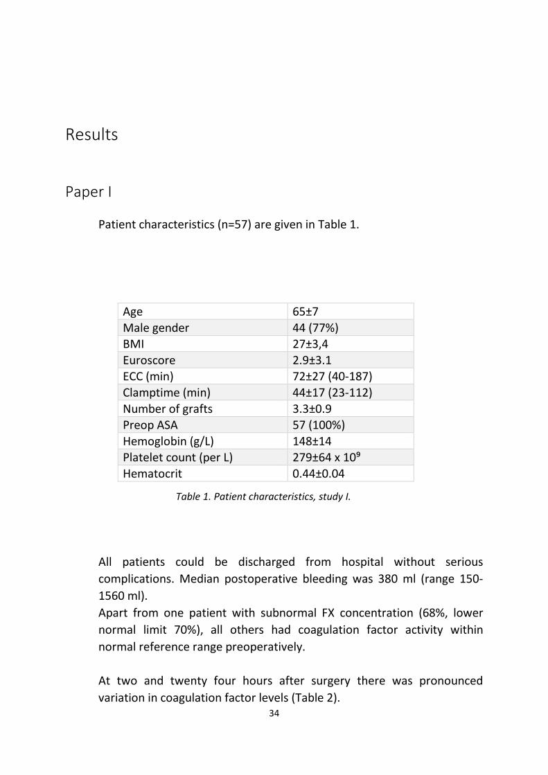
Patient characteristics (n=57) are given in Table 1.
Table 1. Patient characteristics, study I.
All patients could be discharged from hospital without serious
complications. Median postoperative bleeding was 380 ml (range 150-
1560 ml).
Apart from one patient with subnormal FX concentration (68%, lower
normal limit 70%), all others had coagulation factor activity within
normal reference range preoperatively.
At two and twenty four hours after surgery there was pronounced
variation in coagulation factor levels (Table 2).
Age 65±7
Male gender 44 (77%) BMI 27±3,4 Euroscore 2.9±3.1 ECC (min) 72±27 (40-187) Clamptime (min) 44±17 (23-112) Number of grafts 3.3±0.9 Preop ASA 57 (100%) Hemoglobin (g/L) 148±14 Platelet count (per L) 279±64 x 10⁹ Hematocrit 0.44±0.04

35
Table 2. Coagulation factor concentrations before surgery and at 2 and 24 hours after
surgery (absolute and adjusted), *=p<0.05, **=p<0.01, ***=p<0.001 vs before surgery.
Before surgery 2 h Postop 24h postop
Fibrinogen (g/L) Absolute
Adjusted
3.7±0.9
2.5±0.7*** 3.1±0.7***
3.9±0.7* 5.2± 1.0***
FII (%) Absolute
Adjusted
108±12
78±13*** 99±13***
79±11*** 106±20
FV (%) Absolute
Adjusted
111±19
76±18*** 96±20***
81±16*** 106±19*
FVII (%) Absolute
Adjusted
121±31
98±28*** 124±32
57±20*** 76±27***
FVIII (%) Absolute
Adjusted
194±66
175±59* 221±70**
263±76*** 347±95***
FIX (%) Absolute
Adjusted
149±24
143±30 180±29***
146±26 194±38***
FX (%) Absolute
Adjusted
104±18
72±17*** 91±18***
70±14*** 93±20***
FXI (%) Absolute
Adjusted
117±25
91±19*** 115±18
83±18*** 110±24*
FXIII (%) Absolute
Adjusted
135±30
97±26*** 122±31***
94±19*** 125±26**

36
Mean hematocrit fell from 44.1 ± 4.0 % preoperatively to 34.7 ± 3.9 %
two hours after surgery and 33.7 ± 3.6 % after 24 hours (both p<0.001 vs
preoperative).
After adjustment for hemodilution, measurements at two hours after
surgery showed increase in FVIII and FIX, unaltered FVII and FXI and
decrease in fibrinogen, FII, FV, FX and FXIII, Figure 3.
Figure 3. Plasma concentration of coagulation factors before, and 2 and 24 hours after
surgery (adjusted for hemodilution), in percents of preoperative value.
At 24 hours, factor levels adjusted for hemodilution demonstrated
increased fibrinogen, FVIII and FIX, unchanged FII and decreased FV, FVII,
FX, FXI and FXIII, all compared to baseline.
The only correlations between absolute factor level and volume of
postoperative blood loss were the weak ones with fibrinogen at 2 hours
50
75
100
125
150
175
200
Preop 2h Postop 24h Postop
%
FVIII
FIB
FIX
FVII
FII
FV
FX
FXI
FXIII
37
postoperatively (r= -0.33, p=0.019) and FXIII, both pre- (r= -0.34,
p=0.009) and postoperatively (r= -0.41, p=0.003).
Postoperative bleeding volume did not correlate to any other clinical or
laboratory preoperative parameters.
Paper II
Patients were the same cohort as in Paper I, with seven patients
excluded.
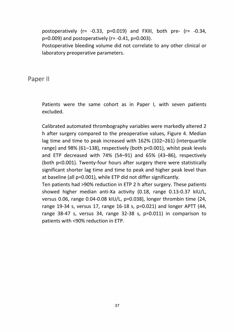
Calibrated automated thrombography variables were markedly altered 2
h after surgery compared to the preoperative values, Figure 4. Median
lag time and time to peak increased with 162% (102–261) (interquartile
range) and 98% (61–138), respectively (both p<0.001), whilst peak levels
and ETP decreased with 74% (54–91) and 65% (43–86), respectively
(both p<0.001). Twenty-four hours after surgery there were statistically
significant shorter lag time and time to peak and higher peak level than
at baseline (all p=0.001), while ETP did not differ significantly.
Ten patients had >90% reduction in ETP 2 h after surgery. These patients
showed higher median anti-Xa activity (0.18, range 0.13-0.37 kIU/L,
versus 0.06, range 0.04-0.08 kIU/L, p=0.038), longer thrombin time (24,
range 19-34 s, versus 17, range 16-18 s, p=0.021) and longer APTT (44,
range 38-47 s, versus 34, range 32-38 s, p=0.011) in comparison to
patients with <90% reduction in ETP.
38
Figure 4. Changes in percent from baseline in CAT parameters. Median values and
interquartile range. ***=p<0.001.
There were moderate correlations between ETP and anti-Xa (r= -0.50,
p=0.01), ETP and thrombin time (r= -0.42, p=0.037) and ETP and APTT (r=
-0.44, p=0.027), all at two hours after operation.
Median postoperative bleeding (380 ml, range 160-1520 ml) did not
correlate with CAT variables at any point. In contrast, postoperative
bleeding volume correlated to TAT (r= 0.57, p<0.001) and F1.2 (r=0.56,
p<0.001) two hours postoperatively.

39
Paper III
All of the study participants completed the study. There was no
difference between Intervention group and control group in any of the
baseline variables, inclusive platelet count, PT INR, APTT, hematocrit and
antithrombin.
Despite randomization, aortic clamp time and ECC time were
significantly longer in control group than in the intervention group,
reflecting more valve surgery patients in that group, Table 5.
Control group (n=30)
Intervention group (n=30)
p
Valve surgery (n)
5 (17%) 2 (7%) 0.23
Total protamine dose (mg)
314 ± 58 319 ± 96 0.78
Total heparin dose (IU)
37167 ± 11573 37150 ± 8734 0.99
Clamp time (min)
57 ± 4 44 ± 3 0.009*
ECC time (min). 87 ± 6 69 ± 4 0.023*
Table 5. Intra- and postoperative characteristics.
40
The intervention group received higher bolus dose heparin (p=0.009),
but the total heparin dose did not differ between the groups (p=0.99).
Fourteen patients in control- and twelve patients in intervention group
received extra heparin during surgery. One patient in each group did not
reach ACT 480 s after the initial heparin bolus dose. The total protamine
dose did not differ significantly (p=0.78).
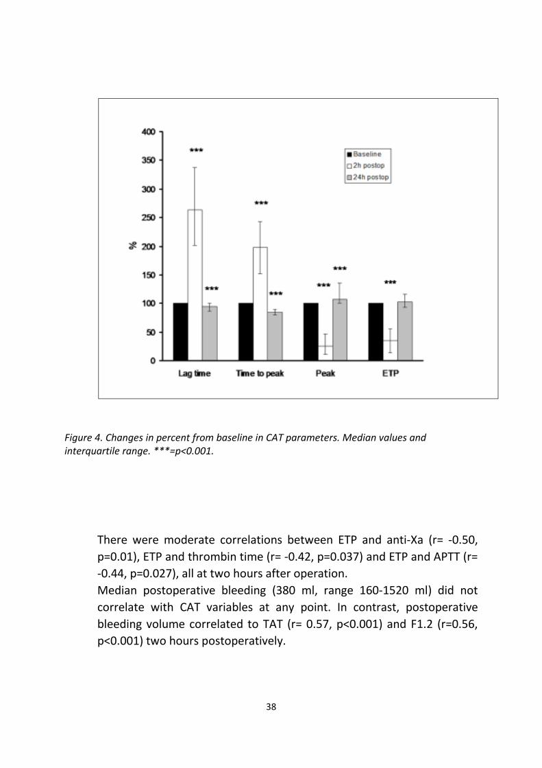
There was a significant reduction in thrombin generation capacity in both
groups postoperatively compared to the preoperative measurements but
to a similar degree in both groups (Figure 5). This change was significant
in all CAT variables (lag time, time to peak, peak value and ETP). The
lowest ETP was registered 4 hours after CPB in both groups. Time to peak
(TTP) was significantly longer in the intervention group at 2 and 4 hours
after surgery but no other statistically significant difference between the
groups was observed.
Figure 5. CAT variables between the groups at four time points. Mean and standard error of
mean. P value for group comparisons with repeated measures ANOVA.
41
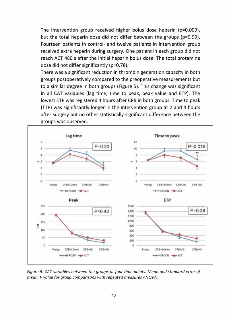
The heparin effect measured as anti-Xa was present in both groups, with
no significant difference (Figure 6). There were significant inverse
correlations between anti Xa levels and ETP 10 min (r= -0.43, p=0.001), 2
hours (r= -0.66, p<0.001) and 4 hours after surgery (r= -0.58, p<0.001).
Postoperative INTEM/HEPTEM ratio correlated also with ETP levels, two
(r=0.44, p=0.001) and four hours (r=0.44, p=0.001) after surgery.
Figure 6. Anti-Xa levels in intervention and control group at four time points. Mean and
standard error of mean. P value for group comparison with repeated measures ANOVA.
The groups did not differ in per- or postoperative bleeding, transfusion
rate, ICU stay or time spent on mechanical respiratory support.

42
Paper IV
The median preoperative and postoperative fibrinogen plasma
concentration was 2.9 g/L (range 1.6–3.4) and 2.0 g/L (range 1.3–2.8)
respectively, p=0.005. There were no significant differences between
recombinant and plasma-derived fibrinogen in any of the
thromboelastometric analyzes at any concentration, Figure 7.
EXTEM-CFT decreased dose dependently after the addition of both
plasma-derived and recombinant fibrinogen (p=0.031 and p=0.007 for
trend, respectively), whereas EXTEM-MCF (p<0.001 both), EXTEM- α
(p=0.002 and p<0.001, respectively), FIBTEM-A10 and FIBTEM-MCF
(p<0.001 all) increased dose dependently. EXTEM-CT, FIBTEM-CT,
FIBTEM-α and FIBTEM LI30 did not change significantly with any of the
fibrinogens.
Figure 3. EXTEM-CT (A), EXTEM-MCF (B), FIBTEM-CT (C), FIBTEM-MCF (D)
A B
C D D

43

44
Discussion
Cardiac surgery with cardiopulmonary bypass (CPB) induces profound
alteration of the whole hemostatic system. Plasma coagulation may contribute
to perioperative coagulopathy but also hemodilution, and platelet dysfunction
may be equally or more important (17, 18). However, most of the studies
measuring coagulation proteins during cardiac surgery are of older date (18,
22-24, 26), reflecting, in some ways not actual clinical and perfusion
management and laboratory methods. That is why we conducted study I, which
aimed to prospectively determine levels of all coagulation factors pre- and
postoperatively in a modern surgery setting.
The absolute levels of all factors measured (apart from FIX) decreased
significantly at two hours after surgery (-4 to -32% of preoperative values), to
an extend greater than the change in hematocrit level solely, implying a certain
consumption mechanism perioperatively. After all, CPB is known to induce
massive thrombin generation (70). During the postoperative course, the
hemostatic balance shifts to a procoagulant state, with increases in fibrinogen,
FVIII and FIX levels. Although the other factors are still statistically lower than
baseline, this difference is small, apart from more pronounced decrease in FVII.
This decrease could theoretically be due to a FVII´s short half-life and its rapid
recruitment by huge amounts of tissue factor, both wound bound and
circulating. The observed variation in factor concentrations does not seem to
be directly associated with postoperative blood loss. The only two factors that
correlated to chest drainage volume at 12 hours were postoperative fibrinogen
and pre- and postoperative FXIII.
The correlation between fibrinogen concentration and postoperative bleeding
has previously been demonstrated by our group (71) and others (72).
Furthermore, prophylactic substitution with fibrinogen concentrate reduces
the blood loss (62). In a recent meta-analysis on use of fresh frozen plasma
(FFP) or fibrinogen in perioperative and massive trauma settings, the latter
seemed to have superior effects regarding blood loss, transfusion rate and
duration of ICU- and hospital stay (73). Fibrinogen substitution is also known to
compensate for hemodilution (74) and thrombocytopenia (75) in animal

45
models. There are several ongoing trials investigating fibrinogen concentrate
use in heart surgery (76).
Findings for FXIII are more contradictory in regard to bleeding, with some
authors reporting relation to bleeding volume (77, 78), while others not (79). A
recent study on substitution with recombinant FXIII before surgery failed to
show any benefit concerning transfusion rate (80). This endpoint is however
elusive and in spite of predefined criteria, transfusion practice in the trial varied
between the study centers.
There is massive thrombin production during the course of cardiac surgery with
CPB (17, 70) with typical bursts immediately after going on CPB and after
reperfusion of the ischemic heart (51, 81). Indirect markers of already formed
thrombin (such as F1.2, D dimer, TAT complex) have predominately being used
in this research. New automated technique for assessing plasma´s potential to
form thrombin has made it potentially easier to study thrombin generation in
surgical setting in real time. By this method, both procoagulants and
anticoagulants in plasma are taken into account and plasma´s ability to trigger
thrombin formation can be quantified (54). However, there are still problems
with intra- and inter-individual variation (82) and lack of standardization (83),
just to name some.
In conducting the study II we speculated that decreased thrombin generation
potential (measured by CAT), would be found postoperatively and that it would
be associated with decline in concentration of individual coagulation factors.
Indeed, the results demonstrated a substantial drop in plasma ability to
generate thrombin two hours after surgery. This finding is in accordance with
some other studies (25, 84-86), but in contrast with others (87, 88), perhaps
due to different sampling time points, overall study design and modifications in
laboratory technique. The concentration of tissue factor (TF) is of importance
for the test result. We deliberately used low TF concentration (1 pM) in order
to improve sensibility of the assay. According to the recommendation of the
manufacturer, the reagent containing 20 pM TF should be used in plasma
containing UFH, but various concentrations have been utilized in research.
Hemker’s group reported recently applying 30 pM TF while testing the cardiac
surgery population (89). Furthermore, heparin presence is actually not
expected, after the protamine neutralization postoperatively.
In our study, there was no correlation between CAT variables and changes in
individual coagulation factor activity. In search for plausible explanation,

46
samples were then tested in respect to heparin effect, showing association
between anti Xa, APTT and thrombin time assays and CAT parameters, thus
implicating inadequate neutralization by protamine and/or heparin rebound.
Increased anti-Xa levels in spite of heparin neutralization have been described
previously (90-92). CAT results in study II did not correlate to postoperative
bleeding volume at any time point.
In light of these results, the study III was designed (in prospective, randomized
manner), in order to optimize per-operative heparin dosing and heparin-
protamine ratio and in that sense improve plasma capacity to generate
thrombin. As the counterpart to the widespread weight based heparin dosing
in the control group, Hepcon HMS device was used in the intervention group,
assessing individual sensitivity to heparin.
In spite of this intervention however, the findings were similar to those in our
previous study, with clear postoperative deterioration of CAT variables, caused
probably by heparin effect, as determined with anti Xa, TT and INTEM/HEPTEM
ratio. There was no significant difference between the study groups in this
regard. Furthermore, similar amounts of heparin and protamine were used
during and after surgery. The groups did not differ in postoperative bleeding,
nor did they in any other clinical parameter. Our population was however
undersized concerning the clinical endpoints.
These results are in contrast with some of the other investigations, which
demonstrated increased heparin and lower protamine doses when guided by
heparin dose response curve (93, 94). That change in given heparin and
protamine dose should imply a hypothetical advantage in terms of more potent
anticoagulation during surgery and at the same time reduction of the negative
effects of protamine overdose on platelet function. Trials in the matter show
great variability, making it very difficult to make direct comparisons.
Regarding the decrease in blood loss after surgery by using protamine titration,
the results are conflicting (95). The most recent meta-analysis found
beneficiary effects of this technique (96). Data in that study were obtained
from four studies, comprising 20 to 247 patients per trial. Each of the trials
described used different protamine dosing regimen in the control group.
In summary, the more expensive and labor intensive titration technique has in
our hands shown no benefit compared to a standard weight-based approach.

47
There has been a revival of fibrinogen as a crucial coagulation factor during
major trauma and surgery. Several guidelines for treatment of trauma have
revised the previously recommended fibrinogen threshold of 1 g/L. The
predominant means for fibrinogen substitution today are cryoprecipitate and
plasma derived fibrinogen concentrate (pdFGN). While cryoprecipitate is still
the treatment of choice for acquired hypofibrinogenemia associated with
major trauma, surgery or postpartum hemorrhage in USA and United Kingdom,
in many European countries purified pdFGN is used instead (97). This product
has recently been licensed for patients with congenital a- and
hypofibrinogenemia also in USA.
Large pools of donor plasma are used in production and it takes up to nine
months from plasma donation to the final product. According to a recent WHO
report (98), there are 92 million blood donations worldwide annually. In 39
countries (out of 159 reporting) blood donations are still not routinely tested
for transfusion transmissible infections. On the other hand, plasma derived
fibrinogen concentrates in the western world have proven extremely safe, and
some 3 million grams of fibrinogen have been administered in Europe since
1985 without viral transmission (97). However, theoretical potential for
transmitting the new infectious agents remains possible.
Regarding plasma, majority of donations comes from 400 centers across US
(99). Giving the fact that manufacturers produce plasma products for the
worldwide market, a possible shortage in any product causes domino effect on
the worldwide supply. Just four months after licensing the pdFGN in Canada,
the demands increased so dramatically that Canada Blood Service was forced
to issue an inventory alert on June 10, 2013 (100), due to the fact that the
current inventory would last only for another three weeks. The use of
fibrinogen concentrate is increasing in US as well. According to (101), the
amount of fibrinogen needed to satisfy 75% of US annual demand (just for
topical administration, as a tissue sealant) is 2650 kg.
Taken all together, the study IV is of interest, investigating for the first time
functional effects of recombinant human fibrinogen. Three different
concentrations of plasma derived and recombinant fibrinogen, targeted to
increase plasma fibrinogen by 0.5; 1 and 1.5 g/L, were tested with
thromboelastometry (TEM) on blood samples collected from cardiac surgery
patients. This method has previously been proven for monitoring fibrinogen
substitution (63, 102, 103). Comparable ex vivo results on fibrin specific and

48
tissue factor activated whole blood clot formation were demonstrated. Similar
findings regarding the viscoelastic properties of a blood clot were subsequently
reported by another group, which used different form of recombinant
fibrinogen, the one obtained from the milk of transgenic cows (101).
Recombinant fibrinogen concentrates remain to be tested in a future research,
in larger scale studies. At present, it is hard to predict which manufacturing
method will prevail (if any).
49
Summary
(1) Concentration of plasma coagulation factors varies after cardiac surgery
with CPB. Apart from FVIII and FIX, all others decrease, secondary to
hemodilution and probably consumption. Factors engaged in blood clot
stability (fibrinogen and FXIII) are the only ones demonstrating association
with postoperative bleeding volume.
(2) Thrombin generation capacity as measured with calibrated automated
thrombography (CAT) decreases profoundly early after cardiac surgery due
to sustained heparin effect.
(3) Heparin/protamine titration technique does not improve thrombin
generation capacity in plasma at two and four hours postoperatively.
(4) Similar effects on clot stability ex vivo were shown after addition of
recombinant human fibrinogen concentrate and plasma derived fibrinogen
concentrate.

50
Acknowledgements
Härmed vill jag rikta ett stort tack till:
Min handledare professor Anders Jeppsson, en enastående vetenskapsman
och kirurg, utan vars entusiasm, kunskap och tålamod inget av detta hade blivit
gjort
Min bihandledare, kollega och chef Fariba Baghaei, för all hjälp med arbetena
och för hennes positiva inställning till allt
Min inofficiella bihandledare Kenny Hansson, för lärorika diskussioner
Min tidigare chef Lennart Stigendal, för allt han lärt mig, både om
koagulationen och livet
Alla mina medförfattare
All personal på Thoraxkirurgiska forskningsenheten, i synnerhet
forskningssköterskorna
Alla kollegor på Hematologi och Medicin
Alla på Koagulationscentrum och laboratoriet
Min familj

51
52
References
1. Cooley DA, Frazier OH. The past 50 years of cardiovascular surgery. Circulation. 2000 Nov 14;102(20 Suppl 4):IV87-93. 2. Cohn LH. Fifty years of open-heart surgery. Circulation. 2003 May 6;107(17):2168-70. 3. Pezzella AT. International cardiac surgery: a global perspective. Semin Thorac Cardiovasc Surg. 2002 Oct;14(4):298-320. 4.http://www.surgeons.org/media/18749201/gale_global_burden_of_cardiac_surgical_disease_racs_version.pdf 5. http://www.ucr.uu.se/swedeheart/index.php/annual-report. 6. Bacchetta MD, Ko W, Girardi LN, Mack CA, Krieger KH, Isom OW, et al. Outcomes of cardiac surgery in nonagenarians: a 10-year experience. Ann Thorac Surg. 2003 Apr;75(4):1215-20. 7. Ferraris VA, Davenport DL, Saha SP, Bernard A, Austin PC, Zwischenberger JB. Intraoperative transfusion of small amounts of blood heralds worse postoperative outcome in patients having noncardiac thoracic operations. Ann Thorac Surg. 2011 Jun;91(6):1674-80. 8. Koch CG, Li L, Sessler DI, Figueroa P, Hoeltge GA, Mihaljevic T, et al. Duration of red-cell storage and complications after cardiac surgery. N Engl J Med. 2008 Mar 20;358(12):1229-39. 9. Besser MW, Klein AA. The coagulopathy of cardiopulmonary bypass. Crit Rev Clin Lab Sci. 2010 Dec;47(5-6):197-212. 10. Alstrom U, Levin LA, Stahle E, Svedjeholm R, Friberg O. Cost analysis of re-exploration for bleeding after coronary artery bypass graft surgery. Br J Anaesth. 2012 Feb;108(2):216-22. 11. Unsworth-White MJ, Herriot A, Valencia O, Poloniecki J, Smith EE, Murday AJ, et al. Resternotomy for bleeding after cardiac operation: a marker for increased morbidity and mortality. Ann Thorac Surg. 1995 Mar;59(3):664-7. 12. Bevan DH. Cardiac bypass haemostasis: putting blood through the mill. Br J Haematol. 1999 Feb;104(2):208-19. 13. Bull BS, Huse WM, Brauer FS, Korpman RA. Heparin therapy during extracorporeal circulation. II. The use of a dose-response curve to individualize heparin and protamine dosage. J Thorac Cardiovasc Surg. 1975 May;69(5):685-9. 14. Young JA, Kisker CT, Doty DB. Adequate anticoagulation during cardiopulmonary bypass determined by activated clotting time and the appearance of fibrin monomer. Ann Thorac Surg. 1978 Sep;26(3):231-40. 15. Nielsen VG. Coagulation crystal ball: why can't we predict bleeding after cardiac surgery? Anesth Analg. 2012 Sep;115(3):490-2. 16. Campbell DJ, Dixon B, Kladis A, Kemme M, Santamaria JD. Activation of the kallikrein-kinin system by cardiopulmonary bypass in humans. Am J of Physiol Regul Integr Comp Physiol. 2001 Oct;281(4):R1059-70. 17. Sniecinski RM, Chandler WL. Activation of the hemostatic system during cardiopulmonary bypass. Anesth Analg. 2011 Dec;113(6):1319-33. 18. Harker LA, Malpass TW, Branson HE, Hessel EA, 2nd, Slichter SJ. Mechanism of abnormal bleeding in patients undergoing cardiopulmonary bypass: acquired transient platelet dysfunction associated with selective alpha-granule release. Blood. 1980 Nov;56(5):824-34. 19. Boisclair MD, Lane DA, Philippou H, Esnouf MP, Sheikh S, Hunt B, et al. Mechanisms of thrombin generation during surgery and cardiopulmonary bypass. Blood. 1993 Dec 1;82(11):3350-7. 20. Burman JF, Chung HI, Lane DA, Philippou H, Adami A, Lincoln JC. Role of factor XII in thrombin generation and fibrinolysis during cardiopulmonary bypass. Lancet. 1994 Oct 29;344(8931):1192-3.
53
21. Paparella D, Galeone A, Venneri MT, Coviello M, Scrascia G, Marraudino N, et al. Activation of the coagulation system during coronary artery bypass grafting: comparison between on-pump and off-pump techniques. J Thorac Cardiovasc Surg. 2006 Feb;131(2):290-7. 22. Moriau M, Masure R, Hurlet A, Debeys C, Chalant C, Ponlot R, et al. Haemostasis disorders in open heart surgery with extracorporeal circulation. Importance of the platelet function and the heparin neutralization. Vox sang. 1977;32(1):41-51. 23. Milam JD, Austin SF, Martin RF, Keats AS, Cooley DA. Alteration of coagulation and selected clinical chemistry parameters in patients undergoing open heart surgery without transfusions. Am J Clin Path. 1981 Aug;76(2):155-62. 24. Porter JM, Silver D. Alterations in fibrinolysis and coagulation associated with cardiopulmonary bypass. J Thorac Cardiovasc Surg. 1968 Dec;56(6):869-78. 25. Davidson SJ, Burman JF, Philips SM, Onis SJ, Kelleher AA, De Souza AC, et al. Correlation between thrombin potential and bleeding after cardiac surgery in adults. Blood Coagul Fibrinolysis. 2003 Feb;14(2):175-9. 26. Gans H, Krivit W. Problems in hemostasis during openheart surgery. IV. On the changes in the blood clotting mechanism during cardiopulmonary bypass procedures. Ann Surg. 1962 Mar;155:353-9. 27. Gelb AB, Roth RI, Levin J, London MJ, Noall RA, Hauck WW, et al. Changes in blood coagulation during and following cardiopulmonary bypass: lack of correlation with clinical bleeding. Am J Clin Path. 1996 Jul;106(1):87-99. 28. Wardrop D, Keeling D. The story of the discovery of heparin and warfarin. Br J Haematol. 2008 Jun;141(6):757-63. 29. Crafoord C, Gallie WE, Bauer G. Heparin in surgery. BMJ. 1947 Sep 27;2(4525):503. 30. Barritt DW, Jordan SC. Anticoagulant drugs in the treatment of pulmonary embolism. A controlled trial. Lancet. 1960 Jun 18;1(7138):1309-12. 31. Basu D, Gallus A, Hirsh J, Cade J. A prospective study of the value of monitoring heparin treatment with the activated partial thromboplastin time. N Engl J Med. 1972 Aug 17;287(7):324-7. 32. Zehnder J, Price E, Jin J. Controversies in heparin monitoring. Am J Hematol. 2012 May;87 Suppl 1:S137-40. 33. Carlsson P, Kjellen L. Heparin biosynthesis. Handb Exp Pharmacol. 2012 (207):23-41. 34. Mulloy B. Structure and physicochemical characterisation of heparin. Handb Exp Pharmacol. 2012 (207):77-98. 35. Mulloy B, Gray E, Barrowcliffe TW. Characterization of unfractionated heparin: comparison of materials from the last 50 years. Thromb Haemost. 2000 Dec;84(6):1052-6. 36. http://www.usp.org/sites/default/files/usp_pdf/EN/USPNF/key-issues/heparinPotency.pdf. 37. Brinkhous KM, Smith HP, Jr., Warner ED, Seegers WH. Heparin and Blood Clotting. Science. 1939 Dec 8;90(2345):539. 38. Astrup T. Heparin and the Inhibition of Blood-Clotting. Science. 1939 Jul 14;90(2324):36. 39. Gray E, Hogwood J, Mulloy B. The anticoagulant and antithrombotic mechanisms of heparin. Handb Exp Pharmacol. 2012 (207):43-61. 40. Lindahl U, Thunberg L, Backstrom G, Riesenfeld J, Nordling K, Bjork I. Extension and structural variability of the antithrombin-binding sequence in heparin. J Biol Chem. 1984 Oct 25;259(20):12368-76. 41. Swedenborg J. The mechanisms of action of alpha- and beta-isoforms of antithrombin. Blood Coagul Fibrinolysis. 1998 Nov;9 Suppl 3:S7-10. 42. Lindahl U, Backstrom G, Hook M, Thunberg L, Fransson LA, Linker A. Structure of the antithrombin-binding site in heparin. PNAS. 1979 Jul;76(7):3198-202. 43. Jaques LB. Protamine--antagonist to heparin. CMAJ. 1973 May 19;108(10):1291-7. 44. Pai M, Crowther MA. Neutralization of heparin activity. Handb Exp Pharmacol. 2012 (207):265-77. 45 http://www.ema.europa.eu/docs/en_GB/document_library/Report/2012/12/W500135918.pdf.

54
46. Hagedorn HC, Jensen BN, Krarup NB, Wodstrup I. Landmark article Jan 18, 1936: Protamine insulinate. By H.C. Hagedorn, B.N. Jensen, N.B. Krarup, and I. Wodstrup. JAMA. 1984 Jan 20;251(3):389-92. 47. Chargaff E OK. Studies on the chemistry of blood coagulation. VI. Studies on action of heparin and other anticoagulants. The influence of protamine on the anticoagulant effect in vivo. J Biol Chem. 1937 (122):153. 48. Schulman S, Beyth RJ, Kearon C, Levine MN, American College of Chest P. Hemorrhagic complications of anticoagulant and thrombolytic treatment: American College of Chest Physicians Evidence-Based Clinical Practice Guidelines (8th Edition). Chest. 2008 Jun;133(6 Suppl):257S-98S. 49. Crawley JT, Zanardelli S, Chion CK, Lane DA. The central role of thrombin in hemostasis. J Thromb Haemost. 2007 Jul;5 Suppl 1:95-101. 50. Siller-Matula JM, Schwameis M, Blann A, Mannhalter C, Jilma B. Thrombin as a multi-functional enzyme. Focus on in vitro and in vivo effects. Thromb Haemost. 2011 Dec;106(6):1020-33. 51. Raivio P, Lassila R, Petaja J. Thrombin in myocardial ischemia-reperfusion during cardiac surgery. Ann Thorac Surg. 2009 Jul;88(1):318-25. 52. Macfarlane RG, Biggs R. A thrombin generation test; the application in haemophilia and thrombocytopenia. J Clin Pathol. 1953 Feb;6(1):3-8. 53. Mann KG, Brummel K, Butenas S. What is all that thrombin for? J Thromb Haemost. 2003 Jul;1(7):1504-14. 54. Hemker HC, Giesen P, AlDieri R, Regnault V, de Smed E, Wagenvoord R, et al. The calibrated automated thrombogram (CAT): a universal routine test for hyper- and hypocoagulability. Pathophysiol Haemost Thromb. 2002 Sep-Dec;32(5-6):249-53. 55. Hemker HC, Al Dieri R, De Smedt E, Beguin S. Thrombin generation, a function test of the haemostatic-thrombotic system. Thromb Haemost. 2006 Nov;96(5):553-61. 56. Blomback B. Fibrinogen: evolution of the structure-function concept. Keynote address at fibrinogen 2000 congress. Ann NJ Acad Sci. 2001;936:1-10. 57. Mosesson MW. Fibrinogen and fibrin structure and functions. J Thromb Haemost. 2005 Aug;3(8):1894-904. 58. Wolberg AS. Determinants of fibrin formation, structure, and function. Curr Opin Hematol. 2012 Sep;19(5):349-56. 59. Fenger-Eriksen C, Ingerslev J, Sorensen B. Fibrinogen concentrate--a potential universal hemostatic agent. Expert Opin Biol Ther. 2009 Oct;9(10):1325-33. 60. Rossaint R, Bouillon B, Cerny V, Coats TJ, Duranteau J, Fernandez-Mondejar E, et al. Management of bleeding following major trauma: an updated European guideline. Crit Care. 2010;14(2):R52. 61. Fenger-Eriksen C, Jensen TM, Kristensen BS, Jensen KM, Tonnesen E, Ingerslev J, et al. Fibrinogen substitution improves whole blood clot firmness after dilution with hydroxyethyl starch in bleeding patients undergoing radical cystectomy: a randomized, placebo-controlled clinical trial. J Thromb Haemost. 2009 May;7(5):795-802. 62. Karlsson M, Ternstrom L, Hyllner M, Baghaei F, Flinck A, Skrtic S, et al. Prophylactic fibrinogen infusion reduces bleeding after coronary artery bypass surgery. A prospective randomised pilot study. Thromb Haemosth. 2009 Jul;102(1):137-44. 63. Levy JH, Szlam F, Tanaka KA, Sniecienski RM. Fibrinogen and hemostasis: a primary hemostatic target for the management of acquired bleeding. Anesth Analg. 2012 Feb;114(2):261-74. 64. Ludlam CA, Powderly WG, Bozzette S, Diamond M, Koerper MA, Kulkarni R, et al. Clinical perspectives of emerging pathogens in bleeding disorders. Lancet. 2006 Jan 21;367(9506):252-61. 65. Norja P, Lassila R, Makris M. Parvovirus transmission by blood products - a cause for concern? Br J Haematol. 2012 Nov;159(4):385-93. 66. Pereira A. Cryoprecipitate versus commercial fibrinogen concentrate in patients who occasionally require a therapeutic supply of fibrinogen: risk comparison in the case of an emerging transfusion-transmitted infection. Haematologica. 2007 Jun;92(6):846-9.
55
67. Grillberger L, Kreil TR, Nasr S, Reiter M. Emerging trends in plasma-free manufacturing of recombinant protein therapeutics expressed in mammalian cells. Biotechnol J. 2009 Feb;4(2):186-201. 68. Roth-Isigkeit A, Borstel TV, Seyfarth M, Schmucker P. Perioperative serum levels of tumour-necrosis-factor alpha (TNF-alpha), IL-1 beta, IL-6, IL-10 and soluble IL-2 receptor in patients undergoing cardiac surgery with cardiopulmonary bypass without and with correction for haemodilution. Clin Exp Immunol. 1999 Nov;118(2):242-6. PubMed PMID: 10540185. 69. Luddington RJ. Thrombelastography/thromboelastometry. Clin Lab Haematol. 2005 Apr;27(2):81-90. 70. Edmunds LH, Jr., Colman RW. Thrombin during cardiopulmonary bypass. Ann Thorac Surg. 2006 Dec;82(6):2315-22. 71. Karlsson M, Ternstrom L, Hyllner M, Baghaei F, Nilsson S, Jeppsson A. Plasma fibrinogen level, bleeding, and transfusion after on-pump coronary artery bypass grafting surgery: a prospective observational study. Transfusion. 2008 Oct;48(10):2152-8. 72. Gravlee GP, Arora S, Lavender SW, Mills SA, Hudspeth AS, Cordell AR, et al. Predictive value of blood clotting tests in cardiac surgical patients. Ann Thorac Surg. 1994 Jul;58(1):216-21. 73. Kozek-Langenecker S, Sorensen B, Hess JR, Spahn DR. Clinical effectiveness of fresh frozen plasma compared with fibrinogen concentrate: a systematic review. Crit Care. 2011;15(5):R239. 74. Martini J, Maisch S, Pilshofer L, Streif W, Martini W, Fries D. Fibrinogen concentrate in dilutional coagulopathy: a dose study in pigs. Transfusion. 2013 May 14. 75. Velik-Salchner C, Haas T, Innerhofer P, Streif W, Nussbaumer W, Klingler A, et al. The effect of fibrinogen concentrate on thrombocytopenia. J Thromb Haemost. 2007 May;5(5):1019-25. 76. Available from: http://clinicaltrials.gov. 77. Chandler WL, Patel MA, Gravelle L, Soltow LO, Lewis K, Bishop PD, et al. Factor XIIIA and clot strength after cardiopulmonary bypass. Blood Coagul Fibrinolysis. 2001 Mar;12(2):101-8. 78. Shainoff JR, Estafanous FG, Yared JP, DiBello PM, Kottke-Marchant K, Loop FD. Low factor XIIIA levels are associated with increased blood loss after coronary artery bypass grafting. J Thorac Cardiovasc Surg. 1994 Sep;108(3):437-45. 79. Blome M, Isgro F, Kiessling AH, Skuras J, Haubelt H, Hellstern P, et al. Relationship between factor XIII activity, fibrinogen, haemostasis screening tests and postoperative bleeding in cardiopulmonary bypass surgery. Thromb Haemosth. 2005 Jun;93(6):1101-7. 80. Karkouti K, von Heymann C, Jespersen CM, Korte W, Levy JH, Ranucci M, et al. Efficacy and safety of recombinant factor XIII on reducing blood transfusions in cardiac surgery: A randomized, placebo-controlled, multicenter clinical trial. J Thorac Cardiovasc Surg. 2013 Jun 29. 81. Chandler WL, Velan T. Estimating the rate of thrombin and fibrin generation in vivo during cardiopulmonary bypass. Blood. 2003 Jun 1;101(11):4355-62. 82. Butenas S, van't Veer C, Mann KG. "Normal" thrombin generation. Blood. 1999 Oct 1;94(7):2169-78. 83. Dargaud Y, Luddington R, Gray E, Negrier C, Lecompte T, Petros S, et al. Effect of standardization and normalization on imprecision of calibrated automated thrombography: an international multicentre study. Br J Haematol. 2007 Oct;139(2):303-9. 84. Coakley M, Hall JE, Evans C, Duff E, Billing V, Yang L, et al. Assessment of thrombin generation measured before and after cardiopulmonary bypass surgery and its association with postoperative bleeding. J Thromb Haemost. 2011 Feb;9(2):282-92. 85. Schols SE, Lance MD, Feijge MA, Damoiseaux J, Marcus MA, Hamulyak K, et al. Impaired thrombin generation and fibrin clot formation in patients with dilutional coagulopathy during major surgery. Thromb Haemost. 2010 Feb;103(2):318-28. 86. Wong RK, Sleep JR, Visner AJ, Raasch DJ, Lanza LA, DeValeria PA, et al. Thrombography reveals thrombin generation potential continues to deteriorate following cardiopulmonary bypass surgery despite adequate hemostasis. J Extra Corpor Techol. 2011 Mar;43(1):19-25.
56
87. Solomon C, Rahe-Meyer N, Sorensen B. Fibrin formation is more impaired than thrombin generation and platelets immediately following cardiac surgery. Thromb Res. 2011 Sep;128(3):277-82. 88. Lison S, Dietrich W, Braun S, Boehm J, Schuster T, Englhard A, et al. Enhanced thrombin generation after cardiopulmonary bypass surgery. Anesth Analg. 2011 Jan;112(1):37-45. 89. Bosch Y, Al Dieri R, ten Cate H, Nelemans P, Bloemen S, Hemker C, et al. Preoperative thrombin generation is predictive for the risk of blood loss after cardiac surgery: a research article. J Cardiothorac Surg. 2013;8:154. 90. Teoh KH, Young E, Blackall MH, Roberts RS, Hirsh J. Can extra protamine eliminate heparin rebound following cardiopulmonary bypass surgery? J Thorac Cardiovasc Surg. 2004 Aug;128(2):211-9. 91. Taneja R, Marwaha G, Sinha P, Quantz M, Stitt L, Gao R, et al. Elevated activated partial thromboplastin time does not correlate with heparin rebound following cardiac surgery. Can J Anesth. 2009 Jul;56(7):489-96. 92. Raivio P, Kuitunen A, Petaja J, Ilveskero S, Lassila R. Monitoring high-dose heparinization during cardiopulmonary by-pass--a comparison between prothrombinase-induced clotting time (PiCT) and two chromogenic anti-factor Xa activity assays. Thromb Haemost. 2008 Feb;99(2):427-34. 93. Despotis GJ, Joist JH, Hogue CW, Jr., Alsoufiev A, Kater K, Goodnough LT, et al. The impact of heparin concentration and activated clotting time monitoring on blood conservation. A prospective, randomized evaluation in patients undergoing cardiac operation. J Thorac Cardiovasc Surg. 1995 Jul;110(1):46-54. 94. Beholz S, Grubitzsch H, Bergmann B, Wollert HG, Eckel L. Hemostasis management by use of Hepcon/HMS: increased bleeding without increased need for blood transfuion. Thorac Cardiovasc Surg 1999 Oct;47(5):322-7. 95. Aziz KA, Masood O, Hoschtitzky JA, Ronald A. Does use of the Hepcon point-of-care coagulation monitor to optimise heparin and protamine dosage for cardiopulmonary bypass decrease bleeding and blood and blood product requirements in adult patients undergoing cardiac surgery? Interact Cardiovasc Thorac Surg. 2006 Aug;5(4):469-82. 96. Wang J, Ma HP, Zheng H. Blood loss after cardiopulmonary bypass, standard vs titrated protamine: A meta-analysis. Neth J Med. 2013 Apr;71(3):123-7. 97. Tanaka KA, Kor DJ. Emerging haemostatic agents and patient blood management. Best Pract Res Clin Anaesth. 2013 Mar;27(1):141-60. 98. http://www.who.int/bloodsafety/global_database/GSBS_Summary_Report_2011.pdf. 99 http://www.ftc.gov/os/caselist/1010153/110601grifolsacmpt.pdf. 100. http://www.bloodservice.ca/CentreApps/Internet/UW_V502_MainEngine.nsf/resources/Plasma+Products+and+Services+2/§file/ppp-Alert-Report-06-10-2013.pdf. 101. Calcaterra J, Van Cott KE, Butler SP, Gil GC, Germano M, van Veen HA, et al. Recombinant human fibrinogen that produces thick fibrin fibers with increased wound adhesion and clot density. Biomacromolecules. 2013 Jan 14;14(1):169-78. 102. Solomon C, Cadamuro J, Ziegler B, Schochl H, Varvenne M, Sorensen B, et al. A comparison of fibrinogen measurement methods with fibrin clot elasticity assessed by thromboelastometry, before and after administration of fibrinogen concentrate in cardiac surgery patients. Transfusion. 2011 Aug;51(8):1695-706. 103. Momeni M, Carlier C, Baele P, Watremez C, Van Dyck M, Matta A, et al. Fibrinogen concentration significantly decreases after on-pump versus off-pump coronary artery bypass surgery: a systematic point-of-care ROTEM analysis. J Cardiothorac Vasc Anesth. 2013 Feb;27(1):5-11.