plaque size heterogeneity: lymphocytic choriomeningitisaem.asm.org/content/20/1/123.full.pdf ·...
TRANSCRIPT

APPuED MICROBOLOGY, JUIY 1970, P. 123-128 Vol. 20, No. ICopyright © 1970 American Society for Microbiology Printed in U.S.A.
Plaque Size Heterogeneity: a Genetic Trait ofLymphocytic Choriomeningitis Virus
A. J. PULKKINEN AND C. J. PFAUDepartment of Microbiology, University of Massachusetts, Amherst, Massachusetts 01002
Received for publication 6 April 1970
All of the ten strains of lymphocytic choriomeningitis virus assayed on BHK 21/1 3S cells showed various degrees of plaque size heterogeneity. The amount of virusreleased from these plaques was usually very small because of rapid photodynamicinactivation by neutral red. When virus from large and small plaques of a specificstrain was plated, the same distribution of plaque size was obtained from eachclone. Although it was shown that surface virus could possibly be randomly dis-tributed at the time of addition of neutral red overlays, no virus could be isolatedfrom nonplaque areas. Two different strains of virus (CA1371 and WE) withmarkedly different plaque size ranges were separated by plaque excision fromplates infected with a mixture of both viruses.
Although several plaquing systems for lym-phocytic choriomeningitis (LCM) virus havebeen reported (16), the recent BHK 21 /13Sagarose suspension assay employed by Sedwickand Wiktor (18) has been the only one found tobe reproducible in our laboratory. With thestandard use of a plaque assay, the need forcloned strains of virus in genetic and biochemicalwork is obvious. Although usually routine, theexperiments demonstrating that cloning can beachieved with LCM virus are presented here forthe following reasons: the virus is unusually sensi-tive to inactivation by neutral red in the presenceor absence of light, and the degree of plaque sizeheterogeneity appears to be a heritable propertyof various strains of virus.
MATERIALS AND METHODSViruses. The origin and passage histories of the
LCM strains are given in Table 1.Cell culture. BHK 21/13S cells from T. J. Wiktor
were received at the 18th-passage level. Cells weregrown in monolayers (for no more than 15 passages)or in suspension. The BHK 21 monolayer mediumwas that used by Vahari et al. (22). Propagationtechniques with Blake bottles were essentially asdescribed by Sedwick and Wiktor (18). Cells wereadapted for suspension by the following technique.Monolayers were dispersed with ethylenediamine-tetraacetic acid (EDTA; 14), and these cells wereused to form another monolayer which was againsimilarly dispersed. The cells treated in this way werethen grown in suspension using minimal essentialmedium (5) with 2X vitamins and amino acidsminus Ca and Mg, and supplemented with 0.1 mg ofFeNO3-9H20 per liter, 5.5 g of D-glucose per liter,
20% tryptose phosphate broth, and 10% heat in-activated (56 C for 30 min) fetal calf serum. Cellswere then taken from cultures at concentrationsbetween 106 and 4.0 X 10⁶/ml for use in the plaqueassay. The log phase of growth contained between0.2 X 106 and 106 cells/ml, having a division time ofapproximately 12 hr. Cultures were always split tobetween 105 and 8.0 X 1io cells/ml with no mediasupplement until a density of 4 X 10⁶ cells/ml wasreached. The heat-inactivated sera used in bothtypes of growth media had to be screened before usesince some lots completely inhibited plaque forma-tion.
Measurement of infectious virus. The LDw assay,with Twin Oak Farms Swiss mice, has been described(16). The plaque assay was that used by Sedwick andWiktor (18), with the following modifications: plateswere incubated in a 2.5% CO2 atmosphere with0.1% sodium bicarbonate the final concentration inthe agarose-overlay medium. Instead of mixing virusdilutions with cell suspensions prior to the additionof agarose, the cell-agarose over-layer was pouredand allowed to harden, and then 0.1 ml of the virusdilution was pipetted directly onto the surface (T. J.Wiktor, unpublished data). In addition to beingequally as sensitive as the original, this modifiedtechnique provided excellent cell viability and enabledus to maintain the plates as long as 4 days beforeinfection. Plaque counts on strains WE, CA1371, andG45 were virtually identical whether plates wereaged 1 or 4 days at the time of infection. All LCMstocks exhibited a linear relation between plaquenumber (the range examined was 0 to 100) and rela-tive virus concentration. When plated in triplicate,dilutions of G45, CAl 371, or WE showed a standarderror of no more than 5%. Falcon tissue culture (no.3002) or bacteriology (no. 1007) grade dishes wereused. The bacteriology grade dishes, known to be
123
on July 5, 2018 by guesthttp://aem
.asm.org/
Dow
nloaded from
PULKKINEN AND PFAU
TABLE 1. Characteristics of ten LCM strains
Origin and primary literature citation
American Type Culture Collection,Rockville, Md. (1)
W. P. Rowe, Bethesda, Md. (17)W. P. Rowe, Bethesda, Md. (10)T. J. Wiktor, Philadelphia, Pa. (25)
R. W. Sidwell, Birmingham, Ala. (19)M. Volkert, Copenhagen, Denmark (23)J. Hotchin, Albany, N.Y. (7)W. P. Rowe, Bethesda, Md. (17)B. E. Kirk, Morgantown, W.Va. (12)F. Lehmann-Grube, Giessen, BRD (8)
Passage historya
Mk6, M/B206-L2
M/B4-LI2M/Br-NoneHDCS26, BHK21/13S1, HDCS2,BHK21 l-None
M/B3-L4M/S7-L8M/B6L,1-L2M/B,KLS4-L30G/B1-L4M/B3-Li
a Passage history notations are those used by Hotchin and Benson (6). Passage to the left of the dashis that in the laboratory furnishing the strain; to the right is that in our laboratory. Abbreviations are
B, brain; G, guinea pig; HDCS, human diploid cell strain; K, kidney; L, L cells; Li, liver; M, mouse;Mk, monkey; and S, spleen.
I PFU/LDw ratios were determined by simultaneous use of a dilution series for injection into ani-mals (6 mice/dilution) and infection of duplicate assay plates.
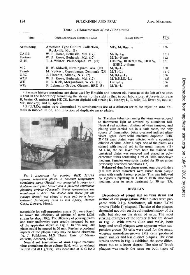
FIG. 1. Apparatus for pourinig BHK 21/13Sagarose suspensioni plates. A constant temperaturecirculating pump (Haake) was connected in series to a
double-walled glass beaker and a jacketed continuouspipetting syringe (Cornwall). Water temperature wasmaintained at 42 C. The double arm sleeve for thesyringe (insert) was closed at both ends by a heat-resistant, fast-drying resin (5 min Epoxy, DevconCorp., Danvers, Mass.).
acceptable for cell-suspension assays (4), were foundto lower the efficiency of plating of some LCMstrains by about 30%. The efficiency of pouring platesand their uniformity were greatly increased by useof the apparatus shown in Fig. 1. In this way, 200plates could be poured in 20 min. Further proceduralaspects of the plaque assay may be found elsewhere(A. J. Pulkkinen, M.S. Thesis, Univ. of Massa-chusetts, Amherst, 1969).
Neutral red inactivation of virus. Liquid medium:virus-containing tissue culture fluid, with or withoutneutral red (0.1 g/liter), was incubated at 37 C for 2
hr. The glass tubes containing the virus were exposedto fluorescent light or covered by aluminum foil.Neutral red addition, dilution of virus samples, andplating were carried out in a dark room, the onlysource of illumination being overhead indirect ultra-violet lights. Semi-solid medium: duplicate BHK21/13S agar plates were infected with a countabledilution of virus. After 4 days, one of the plates wasstained with neutral red in the usual manner (18)At 4 hr, the cell layer from both the stained andunstained plates was removed and placed in poly-carbonate tubes containing 1 ml of BHK monolayermedium. Samples were sonic treated for 30 sec underpreviously described conditions (15).
Release of virus from plaque areas. Agarose cylinders(1.0 mm inner diameter) were exised from plaqueareas with sterile Pasteur pipettes. This was followedby vigorous pipetting in 1 ml of BHK monolayermedium, prior to sonic treatment for 30 sec (15).
RESULTS
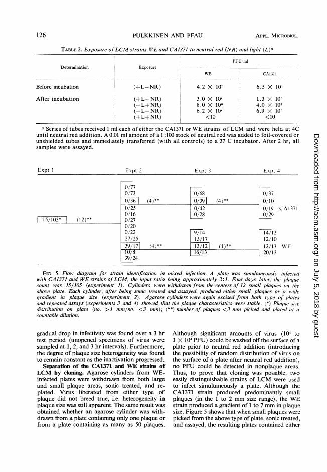
Dependence of plaque size on virus strain andmethod of cell propagation. When plates were pre-pared with 0.1 % bicarbonate, all tested LCMstrains (Table 1) plaqued. The size of the plaquesdepended not only on the cultivation history of thecells, but also on the strain of virus. The moststriking examples of the former factor are shownin Fig. 2. With strains G-45 and WCP, bothlarge and small plaques were apparent when sus-
pension-grown (S) cells were used for the assay,whereas monolayer-grown (M) cells producedmuch smaller and less distinct plaques. The virusstrains shown in Fig. 3 exhibited the same differ-ences but to a lesser degree. The size of Traubplaques was nearly the same on both types of
Virus
Armstrong
CA1371FortnerG-45
M-7TraubUBCWCPWEWE3
PFU/LDbob
1:6
1:121:21:1
1:81:191:61:231:61:6
124 APPL. MICROBIOL.
.l..f.m..,.!., .:rl."?4..,:
'k.
on July 5, 2018 by guesthttp://aem
.asm.org/
Dow
nloaded from

HETEROGENEITY OF LCM VIRUS PLAQUES
rFo. 4. WE3, WE, M-7, Fortner (Fo), and UBCFIG. 2. G45 (right side) and WCP (left side) strains ofLCM 96 hr postinfection. All cells used for
strains ofLCM 96 hr postinfection. The cells used for the assays were grown in suspension.the assays in the upper half of the picture were grownin suspension(S), whereas those in the lower half weregrown in monolayes .(..l
grown in monolayers (M).
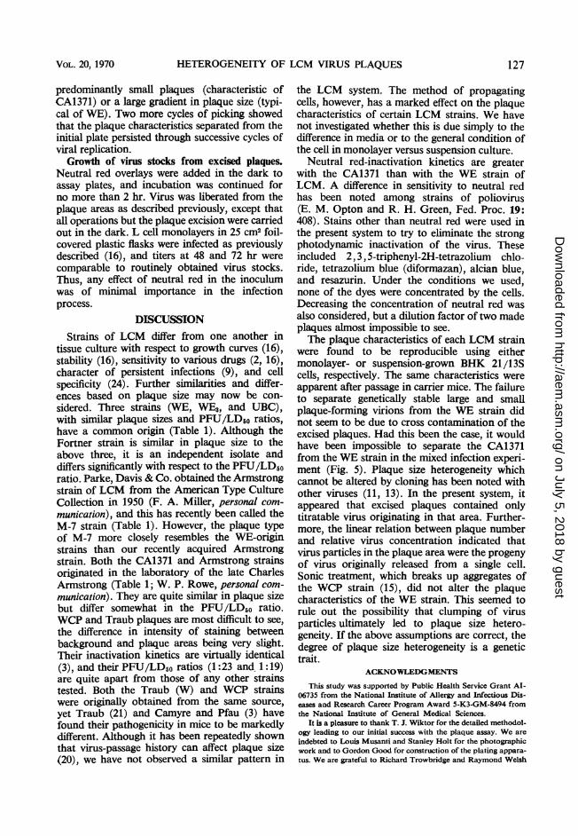
FIG. 3. Armstrong (Arm.), CA1371 (CA), andTraub (Tr) strains of LCM 96 hr postinfection. Thecells used for the assay in the upper half of the picturewere grown in suspension (S), whereas those in thelower half were grown in monolayers (M).
cells, but the plaques on M cells had more sharplydefined edges. With strain CA1371, the plaquesize gradient was larger on S than onM cells. Thestrains shown in Fig. 4 (FO, M-7, WE, WE3, andUBC) were not strikingly dependent on themethod of culture and exhibited a plaque sizegradient from 1 to 7 mm.
Neutral red sensitivity of LCM. Since theamount of virus recoverable from excised plaquesseemed unususally low, 6 X 10² to 60 X 102plaque-forming units (PFU) depending on thestrain of virus used, the effect of neutral red on thesystem was examined. Plates were exposed to adilution of WE virus that would yield about 40plaques. After the usual 4-day incubation period,one plate was stained with neutral red. All plateswere incubated for an additional 4 hr at 37 C.The cell layer (plus neutral red layer) was strippedfrom the plates, sonic treated in liquid medium,and assayed. A typical result was that sonic-treated material of cells never exposed to neutralred yielded 3 X 10 PFU whereas those overlayedwith the dye contained 1.2 X 10⁴ PFU. Noattempt was made to control the amount of il-lumination after neutral red addition. This neutralred sensitivity was then examined by using super-natants from L cell monolayers infected witheither CA1371 or WE strain of virus. The results(Table 2) show that both strains of virus wereinactivated in the presence of neutral red afterincubation in the light and in the dark. In the darkwith neutral red, WE-LCM lost 80% of its in-fectivity, whereas the CA1371 strain lost 99%.This sensitivity to neutral red was magnifiedgreatly in the light; no infectivity was detectablein either strain at the end of the incubation period.It could be argued that inactivation of LCM byneutral red minus light was due to photodynamicinactivation during dilution or incubation of theassay plates. To rule this out, the kinetics of in-activation of the WE strain were followed. A
125VOL. 20, 1970
on July 5, 2018 by guesthttp://aem
.asm.org/
Dow
nloaded from
PULKKINEN AND PFAU
TABLE 2. Exposure ofLCM strainis WE antd CA1371 to neutral red (NR) anld light (L)a
PFU/mlDetermination Exposure
WE CA1371
Before incubation I (+L-NR) 4.2 X 10⁷ 6.5 X> 10
After incubation (+L-NR) 3.0 X 10⁷ 1.3 X 10⁵(-L+NR) 8.0 X 10⁶ 4.0 X 10:(-L-NR) 6.2 X 10⁷ 6.9 X 10 )(+L+NR) <10 <10
a Series of tubes received 1 ml each of either the CA1371 or WE strains of LCM and were held at 4Cuntil neutral red addition. A 0.01 ml amount of a 1:100 stock of neutral red was added to foil-covered orunshielded tubes and immediately transferred (with all controls) to a 37 C incubator. After 2 hr, allsamples were assayed.
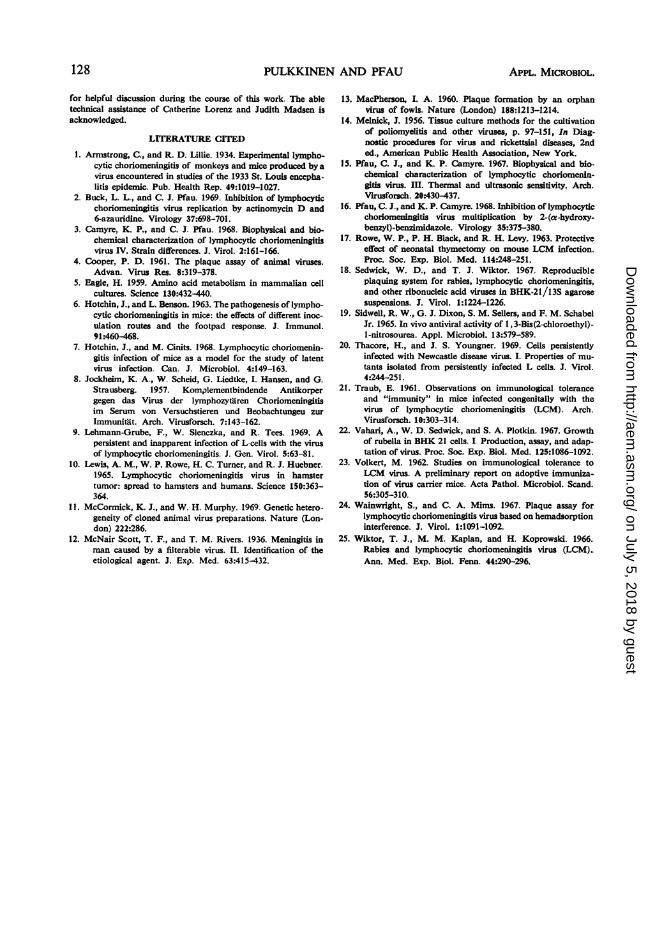
Expt I Exlpt 2 Expt 3 Expt 4
0/770/73 0/68 0/370/36] (4)** 0/391 (4)** 0/100/25 0/42 0/19 CA13710/16 0)/28 0/29
15/105* (12)** 0/270/200/22 9/14 14/1227/25 13/17 12/1039/17 (4)** 13/121 (4)** 12/13 WE10/8 13 20/1339/24
FIG. 5. Flow diagram for strainz identificationz in mixed infection. A plate was simultaneouslj ilnfectedwith CA1371 and WE strains ofLCM, the input ratio being approximately 2:1. Four days later, the plaquecount was 15/105 (experiment 1). Cylinders were withdrawn from the centers of 12 small plaques oni theabove plate. Each cylinder, after being sonic treated and assayed, produced either small plaques or a widegradient in plaque size (experiment 2). Agarose cylinders were again excised from both type of platesand repeated assays (experiments 3 and 4) showed that the plaque characteristics were stable. (*) Plaquie sizedistribution on plate (no. >3 mm/no. <3 mm); (**) number of plaques <3 mm picked anld plated (it acounMtable dilution.
gradual drop in infectivity was found over a 3-hrtest period (unopened specimens of virus weresampled at 1, 2, and 3 hr intervals). Furthermore,the degree of plaque size heterogeneity was foundto remain constant as the inactivation progressed.
Separation of the CA1371 and WE strains ofLCM by cloning. Agarose cylinders from WE-infected plates were withdrawn from both largeand small plaque areas, sonic treated, and re-plated. Virus liberated from either type ofplaque did not breed true, i.e. heterogeneity inplaque size was still apparent. The same result wasobtained whether an agarose cylinder was with-drawn from a plate containing only one plaque orfrom a plate containing as many as 50 plaques.
Although significant amounts of virus (104 to3 X 104 PFU) could be washed off the surface of aplate prior to neutral red addition (introducingthe possibility of random distribution of virus onthe surface of a plate after neutral red addition),no PFU could be detected in nonplaque areas.Thus, to prove that cloning was possible, twoeasily distinguishable strains of LCM were usedto infect simultaneously a plate. Although theCA1371 strain produced predominantly smallplaques (in the 1 to 2 mm size range), the WEstrain produced a gradient of 1 to 7 mm in plaquesize. Figure 5 shows that when small plaques werepicked from the above type of plate, sonic treated,and assayed, the resulting plates contained either
126 APPL. MICROBIOL.
on July 5, 2018 by guesthttp://aem
.asm.org/
Dow
nloaded from
HETEROGENEITY OF LCM VIRUS PLAQUES
predominantly small plaques (characteristic ofCA1371) or a large gradient in plaque size (typi-cal of WE). Two more cycles of picking showedthat the plaque characteristics separated from theinitial plate persisted through successive cycles ofviral replication.Growth of virus stocks from excised plaques.
Neutral red overlays were added in the dark toassay plates, and incubation was continued forno more than 2 hr. Virus was liberated from theplaque areas as described previously, except thatall operations but the plaque excision were carriedout in the dark. L cell monolayers in 25 cm2 foil-covered plastic flasks were infected as previouslydescribed (16), and titers at 48 and 72 hr werecomparable to routinely obtained virus stocks.Thus, any effect of neutral red in the inoculumwas of minimal importance in the infectionprocess.
DISCUSSIONStrains of LCM differ from one another in
tissue culture with respect to growth curves (16),stability (16), sensitivity to various drugs (2, 16),character of persistent infections (9), and cellspecificity (24). Further similarities and differ-ences based on plaque size may now be con-sidered. Three strains (WE, WE3, and UBC),with similar plaque sizes and PFU/LD50 ratios,have a common origin (Table 1). Although theFortner strain is similar in plaque size to theabove three, it is an independent isolate anddiffers significantly with respect to the PFU/LD50ratio. Parke, Davis & Co. obtained the Armstrongstrain of LCM from the American Type CultureCollection in 1950 (F. A. Miller, personal com-munication), and this has recently been called theM-7 strain (Table 1). However, the plaque typeof M-7 more closely resembles the WE-originstrains than our recently acquired Armstrongstrain. Both the CA1371 and Armstrong strainsoriginated in the laboratory of the late CharlesArmstrong (Table 1; W. P. Rowe, personal com-munication). They are quite similar in plaque sizebut differ somewhat in the PFU/LD50 ratio.WCP and Traub plaques are most difficult to see,the difference in intensity of staining betweenbackground and plaque areas being very slight.Their inactivation kinetics are virtually identical(3), and their PFU/LD50 ratios (1:23 and 1:19)are quite apart from those of any other strainstested. Both the Traub (W) and WCP strainswere originally obtained from the same source,yet Traub (21) and Camyre and Pfau (3) havefound their pathogenicity in mice to be markedlydifferent. Although it has been repeatedly shownthat virus-passage history can affect plaque size(20), we have not observed a similar pattern in
the LCM system. The method of propagatingcells, however, has a marked effect on the plaquecharacteristics of certain LCM strains. We havenot investigated whether this is due simply to thedifference in media or to the general condition ofthe cell in monolayer versus suspension culture.
Neutral red-inactivation kinetics are greaterwith the CA1371 than with the WE strain ofLCM. A difference in sensitivity to neutral redhas been noted among strains of poliovirus(E. M. Opton and R. H. Green, Fed. Proc. 19:408). Stains other than neutral red were used inthe present system to try to eliminate the strongphotodynamic inactivation of the virus. Theseincluded 2,3, 5-triphenyl-2H-tetrazolium chlo-ride, tetrazolium blue (diformazan), alcian blue,and resazurin. Under the conditions we used,none of the dyes were concentrated by the cells.Decreasing the concentration of neutral red wasalso considered, but a dilution factor of two madeplaques almost impossible to see.The plaque characteristics of each LCM strain
were found to be reproducible using eithermonolayer- or suspension-grown BHK 21/13Scells, respectively. The same characteristics wereapparent after passage in carrier mice. The failureto separate genetically stable large and smallplaque-forming virions from the WE strain didnot seem to be due to cross contamination of theexcised plaques. Had this been the case, it wouldhave been impossible to separate the CA1371from the WE strain in the mixed infection experi-ment (Fig. 5). Plaque size heterogeneity whichcannot be altered by cloning has been noted withother viruses (11, 13). In the present system, itappeared that excised plaques contained onlytitratable virus originating in that area. Further-more, the linear relation between plaque numberand relative virus concentration indicated thatvirus particles in the plaque area were the progenyof virus originally released from a single cell.Sonic treatment, which breaks up aggregates ofthe WCP strain (15), did not alter the plaquecharacteristics of the WE strain. This seemed torule out the possibility that clumping of virusparticles ultimately led to plaque size hetero-geneity. If the above assumptions are correct, thedegree of plaque size heterogeneity is a genetictrait.
ACKNOWLEDGMENTS
This study was su,pported by Public Health Service Grant AI-06735 from the National Institute of Allergy and Infectious Dis-eases and Research Career Program Award 5-K3-GM-8494 fromthe National Institute of General Medical Sciences.
It is a pleasure to thank T. J. Wiktor for the detailed methodol-ogy leading to our initial success with the plaque assay. We areindebted to Louis Musanti and Stanley Holt for the photographicwork and to Gordon Good for construction of the vlating appara-tus. We are grateful to Richard Trowbridge and Raymond Welsh
VOL. 20, 1970 127
on July 5, 2018 by guesthttp://aem
.asm.org/
Dow
nloaded from
128 PULKKINEN AND PFAU
for helpful discussion during the course of this work. The abletechnical assistance of Catherine Lorenz and Judith Madsen isacknowledged.
LITERATURE CITD
1. Armstrong, C., and R. D. Lillie. 1934. Experimental lympho-cytic choriomeningitis of monkeys and mice produced by avirus encountered in studies of the 1933 St. Louis encepha-litis epidemic. Pub. Health Rep. 49:1019-1027.
2. Buck, L. L., and C. J. Pfau. 1969. Inbibition of lymphocyticchoriomeningitis virus replication by actinomycin D and6-azauridine. Virology 37:698-701.
3. Camyre, K. P., and C. J. Pfau. 1968. Biophysical and bio-chemical cbaracterization of lymphocytic choriomeningitisvirus IV. Strain differences. J. Virol. 2:161-166.
4. Cooper, P. D. 1961. The plaque assay of animal viruses.Advan. Virus Res. 8:319-378.
5. Eagle, H. 1959. Amino acid metabolism in mammalian cellcultures. Science 130:432-440.
6. Hotchin, J., and L. Benson. 1963. The pathogenesis of lympho-cytic choriomeningitis in mice: the effects of different inoc-ulation routes and the footpad response. J. Immunol.91:460-468.
7. Hotchin, J., and M. Cinits. 1968. Lymphocytic choriomenin-gitis infection of mice as a model for the study of latentvirus infection. Can. J. Microbiol. 4:149-163.
8. Jockheim, K. A., W. Scheid, G. Liedtke, I. Hansen, and G.Strausberg. 1957. Komplementbindende Antikorpergegen das Virus der lymphozytairen Choriomeningitisim Serum von Versuchstieren und Beobachtungeu zurImmunitait. Arch. Virusforsch. 7:143-162.
9. Lehmann-Grube, F., W. Slenczka, and R. Tees. 1969. Apersistent and inapparent infection of L-cells with the virusof lymphocytic choriomeningitis. J. Gen. Virol. 5:63-81.
10. Lewis, A. M., W. P. Rowe, H. C. Turner, and R. J. Huebner.1965. Lymphocytic choriomeningitis virus in hamstertumor: spread to hamsters and humans. Science 150:363-364.
11. McCormick, K. J., and W. H. Murphy. 1969. Genetic hetero-geneity of cloned animal virus preparations. Nature (Lon-don) 222:286.
12. McNair Scott, T. F., and T. M. Rivers. 1936. Meningitis inman caused by a filterable virus. II. Identification of theetiological agent. J. Exp. Med. 63:415-432.
APPL. MIcRoBIoL.
13. MacPherson, I. A. 1960. Plaque formation by an orphanvirus of fowls. Nature (London) 188:1213-1214.
14. Melnick, J. 1956. Tissue culture methods for the cultivationof poliomyelitis and other viruses, p. 97-151, In Diag-nostic procedures for virus and rickettsial diseases, 2nded., American Public Health Association, New York.
15. Pfau, C. J., and K. P. Camyre. 1967. Biophysical and bio-chemical characterization of lymphocytic choriomenin-gitis virus. III. Thermal and ultrasonic sensitivity. Arch.Virusforsch. 20:430-437.
16. Pfau, C. J., and K. P. Camyre. 1968. Inhibition of lymphocyticchoriomeningitis virus multiplication by 2-(a-hydroxy-benzyl)-benzimidazole. Virology 35:375-380.
17. Rowe, W. P., P. H. Black, and R. H. Levy. 1963. Protectiveeffect of neonatal thymectomy on mouse LCM infection.Proc. Soc. Exp. Biol. Med. 114:248-251.
18. Sedwick, W. D., and T. J. Wiktor. 1967. Reproducibleplaquing system for rabies, lymphocytic choriomeningitis,and other ribonucleic acid viruses in BHK-21/13S agarosesuspensions. J. Virot. 1:1224-1226.
19. Sidwell, R. W., G. J. Dixon, S. M. Sellers, and F. M. SchabelJr. 1965. In vivo antiviral activity of 1, 3-Bis(2-chloroethyl)-1-nitrosourea. Appl. Microbiol. 13:579-589.
20. Thacore, H., and J. S. Youngner. 1969. Cells persistentlyinfected with Newcastle disease virus. I. Properties of mu-tants isolated from persistently infected L cells. J. Virol.4:244-251.
21. Traub, E. 1961. Observations on immunological toleranceand "immunity" in mice infected congenitally with thevirus of lymphocytic choriomeningitis (LCM). Arch.Virusforsch. 10:303-314.
22. Vahari, A., W. D. Sedwick, and S. A. Plotkin. 1967. Growthof rubella in BHK 21 cells. I Production, assay, and adap-tation of virus. Proc. Soc. Exp. Biol. Med. 125:1086-1092.
23. Volkert, M. 1962. Studies on immunological tolerance toLCM virus. A preliminary report on adoptive immuniza-tion of virus carrier mice. Acta Pathol. Microbiol. Scand.56:305-310.
24. Wainwright, S., and C. A. Mims. 1967. Plaque assay forlymphocytic choriomeningitis virus based on hemadsorptioninterference. J. Virol. 1:1091-1092.
25. Wiktor, T. J., M. M. Kaplan, and H. Koprowski. 1966.Rabies and lymphocytic choriomeningitis virus (LCM).Ann. Med. Exp. Biol. Fenn. 44:290-296.
on July 5, 2018 by guesthttp://aem
.asm.org/
Dow
nloaded from