planning knee replacement on xrays
TRANSCRIPT

PLANNING & TEMPLATE
VAIBHAV BAGARIAJoint Replacement Surgeon
Sir HN Reliance Foundation HospitalGirgaum, Mumbai

GOALS
• Well Aligned
• Well Balanced
• Linear Patellar Tracking
• Infection free healing

EXPECTATIONS
• REDUCE PAIN
• IMPROVE FUNCTION
• IMPROVE QUALITY OF LIFE

PREOPERATIVE WORKUP
• PATIENT SELECTION
• PREOP MEDICAL ASSESMENT
• CLINICAL EXAM
• RADIO EXAM
• IMPLANT SELECTION
• COUNSELLING AND CONSENTING

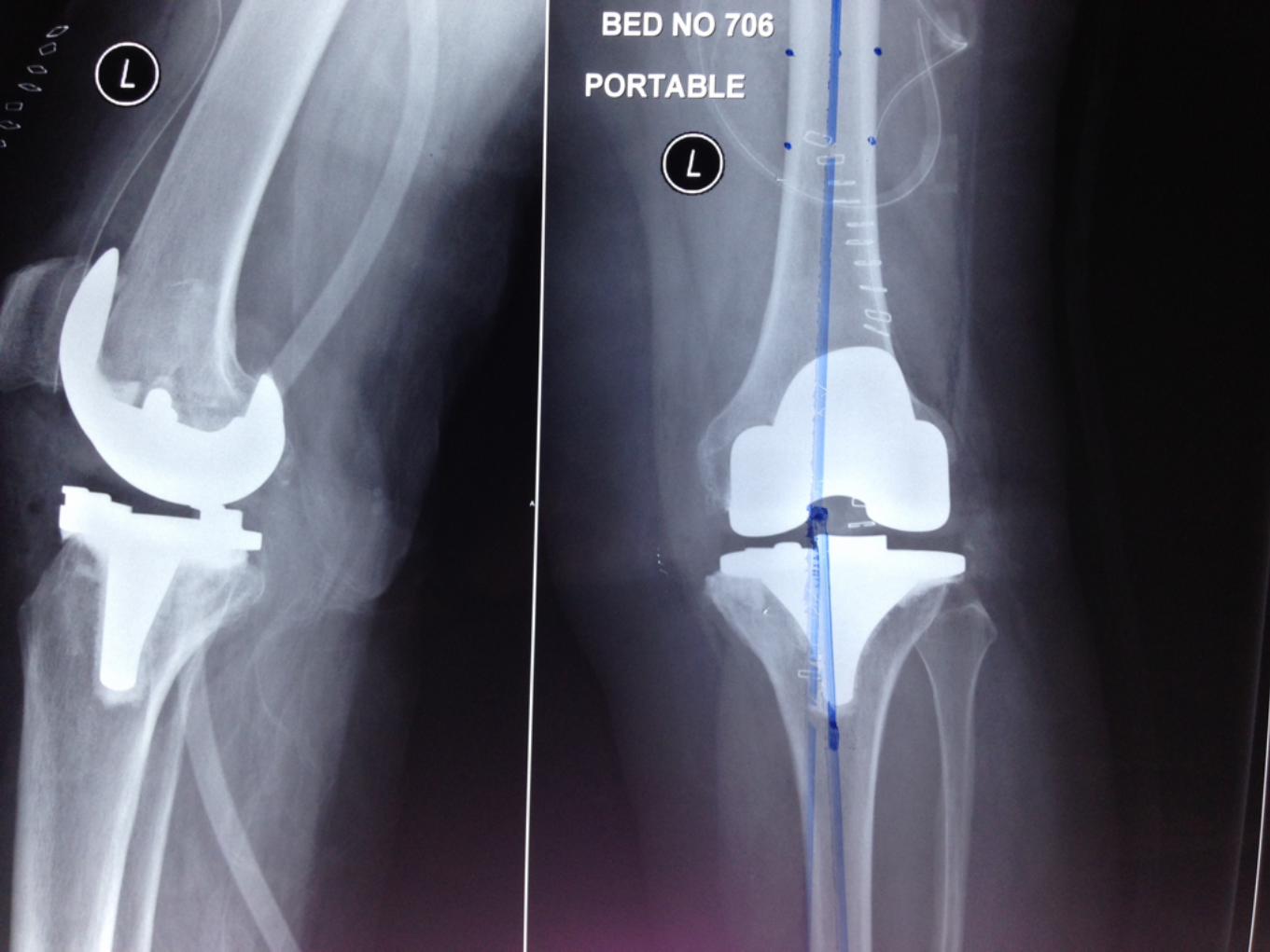
TECHNICAL GOALS
• RESTORATION OF THE NEUTRAL MECHANICAL ALIGNMENT
• PRESERVATION ( RESTORATION) OF JOINT LINE
• PERIARTICULAR LIGAMENT BALANCING
• MAINTAING LINEAR PATELLAR TRACKING AND Q ANGLE

IMAGING
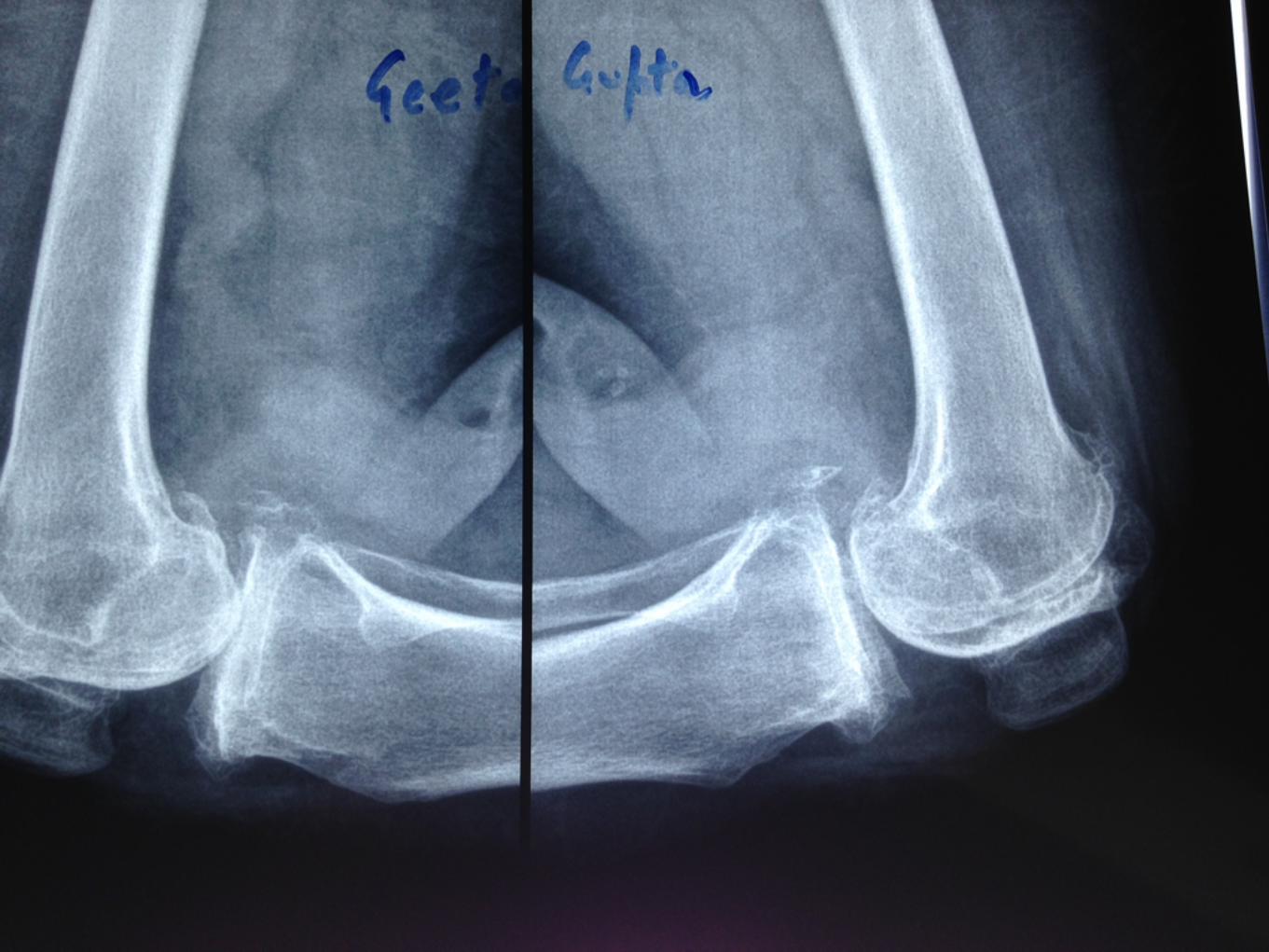
• STANDARD AP/ LAT/ SKYLINE VIEWS
• SCANNOGRAM - PREFERABLE BUT NOT MANDATORY

SCANNOGRAM
• EXTREMES OF HEIGHT
• ALTERED HIP/FEMORAL NECK ANATOMY: DYSPLASIA, TRAUMA, INFECTION, PREVIOUS OPERATION
• OBVIOUS CLINICAL DEFORMITY OF TIBIA AND FEMORAL SHAFTS

SUPPLEMENTARY IMAGING
• TUNNEL VIEW: POSTERIOR WB SURFACE, OCD, LOOSE BODIES
• CT/MRI: COMPLEX PRIMARY FOLLOWING TRAUMA, DYSPLASIA, ALTERED BONE STOCK OR PATIENT SPECIFIC INSTRUMENTATION

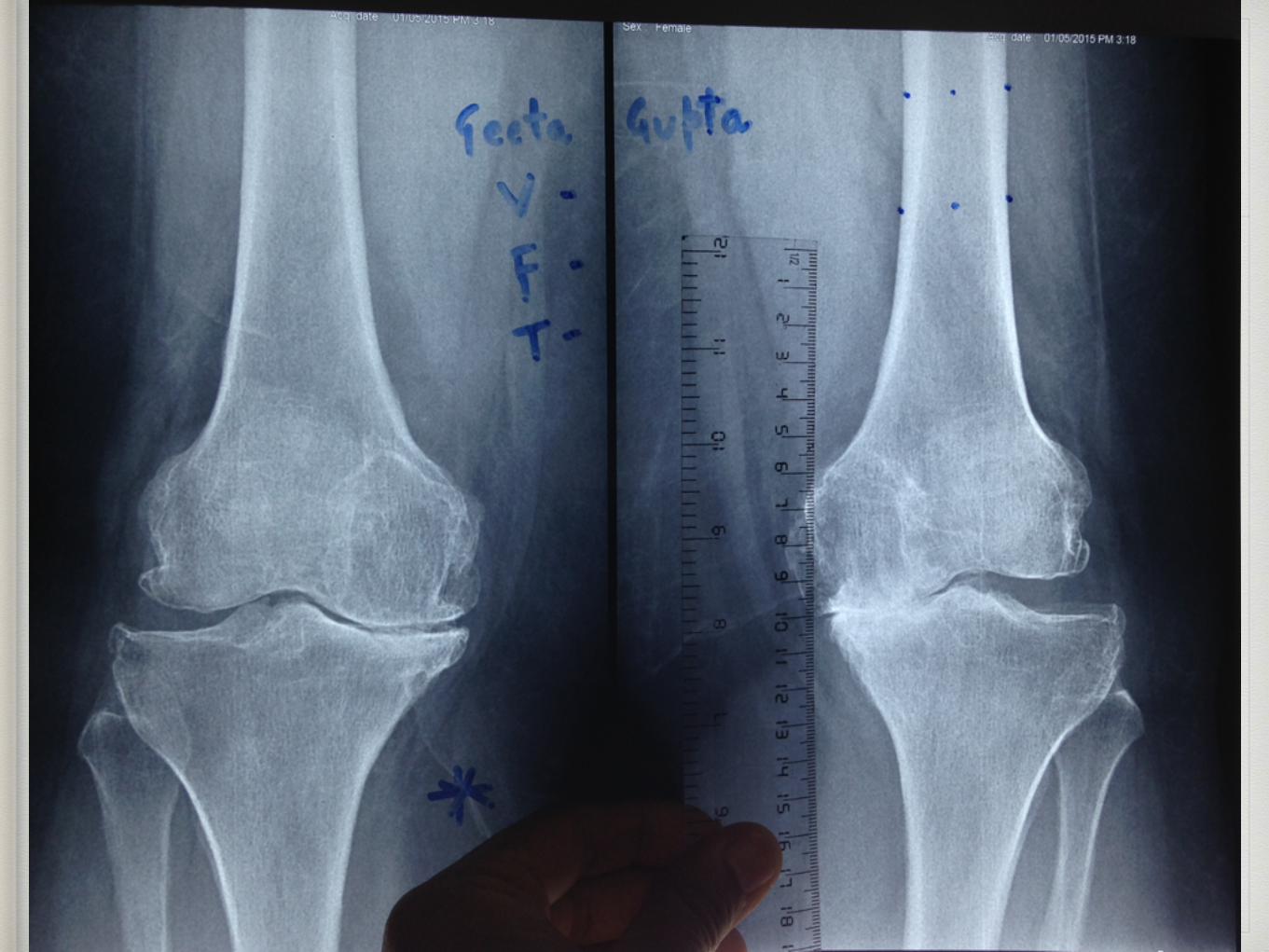
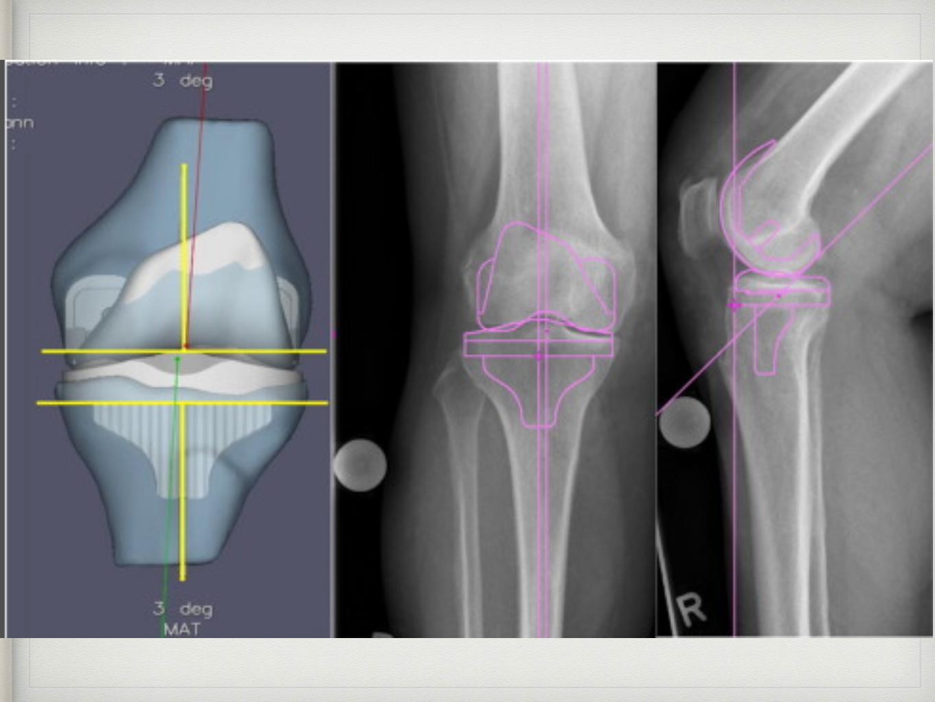
What to see?
• Determine the femoral & tibial cuts and cut angles
• Position of the femoral canal entry point
• Anticipating the ligament release
• Identifying the bone defects, joint subluxation or lig laxity
• Tempting prosthetic components
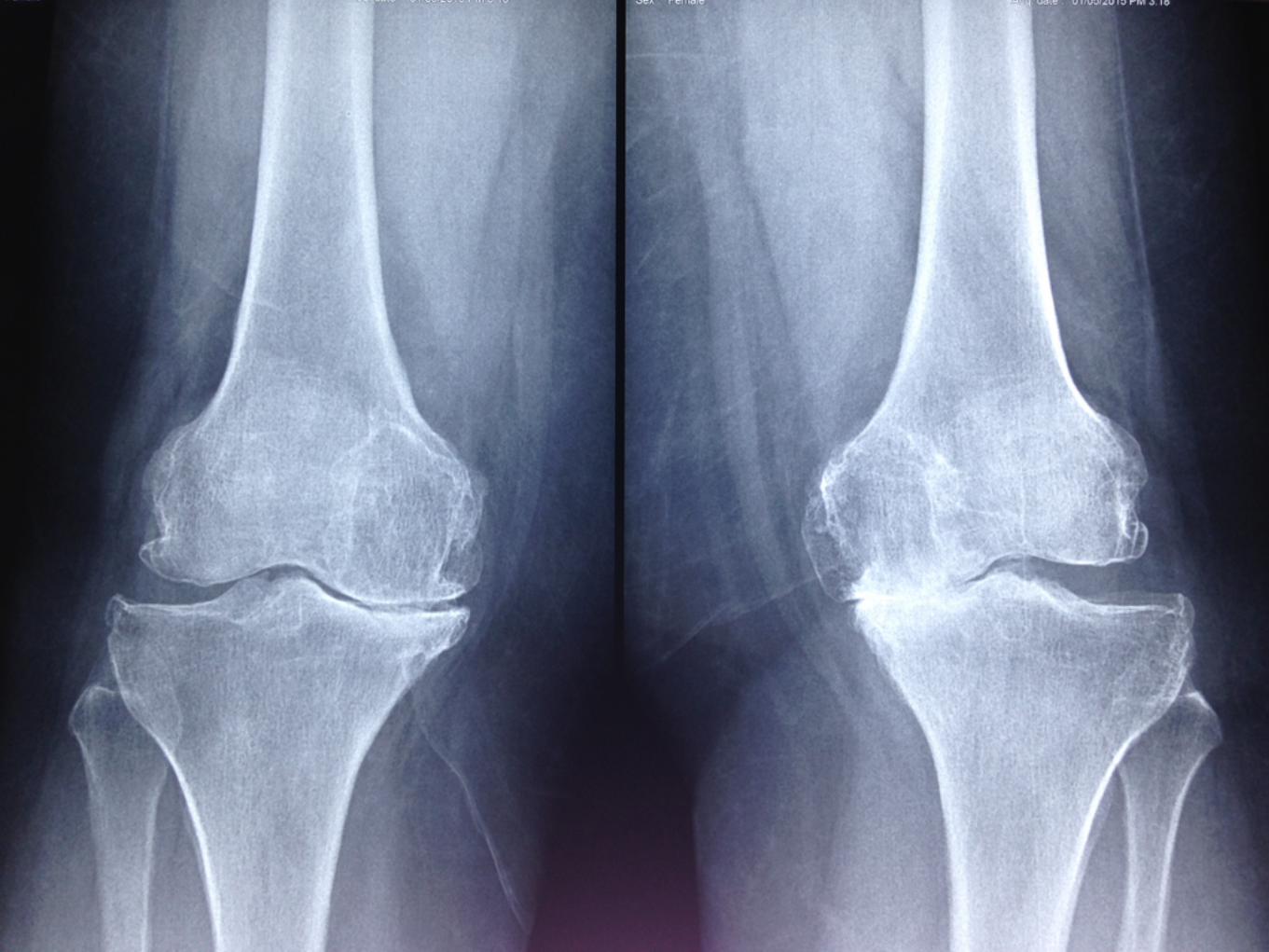
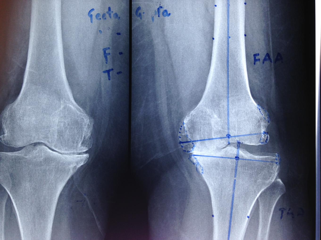
AP View
• Joint Compartments affected & Deformity
• Osteophytes: Imp for releases
• Evidence of previous surgery
• Alignment & Templating

Lateral view
• Flexion deformity
• Osteophytes
• Alignment
• Subchondral sclerosis
• Vessels
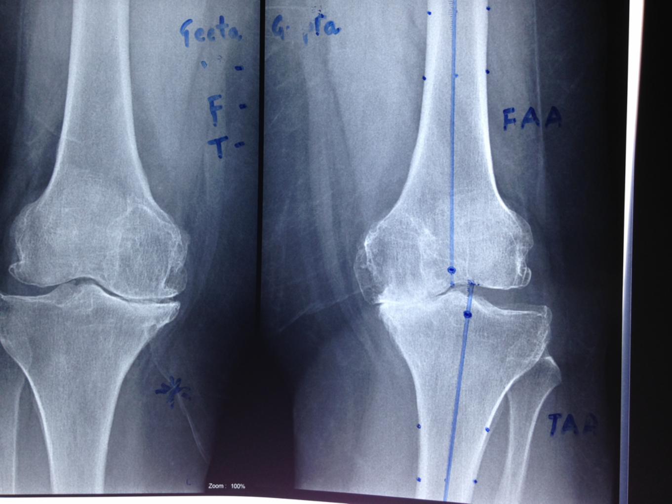
On femur: FAA, FMA, VCA
• Femoral Anatomical axis: Line that bisects the Intramedulary canal
• Exit point determines the entry point of IM jig
• Draw Valgus Cut Angle: 5 - 7 degree decreasing with patient height.
• VCA is perpendicular to femoral mechanical axis ( FMA)


Tibia
• Anatomic axis corresponds to Mechanical Axis
• IM or EM Jigs Used
• The cut is perpendicular to both
• If deformity MA is to be considered

Tibial Axis


QUIZ TIME


Take Home Message
• FAA
• TAA
• Valgus Cut
• Tibial Cut
• Osteophyte
• Implant Sizing & Positioning
Proper preoperative
planning prevents piss-
poor performance