picot: a trx- and grx-like protein in search of a … a trx- and grx-like protein in search of a...
TRANSCRIPT
48 The Open Enzyme Inhibition Journal, 2008, 1, 48-51
1874-9402/08 2008 Bentham Open
Open Access
PICOT: A Trx- and Grx-Like Protein in Search of a Function
Ariel Ohayon, Guangyu Dong, Sagit Cohen, Moran Galperin, Alice Givoni, Keren Moyal and
Noah Isakov*
The Shraga Segal Department of Microbiology and Immunology, Faculty of Health Sciences and the Cancer Research
Center, Ben Gurion University of the Negev, P.O.B. 653, Beer Sheva 84105, Israel
Abstract: The PKC-interacting cousin of thioredoxin (PICOT) protein was discovered based on its ability to
bind PKC in human T lymphocytes. Overexpression of PICOT was found to impose negative regulatory ef-
fects on PKC -dependent functions. This included the inhibition of PKC -dependent activation of c-Jun N-
terminal kinase (JNK) and the activator protein 1 (AP-1) and nuclear factor kappa B (NF- B) transcription
factors. PICOT is a modular protein consisting of a single thioredoxin (Trx)-like homology domain (HD) and
two highly homologous PICOT-HDs. The overall structure of each of the three domains resembles the ca-
nonical thioredoxin fold, which is common in enzymes that catalyze disulfide bond formation. Nevertheless,
the three PICOT domains lack essential catalytic cysteine residues and their mode of activity is therefore un-
clear. PICOT is involved in the regulation of heart muscle function. Its overexpression in the heart of trans-
genic mice increased the ventricular function and cardiomyocyte contractility, and inhibited the overall car-
diac hypertrophy induced by pressure overload. The effects of PICOT on the cardiac tissue are likely to be
mediated via the muscle LIM protein (MLP), which was shown to interact with PICOT in cardiomyocytes,
and colocalize with PICOT at the Z-disc of the sarcomer. PICOT interaction with MLP interfered with bind-
ing of the latter protein to the Ca2+
-dependent Ser/Thr phosphatase, calcineurin, causing the displacement of
calcineurin from the Z-disc. As a result, PICOT inhibited the calcineurin-mediated dephosphorylation and
nuclear translocation of nuclear factor of activated T cells (NF-AT), and the transcriptional activation of NF-
AT regulated genes. Whether the effects of PICOT are dependent on PKC, and whether it can mediate cata-
lytic activity and/or operate as an adaptor protein are only few of the open questions related to the biological
mechanism of action of PICOT.
INTRODUCTION
Studies aimed at the identification of protein kinase C (PKC) theta (PKC ) regulatory molecules, have led Witte et al. to the discovery of a new gene product, termed PICOT (PKC-interacting cousin of thioredoxin), which was found to possess the ability to associate with PKC in human T lym-phocytes [1]. PICOT was first isolated using a yeast-two hybrid screen of human Jurkat T cell cDNA library with bait that consisted of a catalytically inactive full-length PKC cDNA.
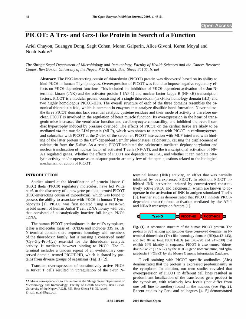
The human PICOT predominates in the cell’s cytoplasm; it has a molecular mass of ~37kDa and includes 335 aa. Its N-terminal domain share sequence homology with members of the thioredoxin family, but is missing a conserved motif (Cys-Gly-Pro-Cys) essential for the thioredoxin catalytic activity. It mediates however binding to PKC . The C-terminal includes a tandem repeat of an evolutionary con-served domain, termed PICOT-HD, which is shared by pro-teins from diverse groups of organisms (Fig. 1) [2].
Transient overexpression of constitutively active PKC in Jurkat T cells resulted in upregulation of the c-Jun N-
*Address correspondence to this author at the Shraga Segal Department of
Microbiology and Immunology, Faculty of Health Sciences, Ben Gurion
University of the Negev, P.O.B. 653, Beer Sheva 84105, Israel;
E-mail: [email protected]
terminal kinase (JNK) activity, an effect that was partially inhibited by overexpressed PICOT. In addition, PICOT in-hibited JNK activation induced by cotransfected constitu-tively active PKC and calcineurin, which are known to co-operate in the activation of JNK in antigen stimulated T cells [3]. Further studies demonstrated that PICOT inhibits PKC -dependent transcriptional activation mediated by the AP-1 and NF- B transcription factors [1].
Fig. (1). A schematic structure of the human PICOT protein. The
protein is 335 aa long and includes three conserved domains: an N-
terminal thioredoxin (Trx)-like homology domain (HD)(aa12-143),
and two 84 aa long PICOT-HDs (aa 145-228 and 247-330) that
exhibit 64% identity in sequence. PICOT is also termed ‘thiore-
doxin-like 2’ (TXNL2) by the HUGO gene nomenclature, and ‘glu-
taredoxin 3’ (Glrx3) by the Mouse Genome Informatics Database.
T cell staining with PICOT specific antibodies (Abs) demonstrated that the protein is expressed predominantly in the cytoplasm. In addition, our own studies revealed that overexpression of PICOT in different cell lines resulted in predominant localization of the transfected gene product in the cytoplasm, with relatively low levels (that differ from one cell line to another) found in the nucleus (see Fig. 2). Recent studies by Park and colleagues [4, 5] demonstrated

PICOT: A Trx- and Grx-Like Protein in Search of a Function The Open Enzyme Inhibition Journal, 2008, Volume 1 49
that PICOT has a significant regulatory role in heart muscle function where it can attenuate stress-induced cardiac hyper-trophy. The present review highlights some of the most re-cent findings on PICOT and discusses the potential biologi-cal function of this protein.
Fig. (2). Localization of overexpressed PICOT in HepG2 cells by
immunofluorescence staining. HepG2 cells were transfected with
pEF-PICOT, cultured for 3 days, fixed, and stained with rabbit anti-
PICOT Abs and a PE-conjugated anti-rabbit IgG (A), phalloidin
that specifically binds filamentous actin (B), and DAPI, which as-
sociates with the minor groove of the double strand DNA (C). A
merged image is shown in D. PICOT localize predominantly in the
cell’s cytoplasm, with low levels at the nucleus.
STRUCTURE OF PICOT AND RELATIONSHIP TO THIOREDOXIN AND GLUTAREDOXIN
The biological role of PICOT has not been identified yet, nor is the exact physiological function of its isolated Trx- or PICOT-HD. Many different proteins from a wide range of species were found to include one or more PICOT-HD as part of the molecule. In a large number of such proteins, the PICOT-HD was found at the protein’s C-terminus, in close proximity to N-terminal Trx- or Grx-like domains [2]. The close physical proximity between PICOT-HD and Trx- or Grx-HD in multiple proteins suggests functional relation-ships between the three domains. Furthermore, PICOT, Trx-HD, and Grx-HD possess an overall similar globular topol-ogy [6]. Each of the three domains possesses the canonical ‘thioredoxin fold domain’, formed by a central mix of 4 or 5 strand -sheets flanked by three or more -helices on either side of the -strands [7, 8] (see Fig. 3). The results imply that formation of the three domains during evolution originated from a single common ancestral gene. It should be men-tioned however that several genes encode short proteins, which are built almost entirely of a PICOT-HD. It appears therefore that PICOT-HD-containing proteins may have bio-logical functions that are independent of Trx or Grx, or that can operate in conjunction with other molecules, such as Trx or Grx.
INVOLVEMENT OF PICOT IN SIGNAL TRANSDUC-TION
The original discovery of PICOT was based on its ability to interact with PKC [1], a critical enzyme for T cell
antigen receptor (TCR)-linked signal transduction pathway [10, 11]. PKC is a Ca
2+-independent PKC isoform that
plays an essential role in reorganization and/or function of the immunological synapse [12, 13]. It cooperates with cal-cineurin in activation of the interleukin-2 (IL-2) gene [3], and is involved in the promotion of signaling pathways that regulate T cell activation and survival [14-16]. Initial studies demonstrated that PICOT can serve as a negative regulator of PKC in T cells, since its overexpression resulted in downregulation of PKC -dependent activation of JNK, con-comitantly with inhibition of the transcription factors AP-1 and NF- B [1].
Fig (3). The 3D structure of the mouse PICOT-HD2 (A) and the
human PICOT Trx-HD (B) were solved by K. Miyamoto et al.
(PDB, 1WIK) and N. Tochio et al. (PDB, 2DIY), respectively. The
figures were prepared using the PyMol Molecular Graphics System
[9].
Physiological activation of T cells is initiated by TCR in-teraction with a major histocompatibility complex (MHC)-bound peptide antigen on the surface of antigen presenting cells (APC). The very early phase of the TCR-linked signal transduction pathway is regulated by protein tyrosine kinases (PTKs), which phosphorylate the cytoplasmic tails of the TCR associated CD3 subunits, as well as a plethora of effec-tor molecules that are involved in signal transduction and induction of the activation response. Many of the effects induced by a physiological activation of tyrosine kinases can be mimicked by cell treatment with reactive oxygen inter-mediates (ROS), such as hydrogen peroxide, that were re-cently found to be produced in vivo and serve as physiologi-cal regulators of lymphocyte activation [17]. Other cell types were also found to produce ROS in response to cytokines and growth factors [18, 19]. One of the major sources for inducible ROS in leukocytes is the NADPH oxidase, which can be activated by several mechanisms, including phos-phorylation by PKC [20].
The fact that PICOT associates with PKC and possesses
Trx- and Grx-like sequences suggested that it might be in-
volved in redox-regulated biochemical pathways. Treatment
of Jurkat T cells with hydrogen peroxide resulted in phos-
phorylation of PICOT on tyrosine residues, an effect that
could be abolished by inhibitors of Src family members of
PTKs [21]. Furthermore, tyrosine phosphorylation of PICOT

50 The Open Enzyme Inhibition Journal, 2008, Volume 1 Ohayon et al.
was also observed in untreated cells following their transfec-
tion with a constitutively active Lck (Lck Y505F). The re-
sults suggest that tyrosine phosphorylation of PICOT can be
directly or indirectly mediated by Lck. They also imply that
PICOT plays a role in cell-activation dependent signaling
pathways, and/or responses to stress signals mediated by
reactive oxygen intermediates.
The fact that PKC is expressed predominantly in hema-
topoietic and muscle cells, in contrast to PICOT, which is
expressed in a wide range of tissues, suggests that PICOT is
likely to be involved in biological functions that are inde-
pendent of PKC .
THE ROLE OF PICOT IN THE REGULATION OF HEART MUSCLE FUNCTION
The potential involvement of PICOT in cellular processes
regulating heart muscle function was demonstrated by Jeong
et al. during studies of the mechanisms leading to cardiac
hypertrophy [4, 5]. These studies were aimed at the identifi-
cation of potential regulators of cardiac hypertrophy, and led
to the observation that PICOT was among several genes un-
dergoing upregulation following transverse aortic constric-
tion-induced cardiac hypertrophy of adult rat hearts. Fur-
thermore, PICOT overexpression in the heart of transgenic
mice inhibited cardiac hypertrophy induced by pressure
overload, concomitantly with increase in ventricular function
and cardiomyocyte contractility.
Jeong et al. have further analyzed the potential binding
partners of PICOT in the muscle tissue, and using a GST-
PICOT pull-down assay in conjunction with mass spec-
trometry they found that PICOT can directly interact with
the muscle LIM domain protein (MLP) [4, 5], a member of a
family of cysteine-rich proteins that mediate protein-protein
interactions [22]. MLP is known to interact with several dif-
ferent proteins, including -actinin, zyxin and calcineurin,
all of which are known to concentrate at the Z-disc of the
sarcomer [23, 24]. Immunofluorescence staining of tissue
section of adult mouse heart using PICOT-specific Abs
demonstrated PICOT localization within the Z-disc [5], re-
sults that were confirmed by our own studies (see Fig. 4). In
addition, PICOT was found to co-localize with MLP and
another known Z-disc protein, the -actinin [5]. MLP inter-
action with calcineurin, a Ca2+
-dependent Ser/Thr phospha-
tase, is known to be essential for calcineurin anchorage to
the Z-disk and upregulation of its catalytic activity. The ac-
tive, calcineurin then dephosphorylates the cytoplasmic NF-
AT transcription factor and promotes its translocation to the
nucleus where it induces the transcription of NF-AT regu-
lated genes [23]. Overexpression of PICOT and its associa-
tion with MLP interfered with MLP binding to calcineurin,
causing displacement of calcineurin from its anchorage site
at the Z disc. As a result, PICOT inhibited calcineurin-
mediated dephosphorylation and subsequent nuclear translo-
cation of NF-AT, as well as the NF-AT-regulated gene tran-
scription. The authors suggest that PICOT inhibits cardiac
hypertrophy by negative regulation of the MLP-calcineurin-
NF-AT signaling pathway via a mechanism, which disrupts
MLP-calcineurin interaction.
Fig. (4). Immunohistochemical staining of adult mouse heart sec-
tions with anti-PICOT Abs. Paraffin embedded sections of adult
mouse heart were deparaffinized, mounted on glass slides, blocked
with BSA, treated with hydrogen peroxide, and stained with rabbit
anti-PICOT Abs plus biotinylated goat anti-rabbit IgG and peroxi-
dase-conjugated streptavidin. Peroxidase activity was demonstrated
by incubation with APC and hydrogen peroxide. Slides were coun-
terstained with Hematoxylin and examined under a bright field
microscope (Olympus I-70) attached to a digital camera (Olympus
DP70).
POTENTIAL ROLE FOR PICOT DURING EMBRYO-INC DEVELOPMENT
Comparative analysis of protein profiles expressed at dif-
ferent stages of embryonic development serves as a useful tool for the identification of molecules that are involved in
specific developmental processes. In order to determine pro-
tein profile changes during embryogenesis Greene et al. [25] have utilized an immobilized pH gradient-based two-
dimensional electrophoresis and identified individual protein
spots by mass spectrometry. Analysis of proteins expressed between days (E) 8.5-to-10.5 of embryogenesis, the time of
formation of the neural tube, revealed a number of changes
in protein patterns at successive embryonic days. PICOT was almost undetectable at E8.5 but was dramatically upregu-
lated at E9.5. Although the onset of PICOT expression corre-
lated with that of PKC [26], the incomplete overlap in tis-sue expression of the two proteins suggest that PICOT may
have developmental roles that are independent of PKC .
CONCLUSIONS
Despite the fact that the exact biological activity of PI-
COT is not yet known, indirect studies suggest that PICOT
plays important roles in the regulation of T lymphocyte acti-
vation and during cardiac muscle responses to hypertrophy-
inducing signals. PICOT is also predicted to be involved in
certain developmental stages in mouse embryos, although
the exact role or the tissue in which it is first expressed are
not know. PICOT was shown to associate with PKC in T
cells, but is also expressed in PKC -negative cell types indi-
cating that PICOT assumes activities independent of PKC .
Further studies of this novel protein and characterization of
its exact biological activity may reveal important clues about

PICOT: A Trx- and Grx-Like Protein in Search of a Function The Open Enzyme Inhibition Journal, 2008, Volume 1 51
PICOT and the biological processes that are mediated by or dependent on PICOT.
ACKNOWLEDGEMENTS
Research in our laboratory is supported in part by the Is-rael Science Foundation, USA-Israel Binational Science Foundation, Israeli-Taiwanese Scientific Research Coopera-tion, Israel Cancer Research Fund, Chief Scientist’s office, Israel Ministry of Health, the Israel Cancer Association through a donation be Ida and Harry Shooster, and a dona-tion by Linda Osofsky. N.I. holds the Joseph H. Krupp Chair in Cancer Immunobiology.
REFERENCES
[1] Witte S, Villalba M, Bi K, Liu Y, Isakov N, Altman A. Inhibition
of the c-Jun N-terminal kinase/AP-1 and NF-kappaB pathways by
PICOT, a novel protein kinase C-interacting protein with a thiore-
doxin homology domain. J Biol Chem 2000; 275(3): 1902-9.
[2] Isakov N, Witte S, Altman A. PICOT-HD: a highly conserved
protein domain that is often associated with thioredoxin and glu-
taredoxin modules. Trends Biochem Sci 2000; 25(11): 537-9.
[3] Werlen G, Jacinto E, Xia Y, Karin M. Calcineurin preferentially
synergizes with PKC-theta to activate JNK and IL-2 promoter in T
lymphocytes. EMBO J 1998; 17(11): 3101-11.
[4] Jeong D, Cha H, Kim E, et al. PICOT inhibits cardiac hypertrophy
and enhances ventricular function and cardiomyocyte contractility.
Circ Res 2006; 99(3): 307-14.
[5] Jeong D, Kim JM, Cha H, et al. PICOT attenuates cardiac hyper-
trophy by disrupting calcineurin-NFAT signaling. Circ Res 2008;
102(6): 711-9.
[6] Herrero E, de la Torre-Ruiz MA. Monothiol glutaredoxins: a com-
mon domain for multiple functions. Cell Mol Life Sci 2007; 64
(12): 1518-30.
[7] Kinch LN, Baker D, Grishin NV. Deciphering a novel thioredoxin-
like fold family. Proteins 2003; 52(3): 323-31.
[8] Qi Y, Grishin NV. Structural classification of thioredoxin-like fold
proteins. Proteins 2005; 58(2): 376-88.
[9] Delano WL. The PyMOL Molecular Graphics System. DeLano
Scientific; San Carlos, CA. 2002.
[10] Altman A, Isakov N, Baier G. Protein kinase Ctheta: a new essen-
tial superstar on the T-cell stage. Immunol Today 2000; 21(11):
567-73.
[11] Isakov N, Altman A. Protein kinase C(theta) in T cell activation.
Annu Rev Immunol 2002; 20: 761-94.
[12] Bi K, Tanaka Y, Coudronniere N, et al. Antigen-induced transloca-
tion of PKC-theta to membrane rafts is required for T cell activa-
tion. Nat Immunol 2001; 2(6): 556-63.
[13] Arendt CW, Albrecht B, Soos TJ, Littman DR. Protein kinase C-
theta;: signaling from the center of the T-cell synapse. Curr Opin
Immunol 2002; 14(3): 323-30.
[14] Bertolotto C, Maulon L, Filippa N, Baier G, Auberger P. Protein
kinase C theta and epsilon promote T-cell survival by a rsk-
dependent phosphorylation and inactivation of BAD. J Biol Chem
2000; 275(47): 37246-50.
[15] Villalba M, Bushway P, Altman A. Protein kinase C-theta mediates
a selective T cell survival signal via phosphorylation of BAD. J
Immunol 2001; 166(10): 5955-63.
[16] Manicassamy S, Gupta S, Huang Z, Sun Z. Protein kinase C-theta-
mediated signals enhance CD4+ T cell survival by up-regulating
Bcl-xL. J Immunol 2006; 176(11): 6709-16.
[17] Reth M. Hydrogen peroxide as second messenger in lymphocyte
activation. Nat Immunol 2002; 3(12): 1129-34.
[18] Meier B, Radeke HH, Selle S, et al. Human fibroblasts release
reactive oxygen species in response to interleukin-1 or tumour ne-
crosis factor-alpha. Biochem J 1989; 263(2): 539-45.
[19] Ohba M, Shibanuma M, Kuroki T, Nose K. Production of hydrogen
peroxide by transforming growth factor-beta 1 and its involvement
in induction of egr-1 in mouse osteoblastic cells. J Cell Biol 1994;
126(4): 1079-88.
[20] Reeves EP, Dekker LV, Forbes LV, et al. Direct interaction be-
tween p47phox and protein kinase C: evidence for targeting of pro-
tein kinase C by p47phox in neutrophils. Biochem J 1999; 344 (Pt 3):
859-66.
[21] Babichev Y, Gelkop S, Witte S, Altman A, Isakov N. The protein
kinase C (PKC) theta interacting protein, PICOT, undergoes tyro-
sine phosphorylation in response to hydrogen peroxide. In: 'Pro-
ceedings of the 2nd congress of the Federation of Immunological
Societies of Asia-Oceania (FIMSA 2000)', Mundozzi Ed., 2000;
11-6.
[22] Bach I. The LIM domain: regulation by association. Mech Dev
2000; 91(1-2): 5-17.
[23] Heineke J, Ruetten H, Willenbockel C, et al. Attenuation of cardiac
remodeling after myocardial infarction by muscle LIM protein-
calcineurin signaling at the sarcomeric Z-disc. Proc Natl Acad Sci
USA 2005; 102(5): 1655-60.
[24] Hoshijima M. Mechanical stress-strain sensors embedded in car-
diac cytoskeleton: Z disk, titin, and associated structures. Am J
Physiol Heart Circ Physiol 2006; 290(4): H1313-25.
[25] Greene ND, Leung KY, Wait R, Begum S, Dunn MJ, Copp AJ.
Differential protein expression at the stage of neural tube closure in
the mouse embryo. J Biol Chem 2002; 277(44): 41645-51.
[26] Wilda M, Ghaffari-Tabrizi N, Reisert I, Utermann G, Baier G,
Hameister H. Protein kinase C isoenzyme: selective expression pat-
tern of protein kinase C-θ during mouse development. Mech
Dev 2001; 103(1-2): 197-200.
Received: April 16, 2008 Revised: May 19, 2008 Accepted: May 19, 2008
© Ohayon et al.; Licensee Bentham Open.
This is an open access article distributed under the terms of the Creative Commons Attribution License (http://creativecommons.org/licenses/by/2.5/), which
permits unrestrictive use, distribution, and reproduction in any medium, provided the original work is properly cited.