phytochemical analysis and antiangiogenic potential of …€¦ · · 2016-08-30abstract - cancer...
TRANSCRIPT

Asia Pacific Journal of Multidisciplinary Research, Vol. 3, No. 5, December 2015 _______________________________________________________________________________________________________________
72 P-ISSN 2350-7756 | E-ISSN 2350-8442 | www.apjmr.com
Phytochemical Analysis and Antiangiogenic
Potential of Gmelina Arborea Roxb. (Paper Tree) Fruit Exocarp Using Duck
Chorioallantoic Membrane (Cam) Assay
Weenalei T. Fajardo1*, Lina T. Cancino
1, Elnora B. Dudang
1, Sharlene
Cherry M. Suratos1, Frienzky B. Macayana
2
1Natural Science Department, College of Arts and Sciences;
2Specialization
Courses Department, College of Teacher Education and Technology,
Pangasinan State University-Lingayen, Philippines [email protected]
Date Received: November 2, 2015; Date Revised: December 15, 2015
Asia Pacific Journal of
Multidisciplinary Research Vol. 3 No.5, 72-79
December 2015 Part IV P-ISSN 2350-7756
E-ISSN 2350-8442
www.apjmr.com
Abstract - Cancer is one of the leading causes of death in the Philippines and in the world. One of
the critical events in the metastasis of cancer is angiogenesis which is the formation of new blood vessels. Prevention of the angiogenesis is necessary for the treatment of such disease. Antiangiogenic chemicals
are needed to prevent the growth of blood vessels. Thus, this research would be beneficial to many people
since it will pave way to the discovery of new drugs against cancer. The study aimed to evaluate the antiangiogenic property of Gmelina arborea fruit exocarp ethanolic
extract (GFEEE) by conducting the Duck Chorioallantoic Membrane (CAM) Assay. The various
concentrations of Gmelina ethanolic extracts and the controls were applied on the tenth day of incubation and on the 12
th day, the eggs were subjected to CAM assay. Analysis of Variance revealed that there was
a significant difference among all the concentrations in terms of percentage CAM vascularity inhibition as compared to the positive control. However, among all the concentrations, 100% concentration of
GFEEE has the highest percentage CAM vascularity inhibition. However, using Tukey’s Multiple
Comparison Tests indicated that there was no significant difference in the percentage vascular inhibition between 75% and 100% concentration which implies that those concentrations have the same
antiangiogenic effect. Moreover, the active constituents present in Gmelina fruit exocarp indicates that
the results of the present study has a clinical effect and can be a potential source of natural antiangiogenic agents that can be possibly used as anti-tumoral agent and can lead to probable settling
of the issue of expensive anticarciongenic drugs.
Keywords: Phytochemical analysis, Chorioallantoic Membrane (CAM) Assay, Gmelina arborea
fruit exocarp ethanolic extract (GFEEE), antiangiogenic potential
INTRODUCTION Cancer has been one of the most feared illnesses
in the world since high mortality rate is observed
during and even after treatment. Furthermore, the chemical and radiation therapy are dreadful
procedures that affect not only the physiological and
emotional states of the affected individuals and their families but is tantamount to finance exhaustion.
In the Philippines, cancer is the third leading cause of morbidity and mortality [1] penetrating the
various strata of the society; it is an equating disease
that does not choose any gender, age and social positions. However, the affluent are the ones who
have the full access to such treatments leaving the
poor unattended largely. These issues had led to the worldwide efforts to discover and develop other
possible cheap sources of anticarcinogenic agents from plant metabolites and animal products.
The ability of the cancer cells to spread to
adjacent or distant organs make the condition life threatening [2]. Tumor growth and metastasis is
dependent on angiogenesis and lymphangiogenesis triggered by chemical signals from tumor cells in a
phase of rapid growth [3]. Storgard, Mikolon and
Stupack [4]defined angiogenesis as a complex biological process involving the generation of blood

Fajardo et al., Phytochemical Analysis and Antiangiogenic Potential of Gmelina Arborea Roxb _______________________________________________________________________________________________________________
73 P-ISSN 2350-7756 | E-ISSN 2350-8442 | www.apjmr.com
Asia Pacific Journal of Multidisciplinary Research, Vol. 3, No. 5, December 2015
vessels from the pre-existing vasculature which is
needed in the pathogenesis of cancer, rheumatoid arthritis and retinal diseases.
Furthermore, tumor angiogenesis is the
proliferation of a network of blood vessels that penetrates into cancerous growths, supplying nutrients
and oxygen and removing waste products. It starts with cancerous tumor cells releasing molecules that
send signals to surrounding normal host tissue. This
signaling activates certain genes in the host that, in turn, make proteins to encourage growth of new blood
vessels (Demers and D’ Arcy, 2010). Therefore any
agent that impedes the formation is said to be antiangiogenic and may have potential
anticarcinogenc effect. Chorioallantoic Membrane (CAM) Assay is a
useful tool to observe angiogenesis in vivo. It is
widely chosen since it is amenable to both intravascular and tropical administration of study
agents, low cost and relatively rapid assay, and can be adapted very easily to study-angiogenesis-dependent
process [4], [5]. The CAM assay uses the avian
chorioallontoic membrane of the 3-day old chicken or duck embryos [6].
Effective and expensive drugs produced
nowadays came from basic researches using plant and plant materials. In fact, more than half of the world’s
population still relies entirely on plants for medicines, and plants supply the active ingredients of most
traditional medical products [7]. Some plants and
plant extracts have been investigated in vivo and were found to exert antitumor or anticancer properties.
These include garlic, Allium sativum (El-Mofty,
1994), Agaricus brazei [8], Astragalus (Chung et al, 1989) and Cassia alata L. [6].
Gmelina arbrorea Roxb., commonly known as paper tree, has been one of the most studied plants for
the biochemical activities of its plant parts involving
its wood, leaf, fruit, flowers and stem. In the study of Ishaku, Ishakeku and Agwale [9], its fruits were found
to have saponins, tannins, reducing sugar, steroids,
flavonoids and glycosides which were claimed to be responsible for its antibacterial activity. Furthermore,
the methanolic extracts of stem bark showed antioxidant activity for it inhibited the formation of
free radicals or scavenging [10]. In addition, the
alcoholic and aqueous leaves extracts have anthelmintic activity for it had increased chloride
conduction of worm muscle membrane resulting to
hyperpolarization and reduction of excitability making
the muscle relaxed and paralyzed (Ambujakshi,
Takkar and Shymnanda, 2009). Also, the crude leaf and stem bark extracts showed antimicrobial activities
against gram positive and gram negative organism and
the activity due to the presence of bioactive compounds such as alkaloids, saponins,
carbohydrates, phenolics, tannins and anthraquinone (El-Mahmood, Doughari and Kiman, 2010).
Moreover, aqueous extracts from fresh fruits, tree bark
and leaves exhibited insecticidal property against legume pod borer and pod sucking bug (Opraeke,
2005). Moreover, there are some folkloric claims that
fresh fruit is toxic and has abortifacient property to farm animals such as cows, buffalos and goats. In
addition, in Region I, only the woods are the concern of farmers since these will be used in the wood craft
industry leaving the other parts not important. With
the vast studies on G. arborea little is known about its antiangiogenic property of its fruit extract which could
eventually pose therapeutic effect against cancer. Thus the research aims to know the antiangiogenic property
of G. arborea exocarp extract through CAM assay and
the active phytochemicals of the plant being studied.
Materials and Methods Research Design
Both descriptive and post-treatment experimental
designs were employed in the research. Collection of G. arborea fruits
Fresh ripe fruits were picked from the tree of
their natural habitat in Capitol Grounds, Lingayen, Pangasinan. The plant species was identified using a
dichotomous key (Sharma, 1999), online program
Floragator (2009) and World Wide Flowering Plant Family Identification (1963) in the Biology
Laboratory, Department of Natural sciences, Pangasinan State University-Lingayen Campus. Its
identity was verified by sending a voucher specimen
in the National Museum-Botany Division in Manila.
Figure 1. Gmelina arborea unripen drupe fruit.

Fajardo et al., Phytochemical Analysis and Antiangiogenic Potential of Gmelina Arborea Roxb _______________________________________________________________________________________________________________
74 P-ISSN 2350-7756 | E-ISSN 2350-8442 | www.apjmr.com
Asia Pacific Journal of Multidisciplinary Research, Vol. 3, No. 5, December 2015
Phytochemical Screening
The phytoactive constituents of fruit exocarp of G. arborea were screened in the Pharmacy Laboratory of
Virgen Milagrosa University, San Carlos City,
Pangasinan. Certification was issued indicating the results were true and correct and that it was conducted
in the same laboratory. Procedures and protocols were followed. Results were analyzed based the chemical
reactions manifested by the addition of various test
solutions and with few methods applied [11].
Preparation of the G. arborea ethanolic fruit exocarp
Fruits were washed and then the seeds were removed from the fruit leaving only the exocarp using
a sterile knife. The exocarp was chopped prior to refluxing of about 50g of the plant sample in a 500mL
Erlenmeyer flask with 300 mL of 80% ethanol for 1
hour in a boiling water bath. The flask was removed, and then the contents were allowed to cool at room
temperature and filtered. A 500 mL solution was made by adding an ethanol sufficiently through the residue
on to the filter paper. The extract was used for the
various phytochemical screening.
Locale of the Study
Initial identification of the plant was done in the Biology Laboratory of Pangasinan State Univesrity-
Lingayen and verified in the National Museum, Botanical Division, Manila and was verified by a
museum researcher. The analysis of secondary
metabolites of Gmelina arborea fruit extract was conducted in Virgen Milagrosa University
Foundation-College of Pharmacy Laboratory and
Duck Chorioallantoic (CAM) Assay was performed in a small hatchery house located at Alvear II, Poblacion,
Lingayen, Pangasinan.
Research Animals
Fifty four (54) three-day fertilized duck embryos were obtained from a reputable poultry farm at #108
Ketegan, Mangatarem, Pangasinan. Egg viability was
determined using the candle method for any sign of embryo formation assisted by Dr. Manuel C. Vallo, a
licensed veterinarian. The eggs were randomly grouped and labeled according to treatment. The eggs
were placed in the incubator at a constant temperature
of 37.50C and at a constant humidity.
Proper disposal After the experiment, the duck embryos were
placed in a plastic bag and autoclaved at a temperature
of 2120C for five hours to avoid contamination. The
embryos were sealed properly and buried in the compost pit.
Duck Chorioallantoic Membrane Assay (CAM) Assay The 3-day old fertilized duck embryos were
incubated for 7 days at 37.50C and 70% humidity.
Prior to windowing, a HEALTHPRO gauze soaked in
70% Band Aid Isopropyl alcohol was wiped in to the
shell of the ducks, A window in the egg shell about 1x1 cm was made to expose the CAM for access to
experimental manipulation. The test plant extract was
absorbed on the sterile filter paper discs. Then, the treated filter paper discs were placed onto the CAM.
The treated eggs were sealed with sterile plastic tape and were incubated for two days. On the 10
th day, the
eggs were subjected for experimental treatments since
the developing CAM vasculature was ready to sprout in response to additional pro-angiogenic stimuli and
were very responsive to antiangiogenic factors. On the 12
th day of incubation, the CAMs were harvested by
removing the hard shell leaving intact the soft
membrane covering the embryo [11]. The shell-less embryo was transferred to a petri dish and 5 mL of the
amniotic fluid was removed using BD 10 mL Syringe
Luer-LokTM
Tip with BD Precision GlideTM
Needle 23G 1 ¼ TW (0.6mm x 32 mm). Duck embryos that
were dead prior to harvest, that is one day after the introducing the soaked filter paper, they were replaced
and same process were performed until they reached
the 12th say of incubation.
Preparation of the Different Extract Concentrations,
Positive Control and Negative Control The 200 grams of air-dried and powdered G.
arborea fruit exocarp was transferred to 500 mL Erlenmeyer flask in 300 mL of 80% Ethanol for one
hour in boiling water bath. The flask was removed and
the contents were allowed to cool at room temperature then filtered. Sufficient ethanol was added through the
residue on to the filter paper to make 500 mL. The
concentrated extract was evaporated through drying and a residue was obtained. The 2.5g, 5.0g, 7.5g, and
10g of the residue were used to make different concentrations. These were dissolved to distilled
Wilkins water to make 10 mL of each concentration
[6]. On the other hand, the positive control was
prepared by dissolving the 200mg of celocoxib
powder in 200 mL of Wilkins distilled water. It was
Fajardo et al., Phytochemical Analysis and Antiangiogenic Potential of Gmelina Arborea Roxb _______________________________________________________________________________________________________________
75 P-ISSN 2350-7756 | E-ISSN 2350-8442 | www.apjmr.com
Asia Pacific Journal of Multidisciplinary Research, Vol. 3, No. 5, December 2015
(a)
(b)
stirred and then filtered (Virrey, et al, 2010). Negative
control was prepared by measuring 10 mL of 80% ethanol and was transferred to a beaker.
Preparation of Filter Paper Discs
The filter paper was punched with a 2-holed puncher to form the paper discs (approx. 5 mm in
diameter). The paper filter paper discs were sterilized by autoclaving. These were soaked to the various
concentrations prior to administration to CAM.
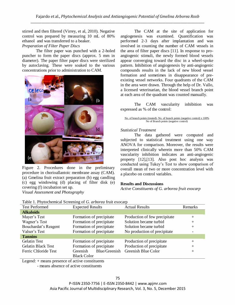
Figure 2. Procedures done in the preliminary
procedure in chorioallantoic membrane assay (CAM).
(a) Gmelina fruit extract preparation (b) egg candling (c) egg windowing (d) placing of filter disk (e)
covering (f) incubation set up.
Visual Assessment and Photography
The CAM at the site of application for
angiogenesis was examined. Quantification was performed 2-3 days after implantation and was
involved in counting the number of CAM vessels in
the area of filter paper discs [11]. In response to pro-angiogenic stimuli, the newly formed blood vessels
appear converging toward the disc in a wheel-spoke pattern. Inhibition of angiogenesis by anti-angiogenic
compounds results in the lack of new blood vessel
formation and sometimes in disappearance of pre-existing vessel networks. Four quadrants of the CAM
in the area were drawn. Through the help of Dr. Vallo,
a licensed veterinarian, the blood vessel branch point at each area of the quadrant was counted manually.
The CAM vascularity inhibition was
expressed as % of the control:
No. of branch points (treated)- No. of branch points (negative control) x 100%
No of Branch points (negative control)
Statistical Treatment
The data gathered were computed and subjected to statistical treatment using one way
ANOVA for comparison. Moreover, the results were
interpreted clinically wherein more than 50% CAM vascularity inhibition indicates an anti-angiogenic
property [12],[13]. Also post hoc analysis was conducted using Tukey’s Test to show comparison of
overall mean of two or more concentration level with
a placebo on control variables.
Results and Discussions Active Constituents of G. arborea fruit exocarp
Table 1. Phytochemical Screening of G. arborea fruit exocarp
Test Performed Expected Results Actual Results Remarks
Alkaloids Mayer’s Test Formation of precipitate Production of few precipitate +
Wagner’s Test Formation of precipitate Solution became turbid + Bouchardat’s Reagent Formation of precipitate Solution became turbid +
Valser’s Test Formation of precipitate No production of precipitate -
Tannins Gelatin Test Formation of precipitate Production of precipitate +
Gelatin Black Test Formation of precipitate Production of precipitate +
Ferric Chloride Test Greenish Blue/Greenish Black Color
Greenish Blue Color +
Legend: + means presence of active constituents
- means absence of active constituents
(c (d
(e (f

Asia Pacific Journal of Multidisciplinary Research, Vol. 3, No. 5, December 2015 _______________________________________________________________________________________________________________
76 P-ISSN 2350-7756 | E-ISSN 2350-8442 | www.apjmr.com
(a) (b
(c) (d)
(e) (f)
Table 1 shows that tannins and alkaloids are
present in Gmelina arborea fruit exocarp ethanolic extract as manifested by the formation of precipitate
when treated with Mayer’s, Wagner’s and Bouchardat’s reagent. In addition, tannins of catechol
type are present as indicated by the greenish-black
coloration upon addition of ferric chloride test solution associated with the precipitation in gelatin-
salt block test. These active constituents maybe
responsible for the anti-angiogenic property of the extract.
Tannins are parts of the diverse chemical groups of polyphenolics that naturally occur in plants such as
flavonoids and phenolic diterpenes [21]. In the study
of Bagchi et al. (2004), Nojiri et al. [15] and Stangl et al. [16], they mentioned that in higher plants, their
polyphenolic components act as antioxidant, antiangiogenic, antiproliferative and anti-
inflammatory as well as vasorelaxants. Moreover, it
was proven that plant polyphenolics inhibit angiogenesis through the regulation of multiple
signaling pathways (Mojzis et al., 2008) such as
angiotensin converting enzyme (ACE) pathway Visual Assessment
Figure 3 provides a general view on the angiogenesis of duck embryo treated with different concentrations of Gmelina extract at (a) 25% (b) 50% (c) 75% and (d) 100%. Duck embryo treated with
Celocoxib is seen in (e) and negative control (ethyl alcohol) is represented in (f). Nine eggs were observed and
used for the counting of blood vessels per group.

Fajardo et al., Phytochemical Analysis and Antiangiogenic Potential of Gmelina Arborea Roxb _______________________________________________________________________________________________________________
77 P-ISSN 2350-7756 | E-ISSN 2350-8442 | www.apjmr.com
Asia Pacific Journal of Multidisciplinary Research, Vol. 3, No. 5, December 2015
0%10%20%30%40%50%60%70%80%90%
25%Gmelina
arborea fruitexocarpextract
50%Gmelina
arborea fruitexocarpextract
75%Gmelina
arborea fruitexocarpextract
100%Gmelina
arborea fruitexocarpextract
PositiveControl
(Celocoxib)
Trial A
Trial B
Trial C
Chorioallantoic Membrane (CAM) Assay
Figure 4. Percentage of CAM Vascularity Inhibition of the Different Concentrations (25%, 50%, 75%, 100%) and Positive Control (celocoxib)
Figure 4 shows the percentage CAM vascularity inhibition of the different concentrations and positive
control (celocoxib). It could be deemed from the table that among the various concentrations of Gmelina
arborea fruit exocarp ethanolic extract, the 100% concentration showed the greatest inhibition property followed by 75%. This means that the mentioned concentrations have anti-angiogenic property because as stated
by Nassar et al. [12] and Aisha et al. [13], agents with 50% or higher CAM vascularity inhibition have anti-
angiogenic property.
Table 2. Analysis of Variance (ANOVA) of the Percentage CAM Vascularity Inhibition SS of Variation SS Df MS F Significance
Between groups 12,882.824 5 2,576.565 243.117 0.000
Within groups 121.176 12 10.598 Total 13,010.001 17
Table 2 shows ANOVA significant comparisons between the different concentrations at f f= 243.117,
significance = 0.000, implying that all the concentrations of G. arborea exocarp fruit exocarp are not
comparable with the percentage inhibition of the Celocoxib treatment (positive) because all the concentrations have greater absolute mean difference compared to critical T range. This implies that the positive control still
has the highest antiangiogenic effect.
Table 3. Tukey’s Multiple Comparison Tests for Percentage CAM Vascular Inhibition of the Different Concentrations of Gmelina arborea Fruit Exocarp Extract Compared to Positive Control Comparison of Trials Absolute Mean Difference Significance
25% and 50% 26.59 0.000*
25% and 75% 43.49 0.000*
25% and 100% 44.70 0.000* 25% and Positive control 60.96 0.000*
50% and 75% 16.90 0.000*
50% and 100% 18.12 0.000*
50% and Positive 34.37 0.000* 75% and 100% 1.22 0.997
75% and Positive control 17.47 0.000*
100% and Positive control 16.26 0.001*
* The mean difference is significant at 0.05 level

Asia Pacific Journal of Multidisciplinary Research, Vol. 3, No. 5, December 2015 _______________________________________________________________________________________________________________
78 P-ISSN 2350-7756 | E-ISSN 2350-8442 | www.apjmr.com
Figure 3 shows Tukey’s Analysis results on the
multiple comparisons on the number of blood vessel branch points upon application of the different
concentrations of G. arborea fruit exocarp extract together with the positive (Celocoxib) and negative
(untreated) control. The data shows, the numbers of
blood vessels significantly decreased as the concentration of G. arborea fruit exocarp extract
increases. Comparison among the different
concentrations showed statistically significant results in the decrease of the number of blood vessels in the
duck embryo treated between 25% and 50%, 25% and 75%, 25% and 100% Gmelina extract. It was also
found significant between the concentrations of 50%
and 75%, 50% and 100%. However, at 75% concentration, the number of blood vessels inhibition
was not significantly different compared to 100%.
Although, the comparisons of various concentrations show significant difference, only the
75% and 100% showed anti-angiogenic property because of 50% and above vascular inhibition [12],
[13].
The reason behind the antiangiogenic property of 75% and 100% Gmelina arborea exocarp fruit crude
extract is that it contains phenolics and alkaloids
which are proven to have antiangiogenic and antiproliferative effects [14], [15], [16]. Also, in the
study of Karagiz et al. [17] it had shown that crude plant extracts are more effective pharmacologically
than isolated active compounds which was claimed to
be due to the synergistic effects of various components present in the extracts.
Furthermore, Kampa, Nifli, Notas and Castanas
[18] claimed that medicinal plants are the most exclusive source of life saving drugs for the majority
of the world’s population since they represent a vast potential resource for anticancer compounds. The
anticancer activity of medicinal plant derived
compounds may result from a number of mechanisms, including effects on cytoskeletal proteins that play a
key role in cell division, inhibition of DNA
topoisomerase enzymes, antiprotease or antioxidant activity, stimulation of the immune system etc. the
value of medicinal plants lies in the potential access to extremely complex molecular structures that would be
difficult to synthesize in the laboratory.
In addition, a study on Premna herbacea Roxb. or Pygmaeopremna herbacea (Roxb.) (Verbenaceae), it
is used for treatment of cancer and rheumatism in Thailand [19]. Plant extracts containing catechin,
epicatechin, quercetin, kaempferol, rutin etc, have
shown to decrease proliferation of breast, pancreatic,
prostate and other cancer cell lines [20]. Thus, with
the presence of catechol and alkaloids, this leads to the inhibition of blood vessel formation in the duck
CAM.
CONCLUSION The treatment of duck chorioallantoic membrane
with the different concentrations of Gmelina arborea
ethanolic extract affected the extent of blood vessel
proliferation. General morphologic observations revealed that there was indeed a remarkable difference
between the blood vessels of the different concentrations and the positive control. Furthermore,
in the number of blood vessel branch points of the
duck embryo especially those treated with 100% concentrations showed reduction in both blood vessel
formation and branching complexity which is much
close to positive control. The findings therefore demonstrate antiangiogenic property of Gmelina
arborea fruit exocarp which may confer its potential as an anticancer agent.
RECOMMENDATION The researchers recommend the structural
elucidation and isolation of the active constituents
responsible for the antiangiogenic property. Furthermore, it is suggested that all concentrations
must be further subjected to more laboratory tests using more samples. Also, in the procedure, the use of
doses maybe used instead to accurately determine the
claimed anti-angiogenic property of that plant sample. Moreover, quantitative evaluation of the degree of
blood vessel branching complexity must be employed
to accurately measure the degree of blood vessel proliferation. Lastly, parallel study using other
biological assays to strengthen the claimed property of the plant sample must be conducted to verify the
results of this study.
Acknowledgements The researchers would like to express their
gratitude to Pangasinan State University for funding the research. Also, Dr. Manuel C. Vallo for assisting
us in the observations of the blood vessels and disposal of the duck embryos.
The following are the contributions of the authors:
Lina T. Cancino and Frienzky B. Macayana for the preparations of the various concentrations and sterile
reagents and glasswares; Elnora B. Dudang and Weenalei T. Fajardo for CAM assay observations;
Sharlene Cherry M. Suratos for statistical treatment

Fajardo et al., Phytochemical Analysis and Antiangiogenic Potential of Gmelina Arborea Roxb _______________________________________________________________________________________________________________
79 P-ISSN 2350-7756 | E-ISSN 2350-8442 | www.apjmr.com
Asia Pacific Journal of Multidisciplinary Research, Vol. 3, No. 5, December 2015
and their interpretations and Weenalei T. Fajardo for
the writing of the manuscript.
REFERENCES [1] Ngelangel C, Wang E (2010). Cancer and the
Philippines cancer control program. Japanese Journal
of Clinical Oncology 32(1) pp S52-S61. Retrieved
from http://goo.gl/VBRq5p
[2] Nishida N, Yano H, Nishida T, Kamura T, Kojiro M (2006). Angiogenesis in Cancer. Vasc Helath Risk
Manag 2(3):213-219.
[1] Folkman J. Tumor angiogenesis theraperutic
implications. N Engl J Med. 1971;285:1182–6. [2] Storgard C, Mikolon D, Stupack. Angiogenesis
assays in the chick embryo.Methods in Molecular
Biology vol 294 pp 123-136. Retrieved from
http://link.springer.com/protocol/10.1385%2F-59259-860-9%3A123#page-1
[3] West D, Thompson D, Selis P, Burbridge.
Angiogenesis Assays using chick chorioallantoic membrane. Methods in Molecular Medicine, Vol 46:
Angiogenesis Prootocols. Retrieved from
http://goo.gl/0qWHv8
[4] Olarte E (2007). The Don Mariano Marcos State University (DMMSU)- Potential antiangiogenic
property of Cassia alata L. hexane Extract on the
embryonic blood vessels of duck, Anaa domesticus
embryos. [5] US National Institute of Gneral Medical Sciences
(2006). Medicines by design. Retrieved from
http://goo.gl/wF1s3A
[6] Takeshi T, Yoshiyuki K, Hiromichii). 2001. Isolation of antitumor compound from Agaricus blazei Murill
and its mechanisms of action. J Nutri 131: 1409-
1413.
[7] Ishaku A, Ishakeku D, Agwale S (2013). Phytochemical screening and antibacterial activity of
Gmelina arborea fruit extracts. International Journal
of Microbiology and Immunology Research.
Retrieved from http://goo.gl/I349Jh
[8] Patil, S. M., Kadam, V. J., & Ghosh, R. (2009). In
vitro antioxidant activity of methanolic extract of
stem bark of Gmelina arborea Roxb.
(Verbenaceae).International Journal of PharmTech Research, 1(4), 1480-1484.
[9] Guevarra B. (2005). A guidebook to Plant Screening:
Phytochemical and Biological. University of Santo
Tomas Publishing House. [10] Nassar et al. (2011). Anti-angiogenic properties of
Koetjapic acid, a Natural Triterpene Isolated from
Sandoricum kooetjaoe Merr from
http://goo.gl/hQosjv
[11] Aisha A et al., (2009). Screening of Anti-angiogenic
Activity of Some Tropical Plants by Rat Aorta Assay
from http://goo.gl/Qx1jQz
[12] Bagchi D, Sen CK, Bagchi M, Atalay M (2004).
Anti-angiogenic, antioxidant, and anti-carcinogenic
properties of a novel anthocyanin rich berry extract formula. Biochemistry (Moscow), 69(1): 75-80.
[13] Nojiri S, Daida H, Inaba Y (2004). Antioxidants and
cardiovascular disease: Still a topic of interest.
Environ. Health Prevent. Med., 9(5): 200-213. [14] Stangl V, Dreger H, Stangl K, Lorenz M (2007).
Molecular targets of tea polyphenols in the
cardiovascular system. Cardiovasc. Res., 73(2): 348-
358. [15] Karagiz AN, Turgut-kara O, Cakir R, Demirgan S.
Ari. Cytotoxic activity of crude extracts from
Astragalus chrysochlorus (Leguminosae). Biotechnol
and Biotechnol. 2007;21(2):220-222
[16] Kampa, M; Nifli, A-P; Notas, G; Castanas, E.
(2007), Polyphenols and cancer cell growth. PubMed
[17] Bravo L. Polyphenols: Chemistry, dietary sources,
metabolism and nutritional significance. Nutr.Rev., 1998, 56: 317-333.
[18] Cragg, G. M., & Newman, D. J. (2005). Plants as a
source of anti-cancer agents. Journal of ethnopharmacology, 100(1), 72-79.
[19] Mohamed AJ et al. (2012). Antioxidant,
antiangiogenic and vasorelaxant activities of
methanolic extract of Clerodendrum serratum (Spreng.) leaves. Journal of Medicinal Plants
Research Vol. 6(3), pp. 348-360. Retrieved June 10,
2015 from http://goo.gl/1jlCvA
COPYRIGHTS Copyright of this article is retained by the author/s, with
first publication rights granted to APJMR. This is an open-
access article distributed under the terms and conditions of the Creative Commons Attribution license (http://creative
commons.org/licenses/by/4.0/