physicalbasisbehindachondroplasia,themostcommon ... ·...
TRANSCRIPT

Physical Basis behind Achondroplasia, the Most CommonForm of Human Dwarfism*□S
Received for publication, December 11, 2009, and in revised form, June 1, 2010 Published, JBC Papers in Press, July 12, 2010, DOI 10.1074/jbc.M109.094086
Lijuan He‡, William Horton§, and Kalina Hristova‡1
From the ‡Department of Materials Science and Engineering, The Johns Hopkins University, Baltimore, Maryland 21218 and the§Department of Molecular and Medical Genetics, Oregon Health and Science University, Research Center, Shriners Hospital forChildren, Portland, Oregon 97239
Fibroblast growth factor receptor 3 (FGFR3) is a receptortyrosine kinase that plays an important role in long bone devel-opment. The G380R mutation in FGFR3 transmembranedomain is known as the genetic cause for achondroplasia, themost common form of human dwarfism. Despite many studies,there is no consensus about the exactmechanismunderlying thepathology. To gain further understanding into the physical basisbehind the disorder, herewemeasure the activation ofwild-typeand mutant FGFR3 in mammalian cells using Western blots,and we analyze the activation within the frame of a physical-chemical model describing dimerization, ligand binding, andphosphorylation probabilities within the dimers. The data anal-ysis presented here suggests that themutation does not increaseFGFR3 dimerization, as proposed previously. Instead, FGFR3activity in achondroplasia is increased due to increased proba-bility for phosphorylation of the unliganded mutant dimers.This finding has implications for the design of targeted molec-ular treatments for achondroplasia.
Fibroblast growth factor receptor 3 (FGFR3) is a receptortyrosine kinase that consists of an extracellular domain withthree immunoglobulin-like motifs, a single transmembrane(TM)2 domain, and an intracellular split tyrosine kinasedomain. Ligands (fibroblast growth factors (fgfs)) and heparinbind to the extracellular domains and stabilize the dimer, withthe ligand inducing a conformational change in the extracellu-lar domain (1). The contact stimulates catalytic activity andresults in the cross-phosphorylation of the two receptors. Thisactivates the catalytic domains for the phosphorylation of cyto-plasmic substrates and triggers signaling cascades (2, 3).Mutations in FGFR3 are known to affect long bone develop-
ment, which proceeds via endochondral ossification along apathway involving differentiation of mesenchymal stem cellsinto cartilage, followed by bone invasion (as the chondrocytes
of the cartilage die, the space is invaded by bone). FGFR3 worksas a negative regulator of bone development by mediating pro-differentiation signals in chondrocytes (4–6). FGFR3 overacti-vation because of mutations alters the terminal differentiationinto hypertrophic chondrocytes, effectively shortening the pro-liferation phase. Furthermore, FGFR3 overactivation can causecancers of the epithelium (7).The G380R mutation in FGFR3 TM domain has been linked
to achondroplasia, the most common form of human dwarfism(8). The incidence rate of achondroplasia is about 1/15,000 livebirths (5). It is an autosomal dominant disorder that interfereswith the maturation of the cartilage growth plate of long bones(6, 9). The phenotype is characterized by short stature, narrow-ing of the lumbar spinal canal, accentuated bowing of the mid-dle and lower part of the back, and trident-shaped hands (5).Affected individuals often exhibit other skeletal as well as neu-rological complications.The physical basis underlying achondroplasia is still under de-
bate, andmultiplemechanismsmay be contributing to pathogen-esis. One establishedmechanism that contributes to FGFR3 over-activation in achondroplasia is the compromiseddown-regulationof themutant receptor. For instance,Monsonego-Ornan et al. (10,11) have found that the rates of internalization and degradation ofthe wild-type and the mutant receptors are different, and as aresult, themutant accumulates at the cell surface and signals overa longer time than thewild type. Furthermore, Cho et al. (12) havereported that theachondroplasiamutation increases theactivityofFGFR3bydisrupting c-Cbl-mediatedubiquitination that serves asa targeting signal for lysosomal degradation and termination ofreceptor signaling.Other studies, however, point to a second mechanism that
might contribute to the pathology, i.e. ligand-independent acti-vation of the mutant receptor. For instance, Webster andDonoghue (13) have studied the effect of the G380R mutationon the kinase and transforming activity of full-length FGFR3and of a chimeric Neu/FGFR3 receptor (consisting of the extra-cellular and catalytic domain of Neu and the TM domain ofFGFR3). They have shown that the mutation increases ligand-independent FGFR3 activation. Li et al. (14) have further shownthat the mutant does not need a ligand to become activated inL6 cells and induces transformations in NIH3T3 cells. Theseauthors concluded that the mutation likely “produces a domi-nant oversignaling receptor that is no longer regulated by FGFbinding.”Recent work suggests that these two mechanisms may be
coupled. Other mutations in FGFR3 that cause skeletal dyspla-
* This work was supported, in whole or in part, by National Institutes of HealthGrant GM068619. This work was also supported by National Science Foun-dation Grant MCB 0718841 and Shriners Hospitals for Children ResearchGrant 10-POR-007.
□S The on-line version of this article (available at http://www.jbc.org) containssupplemental Figs. S1–S6 and Equations S1–S9, and additional references.
1 To whom correspondence should be addressed: Dept. of Materials Scienceand Engineering, The Johns Hopkins University, 3400 N. Charles St., Balti-more, MD 21218. Tel.: 410-516-8939; Fax: 410-516-5293; E-mail:[email protected].
2 The abbreviations used are: TM, transmembrane; RTK, receptor tyrosinekinase.
THE JOURNAL OF BIOLOGICAL CHEMISTRY VOL. 285, NO. 39, pp. 30103–30114, September 24, 2010© 2010 by The American Society for Biochemistry and Molecular Biology, Inc. Printed in the U.S.A.
SEPTEMBER 24, 2010 • VOLUME 285 • NUMBER 39 JOURNAL OF BIOLOGICAL CHEMISTRY 30103
by guest on June 13, 2018http://w
ww
.jbc.org/D
ownloaded from

sias also have been shown to impede the trafficking or down-regulation of the FGFR3mutants, effectively prolonging signal-ing (11, 12, 15, 16). Importantly, the relative magnitudes ofthese trafficking and down-regulation defects have been shownto be proportional to the activation of the mutants, the higherthe activation, the longer the lifetime of the active FGFR3dimers in the cell (10, 11, 15). One interpretation of these find-ings may be that the trafficking/down-regulation defects are aconsequence of the increased activation, although the oppositeis also possible. The increased activation, on the other hand, ishypothesized to be due to increased FGFR3 dimerization (13,15, 17). However, this hypothesis has not been validated thusfar.To test directly the hypothesis that the dimerization propen-
sity of the FGFR3 TM domain changes in the presence of theachondroplasia mutation, we previously characterized thedimerization of the isolated wild-type andmutant TMdomainsof FGFR3 in lipid bilayers (18). Unexpectedly, we found that thedimerization free energies are the same for thewild type and themutant. The results for this mutation contrasted with ourresults for a different pathogenic mutation in FGFR3 TMdomain, A391E (19). The A391E mutation is known as thegenetic cause for Crouzon syndrome with acanthosis nigricanscharacterized by the following three phenotypic features: 1)mild disturbances of the growth plate of the long bones; 2)premature ossification of the skull (craniosynostosis); and 3)skin hyperpigmentation and hyperkeratosis. Unlike the A391Emutation that increased the dimerization propensity by �1.3kcal/mol (19), the G380Rmutation did not affect the dimeriza-tion energetics of FGFR3 TM domain (18).In our search for the physical basis behind achondroplasia,
here we revisit the effect of the G380R mutation in cellularsystems. We seek to determine whether the increase in FGFR3activation occurs due to enhanced dimerization or due to adifferent physicalmechanism.Wedo this using a new approachthat bridges biophysics and cell biology and has the power toprovide mechanistic understanding of the effect of pathogenicmutations on different steps in FGFR3 activation. In thisapproach, FGFR3 dimerization is considered as a two-stepprocess, ligand-independent dimerization followed by ligand-mediated dimer stabilization, and liganded and unligandeddimers are assigned different phosphorylation probabilities.We have already demonstrated the feasibility of such anapproach in our previous work by demonstrating that RTKactivity can bemodeled and predicted based on the laws ofmassaction (20). In this previous work, we investigated the A391Emutation within a Neu_FGFR3 chimeric system, and we dem-onstrated that the A391E mutation increases the activationpropensity of the chimeric Neu_FGFR3 receptor by �0.7 kcal/mol (20). Here, we first assess the effect of the achondroplasiamutation on the phosphorylation of the chimeric Neu_FGFR3receptor in CHO cells. Next, we investigate the effect of themutation on the activation of the full-length FGFR3 receptor, asa function of ligand (fgf1) concentration in HEK 293 cells. Weobserve higher phosphorylation of the mutant FGFR3 than thewild type in the absence of ligand and at low ligand concentra-tions. We demonstrate that the achondroplasia mutation doesnot increase the dimerization propensity of the receptor.
Instead, the data analysis presented here suggests that themutation affects the cross-phosphorylation within the unligan-ded dimer. Thus, we propose that the underlying reason for theincrease in FGFR3 activation in achondroplasia is not increaseddimerization but an elevated phosphorylation within the unli-ganded FGFR3 dimers, most probably due to a structuralchange.
EXPERIMENTAL PROCEDURES
Materials and Experimental Methods
Plasmids—The plasmids used in this study, Neu_FGFR3/WT, Neu_FGFR3/G380R, FGFR3/WT, and FGFR3/G380R,were a generous gift from D. J. Donoghue, University of Cali-fornia, San Diego. Neu_FGFR3 is a chimeric receptor com-posed of the extracellular and intracellular domain of Neu andtheTMdomain of FGFR3. The chimericNeu_FGFR3 receptorswere encoded in the pSV2 vector. The FGFR3/WT and FGFR3/G380R plasmids were in the pcDNA 3.1� vector.Western Blots—Chinese hamster ovary (CHO) cells were
transfectedwith various amounts of plasmids encodingNeu_F-GFR3/WT and Neu_FGFR3/G380R using LipofectamineTM2000 (Invitrogen) according to the manufacturer’s protocol.Cells were cultured for 24 h following transfection, and thentreated with lysis buffer (25 mM Tris-Cl, 0.5% Triton X-100, 20mM NaCl, 2 mM EDTA, 2 mM NaVO4, and protease inhibitor;Roche Applied Science). The supernatant was collected follow-ing centrifugation at 15,000� g for 15min at 4 °C. Lysates wereloaded into 3–8% NuPAGE� Novex� Tris acetate mini gels(Invitrogen). After the proteins were transferred onto a nitro-cellulosemembrane, they were stained withNeu C-18 antibod-ies (Sc-284, Santa Cruz Biotechnology) or anti-phosphoty-rosine (anti-Tyr(P), clone 4G10�, Upstate Biotechnology, Inc.)antibodies, followed by anti-rabbit or anti-mouse HRP-conju-gated secondary antibodies (Promega). The bands were visual-ized using ECLTMdetection reagent (GEHealthcare) and quan-tified using ImageQuant TL.Human embryonic kidney cells (HEK 293) were transfected
with plasmids encoding FGFR3/WT and FGFR3/G380R us-ing FuGENE HD (Roche Applied Science) according to themanufacturer’s protocol. Cells were cultured in normalmedium for 24 h following transfection and then starved inserum-free medium for 24 h. The effect of ligand was mon-itored by incubating the cells in medium supplemented withfgf1 (Millipore, MA). The receptors in the HEK 293 cell lysateswere probed with FGFR3 (H-100) antibodies (sc-9007, SantaCruz Biotechnology) or phospho-Tyr-FGF receptor antibodies(anti-Tyr-653/Tyr-654, Cell Signaling Technology), followedby anti-rabbit HRP-conjugated antibodies (Promega). Anti-ac-tin antibodies (Sigma)were used to stain actin and thus confirmequal loading of protein lysates from different samples.Immunostaining—Cells were cultured in the incubator for
24 h after transfection. After fixing with 3.7% paraformalde-hyde, the cells were blocked using bovine serum albumin (BSA)for 1 h. Surface localization of Neu_FGFR3 in CHO cells wasdetected after incubation with anti-ErbB2 (mouse mAb (7.16.4),Calbiochem), followed by secondary anti-mouse fluoresce-in-conjugated IgG. Surface localization of FGFR3 in HEK
Physical Basis behind Achondroplasia
30104 JOURNAL OF BIOLOGICAL CHEMISTRY VOLUME 285 • NUMBER 39 • SEPTEMBER 24, 2010
by guest on June 13, 2018http://w
ww
.jbc.org/D
ownloaded from

293 cells was detected with anti-FGFR3 (H-100, Santa CruzBiotechnology), followed by Alexa-488-conjugated anti-rabbitIgG (Invitrogen).Cross-linking—In cross-linking experiments, dimeric recep-
tors were cross-linked with either a membrane-permeablelinker (EGS, Pierce) or a membrane-impermeable one (bis(sul-fosuccinimidyl) suberate, Pierce). Twenty four hours aftertransfection, cells were incubated with 2mM cross-linker for 30min to 1 h at room temperature and then quenched in 20 mM
Tris-HCl for 15 min. After a rinse with ice-cold PBS, the cellswere lysed, and the receptors were detected using Westernblotting. The cross-linked fraction was calculated as SD/S �SD/(SM � SD), where SD is the intensity of the dimeric band, andSM is the intensity of the monomeric band.Titration with fgf1—Human embryonic kidney cells were
transfected with plasmids encoding FGFR3/WT or FGFR3/G380R. Cells were cultured in normal medium for 24 h follow-ing transfection and then starved in serum-free medium for24 h. Different concentrations of fgf1 (Millipore, MA), rangingfrom 5 to 5000 ng/ml, were added to the serum-free medium.After incubating for 10 min with ligand, cells were lysed, asdescribed above, and analyzed using Western blotting.Quantification of Western Blots—The Western blot films
were scanned and processed with ImageQuant TL. At leastthree sets of independent experiments were performed todetermine the averages and the standard deviations. The load-ing of the gels was adjusted such that all the band intensitieswerewithin the so-called linear range, where the staining inten-sities were proportional to the protein concentrations (20).
Physical-Chemical Model of FGFR3 Activation
FGFR3 lateral dimerization is a regulator of FGFR3 activity.Ligands, such as fgf1, bind to the dimer and regulate the dimer-ization process. We consider this dimerization process as atwo-step reaction scheme describing ligand-independentdimerization followed by ligand-mediated dimer stabilizationas shown in Reactions 1 and 2,
M � M ¢O¡K1
d �ligand-independent dimerization�
REACTION 1
d � L2 ¢O¡K2
D �ligand-dependent dimer stabilization�
REACTION 2
Here,M represents the monomer; d represents the unligandedFGFR3 dimer, and D represents the ligand-bound (liganded)dimer. In this model, the ligand is pre-dimerized when it bindsto the unliganded dimer; predimerization of fgf1 occurs onheparan sulfates on the cell surface (21, 22). Reactions 1 and 2are coupled; ligand binding depletes the unliganded dimers,and this in turn decreases the monomer concentrations.The two reaction constants,K1 andK2, describe ligand-inde-
pendent dimerization and ligand-mediated dimer stabilization,respectively. They are defined as shown in Equations 1 and 2,
K1 � �d�/�M�2 (Eq. 1)
and
K2 � �D�/�L2��d� (Eq. 2)
In addition, the mass balance equations for the total receptorconcentration [TR] and the total ligand concentration [TL] areshown in Equations 3 and 4,
�TL� � 2�L2� � 2�D� (Eq. 3)
�TR� � �M� � 2�d� � 2�D� (Eq. 4)
In these equations, we define the concentrations of TR, M, D,and d as the average number of receptors on the cell surfaceper cell. Thus, [TR] is the total number of receptors per cell,whereas [M], [d], and [D] are the number of monomers, unli-ganded dimers, and liganded dimers per cell, respectively.Knowing the number of cells and the three-dimensional ligandconcentrations used in the experiment, we also can calculatethe average number of ligands per cell, [TL]. The ligand exist inthree different states as follows: free in solution, bound to hep-arin sulfate on the cell surface, and bound to the receptor dimerin the presence of heparin sulfate. 2[L2] is the average numberof ligands per cell that are not bound to the receptor dimers (seeequation 5). Here we do not consider explicitly the equilibriumbetweenmonomeric and dimeric ligands, as well as the equilib-rium between ligands that are free in solution or bound to thecell surface.If we know the total ligand and receptor concentrations, [TL]
and [TR] (the two parameters that can be varied in the experi-ments), and if we assume that we know the two reaction con-stants K1 and K2, then the four equations (Equations 1–4) canbe used to determine the four unknowns [L2], [M], [d], and [D],and thus predict the monomeric and dimeric fractions ofFGFR3 receptors as a function of total ligand concentration,[TL] for fixed K1, K2, and [TR]. As shown in the supple-mentalmaterial, the problem is reduced to a fourth-order equa-tion for [M], which yields a single real positive root. Once [M] isdetermined from this equation, [d] and [D] can be determinedtoo (see supplemental material). Fig. 1 shows the prediction forthe dimeric fractions, 2[d] and 2[D], for different values of K1and K2 and a fixed value of [TR]. The predictions show how themonomeric and dimeric fractions change when K1 and K2 arechanged. They also illustrate how the ligand is reducing themonomeric fraction by depleting the unliganded dimers.Although Reactions 1 and 2 and Equations 1–4 describe the
lateral interactions between receptors as well as ligand binding,they do not account for the last step in the activation process,cross-phosphorylation within the dimer. Here, we account forthis last step with the help of two additional parameters, theprobabilities for phosphorylation in the unliganded and ligan-ded dimers, d and D. The concentration of phosphorylatedreceptors is then calculated according to Equation 5,
�P� � 2d�d� � 2D�D� (Eq. 5)
The concentration of phosphorylated receptors [P] can bemeasured usingWestern blots with antibodies that are specific
Physical Basis behind Achondroplasia
SEPTEMBER 24, 2010 • VOLUME 285 • NUMBER 39 JOURNAL OF BIOLOGICAL CHEMISTRY 30105
by guest on June 13, 2018http://w
ww
.jbc.org/D
ownloaded from

for phosphorylated tyrosines. The measured phosphostaining,however, depends on the particular antibody used, exposuretime, etc,. and does not yield real concentrations of phosphor-ylated receptors. However, the phosphorylated fraction can bemeasured if experiments are carried out over a very wide rangeof ligand concentrations, including very high ligand concentra-tions that drive all receptors into their liganded dimeric states(i.e. 2[D]sat � [TR], see predictions in Fig. 1). At such a highligand concentration, we expect to reach saturation in FGFR3phosphorylation, such that the phosphostaining levels stay attheir constant maximum values and are not further increasedwhen more ligand is added.At saturating ligand concentrations, we write 2[D]sat � [TR]
and thus we obtain Equation 6,
�P�sat � 2D�D�sat � D�TR� (Eq. 6)
Therefore, the ratio of phospho-Tyr-staining measured at par-ticular ligand concentration to the phospho-Tyr-staining atsaturation yields the phosphorylated fraction of receptors asshown in Equation 7,
�P�
�P�sat�
2d
D
�d�
�TR��
2D
D
�D�
�TR��
d
D
2�d�
�TR��
2�D�
�TR�(Eq. 7)
Equation 7 shows that the phosphorylated fraction as a functionof ligand concentration is the sum of the predicted ligandeddimeric fraction and the unliganded dimeric fraction (predic-tions for both are shown in Fig. 1), the latter multiplied byd/D.The probabilities for phosphorylation, d and D, are un-
known, but the ratio of the two probabilities can be determinedif the concentrations of phosphorylated receptors and dimericreceptors are measured. At saturating ligand concentrations,only liganded dimers are expected to be present, and the con-centration of phosphorylated receptors is given by Equation 6.In the absence of ligand, the concentration of phosphorylatedreceptors is shown in Equation 8,
�P�0 � d2�d�0 (Eq. 8)
Taking the ratio of Equations 8 and 6, we obtain Equations 9and 10,
�P�0
�P�sat�
d
D
�d�0
�D�sat(Eq. 9)
Therefore,
d
D�
�D�sat
�d�0
�P�sat
�P�0
(Eq. 10)
The ratio [P]sat/[P]0 is easily measured using Western blotting.As discussed above, it is the ratio of phospho-Tyr-stainingintensities at saturating ligand concentration and in theabsence of ligand. Dimeric fractions, however, are difficult tomeasure directly, and thus obtaining an estimate of [D]sat/[d]0is a challenge. Usually, dimerization is probed using cross-link-ing, and in this study we approximate the ratios of the dimericfractions at saturating ligand concentrations and in the absenceof ligand, [D]sat/[d]0, with the ratio of cross-linked receptorfractions under the same conditions.
RESULTS
Expression of Neu_FGFR3 and FGFR3 Is Not Affected by theG380R Mutation
First, we investigated whether the G380R mutation affectsthe expression of the receptors. CHO cells were transfectedwith plasmids encoding Neu_FGFR3 (wild-type and mutant).The chimeric receptor Neu_FGFR3 is composed of the extra-cellular and intracellular domains of Neu and the TM domainof FGFR3.HEK293 cells were transfectedwith plasmids encod-ing full-length human FGFR3 (wild type and mutant). Aftertransfection, the expression of the receptors on the cell mem-brane was detected by immunostaining. The primary antibod-ies used were anti-ErbB2 and anti-FGFR3 (H-100), whichdetect the extracellular domains of Neu and FGFR3, respec-tively. Staining results showed that the localization of Neu_F-GFR3 and FGFR3 in the membrane was not affected by theG380R mutation (Fig. 2, A–D). This result is consistent withwork from other laboratories, documenting that the mutationdoes not affect immunostaining patterns (13).
FIGURE 1. Predictions of monomeric and dimeric fractions as a function oftotal ligand concentration [TL] for different values of K1 and K2. The pre-dictions are for a fixed total receptor concentration, [TR].
Physical Basis behind Achondroplasia
30106 JOURNAL OF BIOLOGICAL CHEMISTRY VOLUME 285 • NUMBER 39 • SEPTEMBER 24, 2010
by guest on June 13, 2018http://w
ww
.jbc.org/D
ownloaded from

The expression levels of Neu_FGFR3 and FGFR3 were fur-ther assessed usingWestern blots and antibodies that recognizethe receptors (anti-Neu C-18 antibodies and anti-FGFR3(H-100) antibodies, respectively). Cells, transfected with iden-tical DNA concentrations, yieldedWestern blot bands of simi-lar intensities, further demonstrating that the mutation doesnot affect the expression and trafficking of Neu_FGFR3 andFGFR3 (see data in Figs. 3 and 5–7).
G380R Mutation Does Not Affect the Phosphorylation ofNeu_FGFR3 in CHO Cells
The Neu_FGFR3 chimera has been previously used byWeb-ster and Donoghue (13) to study the consequences of theG380R mutation in NIH3T3 and COS7 cells. These authorsobserved that the G380R mutation substantially increases thephosphorylation of the chimera. Here, we re-examined thephosphorylation of the chimera in CHO cells, under the sameconditions used in a recent study of a Neu_FGFR3 chimeracarrying theA391Emutation linked toCrouzon syndromewithacanthosis nigricans (20). As demonstrated in Ref. 20, theA391E mutation increased Neu_FGFR3 phosphorylation, asprobed by the anti-Tyr(P) 4G10� antibody.
CHO cells were transfected with different amounts of plas-mids encoding Neu_FGFR3/WT or Neu_FGFR3/G380R. Theexpression levels and phosphorylation levels were measured24 h after transfection. Fig. 3 and supplemental Fig. S1 show theband staining intensities of the anti-Neu C-18 and anti-Tyr(P)4G10� antibodies, which report the total expression and phos-phorylation levels of the receptors, respectively.We see that, inour hands, the G380Rmutation does not affect the phosphory-lation of the mutant Neu_FGFR3 chimera (see also supple-mental Fig. S2). This result is different from our previous result
for the A391E mutation, which increased Neu_FGFR3 phos-phorylation (20).Wenext investigatedwhether the cross-linking propensity of
Neu_FGFR3 is affected by the G380R mutation, using EGS as across-linking agent and following the protocol described in Ref.20. Note that here we use EGS, a membrane-permeable cross-linker. The rationale behind this approach is that on Westernblots we cannot distinguishNeu intracellular and plasmamem-brane pools (unlike full-length FGFR3, where the two tools areseparated by molecular weight, see below). Western blots werestained with anti-Neu C-18 antibodies and are shown in Fig. 4.Two bands were observed, corresponding to noncross-linkedand cross-linkedNeu_FGFR3. No apparent difference in cross-linking was observed due to the G380R mutation. After quan-tifying the bands, we calculated the cross-linked fractionaccording to SD/S � SD/(SM � SD), where SD is the intensity ofthe dimeric band, and SM is the intensity of the monomericband. The cross-linked fractions of Neu_FGFR3 and Neu_FGFR3/G380R are similar, 0.20 0.08 and 0.28 0.07, respec-tively. Therefore, the cross-linking propensities of Neu_FGFR3and Neu_FGFR3/G380R are the same within experimentalerror. This result is in contrast to our previous study of theA391E mutant, which exhibited a substantial increase in cross-linking (20), as compared with wild-type.
G380R Mutation Increases the Phosphorylation of Full-lengthFGFR3 in the Absence of Ligand
Next, we investigated whether the G380R mutation affectsthe phosphorylation of full-length FGFR3. Plasmids encodingwild-type and mutant FGFR3 were transfected into HEK 293cells using FuGENE HD. One day after transfection, cells werestarved in serum-free medium for 24 h. The activation levelsand the expression levels in the absence of ligand were mea-
FIGURE 3. Western blots showing staining intensities of anti-Neu C-18antibodies and anti-Tyr(P) 4G10 antibodies for Neu_FGFR3 and Neu_F-GFR3/G380R. CHO cells were transfected with different amounts of DNA (1,2, or 4 �g) and lysed 24 h after transfection. Anti-Neu C-18 antibodies wereused to detect the total expression levels of the receptors, whereas anti-Tyr(P)4G10 antibodies were used to detect the activation levels. The expressions ofthe wild-type and the mutant receptors are same. The phosphorylation of themutant receptor is similar to that of the wild type. Although in this particulargel the phosporylation of the mutant appears slightly higher, replicate exper-iments sometimes showed slightly higher phosphorylation for the wild type.
FIGURE 2. Immunostaining of cells expressing Neu_FGFR3 and full-length FGFR3. The mutation does not affect the location of Neu_FGFR3 andFGFR3 on the cell membrane. A, Neu_FGFR3 in CHO cells. B, Neu_FGFR3/G380R in CHO cells. C, FGFR3 in HEK 293 cells. D, FGFR3/G380R in HEK 293cells.
Physical Basis behind Achondroplasia
SEPTEMBER 24, 2010 • VOLUME 285 • NUMBER 39 JOURNAL OF BIOLOGICAL CHEMISTRY 30107
by guest on June 13, 2018http://w
ww
.jbc.org/D
ownloaded from

sured while varying the DNA amount used to transfect the cells(Fig. 5). The total number of receptors was assessed usingWestern blotting and antibodies that are specific for FGFR3and recognize both phosphorylated and unphosphorylatedFGFR3. In particular, we used the anti-N-terminal FGFR3 anti-body (H-100) (Santa Cruz Biotechnology). Only the activatedreceptors, on the other hand, were detected using antibodiestargeting phosphorylated tyrosines in the catalytic domain ofFGFR3. Here used the specific anti-Tyr-653/Tyr-654 FGFreceptor antibody (Cell Signaling). Although this antibody hasbeen generated to recognize phosphorylated Tyr-653 and Tyr-654 in FGFR1, it also specifically recognizes the analogous Tyr-647 and Tyr-648 in FGFR3, because the amino acid sequencesof the two receptors in the vicinity of these two tyrosines are thesame.Fig. 5 shows the band staining intensities of anti-FGFR3
(H-100) and anti-Tyr-653/Tyr-654, which report the totalexpression and phosphorylation levels of FGFR3, respectively.In each case we observe two bands that are reactive to bothantibodies. This is not surprising, because FGFR3 has threeisoforms,with various degrees ofN-glycosylation as follows: theimmature unglycosylated 98-kDa receptor, the intermediate120-kDa form that is sensitive to endo-�-N-acetylglucosamini-dase H and is found in the endoplasmic reticulum/cis-Golgi,and the fully glycosylated mature 130-kDa form located pre-dominantly on the plasmamembrane and activated in the pres-ence of the ligand (23). In our own hands, treatment with pep-tide:N-glycosidase, which cleaves all sugars, eliminated boththe 130- and 120-kDa bands and produced a single band atabout 100 kDa. Endo-�-N-acetylglucosaminidase H treatmenthad no effect on the 130-kDa band but eliminated the 120-kDaband. The results shown in Fig. 5 demonstrate that the G380Rmutation significantly increases the activation level of themature band of FGFR3, in the absence of ligand.
G380R Mutation Increases the Phosphorylation of Full-lengthFGFR3 at Low Ligand Concentrations, but Has No Effect atHigh Ligand Concentrations
We next characterized the phosphorylation of wild-typeFGFR3 and the G380R mutant as a function of fgf1 concentra-tion. HEK cells were incubated with serum-free medium sup-plemented by different concentrations of fgf1, ranging from5 to5000 ng/ml. After a 10-min incubation with fgf1, the cells were
lysed. Western blotting was used to measure the phosphoryla-tion levels and the total expression levels of wild-type FGFR3and the G380R mutant, as a function of ligand concentration.Fig. 6 shows one representative Western blot experiment.
The Western blot shows both staining with the anti-FGFR3antibody (monitoring expression) and with the anti-Tyr-653/Tyr-654 antibody (monitoring phosphorylation). In this exper-iment, as expected, only the mature FGFR3 located predomi-nantly on the cell surface responds to the ligand, although thelower molecular weight FGFR3 located in the endoplasmicreticulum was not affected. Inspection of the high molecularweight band shows that the phosphorylation of the G380Rmutant is higher than the phosphorylation of the wild type inthe absence of ligand and at low ligand concentrations. Thedifference gradually decreases as the concentration of ligandincreases, and no difference can be observed at the highestligand concentrations. Thus, theG380Rmutation increases thephosphorylation of the mature FGFR3 in the absence of ligandand at low ligand concentration. The ratios of the Tyr-653/Tyr-654 band intensities for themutant and thewild type are shownin Fig. 6B, together with the standard errors from three inde-pendent experiments. By analysis of variance, there is a statis-tically significant difference in the phosphorylation of themutant and the wild type at low ligand concentration (p �0.001).In the supplemental material, we describe the creation and
characterization of two stable lines expressing thewild type andthe mutant, HEK293-fWT and HEK293-fG380R. Experimentswith these stable cell lines confirm that the phosphorylation ofthe mutant is higher than the phosphorylation of the wild typein the absence of ligand.In Fig. 6, we see that for ligand concentrations above 1250
ng/ml (i.e. 4.8 � 107 ligands per cell), the intensity of the Tyr-653/Tyr-654 bands does not increase when the concentrationof ligand is further increased, i.e. a plateau in phosphorylationwas observed. Such a behavior is expected based on the physi-cal-chemical model proposed here. As shown in Fig. 1, at high
FIGURE 4. Western blots of Neu_FGFR3 and Neu_FGFR3/G380R afterincubation with a cross-linking agent (EGS, Pierce) for 1 h at room tem-perature and staining with anti-Neu antibodies. The G380R mutation doesnot change the propensity for cross-linking.
FIGURE 5. Western blots showing staining intensities for anti-FGFR3 anti-bodies and anti-P-FGFR3 (anti-Tyr-654/Tyr-654) antibodies, for wild-type FGFR3, and the FGFR3/G380R mutant. HEK 293 cells were transfectedwith different amounts of DNA (1, 2, or 4 �g). The lysate, loaded in each well,contained 10 �g of total protein. The expressions of the wild-type and themutant receptors are the same. However, the phosphorylation of the mutantreceptor is higher than of the wild type.
Physical Basis behind Achondroplasia
30108 JOURNAL OF BIOLOGICAL CHEMISTRY VOLUME 285 • NUMBER 39 • SEPTEMBER 24, 2010
by guest on June 13, 2018http://w
ww
.jbc.org/D
ownloaded from

ligand concentrations, all receptors that are exposed to ligandand capable of binding ligand are expected to be in their ligan-ded dimeric state.In these experiments, special attention was paid to ensure
that we achieve saturation in receptor phosphorylation and notsimply gel saturation. Western blot protocols were developedsuch that the band intensities were always within the so-calledlinear range, i.e. the band intensities were proportional to thereceptor concentrations. Detailed discussion of how to ensurethat the data are in the linear range is given in the supplement toour previous paper (20). Furthermore, saturation was observedeven when gel loading was decreased and when gel exposuretimeswere deliberately very short, such that theweak phospho-Tyr-staining bands at low ligand concentration were barelyobservable. Thus, the observed saturation in Tyr-653/Tyr-654staining intensity in Fig. 6 is true saturation and is not due to gelprocessing.The fact that the intensities of the Tyr-653/Tyr-654 bands at
high ligand concentration were the same for the wild type andthe mutant suggests that the probability for phosphorylationwithin the liganded dimers, D, is the same for the wild-typeand the mutant (see Equation 6). Thus, the mutation is notaffecting the phosphorylation of the liganded dimer.
G380R Mutation Does Not Affect the Cross-linking Propensityof Full-length FGFR3
Wealso investigated if the cross-linking propensity of FGFR3was affected by the G380Rmutation. Experiments were carriedout in the absence of ligand and at saturating ligand concentra-tions.We used bis(sulfosuccinimidyl) suberate to cross-link thereceptors on the cellmembrane. After cell lysis, we ranWesternblots while staining with the anti-FGFR3 antibody. The results
are shown in Fig. 7. We saw a modest increase (�50%) in theintensity of the cross-linked band in the presence of saturatingligand concentration for both FGFR3 and FGFR3/G380R. Thecross-linked band became wider and encompassed highermolecular weights. This is not surprising, because the cross-linker is nonspecific and can cross-link the ligand with the re-ceptor dimer.Fig. 7 further shows that theG380Rmutation has no effect on
FGFR3 cross-linking, with or without fgf1 stimulation. Quanti-fication of the gels yielded the cross-linked fractions for thewildtype and the mutant. In the absence of ligand, the cross-linkedfractions were 0.23 0.04 and 0.25 0.034 for the wild-typeand the mutant, respectively, and thus they were identicalwithin experimental error. These results suggest that theincrease in phosphorylation observed in Fig. 6was not due to anincrease in cross-linking propensities. Cross-linking propensi-ties correlate with dimerization propensities, and the resultssuggest that the achondroplasia mutation does not affectdimerization.
Quantitative Analysis of Western Blot Results
Calculation of [P]sat/[P] as a Function of Ligand Concen-tration—To calculate [P]sat/[P], for the wild-type and themutant, three independent titration experiments, such as theone in Fig. 6, were performed. For each titration, the point withthe highest phospho-Tyr-staining band intensity was assignedthe saturation value [P]sat for this particular titration. The val-ues of [P]/[P]sat from the three experiments were averaged andare shown in Fig. 8.Calculation of d/D—The values of d/D for the wild-
type and the mutant are needed to fit the data in Fig. 8 to thephysical-chemical model described by Reactions 1 and 2 and
FIGURE 6. A, Western blots, used to measure the phosphorylation levels and the total expression levels of FGFR3 and FGFR3/G380R as a function of fgf1concentrations. HEK 293 cells were transfected with 2 �g of DNA encoding FGFR3 or FGFR3/G380R, using FuGENE HD. 24 h later, cells were starved inserum-free medium for another 24 h. Then they were stimulated by different concentrations of fgf1, ranging from 5 to 5000 ng/ml. B, relative increase inphosphorylation due to the G380R mutation, as a function of ligand concentration. The ratios of Tyr-653/Tyr-654 band intensities [P]mut/[P]WT, as a function ofligand concentration, were determined from three independent Western blot experiments.
Physical Basis behind Achondroplasia
SEPTEMBER 24, 2010 • VOLUME 285 • NUMBER 39 JOURNAL OF BIOLOGICAL CHEMISTRY 30109
by guest on June 13, 2018http://w
ww
.jbc.org/D
ownloaded from

Equations 1–7. The ratio d/D can be calculated using Equa-tion 10 if the ratios [P]sat/[P]0 and [D]sat/[d]0 are known.
TheWestern blot results shown in Fig. 7 allow us to calculatethe ratio of the cross-linked bands at saturating ligand concen-tration and in the absence of ligand. Here, we use this ratio toestimate the ratio of the dimeric fractions [D]sat/[d]0. The aver-age and the standard deviation from three independent exper-iments are shown in Table 1.
The data in Fig. 8 yield both the phosphorylation at saturat-ing ligand concentration, [P]sat, and at zero ligand concentra-tion, [P]0 and thus, the ratio [P]sat/[P]0. The values from threeindependent experiments at identical conditions (same DNAamounts used for transfection and same ligand concentration)are given in Table 1.Based on the calculated values of [P]sat/[P]0 and [D]sat/[d]0,
we next calculate the ratio d/D for the wild-type and themutant, according to Equation 10. The results are given inTable 1. We show that there are statistically significant differ-ences between the values of d/D for the wild type andmutant. The ratio (d
mut/Dmut)�(D
WT/dWT)was about 2.5.
Because the values of D were the same for the wild type andmutant (D
mut � DWT, see above), we conclude that d
mut/d
WT � 2.5, and thus the achondroplasia mutation increasesd, the probability for Tyr-647/Tyr-648 phosphorylationwithin the unliganded dimer, by a factor of 2.5.Fit of the Phosphorylation Data to theModel and Calculation
of K1 and K2—Using the values of d/D calculated above, theactivationmodel given by Reactions 1 and 2 and Equations 1–7was fitted to the experimental data in Fig. 8 using aMatlab code.There were three unknowns in the fit, K1, K2, and [TR]. Initialguesses for these three unknownparameterswere inputted intothe Matlab code, and the parameters were varied until the cal-culated [P]sat/[P] as a function of total ligand concentration,[TL], provided the best description of the experimental data.The optimal parameters determined in the fit did not dependon the initial conditions, indicative of robust fits. The results areshown inTable 2. The number of receptors per cell, [TR], deter-mined in the fit, was 5 � 106 for both the wild type and themutant. Importantly, in the two independent fits for the wildtype and the mutant, we obtain the same value of [TR], consis-tent with the fact that the expression of the wild type and themutant is the same. The value is similar to measurements ofRTK concentrations in cells that overexpress them (20).The results of the fit are shown in Table 2. The optimalK2 for
the wild type and the mutant are the same within experimentalerror. Therefore, the value ofK2 does not change because of themutation, i.e. the mutation does not affect ligand-induceddimer stabilization and therefore ligand binding. Also, there isno statistically significant change in K1. Thus, the mutationdoes not increase the dimerization propensity. The latter resultis consistent with the cross-linking results shown in Fig. 7,demonstrating no observable effects of the mutation on cross-linking. The increase in phosphorylation in the absence ofligand is thus primarily due to the different values ofd, i.e. dueto the different phosphorylations of the unliganded dimers ofthe wild type and the mutant.
Effect of the Achondroplasia Mutation on FGFR3 TM DimerStructure
Insights into the observed change in phosphorylation prob-ability in the unliganded FGFR3 dimer may come from theexperiments of Bell et al. (24), who showed that in the absenceof ligand the rotation of the RTK TM dimer interface leads toperiodic oscillations in kinase activity. Furthermore, insertingresidues in the C-terminal TM flanking region, which causedthe kinase domain to rotate with respect to the TM domain,
FIGURE 7. Western blots of cross-linked FGFR3 and FGFR3/G380R in theabsence of fgf1 and in the presence of saturating fgf1 concentrations.Cells were incubated with bis(sulfosuccinimidyl) suberate cross-linking agent(Pierce) for 30 min at room temperature prior to lysis. The blot was stainedwith anti-FGFR3 antibodies.
FIGURE 8. Phosphorylated fractions for the wild type and the mutant,obtained from Western blots such as the one shown in Fig. 6. The data arefitted to the theoretical curves describing phosphorylated fractions (seeEquation 7). There are three unknowns in each fit, [TR], K1 and K2, which arevaried to achieve the best fit to the data. The optimal values are shown inTable 2.
Physical Basis behind Achondroplasia
30110 JOURNAL OF BIOLOGICAL CHEMISTRY VOLUME 285 • NUMBER 39 • SEPTEMBER 24, 2010
by guest on June 13, 2018http://w
ww
.jbc.org/D
ownloaded from

restored the kinase activity of inactive receptors. These resultssuggested that the RTK TM dimer structure determines thepositions of the two catalytic domains with respect to eachother and controls their phosphorylation efficiency in theabsence of ligand. Therefore, the increase in phosphorylationthat we observe due to the mutation may be due to a structuralchange.We have already reported a computer model of the mutant
FGFR3 TMdimer structure (25) created with the programCHI(26–28). The model suggests that cation-� interactions occurbetween Arg-380 and three aromatic residues, Phe-384 on thesame helix and Tyr-379 and Phe-383 on the neighboring helix.These cation-� interactions may compensate for the electro-static repulsion between the twoArg residues, such that dimer-ization is not altered due to the mutation (25).To gain insight into the effect of the G380R mutation on
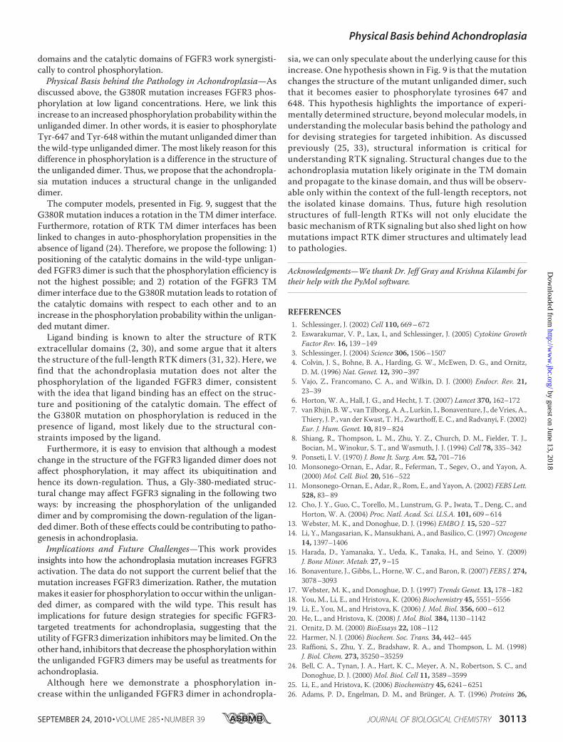
FGFR3 dimer structure, herewe compared the structures of themutant (Fig. 9, green) and wild-type (cyan) TM dimers, bothdetermined with the program CHI, by aligning one of the heli-ces in the wild-type dimer with the respective helix in themutant dimer. The result is shown in Fig. 9 and reveals thedifferences inTMdimer structures. The amino acids thatmedi-ate helix-helix contacts in the wild-type dimer are Leu-377,Val-381, Phe-384, and Ile-387, whereas themutant dimer inter-face is composed of Ile-376, Arg-380, Phe-383, Ile-387, Val-390,and Thr-394. In both structures, specific interactions betweenthe helices are mediated by side chains.To visualize the rotation in the dimer structure, in Fig. 9 we
have colored Ala-391 (which is close to the C terminus of theTM domain) in both structures as follows: yellow for the wild-type and red for the mutant. We see that the two alanines inposition 391 face each other directly in the wild-type structurebut are rotated away from each other in the mutant structure.Such a rotation, according to the experiments of Bell et al. (24),leads to a rotation of the catalytic domains with respect to eachother and to a change in their kinase activity.
DISCUSSION
Novel Findings—The main findings in this paper are as fol-lows. 1) The achondroplasia mutation does not affect the phos-phorylation of Neu_FGFR3, as probed by the 4G10 antibody. 2)The achondroplasia mutation does not affect the cross-linking
ofNeu_FGFR3. 3) The achondroplasiamutation does not affectthe phosphorylation of FGFR3 at high ligand concentration, asprobed by the Tyr-653/Tyr-654 antibody, which specificallyrecognizes Tyr-647 and Tyr-648 in FGFR3. 4) The mutationdoes not affect the cross-linking of FGFR3 at any ligand con-centration. 5) The mutation increases the phosphorylation ofFGFR3 at no and at low ligand concentration, as probed by theTyr-653/Tyr-654 antibody.Thus, the only effect that we observe due to the mutation is
an increase in the fraction of receptors that are phosphorylatedat Tyr-653/Tyr-654 in the absence of ligand and at low ligandconcentration. The two tyrosines, Tyr-647 and Tyr-648, thatare recognized by the Tyr-653/Tyr-654 antibody are essentialfor biological function, and thus the observed increase in theirphosphorylation probability is likely related to pathogenesis inachondroplasia.Here, we do not study the effect of the G380R mutation on
FGFR3 trafficking or down-regulation. We address the under-lying reason for the increased FGFR3 phosphorylation becauseof the mutation. It has been hypothesized that this reason isincreased FGFR3 dimerization. To address this hypothesis, wepreviously determined the dimerization propensity of the iso-lated FGFR3 TM domain in liposomes (18). We observed thatthe mutation does not alter the dimerization energetics of the
TABLE 1Measured ratios of cross-linked fractions �D�sat/�d�0 and phospho-Tyr-staining �P�sat/�P�0, as well as calculated phosphorylation probabilityratios �d/�D, for the wild-type FGFR3 and the G380R mutant
�D�sat/�d�0 �P�sat/�P�0 �d/�D
Wild type 1.51 0.05 6.24 0.50 0.24 0.07G380R mutant 1.50 0.44 2.50 0.60 0.60 0.09
TABLE 2Optimal values for the total receptor concentration �TR�, dimerization constant K1, and ligand-binding constant K2 determined by fitting thetheoretical predictions of phosphorylated fractions, given by Reactions 1 and 2 and Equations 1–7, to the experimentally measured values inFig. 8The differences in the optimal values of K1 and K2 for the wild type and mutant are not significant (p � 0.05). Thus, the mutation is not affecting dimerization or ligandbinding.
�TR� K1 K2
Wild type 4.9 0.9�106 0.07 0.06 0.2 0.1G380R mutant 4.7 0.2�106 0.02 0.01 0.3 0.1
FIGURE 9. Model structures of the mutant (green) and wild-type (cyan)FGFR3 TM domain dimers. One of the helices in the wild-type dimer isaligned with the respective helix in the mutant dimer. To visualize the rota-tion in the dimer structure, we have colored Ala-391 (which is close to the Cterminus of the TM domain) in both structures, yellow for the wild type andred for the mutant. These amino acids face each other directly in the wild-typestructure but are rotated away from each other in the mutant structure.
Physical Basis behind Achondroplasia
SEPTEMBER 24, 2010 • VOLUME 285 • NUMBER 39 JOURNAL OF BIOLOGICAL CHEMISTRY 30111
by guest on June 13, 2018http://w
ww
.jbc.org/D
ownloaded from

isolated FGFR3 TM domain. In this study, we do not see aneffect of the mutation on FGFR3 and Neu_FGFR3 cross-link-ing. By fitting the Western blot phosphorylation data to theactivation model described by Reactions 1 and 2 and Equations1–7, we further show that the mutation does not affect thedimerization constant K1. Thus, our results do not support thehypothesis that the mutation enhances FGFR3 dimerization.The only parameter of the activation model affected by the
achondroplasia mutation is d, the probability for phosphoryl-ation in the unliganded dimer. The increase in d leads to theobserved increase in FGFR3 phosphorylation in the absence ofligand and at low ligand concentrations.Discussion of the FGFR3 Activation Model—The heart of the
physical-chemical model used here to fit the Western blotresults are the two coupled Reactions 1 and 2 describing dimer-ization and ligand binding. Thus, the model accounts for whatis often called “basal or constitutive” activation of FGFR3 in theabsence of ligand (13). Indeed, Reaction 1 is driven to the right,toward the unliganded dimeric state, by increasing the totalreceptor concentration.To account for phosphorylation within the unliganded and
liganded dimers, here we introduce the probabilities for phos-phorylation, d and D. Both values are non-zero, because weobserve phosphorylation both in the absence and presence ofligand. Note that even the immature FGFR3 population locatedin the endoplasmic reticulum can be phosphorylated (16).In themodel,d andD are experimentally determined. The
phosphorylation level at high ligand concentrations yieldsinformation about the value ofD. Although the absolute valueofD cannot be determined, the ratiosd/D for the wild typeand the mutant are determined by comparing phosphorylationand cross-linking data in the absence of ligand and at saturatingligand concentrations (see Table 1). Furthermore, the fact thatthe Tyr-653/Tyr-654 band intensities are the same for the wildtype and the mutant at saturating ligand concentrations sug-gests that the values ofD are the same for thewild type and themutant.A crude assumption in the data analysis is the use of cross-
linked fractions as ameasure of dimeric fractions. This assump-tion is not always valid, because the probability for cross-linkingdepends on both dimerization propensity and dimer structure.The latter is an important determinant of cross-linking efficien-cies, because the cross-linker reacts with amines in very closeproximity.We approximate dimericwith cross-linked fractionsbecause there are no experimental methods available that yieldabsolutemeasures of RTK dimeric fractions on cell surfaces. Asquantitative FRET methods become more sophisticated, theymight eventually allow us tomeasure [D]sat/[d]0 directly.Whensuch measurements become feasible and available, the ratiosd/D for thewild type and themutant can be recalculated andtheWestern blot data in Fig. 8 refitted, for better estimates ofK1and K2.We know that the ligand indices a large structural change in
the extracellular domain, and thus it is possible that the effi-ciency of the cross-linker is different for unliganded and ligan-ded dimers (2, 29). Thus, our estimates of [D]sat/[d]0 may bevery crude. At the same time, the G380R mutation is notexpected to have a large effect on the structure of the extracel-
lular domain, and thus we can expect that the ratios [d]0mut/[d]0WT, [D]satmut/[D]satWT, and d
mut/dWT are not much
affected by approximating cross-linked with dimeric fractions.Thus, the conclusions of this study should not be affected byour inability to directly measure dimeric fractions.A second crude assumption of the model is the assumption
that the mature FGFR3 is located predominantly in the plasmamembrane. In fact, a fraction of the mature FGFR3 is expectedto be found intracellularly. For instance, FGFR3 has beenshown to localize and signal in the endosomal compartments,where it can be either destroyed or recycled back to the mem-brane (12). The model we present here is simplified, because itdoes not take into account the different mature FGFR3 pools(trans-Golgi, plasma membrane, endosomes). Yet the modelfits the experimental data very well, as demonstrated in Fig. 8.Comparison with Other Studies and New Insights—Our
results with the chimeric Neu_FGFR3 receptor are differentfrom a previous study by Webster and Donoghue (13), whoobserved an increase in phosphorylation due to the G380Rmutation. The observed difference may be due to experimentaldetails. For instance, we used CHO cells for these experiments,andWebster and Donoghue (13) used NIH3T3 cells and COS7cells. They assayed phosphorylation after immunoprecipita-tion, while here we probe the phosphorylation in cells. It ispossible that phosphorylation probabilities are different withinthe immunoprecipitate and the cellular membrane. Note that asimilar mutation in FGFR3 TM domain, A391E, shows anincrease in phosphorylation and cross-linking under the verysame conditions used here to probe the effect of the G380Rmutation (20).In this study, we use a new approach to gain insight into
the effect of the G380R mutation on receptor function.For the first time, we use a physical-chemical model to inter-pret the phosphorylation of full-length FGFR3. We fit theexperimental data to the model, which describes phospho-rylation in terms of propensities for dimerization and ligandbinding, and in terms of phosphorylation probabilities.Although the model is relatively simple, we see that it fits thedata very well. Thus, it allows us to extract apparent thermo-dynamic parameters and reveal the effect of the mutation ondifferent steps in FGFR3 activation. To be able to do this work,we titrate ligand over a very wide concentration range, whichallows themeasurement of phosphorylated fractions. Althoughsuch high ligand concentrations are nonphysiological, theyallow us to observe a plateau in receptor phosphorylation andthusmeasure the phosphorylated fractions at lower, physiolog-ically relevant ligand concentrations.By using two different receptors, the chimeric Neu_FGFR3
and the full-length FGFR3, we gain additional insight into theeffect of the G380R mutation on FGFR3 activation. Althoughthe mutation does not affect the phosphorylation within thechimeric receptor dimer, it increases the phosphorylationprobability within the full-length FGFR3 unliganded dimer.Thus, the effect of the mutation appears to propagate into thekinase domain of FGFR3 but not into the kinase domain of Neuwithin the Neu_FGFR3 chimera. The fact that the effect isobserved only in full-length FGFR3 suggests that the TM
Physical Basis behind Achondroplasia
30112 JOURNAL OF BIOLOGICAL CHEMISTRY VOLUME 285 • NUMBER 39 • SEPTEMBER 24, 2010
by guest on June 13, 2018http://w
ww
.jbc.org/D
ownloaded from
domains and the catalytic domains of FGFR3 work synergisti-cally to control phosphorylation.Physical Basis behind the Pathology in Achondroplasia—As
discussed above, the G380R mutation increases FGFR3 phos-phorylation at low ligand concentrations. Here, we link thisincrease to an increased phosphorylation probability within theunliganded dimer. In other words, it is easier to phosphorylateTyr-647 andTyr-648within themutant unliganded dimer thanthe wild-type unliganded dimer. Themost likely reason for thisdifference in phosphorylation is a difference in the structure ofthe unliganded dimer. Thus, we propose that the achondropla-sia mutation induces a structural change in the unligandeddimer.The computer models, presented in Fig. 9, suggest that the
G380R mutation induces a rotation in the TM dimer interface.Furthermore, rotation of RTK TM dimer interfaces has beenlinked to changes in auto-phosphorylation propensities in theabsence of ligand (24). Therefore, we propose the following: 1)positioning of the catalytic domains in the wild-type unligan-ded FGFR3 dimer is such that the phosphorylation efficiency isnot the highest possible; and 2) rotation of the FGFR3 TMdimer interface due to the G380Rmutation leads to rotation ofthe catalytic domains with respect to each other and to anincrease in the phosphorylation probability within the unligan-ded mutant dimer.Ligand binding is known to alter the structure of RTK
extracellular domains (2, 30), and some argue that it altersthe structure of the full-length RTK dimers (31, 32). Here, wefind that the achondroplasia mutation does not alter thephosphorylation of the liganded FGFR3 dimer, consistentwith the idea that ligand binding has an effect on the struc-ture and positioning of the catalytic domain. The effect ofthe G380R mutation on phosphorylation is reduced in thepresence of ligand, most likely due to the structural con-straints imposed by the ligand.Furthermore, it is easy to envision that although a modest
change in the structure of the FGFR3 liganded dimer does notaffect phosphorylation, it may affect its ubiquitination andhence its down-regulation. Thus, a Gly-380-mediated struc-tural change may affect FGFR3 signaling in the following twoways: by increasing the phosphorylation of the unligandeddimer and by compromising the down-regulation of the ligan-ded dimer. Both of these effects could be contributing to patho-genesis in achondroplasia.Implications and Future Challenges—This work provides
insights into how the achondroplasia mutation increases FGFR3activation. The data do not support the current belief that themutation increases FGFR3 dimerization. Rather, the mutationmakes it easier for phosphorylation to occurwithin the unligan-ded dimer, as compared with the wild type. This result hasimplications for future design strategies for specific FGFR3-targeted treatments for achondroplasia, suggesting that theutility of FGFR3 dimerization inhibitorsmay be limited. On theother hand, inhibitors that decrease the phosphorylationwithinthe unliganded FGFR3 dimers may be useful as treatments forachondroplasia.Although here we demonstrate a phosphorylation in-
crease within the unliganded FGFR3 dimer in achondropla-
sia, we can only speculate about the underlying cause for thisincrease. One hypothesis shown in Fig. 9 is that the mutationchanges the structure of the mutant unliganded dimer, suchthat it becomes easier to phosphorylate tyrosines 647 and648. This hypothesis highlights the importance of experi-mentally determined structure, beyondmolecular models, inunderstanding the molecular basis behind the pathology andfor devising strategies for targeted inhibition. As discussedpreviously (25, 33), structural information is critical forunderstanding RTK signaling. Structural changes due to theachondroplasia mutation likely originate in the TM domainand propagate to the kinase domain, and thus will be observ-able only within the context of the full-length receptors, notthe isolated kinase domains. Thus, future high resolutionstructures of full-length RTKs will not only elucidate thebasic mechanism of RTK signaling but also shed light on howmutations impact RTK dimer structures and ultimately leadto pathologies.
Acknowledgments—We thank Dr. Jeff Gray and Krishna Kilambi fortheir help with the PyMol software.
REFERENCES1. Schlessinger, J. (2002) Cell 110, 669–6722. Eswarakumar, V. P., Lax, I., and Schlessinger, J. (2005) Cytokine Growth
Factor Rev. 16, 139–1493. Schlessinger, J. (2004) Science 306, 1506–15074. Colvin, J. S., Bohne, B. A., Harding, G. W., McEwen, D. G., and Ornitz,
D. M. (1996) Nat. Genet. 12, 390–3975. Vajo, Z., Francomano, C. A., and Wilkin, D. J. (2000) Endocr. Rev. 21,
23–396. Horton, W. A., Hall, J. G., and Hecht, J. T. (2007) Lancet 370, 162–1727. van Rhijn, B.W., vanTilborg, A. A., Lurkin, I., Bonaventure, J., deVries, A.,
Thiery, J. P., van der Kwast, T. H., Zwarthoff, E. C., and Radvanyi, F. (2002)Eur. J. Hum. Genet. 10, 819–824
8. Shiang, R., Thompson, L. M., Zhu, Y. Z., Church, D. M., Fielder, T. J.,Bocian, M., Winokur, S. T., and Wasmuth, J. J. (1994) Cell 78, 335–342
9. Ponseti, I. V. (1970) J. Bone Jt. Surg. Am. 52, 701–71610. Monsonego-Ornan, E., Adar, R., Feferman, T., Segev, O., and Yayon, A.
(2000)Mol. Cell. Biol. 20, 516–52211. Monsonego-Ornan, E., Adar, R., Rom, E., and Yayon, A. (2002) FEBS Lett.
528, 83–8912. Cho, J. Y., Guo, C., Torello, M., Lunstrum, G. P., Iwata, T., Deng, C., and
Horton, W. A. (2004) Proc. Natl. Acad. Sci. U.S.A. 101, 609–61413. Webster, M. K., and Donoghue, D. J. (1996) EMBO J. 15, 520–52714. Li, Y., Mangasarian, K., Mansukhani, A., and Basilico, C. (1997)Oncogene
14, 1397–140615. Harada, D., Yamanaka, Y., Ueda, K., Tanaka, H., and Seino, Y. (2009)
J. Bone Miner. Metab. 27, 9–1516. Bonaventure, J., Gibbs, L., Horne,W. C., and Baron, R. (2007) FEBS J. 274,
3078–309317. Webster, M. K., and Donoghue, D. J. (1997) Trends Genet. 13, 178–18218. You, M., Li, E., and Hristova, K. (2006) Biochemistry 45, 5551–555619. Li, E., You, M., and Hristova, K. (2006) J. Mol. Biol. 356, 600–61220. He, L., and Hristova, K. (2008) J. Mol. Biol. 384, 1130–114221. Ornitz, D. M. (2000) BioEssays 22, 108–11222. Harmer, N. J. (2006) Biochem. Soc. Trans. 34, 442–44523. Raffioni, S., Zhu, Y. Z., Bradshaw, R. A., and Thompson, L. M. (1998)
J. Biol. Chem. 273, 35250–3525924. Bell, C. A., Tynan, J. A., Hart, K. C., Meyer, A. N., Robertson, S. C., and
Donoghue, D. J. (2000)Mol. Biol. Cell 11, 3589–359925. Li, E., and Hristova, K. (2006) Biochemistry 45, 6241–625126. Adams, P. D., Engelman, D. M., and Brunger, A. T. (1996) Proteins 26,
Physical Basis behind Achondroplasia
SEPTEMBER 24, 2010 • VOLUME 285 • NUMBER 39 JOURNAL OF BIOLOGICAL CHEMISTRY 30113
by guest on June 13, 2018http://w
ww
.jbc.org/D
ownloaded from

257–26127. Adams, P. D., Arkin, I. T., Engelman, D.M., and Brunger, A. T. (1995)Nat.
Struct. Biol. 2, 154–16228. Adams, P. D., and Brunger, A. T. (1997) in Membrane Protein Assembly
(von Heijne, G., ed) pp. 251–265, R.G. Landes Co., Austin, TX29. Schlessinger, J. (2003) Science 300, 750–75230. Olsen, S. K., Ibrahimi, O. A., Raucci, A., Zhang, F., Eliseenkova, A. V.,
Yayon, A., Basilico, C., Linhardt, R. J., Schlessinger, J., and Mohammadi,M. (2004) Proc. Natl. Acad. Sci. U.S.A. 101, 935–940
31. Moriki, T., Maruyama, H., and Maruyama, I. N. (2001) J. Mol. Biol. 311,1011–1026
32. Yu, X., Sharma, K. D., Takahashi, T., Iwamoto, R., and Mekada, E. (2002)Mol. Biol. Cell 13, 2547–2557
33. Li, E., and Hristova, K. (2010) Cell Adhes. Migrat. 4, 249–254
Physical Basis behind Achondroplasia
30114 JOURNAL OF BIOLOGICAL CHEMISTRY VOLUME 285 • NUMBER 39 • SEPTEMBER 24, 2010
by guest on June 13, 2018http://w
ww
.jbc.org/D
ownloaded from

Lijuan He, William Horton and Kalina HristovaDwarfism
Physical Basis behind Achondroplasia, the Most Common Form of Human
doi: 10.1074/jbc.M109.094086 originally published online July 12, 20102010, 285:30103-30114.J. Biol. Chem.
10.1074/jbc.M109.094086Access the most updated version of this article at doi:
Alerts:
When a correction for this article is posted•
When this article is cited•
to choose from all of JBC's e-mail alertsClick here
Supplemental material:
http://www.jbc.org/content/suppl/2010/07/12/M109.094086.DC1
http://www.jbc.org/content/285/39/30103.full.html#ref-list-1
This article cites 32 references, 8 of which can be accessed free at
by guest on June 13, 2018http://w
ww
.jbc.org/D
ownloaded from