physical therapy rehabilitation following tlif - …chronicpainsite.com/articles/ptrf.pdf ·...
TRANSCRIPT
Physical Therapy Rehabilitation Following TLIF
A Case Series Approach
By Michael R. Noonan P.T., D.P.T November 18, 2005
0
Introduction The spinal fusion procedure was introduced as a treatment option for chronic LBP nearly 70 years ago; however the literature reveals divergent opinions about when fusion is indicated and how it should be performed. Furthermore, the significance of the role of postoperative rehabilitation following spinal fusion may be underestimated and there exists no consensus on the design of a program specific for rehabilitation (1-Christensen FB 2004).
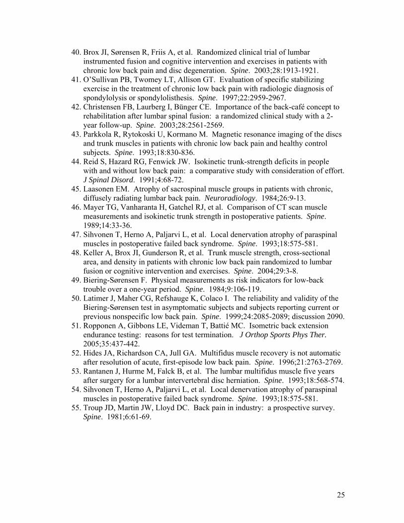
The rate of lumbar fusion surgery in the U.S. increased 100% in the 1980’s and more than 220% from 1990 to 2001. Of coincidence is the FDA approval of intervertebral fusion cages in the U.S. in 1996. Also of note is the 113% increase in lumbar fusions from 1996 to 2001, compared to 13% and 15% increases in hip and knee replacements respectively during that same time period. The over 60 age group involving fusions has risen most rapidly by 230% from 1988 to 2001. Lumbar fusion is among the most rapidly increasing of all major surgical procedures, and also one of the most expensive (2-Deyo 2005). Refer to Figure 1 and Table 1. Satisfactory results after different surgical fusion techniques used in patients with low back pain has been shown in the literature to vary between 16-95% (3-Fritzell et al. 2001). The reported “overall” improvement in the Swedish Lumbar Spine Study (a multicenter randomized controlled trial) was 63% and statistically, all primary outcome measures in the study were significantly in favor of surgery. In that study, patients in the surgical group were rehabilitated according to “local preferences”, with a focus on early mobilization and informational support. Yet no specific or extensive exercise program was used routinely.
Low back pain (LBP) not only is the second most common symptom reported by persons visiting their primary care physician (4- Hart et al 1995), but it’s rather costly too. The most up-to-date information indicates that LBP accounts for $91 billion annually on our health care budget of $1.2 trillion; accounting for 13.6% of gross domestic product (5-Blumenthal 2001) Estimates of at least one lifetime episode of low back pain among Americans are 80%. On average, individuals with back pain incurred health care expenditures about 60% higher than individuals without back pain. (6-Luo et al. 2004).
Although most acute low back pain episodes (80-90%) subside within 6 to 12 weeks, recurrence is common in half of these patients, and about 10% of patients have chronic symptoms (7-Croft et al. 1998). The average age of patients with LBP is 44.5 years, with a majority of claimants (56.5%) being women (8-Vogt et al. 2005).
More disconcerting is that 5-10% of the people who become disabled with chronic LBP account for 75-90% of the cost (9-Indahl et al. 1995). Despite a large number of pathological conditions that can give rise to back pain, 85% are classified as having “non specific low back pain”. (10-Dillingham 1995). More recently, there has been an increased focus on the different subgroups within this population, with lumbar segmental instability representing one of these groups (11-Friberg 1987). The radiological diagnosis of spondylolisthesis and spondylolysis, in subjects with chronic LBP attributable to this finding, has been considered to be one of the most obvious manifestations of lumbar instability (12-Nachemson 1991;13- Pope et al. 1992, 14-Friberg 1989). A number of studies have reported increased and abnormal
1
intersegmental motion in subject with chronic LBP, often in the absence of other radiological findings (15-Gertzbein 1991). The most common reasons for lumbar fusion in order of prevalence are possible instability (spondylolysis or spondylosisthesis), degenerative disc disease (including HNP), and spinal stenosis (central canal and/or foraminal). Less common indications are fractures, neoplasms, infections and inflammatory diseases (2-Deyo 2005), and also intraoperative removal of more than one facet joint that renders the segmental level unstable as in cases of severe foraminal stenosis during lumbar decompression surgery (16-Jenis L, and An H. 2000). From a clinical perspective, disc degeneration is believed to be a source of chronic pain, and over 90% of surgical spine procedures are performed because of consequences of the degenerative process. Disc degeneration can lead to secondary clinical problems, including disc herniation, spinal stenosis, and degenerative spondylolisthesis (17-An et al. 2004).
This dramatic increase in lumbar fusion rates over the past decade and associated costs of care and rate of disability creates not only an opportunity but a responsibility for the physical therapist to better understand the surgical approach involved. This will in turn allow physical therapists to develop a biomechanical based rehabilitation program that not only protects the repair and optimizes remaining physical function, but also educates the patient about their new spinal loading and range of motion limitations, both early on and long term, to help prevent future disability.
The purpose of this case series is to compare 3 patients with similar impairments and functional limitations who underwent a nearly identical lumbar fusion by the same surgeon and treated postoperatively by the same physical therapist. Particular attention is placed on the rehabilitation exercises, as evidence in favor of any specific exercise is lacking.
2
Lumbar Fusion Overview Posterolateral intertransverse fusion (PLF): bone is placed to join decorticated transverse processes of adjacent vertebrae using bone graft and bone morphogenic protein (BMP). This procedure is often combined with pedicle screw fixation. (18-DeRosa 2005) Pedicle screws fixation: screws are placed horizontally through the pedicles and into the vertebral body of each vertebra of adjacent segments. Then vertically placed rods are coupled to the screw in order to fixate the adjacent segments. (18-DeRosa 2005) Interbody fusions: these types of procedures involve the bone-disc-bone interface.
• Anterior lumbar interbody fusion (ALIF): an anterior approach through
the abdomen is utilized for placement of a large bone graft or intervertebral body device, such as titanium, plastic, or carbon fiber cage, after diskectomy is performed. (18-DeRosa 2005)
• Posterior lumbar interbody fusion (PLIF): a posterior approach involving
a laminectomy then diskectomy with insertion of a spacer with autogenous bone graft into the space left after disc removal. First attempted by Cloward in 1940 and later revised by Lin in 1977 (19-C. Humphreys 2001). Pedicle screw fixation is performed to gain immediate segmental rigidity while the fusion heals.
• Transforaminal lumbar interbody fusion (TLIF): a recent variation of the
PLIF. Developed by Harms in approximately 1997 (20-Harms 1998), it uses a posterior approach to the spine, but accesses the disc space using a unilateral facetectomy via a path that runs through the far lateral portion of the vertebral foramen, allowing the complete removal of the disc and placement of an interbody support transforaminally, without extensive mobilization of the thecal sac and nerve roots. Pedicle screw fixation is performed to gain immediate segmental rigidity while the fusion heals (19-C. Humphreys 2001, 21-N. Figueiredo 2004).
*See Figure 2. A-G for detailed steps in TLIF procedure. Also see Table 2 for potential advantages and disadvantages of the TLIF procedure.
3
*All three case studies presented in this paper involved a TLIF procedure, described as follows:
Surgical Procedure*
The patient has a Grade II spondylolisthesis at L4-5 as well as a lateral disc
displacement of the L5-S1 disc on the left with displacement of the S1 nerve root. Once under general anesthesia and intubated, the team of 5 and the surgeon
transfer the patient from supine to prone onto a Wilson frame in log-roll fashion. Hips and knees are flexed 45° and spine is maintained in neutral posture while face is padded by surgical foam pillow to allow access by anesthesiologist. Arms and shoulders are flexed and abducted at shoulder height and ankles are padded on dorsal surface. The low back is prepped and draped in standard sterile fashion.
L4 through S1 is marked with a surgical pen and confirmed by placing a metal probe on the skin and taking a picture with the Imaging Intensifier “ii”. The ii is a flouroscope that can rotate nearly 360° around patient, up and down and side to side. It’s used frequently throughout the surgery to confirm bony landmarks and placement of hardware. Electrocautery and gel foam are used to control bleeding during dissection.
An incision is made from L4 to S1 and the fascial planes are carefully dissected. The paraspinals (iliocostalis lumborum, longissimus thoracis and multifidus) are then elevated as one mass from the spinous processes and lamina in a subperiosteal dissection technique. The exposure is carried out laterally over the facet joints and out to the base of the transverse processes on either side. The incision is extended to L3, including additional muscular fascial release to be “less traumatic” on the musculature (i.e. paraspinals). A depth of >2 inches is obtained before the facets can be visualized. After removal of hypertrophic ligamentum flavum and the supra/interspinous ligaments of L4-S1, attention is first placed on the right side and a partial facetectomy and laminoforaminotomy is performed. Next, fixation on the right begins with insertion of a Pedicle Screw with a hand drill into L4, followed by L5 and then S1. The landmarks were first identified with a sharp metal probe and checked by the ii. Next, one rod is placed over the “gutter like” ends of the pedicle screws (3), and then partially tightened with special nuts (3), to complete one side of the fusion.
Next, attention is placed on the left side with complete facetectomy and laminoforaminotomy of L4-5 after ligamentous removal as on the right. Then radical transforaminal discectomies were performed at L4-5, followed by L5-S1. The endplates of L4-S1 were prepared for arthrodesis by decorticating them using an assortment of curets, rasps, rongeurs and the Midas Rex drill.
The disc spaces are now ready for the “Boomerang” interbody spacer. The “Boomerang” is made by Medtronic (www.medtronicsofamordanek.com). It is essentially an implant made of synthetic plastic that houses autologous bone and BMP (bone morphogenic protein; a recombinant protein that stimulates bone metabolism). The bone harvested from the facetectomies is placed on a collagen sponge that has been soaked in BMP and then rolled into a tight wrap and placed into the Boomerang. This is placed into the disc spaces of L4-5 and L5-S1, along with additional autogolous bone and BMP, to complete the fusion anteriorly.
4
Now, the pedicle screws are placed on the left into L4-S1, followed by the rod and nuts as on the right. Both sides are tightened and then connected with 3 cross-links attaching the nuts of L-4, L-5, and then S1 to complete the fusion posteriorly.
This fusion creates a “Box-like” or 360° affect with the Boomerang and autogolous bone anteriorly (anterior pillar), pedicle screws and rods laterally, and cross-links posteriorly (posterior pillar).
The surgical area is then closed with sutures to repair the fascial layers. The thoracolumbar fascia is reattached to the remaining midline structures in multiple layers with dissolvable sutures, up to and including the skin. Then 3 coats of “dermabond” glue (Johnson & Johnson) are used to seal the epidermis. Total surgical time 5 hrs.
Case Series Descriptions Subjects:
Three adults with chronic low back pain and lumbar instability underwent lumbar decompression and fusion via TLIF by the same surgeon. Each had evidence of spondylolisthesis with stenosis and/or HNP. Each patient’s referral diagnosis was refined through constructing a diagnosis for physical therapy that consists of hypothetical relationships among pathology, impairment, functional limitation, and disability according to the Guide to Physical Therapist Practice (22) as Musculoskeletal: 4F Impaired joint mobility, motor function, muscle performance, range of motion, and reflex integrity associated with spinal disorders. None had previous lumbar surgeries, but all three failed conservative measures (epidural steroid injections, chiropractic treatment, and physical therapy). All three were hospitalized where they received in-patient physical therapy and early ambulation the day after surgery. Each began outpatient therapy 4-6 weeks post-operatively. All three patients had similar impairments and functional limitations and followed the surgeon’s guidelines listed in Table 3, with variations listed separately under each case study:
5
Case Study 1(GL) Patient History: An 84-year-old woman underwent lumbar decompression and fusion at L3-4 and L4-5, utilizing a TLIF approach and pedicle screw fixation at both levels. She was hospitalized for 12 days and received daily PT/OT to regain strength and independence as she lives alone in a single story apartment, and then 2 weeks of home health PT.
Her initial symptoms began insidiously in 1999 with LBP only and she sought chiropractic manipulation for 5 months, but reported minimal relief. On 1/21/00, standard radiographs revealed mild to moderate degenerative changes L2 thru S2, lumbarization of S1, Grade 1 spondylolisthesis L4-5 and L5-S1, and mild osteoarthritis both hips. For the next few years she continued to receive intermittent treatment of non–manipulative manual therapy, massage, electrical stimulation and ultrasound from a PT/DC and another DC to her low back, shoulders, and right knee. On 7/29/02 an MRI revealed moderately-severe central stenosis reflecting degenerative listhesis at L3-4 and mild-moderate central stenosis at L4-5. In late 2003 she began receiving cortisone injects for moderate-severe right knee osteoarthritis every 3 to 6 months, to present, after a tibial plateau fracture from a MVA on 5/22/03. In early 2004 she received a series of two epidural injections with relief of LBP for several months, but by 12/04 her LBP was worsening progressively and more severely and bilateral leg pain (posterior thighs to the knees) had become constant and forced her to using a walker. On 1/11/05, hip radiographs showed mild-to-moderate osteoarthritis and she chose pool therapy over low back surgery, but obtained no relief. In general, her LBP worsened and lessened for the past six years without recollection of any specific event that may have caused her pain, but it was the onset of leg pain for 6 months that led to the above said surgery on 5/9/05. The initial physical therapy evaluation was performed 5 ½ weeks post-surgically. The patient’s primary complaint was constant “ache” across the lumbosacral area as revealed on the body chart which she reported was 3/10 to 4/10 on the numeric pain rating scale (NPRS). She denied any leg pain since surgery and a thorough health history screen indicated osteoarthritis in hips and right knee and osteoporosis, and age related urinary incontinence. Medications included Tylenol with codeine for back pain t.i.d., synthroid, and over-the-counter antacids, laxatives and Actifed. She is retired and a non-smoker. She lives alone (widowed) and her primary goal was to resume golf once a week, and occasional day trips once or twice a month. She resumed driving 11 days prior to her therapy visit (4 weeks post-op) and was slowly resuming her independence. Observation: The patient ambulated with a FWW and wore a semi-rigid back brace. Her gait was steady with a widened base of support and she reported tiring easily with limited distances from room to room in her apartment. She was able to transfer sit to stand well,
6
but needed assistance sit↔sidelying↔supine due to LBP. In standing she demonstrated a flat back posture with a slight forward lean with mild genu-valgus bilaterally and level hips in frontal plane. She was able to safely take a few steps without the walker in the treatment room with her hands on treatment table and chair. The incision was healing well with no signs of infection. Tests and Measures: See tables 4a and 4b for impairments/functional limitations and associated tests and measures with their results. Evaluation and Treatment (Plan of Care): The patient demonstrated an expected amount of post-operative stiffness, ROM limitations and functional deficits with minimal ache status-post fusion L3-L5. She was surprisingly independent for a woman her age and given the extensiveness of her procedure. Of concern was the osteoarthritis in her hips and right knee documented radiographically as increased demand on the lower extremities was required to avoid bending and loading the lumbar spine to protect the fusion. Treatment interventions included the following: Therapeutic exercises; Moist heat; Soft tissue mobilization; Biomechanical counseling/posture principles for ADL’s and IADL’s were emphasized for the duration of therapy. Treatment was recommended twice a week for 6 to 8 weeks. Intervention: Patient was seen 2 visits per week for 6 ½ weeks for a total of 13 sessions. She was also given a home exercise program (HEP) of twice daily stretches, and a walking program. The following protocol was developed by the therapist at this clinic with the surgeon involved: Rehabilitation Phase I (5-7 weeks postoperative)
• Protection Fusion & Movement Extremities: – Moist heat in supine, soft tissue mobilization thoracolumbar paraspinals
and scar cross fiber friction in supported sitting against treatment table for comfort without imparting stress to the fused area.
– Therapeutic exercises in supine for support while on moist heat as follows: Single and double knees-to-chest, pelvic tilts, neurodynamic glides, hip group and gastroc-soleus stretching and the abdominal drawing in maneuver (ADIM).
– Functional Activities: golfer’s lift &/or reacher, squat &/or ½ kneel, pivot not twist with neutral spine with all transfers and bed mobility.
– Initiate CV conditioning; treadmill & recumbent bike 3-5 minutes each. – Wean from walker and brace after X-ray showed healing and steady gait. – Continue bone stimulator. – Frequency 2x/wk. x 3 wks. (6 visits).
7
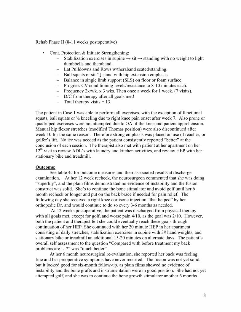
Rehab Phase II (8-11 weeks postoperative)
• Cont. Protection & Initiate Strengthening: – Stabilization exercises in supine → sit → standing with no weight to light
dumbbells and theraband. – Lat Pulldowns and Rows w/theraband seated/standing. – Ball squats or sit ↑↓ stand with hip extension emphasis. – Balance in single limb support (SLS) on floor or foam surface. – Progress CV conditioning levels/resistance to 8-10 minutes each. – Frequency 2x/wk. x 3 wks. Then once a week for 1 week. (7 visits). – D/C from therapy after all goals met! – Total therapy visits = 13.
The patient in Case 1 was able to perform all exercises, with the exception of functional squats, ball squats or ½ kneeling due to right knee pain onset after week 7. Also prone or quadruped exercises were not attempted due to OA of the knee and patient apprehension. Manual hip flexor stretches (modified Thomas position) were also discontinued after week 10 for the same reason. Therefore strong emphasis was placed on use of reacher, or golfer’s lift. No ice was needed as the patient consistently reported “better” at the conclusion of each session. The therapist also met with patient at her apartment on her 12th visit to review ADL’s with laundry and kitchen activities, and review HEP with her stationary bike and treadmill. Outcome: See table 4c for outcome measures and their associated results at discharge examination. At her 12 week recheck, the neurosurgeon commented that she was doing “superbly”, and the plain films demonstrated no evidence of instability and the fusion construct was solid. She’s to continue the bone stimulator and avoid golf until her 6 month recheck or longer and put on the back brace if needed for pain relief. The following day she received a right knee cortisone injection “that helped” by her orthopedic Dr. and would continue to do so every 3-6 months as needed.
At 12 weeks postoperative, the patient was discharged from physical therapy with all goals met, except for golf, and worse pain 4/10, as the goal was 2/10. However, both the patient and therapist felt she could eventually reach these goals through continuation of her HEP. She continued with her 20 minute HEP in her apartment consisting of daily stretches, stabilization exercises in supine with 3# hand weights, and stationary bike or treadmill an additional 15-20 minutes on alternate days. The patient’s overall self assessment to the question “Compared with before treatment my back problems are …?” was “much better”.
At her 6 month neurosurgical re-evaluation, she reported her back was feeling fine and her preoperative symptoms have never recurred. The fusion was not yet solid, but it looked good for six-month follow-up, as plain films showed no evidence of instability and the bone grafts and instrumentation were in good position. She had not yet attempted golf, and she was to continue the bone growth stimulator another 6 months.
8
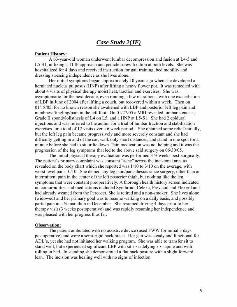
Case Study 2(JE) Patient History: A 63-year-old woman underwent lumbar decompression and fusion at L4-5 and L5-S1, utilizing a TLIF approach and pedicle screw fixation at both levels. She was hospitalized for 4 days and received instruction for gait training, bed mobility and dressing stressing independence as she lives alone.
Her initial symptoms began approximately 10 years ago when she developed a herniated nucleus pulposus (HNP) after lifting a heavy flower pot. It was remedied with about 4 visits of physical therapy moist heat, traction and exercises. She was asymptomatic for the next decade, even running a few marathons, with one exacerbation of LBP in June of 2004 after lifting a couch, but recovered within a week. Then on 01/18/05, for no known reason she awakened with LBP and posterior left leg pain and numbness/tingling/pain in the left foot. On 01/27/05 a MRI revealed lumbar stenosis, Grade II spondylolisthesis of L4 on L5, and a HNP at L5-S1. She had 2 epidural injections and was referred to the author for a trial of lumbar traction and stabilization exercises for a total of 12 visits over a 6 week period. She obtained some relief initially, but the left leg pain became progressively and more severely constant and she had difficulty getting in and of the car, walk only short distances, and stand in one spot for a minute before she had to sit or lie down. Pain medication was not helping and it was the progression of the leg symptoms that led to the above said surgery on 06/30/05. The initial physical therapy evaluation was performed 3 ½ weeks post-surgically. The patient’s primary complaint was constant “ache” across the incisional area as revealed on the body chart which she reported was 1/10 to 3/10 on the average, with worst level pain 10/10. She denied any leg pain/parasthesias since surgery, other than an intermittent pain in the center of the left posterior thigh, but nothing like the leg symptoms that were constant preoperatively. A thorough health history screen indicated no comorbidities and medications included Synthroid, Celexa, Prevacid and Flexeril and had already weaned from the Percocet. She is retired and a non-smoker. She lives alone (widowed) and her primary goal was to resume walking on a daily basis, and possibly participate in a ½ marathon in December. She resumed driving 4 days prior to her therapy visit (3 weeks postoperative) and was rapidly resuming her independence and was pleased with her progress thus far. Observation: The patient ambulated with no assistive device (used FWW for initial 3 days postoperative) and wore a semi-rigid back brace. Her gait was steady and functional for ADL’s, yet she had not initiated her walking program. She was able to transfer sit to stand well, but experienced significant LBP with sit ↔ sidelying ↔ supine and with rolling in bed. In standing she demonstrated a flat back posture with a slight forward lean. The incision was healing well with no signs of infection.
9
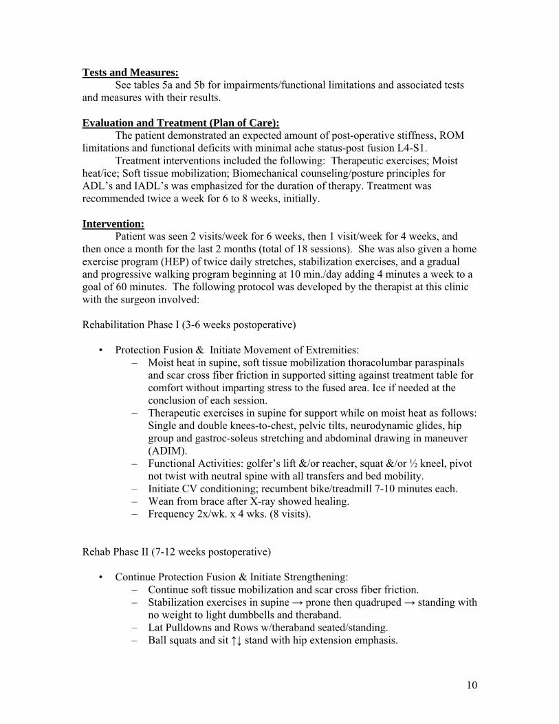
Tests and Measures: See tables 5a and 5b for impairments/functional limitations and associated tests and measures with their results. Evaluation and Treatment (Plan of Care): The patient demonstrated an expected amount of post-operative stiffness, ROM limitations and functional deficits with minimal ache status-post fusion L4-S1. Treatment interventions included the following: Therapeutic exercises; Moist heat/ice; Soft tissue mobilization; Biomechanical counseling/posture principles for ADL’s and IADL’s was emphasized for the duration of therapy. Treatment was recommended twice a week for 6 to 8 weeks, initially. Intervention: Patient was seen 2 visits/week for 6 weeks, then 1 visit/week for 4 weeks, and then once a month for the last 2 months (total of 18 sessions). She was also given a home exercise program (HEP) of twice daily stretches, stabilization exercises, and a gradual and progressive walking program beginning at 10 min./day adding 4 minutes a week to a goal of 60 minutes. The following protocol was developed by the therapist at this clinic with the surgeon involved: Rehabilitation Phase I (3-6 weeks postoperative)
• Protection Fusion & Initiate Movement of Extremities: – Moist heat in supine, soft tissue mobilization thoracolumbar paraspinals
and scar cross fiber friction in supported sitting against treatment table for comfort without imparting stress to the fused area. Ice if needed at the conclusion of each session.
– Therapeutic exercises in supine for support while on moist heat as follows: Single and double knees-to-chest, pelvic tilts, neurodynamic glides, hip group and gastroc-soleus stretching and abdominal drawing in maneuver (ADIM).
– Functional Activities: golfer’s lift &/or reacher, squat &/or ½ kneel, pivot not twist with neutral spine with all transfers and bed mobility.
– Initiate CV conditioning; recumbent bike/treadmill 7-10 minutes each. – Wean from brace after X-ray showed healing. – Frequency 2x/wk. x 4 wks. (8 visits).
Rehab Phase II (7-12 weeks postoperative)
• Continue Protection Fusion & Initiate Strengthening: – Continue soft tissue mobilization and scar cross fiber friction. – Stabilization exercises in supine → prone then quadruped → standing with
no weight to light dumbbells and theraband. – Lat Pulldowns and Rows w/theraband seated/standing. – Ball squats and sit ↑↓ stand with hip extension emphasis.
10
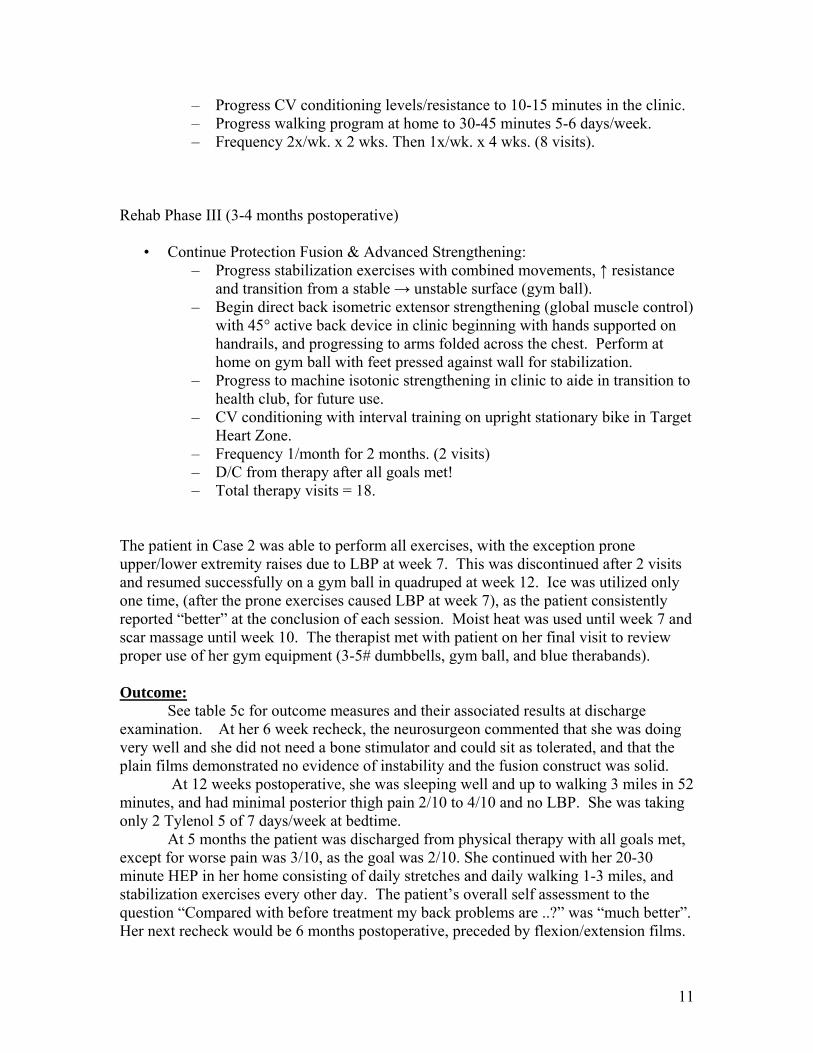
– Progress CV conditioning levels/resistance to 10-15 minutes in the clinic. – Progress walking program at home to 30-45 minutes 5-6 days/week. – Frequency 2x/wk. x 2 wks. Then 1x/wk. x 4 wks. (8 visits).
Rehab Phase III (3-4 months postoperative) • Continue Protection Fusion & Advanced Strengthening:
– Progress stabilization exercises with combined movements, ↑ resistance and transition from a stable → unstable surface (gym ball).
– Begin direct back isometric extensor strengthening (global muscle control) with 45° active back device in clinic beginning with hands supported on handrails, and progressing to arms folded across the chest. Perform at home on gym ball with feet pressed against wall for stabilization.
– Progress to machine isotonic strengthening in clinic to aide in transition to health club, for future use.
– CV conditioning with interval training on upright stationary bike in Target Heart Zone.
– Frequency 1/month for 2 months. (2 visits) – D/C from therapy after all goals met! – Total therapy visits = 18.
The patient in Case 2 was able to perform all exercises, with the exception prone upper/lower extremity raises due to LBP at week 7. This was discontinued after 2 visits and resumed successfully on a gym ball in quadruped at week 12. Ice was utilized only one time, (after the prone exercises caused LBP at week 7), as the patient consistently reported “better” at the conclusion of each session. Moist heat was used until week 7 and scar massage until week 10. The therapist met with patient on her final visit to review proper use of her gym equipment (3-5# dumbbells, gym ball, and blue therabands). Outcome: See table 5c for outcome measures and their associated results at discharge examination. At her 6 week recheck, the neurosurgeon commented that she was doing very well and she did not need a bone stimulator and could sit as tolerated, and that the plain films demonstrated no evidence of instability and the fusion construct was solid.
At 12 weeks postoperative, she was sleeping well and up to walking 3 miles in 52 minutes, and had minimal posterior thigh pain 2/10 to 4/10 and no LBP. She was taking only 2 Tylenol 5 of 7 days/week at bedtime.
At 5 months the patient was discharged from physical therapy with all goals met, except for worse pain was 3/10, as the goal was 2/10. She continued with her 20-30 minute HEP in her home consisting of daily stretches and daily walking 1-3 miles, and stabilization exercises every other day. The patient’s overall self assessment to the question “Compared with before treatment my back problems are ..?” was “much better”. Her next recheck would be 6 months postoperative, preceded by flexion/extension films.
11
Case Study 3(MZ) Patient History: An 84-year-old woman underwent lumbar decompression and fusion at L3-4 and L4-5, utilizing a TLIF approach and pedicle screw fixation at both levels. She was hospitalized for 5 days and received daily PT to regain strength and independence as she lives alone in a single story home. She also had 2 visits by a home health nurse. Her initial symptoms began several years ago after performing routine household tasks, presenting as a back ache as if she had over worked. This continued intermittently until about 4 years ago while in Mexico when she had a sudden and terrible attack of pain and could not straighten up and had to head for home immediately. By the time she reached home the pain subsided. She reported that it was not in her nature to complain so she just let it pass. While at a routine physical in January 2005, she told her primary care physician once again about it. By this time she had constant LBP and right LE pain to the ankle and was forward flexed most of the time. He ordered x-rays, and later an MRI. Upon reviewing the reports, he referred her to a neurosurgeon. The initial physical therapy evaluation was performed 3 ½ weeks post-surgically. The patient’s primary complaint was constant “ache” across the lumbosacral area as revealed on the body chart which she reported was 1/10 to 2/10 on the numeric pain rating scale (NPRS). She denied any leg pain since surgery and a thorough health history screen indicated and age related urinary incontinence controlled with medication, and Type II insulin dependent diabetes that has been well managed for the past 25 years. Medications included an occasional ¼ tablet Tylenol with codeine for back pain. She is retired and a non-smoker. She lives alone (divorced) and her primary goal was to resume driving, light house cleaning and grocery shopping, and her independence. Her daughter checked in on her daily, primarily during her showers, although she was independent with use of a walker and shower bench. Observation: The patient ambulated with a FWW and wore a semi-rigid back brace. Her gait was steady with a widened base of support and she reported tiring easily with limited distances from room to room in her home and overall felt “wobbly”. She was able to transfer sit to stand well, but needed assistance sit↔sidelying↔supine due to LBP. In standing she demonstrated a flat back posture with a slight forward lean, but hips were level in the frontal plane. She was able to safely take a few steps without the walker in the treatment room with her hands on treatment table and chair. The incision was healing well with scabbing in place no signs of infection
12
Tests and Measures: See tables 6a and 6b for impairments/functional limitations and associated tests and measures with their results. Evaluation and Treatment (Plan of Care): The patient demonstrated an expected amount of post-operative stiffness, ROM limitations and functional deficits with minimal ache status-post fusion L3-L5. She was slowly resuming her independence, and did rely on her daughter for driving, shopping and minimal bathing assistance. Treatment interventions included the following: Therapeutic exercises; Moist heat; Soft tissue mobilization; Biomechanical counseling/posture principles for ADL’s and IADL’s were emphasized for the duration of therapy. Treatment was recommended twice a week for 6-8 weeks. Intervention: Patient was seen 2 visits per week for 4 weeks, followed by once a week for 4 weeks for a total of 12 sessions. She was also given a home exercise program (HEP) of twice daily stretches, and walking. The following protocol was developed by the therapist at this clinic with the surgeon involved: Rehabilitation Phase I (3-6 weeks postoperative)
• Protection Fusion & Movement Extremities: – Moist heat in supine with diaphragmatic breathing techniques for
relaxation response, soft tissue mobilization thoracolumbar paraspinals and scar cross fiber friction in supported sitting against treatment table for comfort without imparting stress to the fused area.
– Therapeutic exercises in supine for support while on moist heat as follows: Single and double knees-to-chest, pelvic tilts, neurodynamic glides, hip group and gastroc-soleus stretching and the abdominal drawing in maneuver (ADIM).
– Functional Activities: golfer’s lift &/or reacher, squat &/or ½ kneel, pivot not twist with neutral spine with all transfers and bed mobility.
– Initiate CV conditioning; recumbent bike & treadmill 3-5 minutes each. – Continue bone stimulator. – Frequency 2x/wk. x 4 wks. (8 visits).
Rehab Phase II (7-10 weeks postoperative)
• Cont. Protection & Initiate Strengthening: – Stabilization exercises in supine → sit → standing with no weight to light
dumbbells and theraband. – Lat Pulldowns and Rows w/theraband seated/standing. – Chair squats (sit ↑↓ stand) with hip extension emphasis. – Balance in single limb support (SLS) on stable surface (floor).
13
– Wean from brace & walker as 6 week X-ray showed healing and gait steady.
– Progress CV conditioning levels/resistance to 8-10 minutes each. – Frequency 1x/wk. x 4 wks. (4 visits). – D/C from therapy after all goals met! – Total therapy visits = 12.
The patient in Case 3 was able to perform all exercises. Prone exercises were not attempted based on previous experience with Case 2 and a sufficient program was in place without adding quadruped exercises. No ice was needed as the patient consistently reported “better” at the conclusion of each session. Moist heat was used until week 7 and scar massage until week 9. The entire course of rehabilitation was uneventful, other than at week 8 when the patient fell when stepping over curb, scrapping the left shin a 4 x 6 inch area. She had completed 2 courses of antibiotics and the wound was healing well at the time of discharge. Nevertheless, she was instructed to follow-up with PCP on a weekly basis due to her diabetes, for precautionary measures. Also, she was re-instructed to use curb cutouts vs. stepping over curbs, and continue with her cane when walking outside. Outcome: See table 6c for outcome measures and their associated results at discharge examination. At her 6 week recheck, the neurosurgeon commented that she was doing “superbly”, and the plain films demonstrated no evidence of instability and the fusion construct was solid. She’s to continue the bone stimulator for a total of 9 months. She was instructed to wean from the brace and walker, and use a cane as needed, and recheck at 12 weeks postoperative with flexion/extension X-rays prior to that visit.
At 10 weeks postoperative, the patient was discharged from physical therapy with all goals met. Pain was intermittent with her worse pain 1/10 in the right lumbosacral area in AM only. She continued with her 20 minute HEP in her home consisting of daily stretches, stabilization exercises in supine with 2# hand weights, chair squats, balance drills, and walking her dogs 25 minutes a day, one at a time. The patient’s overall self assessment to the question “Compared with before treatment my back problems are …?” was “much better…100% better”.
14
Interventions
For treatment to improve gait, locomotion, and balance, treadmill walking in the
clinic and a home walking program were instituted on a gradual and progressive basis from 5 to 20 minutes. Balance drills were performed in SLS on the floor in front of an external bar for support and patient was encouraged to use as little contact as necessary, alternating from left to right foot every 10-15 seconds for 5 minutes.
The use of moist heat during each therapy session was used to relax the tissue for soft tissue mobilization, increase tissue extensibility during stretching, and decrease pain through a sedative affect. The heat was combined with the stretching exercises after a 5 minute preheat at the beginning of each session for the first 10-12 visits, for a total of 20 minutes. Ice was used post-treatment for 10 minutes, after nearly every visit, to decrease inflammation and also decrease pain through a sedative affect. This is common practice in physical therapy and the patient was also instructed to utilize heat and ice at home, and encouraged to continue medication use as prescribed.
To increase general trunk mobility, and improve length of surgically involved muscles, gentle stretching of single and double knees to chest, and posterior pelvic tilts were performed in a supine and supported position with care not to put stress on the fusion and avoid LBP. Hold time of 20 seconds for 3 repetitions twice daily were used as a total of 2 minutes hold time is commonly recommended.
Lower extremity stretching for hamstrings with combined ankle pumps (neurodynamic glides), and hip external rotation was performed in supine while on moist heat, while hip flexor stretches were performed in ½ kneeling or supine (modified Thomas position) and calf stretches in standing, hold time of 20 seconds for 3 repetitions and twice daily. The goal was to maintain spine neutral and gain mobility through the hips and lower extremities to divert stress away from the spine in both the sagittal and transverse planes. The combined ankle pumps with the hamstring stretches with knee slightly bent (in supine to support/protect the fusion) were performed to increase neurodynamic gliding of the sciatic nerve and to prevent adhesions from forming on the neural tissue, being careful not to reproduce pain. See figure 3 (a & b).
Patients were comfortably positioned on a stool facing an elevated plinth and leaned slightly forward with upper-body weight supported on pillows. The therapist approached the patient from behind on a low stool to perform soft tissue mobilization and scar massage, making sure not to stress the fusion, for approximately 8-10 minutes after the moist heat was removed.
Stabilization exercises were initiated during the first phase of rehabilitation using the abdominal drawing in maneuver (ADIM) in hooklying, quadruped and standing positions as described by Richardson and Jull in 1995 (23- 1995), and O’Sullivan et al in 1998 (24-1998). A pressure biofeedback unit called the STABILIZER™ (Chattanooga Group, Inc, Hixson, TN) was used to train the patient how to activate their local muscle system, i.e. transversus abdominis and internal oblique muscles (and co-contraction of the multifidus) without contribution from the rectus abdominis where the treatment objective is to enhance the dynamic control and stability of the spine. Exercises in hooklying included bridges, arms overhead, unilateral and alternating lower extremity raises, and combining these movements such as in “dead bug” exercises. Also alternating upper and
15
lower extremity raises in quadruped such as in “bird dog” exercises. Each new exercise began with 1 set of 5-10 repetitions and was progressed to 2 sets of 15-20 repetitions, three times a week.
Strengthening of the global muscle system and other important muscles was introduced in the second phase of rehabilitation (7-12 weeks postoperative), specifically the superficial musculature with attachments to the Thoracolumbar Fascia (TLF); global muscles of external oblique, erector spinae, latissimus dorsi, and the gluteus maximus and medius. Increased tension (strength) to the above musculature increases pull on the TLF and the TLF provides posterior support to the lumbar spine. Initially, therabands, light dumbbells (1-4 #’s), and ball squats are used, with progression to isotonic machines exercises (lat pulldown, seated rows (10-20 #’s), and leg press (40-50 #’s), if appropriate. Each new exercise began with 1 set of 5-10 repetitions and was progressed to 2 sets of 15-20 repetitions, three times a week.
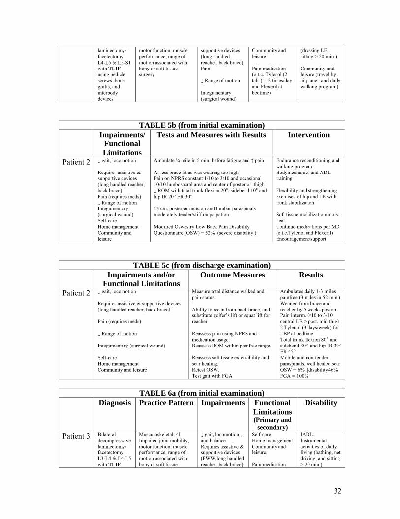
Functional Activities included instructing the patient in a “golfer’s lift” and squat and/or ½ kneel lift with emphasis on maintaining a neutral spine while flexing/extending through the hips to retrieve objects off tables/chairs/or floors and with transfers sit ↑↓ stand. Emphasis on pivoting through the hips (IR/ER) and not twisting the spine was instructed, as was log rolling (non-segmental) with bed mobility and supine ↔ sideling↔ sitting in/out of bed. In general, the same guidelines were followed for intradiscal pressures in the disc in daily life (25-Wilke et al. 1999; 26-Nachemson 1964). See table 7 and figure 4. Similar values were obtained in a study evaluating Loads on an Internal Spinal Fixation Device During Physical Therapy (27-Rohlmann et al. 2002) when a modified fixation device was implanted posteriorly followed by an anterior interbody fusion.
Cardiovascular conditioning was performed initially on a recumbent stationary bike, beginning at 3-5 minutes at a comfortable pace, while monitoring patients perceived level of exertion (at or below somewhat hard level). Patients were progressed 2-4 minutes/week until reaching 8-10 minutes in the clinic, and 15-20 minutes at home if a bike was available. The treadmill or walking outside was incorporated by the 2nd week of rehab in a similar fashion.
16
Results
The selection of tests and measures used to assess patients’ impairments and functional limitations on initial examination was based on clinical observations and experience of the treating therapist as well as the patient’s postoperative stage of recovery and referring physician’s precautions.
Gait, locomotion, and balance were assessed using observation of the patient ambulating from the lobby to the treatment room, a distance of 50 feet. Use of assistive and supportive devices was noted. Subjective reports of endurance limits were recorded and as treatment progressed in the ensuing weeks, the treadmill was introduced to quantify time and distance covered for outcomes purposes. Balance was assessed by having the patient perform single limb support (SLS) and then noting the time in seconds before losing balance and the use of external support. In all 3 cases balance was improved.
Pain was monitored throughout the course of rehabilitation using an 11-point numeric pain rating scale (NPRS), where 0 represents no pain and 10 represents the worse pain imaginable. Patients initially rated their pain intensity by marking their pain level on a written scale on the intake form for current level, lowest level in the past week, and highest level in the past week. From then on, verbal ratings were used until discharge. All 3 cases consistently had decreased pain ratings, from constant to intermittent 3/10 or lower. Medication usage dropped significantly or to not at all in all 3 cases as well.
Range of motion (ROM) was measured using an inclinometer. Due to the nature of the surgery (i.e. fusion), the patient was instructed to avoid pain, move slowly, and not to overdo it. For forward flexion, the inclinometer base was placed on the T12-L1 spinous process in the sagittal plane. The inclinometer was placed on this mark and zeroed out, then the patient was instructed as above to “slowly bend forward as far as you comfortably can, but not to the point of pain” and the reading on the inclinometer was recorded. For side-bending measurements the inclinometer was placed in the frontal plane on the same T9-T12 spinous process and then zeroed out. The patient was instructed to “slowly bend to the right as far as you comfortably can, but not to the point of pain” and the reading on the inclinometer was recorded, and then repeated with side-bending to the left. All 3 cases improved in regards to total trunk motion, an average of 50° for forward bend, and 15° for side bending.
A Modified Oswestry Low Back Pain Disability Questionnaire (OSW) was administered and scored on intake and discharge. The OSW is a LBP questionnaire consisting of 10 questions used to measure low back disability as a combination of physical and social restrictions with daily living, stage a patient’s acuity status, and monitor change over time (28-Delitto et al. 1995; 29-Fritz et al. 2001). The score is presented in a percentage, 0-100%, with higher scores indication greater levels of disability due to LBP. Each of the 10 questions is scored on a 0-5 scale, 5 representing the greatest disability, and then added together and multiplied by two to get a percent score. The average decrease for all three cases was between 16-46%.
A neurological screen was performed at intake to assess sensation, deep tendon reflexes at L4, L5, and S1 and strength using manual muscle testing of the bilateral hips
17
and lower extremities, with close attention to distal muscle strength. Also, neurodynamic testing and treatment was carefully performed by use of a SLR in supine with ankle dorsiflexion. Active movement was used as a glider for treatment, based on principles of sliders and tensioners (30-Butler 1991). Slump test was not used as it was felt by the author that this may stress the fusion. Normal findings were documented for all 3 cases on initial exam, therefore this was not repeated throughout treatment or at discharge, but would have been had any initial findings been abnormal.
Passive hip and knee ROM was assessed with the patient supine with spine in neutral and findings documented using standard goniometry methods.
Integumentary system (surgical wound) and adjacent soft-tissue was assessed by the following palpation methods: muscle tone assessment being increased/decreased/normal; presence/absence of muscle spasm; tenderness none/minimal/moderate/severe; skin rolling stiff/mobile; temperature cool/warm/hot; and by visual inspection for wound healing assessing closure/gapping; drainage/dryness; color pinkish/redness/streaking/; texture thickened/smooth; and overall length measured with a tape measure in centimeters. Each case showed progressive improvement with the incision and underlying soft tissue being non-tender and normal muscle tone by discharge.
Finally, patients were questioned on intake about their overall status regarding their progress since date of surgery (DOS) as to whether or not they were improving and if surgery had helped their preoperative pain (back or leg pain). At discharge, they were asked for overall assessment to the question “Compared with before treatment my back problems are: much better, better, unchanged, or worse.” In this case series, similar results were achieved with pain scores decreasing from constant to intermittent, OSW score reduced by 16% and 46% respectively, and overall assessment of “much better” reported at 3- month follow-up.
18
Discussion For future studies, a more standardized method to assess gait and locomotion,
such as the timed up-and-go (TUG), 5 minute walk test or timed 50-foot walk at fastest speed, may be recommended. These tests have excellent reliability and validity and good clinical use, however exclusion criteria used in their accompanying study included major surgery within the past year, spondylolisthesis, or spondylolysis (31-Simmonds et al 1998). The functional gait assessment (FGA) and TUG was used in the 3rd case study and results showed significant improvements. See table 6b and 6c. This test also demonstrates acceptable reliability and validity, but with other balance measures used for patients with vestibular disorders (32-Wrisley).
The NPRS worked well with this patient population. When similar methods were used to assess pain intensity, acceptable ranges of reliability (ICC range = 0.66-0.93) have been observed for patients with LBP (33-Roach KE 1997). In addition, 11-point rating scales have demonstrated sufficient levels of discrimination when patients with chronic pain rated pain intensity (34-Jensen MP 1994).
ROM of the trunk was not a key component of this study, in order to protect the fusion; however improvement was noted in all 3 cases. With the inclinometer placed at the T12-L1 level, above the fused area, the fusion would not be compromised, as this method assesses total trunk flexion, and not just lumbar ROM, which was the goal of the examiner. The interrater reliability for this method is excellent (ICC=.87-.95 in 60 subjects with chronic LBP (35-Waddell et al 1992).
Stabilization exercises were felt to be a key component for the success of all 3 cases. Contractions as low as 25% maximum voluntary contraction (MVC) are able to provide maximal joint stiffness (36-Hoffer and Andreassen 1981). Therefore the addition of high external loading, which is typically required for strength changes, is not suitable for the development of muscle stiffness for joint support. For these reasons, positions and exercises involving minimal external loading are the ideal when rehabilitating the local muscles for lumbar spine stabilization. It has been proposed that contraction of the transverses abdominis applies tension on the thoracolumbar fascia, which in turn, constrains the three lumbar back muscles (multifidus, longissmus thoracis and iliocostalis lumborum) and exerts a pushing force on these muscles to promote lumbosacral stiffness, known as a “hydraulic amplifier mechanism” (37-Gracovetsky 1977).
Once the patient masters the ADIM for 10 repetitions for 10 seconds without fatigue, the exercise can be progressed to slowly increase loads and functional demands and integrate the local and global muscle systems (23-Richardson et al. 1995). Local muscles refer to those attaching directly to the lumbar vertebrae and responsible for segmental stability and control of the lumbar segments, whereas global describes the large torque producing muscles linking the pelvis to the thoracic cage, providing general trunk stabilization (38-O’Sullivan 2000).
The OSW has been shown to be an excellent measurement of outcome for LBP, with a minimal clinically important difference (MCID) of approximately 6 points (29-Fritz et al 2001). Others have reported a minimum detectable change (MDC) of at least 10.5 (and possibly as much as 15 points), to be 90% confident that real change had occurred (39-Davidson et al 2002).
19
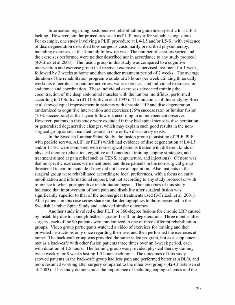
Information regarding postoperative rehabilitation guidelines specific to TLIF is lacking. However, similar procedures, such as PLIF, may offer valuable suggestions. For example, one study involving a PLIF procedure at L4-L5 and/or L5-S1 with evidence of disc degeneration described how surgeons customarily prescribed physiotherapy, including exercises, at the 3-month follow-up visit. The number of sessions varied and the exercises performed were neither described nor in accordance to any study protocol (40-Brox et al 2003). The fusion group in this study was compared to a cognitive intervention and exercise group that received extensive supervised treatment for 1 week, followed by 2 weeks at home and then another treatment period of 2 weeks. The average duration of the rehabilitation program was about 25 hours per week utilizing three daily workouts of aerobics or outdoor activities, water exercises, and individual exercises for endurance and coordination. These individual exercises advocated training the cocontraction of the deep abdominal muscles with the lumbar multifidus, performed according to O’Sullivan (41-O’Sullivan et al 1997). The outcomes of this study by Brox et al showed equal improvement in patients with chronic LBP and disc degeneration randomized to cognitive intervention and exercises (76% success rate) or lumbar fusion (70% success rate) at the 1-year follow-up, according to an independent observer. However, patients in this study were excluded if they had spinal stenosis, disc herniation, or generalized degenerative changes, which may explain such good results in the non-surgical group as such isolated lesions to one or two discs rarely exists.
In the Swedish Lumbar Spine Study, the fusion group (consisting of PLF, PLF with pedicle screws, ALIF, or PLIF) which had evidence of disc degeneration at L4-L5 and/or L5-S1 were compared with non-surgical patients treated with different kinds of physical therapy (education, cognitive and functional training, coping strategies, and treatment aimed at pain relief such as TENS, acupuncture, and injections). Of note was that no specific exercises were mentioned and three patients in the non-surgical group threatened to commit suicide if they did not have an operation. Also, patients in the surgical group were rehabilitated according to local preferences, with a focus on early mobilization and informational support, but not according to any study protocol or with reference to when postoperative rehabilitation began. The outcomes of this study indicated that improvement of both pain and disability after surgical fusion was significantly superior to that of the non-surgical treatments used (3-Fritzell et al. 2001). All 3 patients in this case series share similar demographics to those presented in the Swedish Lumbar Spine Study and achieved similar outcomes.
Another study involved either PLIF or 360-degree fusions for chronic LBP caused by instability due to spondylolisthesis grades I or II, or degeneration. Three months after surgery, each of the 90 patients were randomized to one of three different rehabilitation groups. Video group participants watched a video of exercises for training and then provided instructions only once regarding their use, and then performed the exercises at home. The back-café group was provided the same video program, but as a supplement met as a back-café with other fusion patients three times over an 8-week period, each with duration of 1.5 hours. The training group was provided physical therapy training twice weekly for 8 weeks lasting 1.5 hours each time. The outcomes of this study showed patients in the back-café group had less pain and performed better at ADL’s, and more resumed working after surgery compared to the other two groups (42-Christensen et al. 2003). This study demonstrates the importance of including coping schemes and the
20
opportunity for exchange of experiences of pain and physical incapacity, doubts, reassurance, and psychological support from similar patients and may put into question the role of intensive exercises after spinal fusion.
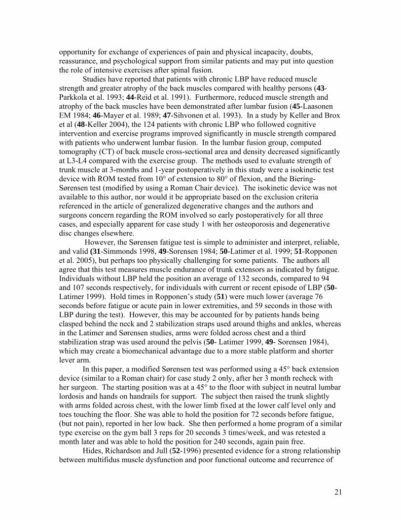
Studies have reported that patients with chronic LBP have reduced muscle strength and greater atrophy of the back muscles compared with healthy persons (43-Parkkola et al. 1993; 44-Reid et al. 1991). Furthermore, reduced muscle strength and atrophy of the back muscles have been demonstrated after lumbar fusion (45-Laasonen EM 1984; 46-Mayer et al. 1989; 47-Sihvonen et al. 1993). In a study by Keller and Brox et al (48-Keller 2004), the 124 patients with chronic LBP who followed cognitive intervention and exercise programs improved significantly in muscle strength compared with patients who underwent lumbar fusion. In the lumbar fusion group, computed tomography (CT) of back muscle cross-sectional area and density decreased significantly at L3-L4 compared with the exercise group. The methods used to evaluate strength of trunk muscle at 3-months and 1-year postoperatively in this study were a isokinetic test device with ROM tested from 10° of extension to 80° of flexion, and the Biering-Sørensen test (modified by using a Roman Chair device). The isokinetic device was not available to this author, nor would it be appropriate based on the exclusion criteria referenced in the article of generalized degenerative changes and the authors and surgeons concern regarding the ROM involved so early postoperatively for all three cases, and especially apparent for case study 1 with her osteoporosis and degenerative disc changes elsewhere.
However, the Sørensen fatigue test is simple to administer and interpret, reliable, and valid (31-Simmonds 1998, 49-Sorensen 1984; 50-Latimer et al. 1999; 51-Ropponen et al. 2005), but perhaps too physically challenging for some patients. The authors all agree that this test measures muscle endurance of trunk extensors as indicated by fatigue. Individuals without LBP held the position an average of 132 seconds, compared to 94 and 107 seconds respectively, for individuals with current or recent episode of LBP (50-Latimer 1999). Hold times in Ropponen’s study (51) were much lower (average 76 seconds before fatigue or acute pain in lower extremities, and 59 seconds in those with LBP during the test). However, this may be accounted for by patients hands being clasped behind the neck and 2 stabilization straps used around thighs and ankles, whereas in the Latimer and Sørensen studies, arms were folded across chest and a third stabilization strap was used around the pelvis (50- Latimer 1999, 49- Sorensen 1984), which may create a biomechanical advantage due to a more stable platform and shorter lever arm.
In this paper, a modified Sørensen test was performed using a 45° back extension device (similar to a Roman chair) for case study 2 only, after her 3 month recheck with her surgeon. The starting position was at a 45° to the floor with subject in neutral lumbar lordosis and hands on handrails for support. The subject then raised the trunk slightly with arms folded across chest, with the lower limb fixed at the lower calf level only and toes touching the floor. She was able to hold the position for 72 seconds before fatigue, (but not pain), reported in her low back. She then performed a home program of a similar type exercise on the gym ball 3 reps for 20 seconds 3 times/week, and was retested a month later and was able to hold the position for 240 seconds, again pain free.
Hides, Richardson and Jull (52-1996) presented evidence for a strong relationship between multifidus muscle dysfunction and poor functional outcome and recurrence of
21
LBP after disc surgery (53-Rantanen et al. 1993; 54-Sihvonen et al. 1993). Multifidus muscle recovery does not occur spontaneously on remission of painful symptoms, and reflex inhibition can occur in the absence of pain, similar to what is often seen in the quadriceps muscles following knee surgeries. The clinical significance is that patients with LBP may appear fully recovered after the initial acute pain subsided, the muscle system had certainly not recovered, which may explain the staggering recurrence rate of 60-80% following first episode of LBP (55-Troup et al. 1981).
Patients overall assessment is probably the most frequently used measure in outpatient rehabilitation for a nearly all conditions. This self assessment is more of global assessment of pain, function, and possibly even customer satisfaction. This was one of the outcome measures used in the Swedish Lumbar Spine Study Group which most closely matches the demographics of the 3 patients in this case study (3-Fritzell 2001). In that study, at the 2- year follow-up back pain was reduced by 33%, disability according to OSW reduced by 25%, and 63% rated themselves as “much better” or “better” in the surgical group of 222 patients. Similar of better results were achieved in this series of case studies.
Conclusion With the rapid rise in lumbar fusion surgery, and the lack of consensus on the
design of a specific rehabilitation program following lumbar spinal fusion found in the literature, future studies are needed to establish general guidelines following this procedure. Attention regarding when to begin physical therapy postoperatively and specific details regarding the exercises utilized, frequency, repetition, and intensity would be most valuable. The early results and outcomes of the case studies presented in this paper are encouraging and consistent with similar studies involving 2-level lumbar spinal fusion for instability and degenerative changes.
For future cases, this author recommends a combined “support group” similar to the back-café study meeting every 4 to 6 weeks from 1-month preoperative to 5-months postoperative, and commence a gentle rehabilitation program after the 6 week check-up, twice weekly for 4 weeks, then once weekly for 4 weeks. This is provided patients are instructed in biomechanical counseling/posture principles for ADL’s and IADL’s postoperatively on days 2 and 3, and are encouraged to ambulate early and often with the appropriate assistive device.
Acknowledgements
The author wishes to thank Carl P. DeRosa, PT, PhD, DPT for review and input of this manuscript before submission. Finally the author would like to express his appreciation to his wife Katherine and three children Karolyn, Jacob, and Joseph for their helpful support and patience during the writing process.
22
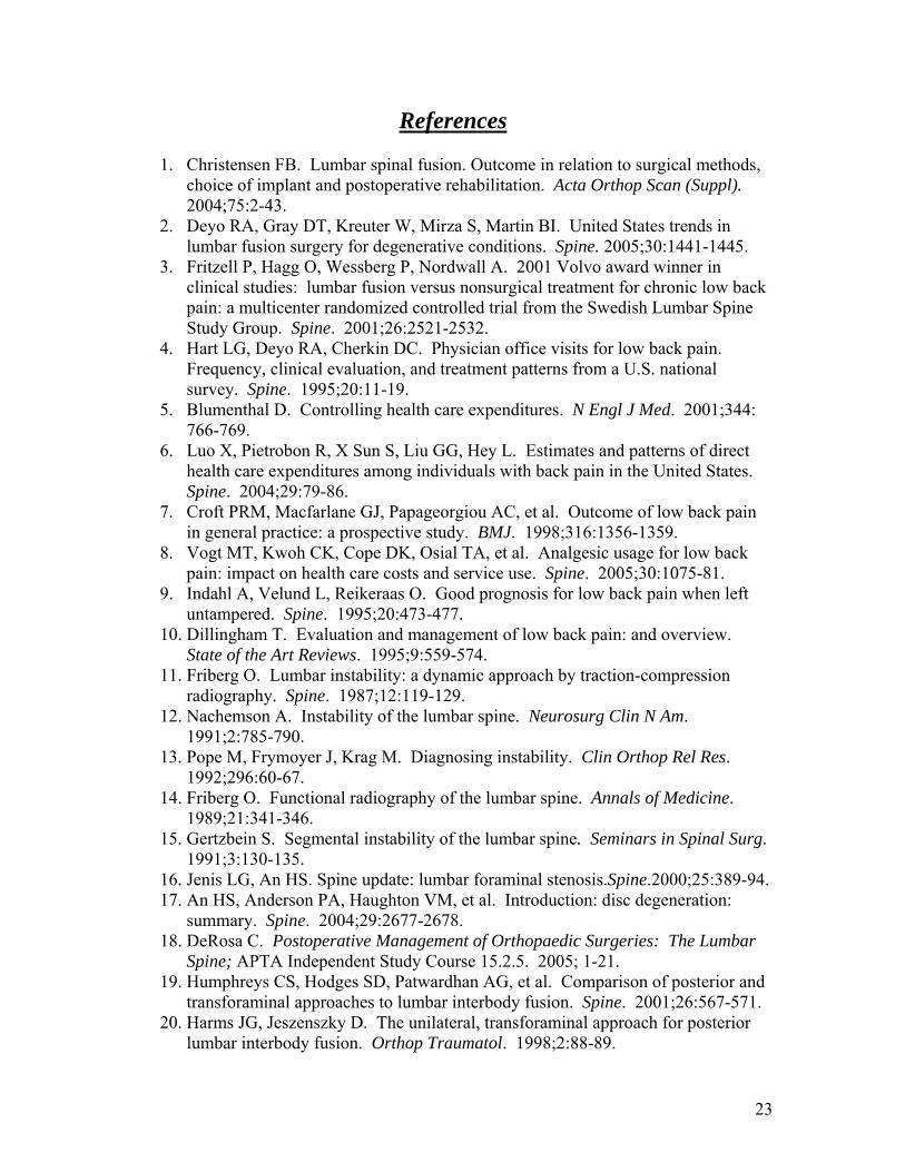
References
1. Christensen FB. Lumbar spinal fusion. Outcome in relation to surgical methods, choice of implant and postoperative rehabilitation. Acta Orthop Scan (Suppl). 2004;75:2-43.
2. Deyo RA, Gray DT, Kreuter W, Mirza S, Martin BI. United States trends in lumbar fusion surgery for degenerative conditions. Spine. 2005;30:1441-1445.
3. Fritzell P, Hagg O, Wessberg P, Nordwall A. 2001 Volvo award winner in clinical studies: lumbar fusion versus nonsurgical treatment for chronic low back pain: a multicenter randomized controlled trial from the Swedish Lumbar Spine Study Group. Spine. 2001;26:2521-2532.
4. Hart LG, Deyo RA, Cherkin DC. Physician office visits for low back pain. Frequency, clinical evaluation, and treatment patterns from a U.S. national survey. Spine. 1995;20:11-19.
5. Blumenthal D. Controlling health care expenditures. N Engl J Med. 2001;344: 766-769.
6. Luo X, Pietrobon R, X Sun S, Liu GG, Hey L. Estimates and patterns of direct health care expenditures among individuals with back pain in the United States. Spine. 2004;29:79-86.
7. Croft PRM, Macfarlane GJ, Papageorgiou AC, et al. Outcome of low back pain in general practice: a prospective study. BMJ. 1998;316:1356-1359.
8. Vogt MT, Kwoh CK, Cope DK, Osial TA, et al. Analgesic usage for low back pain: impact on health care costs and service use. Spine. 2005;30:1075-81.
9. Indahl A, Velund L, Reikeraas O. Good prognosis for low back pain when left untampered. Spine. 1995;20:473-477.
10. Dillingham T. Evaluation and management of low back pain: and overview. State of the Art Reviews. 1995;9:559-574.
11. Friberg O. Lumbar instability: a dynamic approach by traction-compression radiography. Spine. 1987;12:119-129.
12. Nachemson A. Instability of the lumbar spine. Neurosurg Clin N Am. 1991;2:785-790.
13. Pope M, Frymoyer J, Krag M. Diagnosing instability. Clin Orthop Rel Res. 1992;296:60-67.
14. Friberg O. Functional radiography of the lumbar spine. Annals of Medicine. 1989;21:341-346.
15. Gertzbein S. Segmental instability of the lumbar spine. Seminars in Spinal Surg. 1991;3:130-135.
16. Jenis LG, An HS. Spine update: lumbar foraminal stenosis.Spine.2000;25:389-94. 17. An HS, Anderson PA, Haughton VM, et al. Introduction: disc degeneration:
summary. Spine. 2004;29:2677-2678. 18. DeRosa C. Postoperative Management of Orthopaedic Surgeries: The Lumbar
Spine; APTA Independent Study Course 15.2.5. 2005; 1-21. 19. Humphreys CS, Hodges SD, Patwardhan AG, et al. Comparison of posterior and
transforaminal approaches to lumbar interbody fusion. Spine. 2001;26:567-571. 20. Harms JG, Jeszenszky D. The unilateral, transforaminal approach for posterior
lumbar interbody fusion. Orthop Traumatol. 1998;2:88-89.
23
21. Figueiredo N, Martins JWG, Arruda AA, et al. TLIF – transforaminal lumbar interbody fusion. Arq Neuropsiquiatr. 2004;62:815-820.
22. American Physical Therapy Association. Guide to Physical Therapist Practice. Second Edition. Phys Ther. 2001;81:9-746.
23. Richardson CA, Jull GA. Muscle control – pain control. What exercises would you prescribe? Manual Ther. 1995;1:2-10.
24. O’Sullivan PB, Twomey L, Allison GT. Altered abdominal muscle recruitment in patients with chronic back pain following a specific exercise intervention. J Orthop Sports Phys Ther. 1998;27:114-124.
25. Wilke HJ, Neef P, Caimi M, et al. New in vivo measurements of pressures in the intervertebral disc in daily life. Spine. 1999;24:755-762.
26. Nachemson AL, Morris JM. In vivo measurements of intradiscal pressure. J Bone Joint Surg Am. 1964;46:1077-1092.
27. Rohlmann A, Graichen F, Bergmann G. Loads on an internal spinal fixation device during physical therapy. Phys Ther. 2002;82:44-52.
28. Delitto A, Erhard RE, Bowling RW. A treatment-based classification approach to low back syndrome: identifying and staging patients for conservative treatment. Phys Ther. 1995;75:470-485; discussion 485-489.
29. Fritz JM, Irrgang JJ. A comparison of a modified Oswestry Low Back Pain Disability Questionnaire and the Quebec Back Pain Disability Scale. Phys Ther. 2001;81:776-788.
30. Butler DS. Mobilization of the Nervous System. Churchill Livingstone, London; 1991.
31. Simmonds MJ, Olson SL, Jones S, et al. Psychometric characteristics and clinical usefulness of physical performance tests in patients with low back pain. Spine. 1998;23:2412-2421.
32. Wrisley DM, Marchetti GF, Kuharsky DK, Whitney SL. Reliability, internal consistency, and validity of data obtained with the Functional Gait Assessment. Phys Ther. 2004;84:906-918.
33. Roach KE, Brown MD, Dunigan KM, et al. Test-retest reliability of patient reports of low back pain. J Orthop Sports Phys Ther. 1997;26:253-259.
34. Jensen MP, Turner JA, Romano JM. What is the maximum number of levels needed in pain intensity measurement? Pain. 1994;58:387-392.
35. Waddell G, Somerville D, Henderson I, Newton M. Objective clinical evaluation of physical impairment in chronic low back pain. Spine. 1992;17:617-628.
36. Hoffer J, Andreassen S. Regulation of soleus muscle stiffness in premamillary cats. J Neurophys. 1981;45:267-285.
37. Gracovetsky S, Farfan H, Lamy C. A mathematical model of the lumbar spine using an optimized system to control muscles and ligaments. Orthop Clin N Am. 1977;85:135-153.
38. O’Sullivan PB. Lumbar segmental ‘instability’: clinical presentation and specific stabilizing exercise management. Manual Ther. 2000;5:2-12.
39. Davidson M, Keating JL. A comparison of five low back disability questionnaires: reliability and responsiveness. Phys Ther. 2002;82:8-24.
24
40. Brox JI, Sørensen R, Friis A, et al. Randomized clinical trial of lumbar instrumented fusion and cognitive intervention and exercises in patients with chronic low back pain and disc degeneration. Spine. 2003;28:1913-1921.
41. O’Sullivan PB, Twomey LT, Allison GT. Evaluation of specific stabilizing exercise in the treatment of chronic low back pain with radiologic diagnosis of spondylolysis or spondylolisthesis. Spine. 1997;22:2959-2967.
42. Christensen FB, Laurberg I, Bünger CE. Importance of the back-café concept to rehabilitation after lumbar spinal fusion: a randomized clinical study with a 2-year follow-up. Spine. 2003;28:2561-2569.
43. Parkkola R, Rytokoski U, Kormano M. Magnetic resonance imaging of the discs and trunk muscles in patients with chronic low back pain and healthy control subjects. Spine. 1993;18:830-836.
44. Reid S, Hazard RG, Fenwick JW. Isokinetic trunk-strength deficits in people with and without low back pain: a comparative study with consideration of effort. J Spinal Disord. 1991;4:68-72.
45. Laasonen EM. Atrophy of sacrospinal muscle groups in patients with chronic, diffusely radiating lumbar back pain. Neuroradiology. 1984;26:9-13.
46. Mayer TG, Vanharanta H, Gatchel RJ, et al. Comparison of CT scan muscle measurements and isokinetic trunk strength in postoperative patients. Spine. 1989;14:33-36.
47. Sihvonen T, Herno A, Paljarvi L, et al. Local denervation atrophy of paraspinal muscles in postoperative failed back syndrome. Spine. 1993;18:575-581.
48. Keller A, Brox JI, Gunderson R, et al. Trunk muscle strength, cross-sectional area, and density in patients with chronic low back pain randomized to lumbar fusion or cognitive intervention and exercises. Spine. 2004;29:3-8.
49. Biering-Sørensen F. Physical measurements as risk indicators for low-back trouble over a one-year period. Spine. 1984;9:106-119.
50. Latimer J, Maher CG, Refshauge K, Colaco I. The reliability and validity of the Biering-Sørensen test in asymptomatic subjects and subjects reporting current or previous nonspecific low back pain. Spine. 1999;24:2085-2089; discussion 2090.
51. Ropponen A, Gibbons LE, Videman T, Battié MC. Isometric back extension endurance testing: reasons for test termination. J Orthop Sports Phys Ther. 2005;35:437-442.
52. Hides JA, Richardson CA, Jull GA. Multifidus muscle recovery is not automatic after resolution of acute, first-episode low back pain. Spine. 1996;21:2763-2769.
53. Rantanen J, Hurme M, Falck B, et al. The lumbar multifidus muscle five years after surgery for a lumbar intervertebral disc herniation. Spine. 1993;18:568-574.
54. Sihvonen T, Herno A, Paljarvi L, et al. Local denervation atrophy of paraspinal muscles in postoperative failed back syndrome. Spine. 1993;18:575-581.
55. Troup JD, Martin JW, Lloyd DC. Back pain in industry: a prospective survey. Spine. 1981;6:61-69.
25
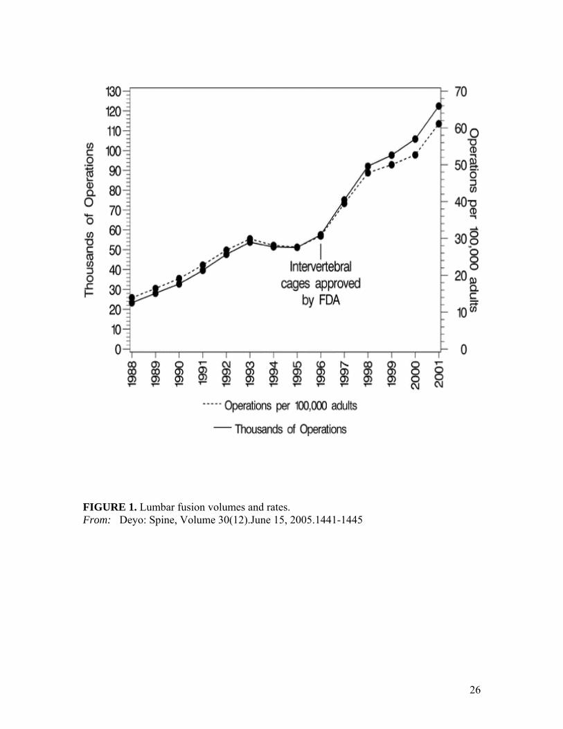
FIGURE 1. Lumbar fusion volumes and rates. From: Deyo: Spine, Volume 30(12).June 15, 2005.1441-1445
26
TABLE 1. Procedure Comparisons From: Deyo: Spine, Volume 30(12).June 15, 2005.1441-1445
27
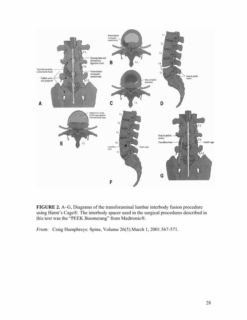
FIGURE 2. A–G, Diagrams of the transforaminal lumbar interbody fusion procedure using Harm’s Cage®. The interbody spacer used in the surgical procedures described in this text was the “PEEK Boomerang” from Medtronic®. From: Craig Humphreys: Spine, Volume 26(5).March 1, 2001.567-571.
28
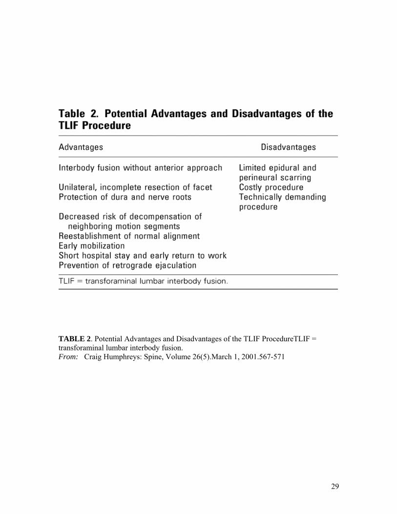
TABLE 2. Potential Advantages and Disadvantages of the TLIF ProcedureTLIF = transforaminal lumbar interbody fusion. From: Craig Humphreys: Spine, Volume 26(5).March 1, 2001.567-571
29
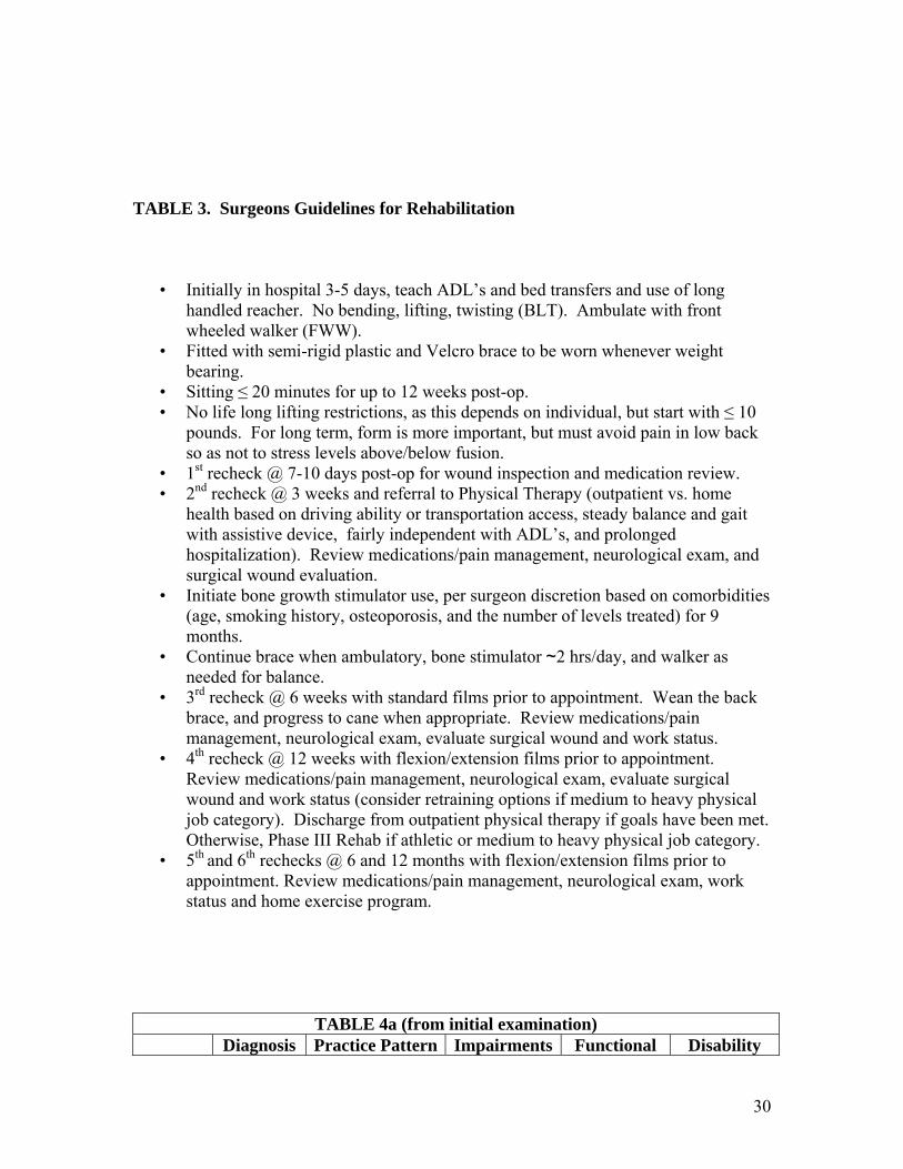
TABLE 3. Surgeons Guidelines for Rehabilitation
• Initially in hospital 3-5 days, teach ADL’s and bed transfers and use of long handled reacher. No bending, lifting, twisting (BLT). Ambulate with front wheeled walker (FWW).
• Fitted with semi-rigid plastic and Velcro brace to be worn whenever weight bearing.
• Sitting ≤ 20 minutes for up to 12 weeks post-op. • No life long lifting restrictions, as this depends on individual, but start with ≤ 10
pounds. For long term, form is more important, but must avoid pain in low back so as not to stress levels above/below fusion.
• 1st recheck @ 7-10 days post-op for wound inspection and medication review. • 2nd recheck @ 3 weeks and referral to Physical Therapy (outpatient vs. home
health based on driving ability or transportation access, steady balance and gait with assistive device, fairly independent with ADL’s, and prolonged hospitalization). Review medications/pain management, neurological exam, and surgical wound evaluation.
• Initiate bone growth stimulator use, per surgeon discretion based on comorbidities (age, smoking history, osteoporosis, and the number of levels treated) for 9 months.
• Continue brace when ambulatory, bone stimulator ~2 hrs/day, and walker as needed for balance.
• 3rd recheck @ 6 weeks with standard films prior to appointment. Wean the back brace, and progress to cane when appropriate. Review medications/pain management, neurological exam, evaluate surgical wound and work status.
• 4th recheck @ 12 weeks with flexion/extension films prior to appointment. Review medications/pain management, neurological exam, evaluate surgical wound and work status (consider retraining options if medium to heavy physical job category). Discharge from outpatient physical therapy if goals have been met. Otherwise, Phase III Rehab if athletic or medium to heavy physical job category.
• 5th and 6th rechecks @ 6 and 12 months with flexion/extension films prior to appointment. Review medications/pain management, neurological exam, work status and home exercise program.
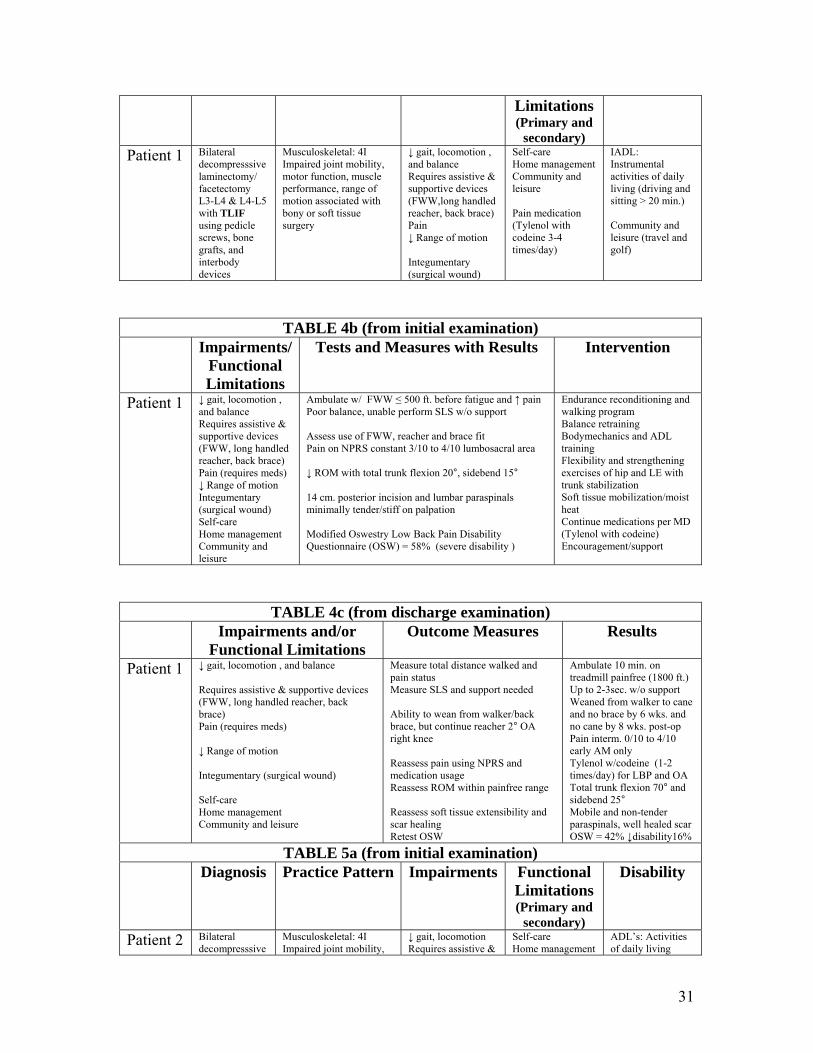
TABLE 4a (from initial examination) Diagnosis Practice Pattern Impairments Functional Disability
30
Limitations (Primary and
secondary) Patient 1 Bilateral
decompresssive laminectomy/ facetectomy L3-L4 & L4-L5 with TLIF using pedicle screws, bone grafts, and interbody devices
Musculoskeletal: 4I Impaired joint mobility, motor function, muscle performance, range of motion associated with bony or soft tissue surgery
↓ gait, locomotion , and balance Requires assistive & supportive devices (FWW,long handled reacher, back brace) Pain ↓ Range of motion Integumentary (surgical wound)
Self-care Home management Community and leisure Pain medication (Tylenol with codeine 3-4 times/day)
IADL: Instrumental activities of daily living (driving and sitting > 20 min.) Community and leisure (travel and golf)
TABLE 4b (from initial examination) Impairments/
Functional Limitations
Tests and Measures with Results Intervention
Patient 1 ↓ gait, locomotion , and balance Requires assistive & supportive devices (FWW, long handled reacher, back brace) Pain (requires meds) ↓ Range of motion Integumentary (surgical wound) Self-care Home management Community and leisure
Ambulate w/ FWW ≤ 500 ft. before fatigue and ↑ pain Poor balance, unable perform SLS w/o support Assess use of FWW, reacher and brace fit Pain on NPRS constant 3/10 to 4/10 lumbosacral area ↓ ROM with total trunk flexion 20°, sidebend 15° 14 cm. posterior incision and lumbar paraspinals minimally tender/stiff on palpation Modified Oswestry Low Back Pain Disability Questionnaire (OSW) = 58% (severe disability )
Endurance reconditioning and walking program Balance retraining Bodymechanics and ADL training Flexibility and strengthening exercises of hip and LE with trunk stabilization Soft tissue mobilization/moist heat Continue medications per MD (Tylenol with codeine) Encouragement/support
TABLE 4c (from discharge examination) Impairments and/or
Functional Limitations Outcome Measures Results
Patient 1 ↓ gait, locomotion , and balance Requires assistive & supportive devices (FWW, long handled reacher, back brace) Pain (requires meds) ↓ Range of motion Integumentary (surgical wound) Self-care Home management Community and leisure
Measure total distance walked and pain status Measure SLS and support needed Ability to wean from walker/back brace, but continue reacher 2° OA right knee Reassess pain using NPRS and medication usage Reassess ROM within painfree range Reassess soft tissue extensibility and scar healing Retest OSW
Ambulate 10 min. on treadmill painfree (1800 ft.) Up to 2-3sec. w/o support Weaned from walker to cane and no brace by 6 wks. and no cane by 8 wks. post-op Pain interm. 0/10 to 4/10 early AM only Tylenol w/codeine (1-2 times/day) for LBP and OA Total trunk flexion 70° and sidebend 25° Mobile and non-tender paraspinals, well healed scar OSW = 42% ↓disability16%
TABLE 5a (from initial examination) Diagnosis Practice Pattern Impairments Functional
Limitations (Primary and
secondary)
Disability
Patient 2 Bilateral decompresssive
Musculoskeletal: 4I Impaired joint mobility,
↓ gait, locomotion Requires assistive &
Self-care Home management
ADL’s: Activities of daily living
31
laminectomy/ facetectomy L4-L5 & L5-S1 with TLIF using pedicle screws, bone grafts, and interbody devices
motor function, muscle performance, range of motion associated with bony or soft tissue surgery
supportive devices (long handled reacher, back brace) Pain ↓ Range of motion Integumentary (surgical wound)
Community and leisure Pain medication (o.t.c. Tylenol (2 tabs) 1-2 times/day and Flexeril at bedtime)
(dressing LE, sitting > 20 min.) Community and leisure (travel by airplane, and daily walking program)
TABLE 5b (from initial examination) Impairments/
Functional Limitations
Tests and Measures with Results Intervention
Patient 2 ↓ gait, locomotion Requires assistive & supportive devices (long handled reacher, back brace) Pain (requires meds) ↓ Range of motion Integumentary (surgical wound) Self-care Home management Community and leisure
Ambulate ¼ mile in 5 min. before fatigue and ↑ pain Assess brace fit as was wearing too high Pain on NPRS constant 1/10 to 3/10 and occasional 10/10 lumbosacral area and center of posterior thigh ↓ ROM with total trunk flexion 20°, sidebend 10° and hip IR 20° ER 30° 13 cm. posterior incision and lumbar paraspinals moderately tender/stiff on palpation Modified Oswestry Low Back Pain Disability Questionnaire (OSW) = 52% (severe disability )
Endurance reconditioning and walking program Bodymechanics and ADL training Flexibility and strengthening exercises of hip and LE with trunk stabilization Soft tissue mobilization/moist heat Continue medications per MD (o.t.c.Tylenol and Flexeril) Encouragement/support
TABLE 5c (from discharge examination) Impairments and/or
Functional Limitations Outcome Measures Results
Patient 2 ↓ gait, locomotion Requires assistive & supportive devices (long handled reacher, back brace) Pain (requires meds) ↓ Range of motion Integumentary (surgical wound) Self-care Home management Community and leisure
Measure total distance walked and pain status Ability to wean from back brace, and substitute golfer’s lift or squat lift for reacher Reassess pain using NPRS and medication usage. Reassess ROM within painfree range. Reassess soft tissue extensibility and scar healing. Retest OSW. Test gait with FGA
Ambulates daily 1-3 miles painfree (3 miles in 52 min.) Weaned from brace and reacher by 5 weeks postop. Pain interm. 0/10 to 3/10 central LB > post. mid thigh 2 Tylenol (3 days/week) for LBP at bedtime Total trunk flexion 80° and sidebend 30° and hip IR 30° ER 45° Mobile and non-tender paraspinals, well healed scar OSW = 6% ↓disability46% FGA = 100%
TABLE 6a (from initial examination)
Diagnosis Practice Pattern Impairments Functional Limitations (Primary and
secondary)
Disability
Patient 3 Bilateral decompresssive laminectomy/ facetectomy L3-L4 & L4-L5 with TLIF
Musculoskeletal: 4I Impaired joint mobility, motor function, muscle performance, range of motion associated with bony or soft tissue
↓ gait, locomotion , and balance Requires assistive & supportive devices (FWW,long handled reacher, back brace)
Self-care Home management Community and leisure. Pain medication
IADL: Instrumental activities of daily living (bathing, not driving, and sitting > 20 min.)
32
using pedicle screws, bone grafts, and interbody devices
surgery Pain ↓ Range of motion Integumentary (surgical wound)
(Tylenol with codeine 1-2 times/week)
Community and leisure (grocery shopping, light housekeeping)
TABLE 6b (from initial examination) Impairments/
Functional Limitations
Tests and Measures with Results Intervention
Patient 3 ↓ gait, locomotion , and balance Requires assistive & supportive devices (FWW, long handled reacher, back brace) Pain (requires meds) ↓ Range of motion Integumentary (surgical wound) Self-care Home management Community and leisure
Ambulate with ≤ 100 ft. before fatigue Poor balance, unable perform SLS w/o support Assess use of FWW, reacher and brace fit Pain on NPRS constant 1/10 to 2/10 lumbosacral area ↓ ROM with total trunk flexion 30°, sidebend 10° 12 cm. posterior incision and lumbar paraspinals minimally tender/stiff on palpation Modified Oswestry Low Back Pain Disability Questionnaire (OSW) = 36% (moderate disability ) Test gait with TUG = 33 seconds, and FGA =40%
Endurance reconditioning and walking program Balance retraining Bodymechanics and ADL training Flexibility and strengthening exercises of hip and LE with trunk stabilization Soft tissue mobilization/moist heat Continue medications per MD (Tylenol with codeine) Encouragement/support
TABLE 6c (from discharge examination) Impairments and/or
Functional Limitations Outcome Measures Results
Patient 3 ↓ gait, locomotion , and balance Requires assistive & supportive devices (FWW, long handled reacher, back brace) Pain (requires meds) ↓ Range of motion Integumentary (surgical wound) Self-care Home management Community and leisure
Measure total distance walked and pain status Measure SLS and support needed Ability to wean from walker/back brace, but continue reacher 2° Reassess pain using NPRS and medication usage Reassess ROM within painfree range Reassess soft tissue extensibility and scar healing Retest OSW, TUG, and FGA
Ambulates 20-25 min./day outside painfree (walks dog) Up to 2-3sec. w/o support Weaned from walker to cane and no brace by 6 wks. and cont. cane when outside Pain interm. 0/10 to 1/10 No Tylenol needed Total trunk flex 70°, SB 25° Mobile and non-tender paraspinals, well healed scar OSW= 6% (↓disability 30%) TUG=12seconds,FGA=60%.
33
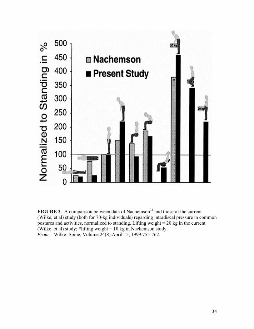
FIGURE 3. A comparison between data of Nachemson31 and those of the current (Wilke, et al) study (both for 70-kg individuals) regarding intradiscal pressure in common postures and activities, normalized to standing. Lifting weight = 20 kg in the current (Wilke, et al) study; *lifting weight = 10 kg in Nachemson study. From: Wilke: Spine, Volume 24(8).April 15, 1999.755-762.
34