physical therapy for cardiopulmonary disorders - kaukau.edu.sa/files/0053233/subjects/physical...
TRANSCRIPT

1
Physical Therapy for
Cardiopulmonary Disorders
Fourth Edition 2011
Dr. Shehab M. Abd El-Kader Associate Professor of Physical Therapy
Contents

2
Subjects Page
*Chronic Obstructive Pulmonary Disease 4
*Restrictive Lung Diseases 18
* Suppurative Lung Diseases 27
* Diabetes Mellitus 31
* Obesity 40
* Role of physiotherapy in cardiothoracic surgery 46
* Complications following cardiothoracic surgery 49 * Congenital heart diseases 54 * Ischemic heart disease 59 * Rheumatic fever 61 * Heart failure 63 * Cardiac Rehabilitation 68 * Role of physiotherapy in intensive care unit 88

3
CHEST PHYSICAL THERAPY
Dr. Shehab M. Abd El-Kader Associate Professor of Physical Therapy
Chronic Obstructive Pulmonary Disease Definition
Chronic obstructive pulmonary disease is a general term that refers to a number of
chronic pulmonary conditions characterized by narrowing and obstruction of airways,
4
increased retention of pulmonary secretions and structural deterioration of alveoli.
This airflow limitation is progressive and not fully reversible.
Several terms are used to describe obstructive lung disease they include:
1- COLD : chronic obstructive lung disease. 2- COAD: chronic obstructive airway dysfunction. 3- COPD : chronic obstructive pulmonary disease.
Diseases Classified as COPD
1- Chronic bronchitis. 2- Emphysema. 3- Asthma. 4- Other diseases such as cystic fibrosis and bronchiectasis usually lead to chronic
obstructive dysfunction. Characteristics of patients with obstructive lung disease
1- Patients exhibit persistent resistance of airflow, which causes prolonged and
often forced expiration.
2- Vital capacity is decreased.
3- Exercise tolerance is markedly diminished. Patients with COPD become
dyspneic with minimal physical exertion.
General clinical problems
1- Frequent episodes of shortness of breath “dyspnea on exertion”.
2- Prolonged and labored expiration. Air gets trapped as airways narrow during
expiration.
3- Chronic accumulation of pulmonary secretions.
4- Decreased endurance and exercise capacity.
5- Associated postural defects.

5
Figure (1): The loss of elastic recoil in lung tissue and the increased airway resistance decrease the expiratory airflow in a patient with chronic obstructive pulmonary disease as compared with the expiratory airflow in a normal subject. Presentation:
Significant overlaps exist in signs and symptoms among the three major diseases of airflow obstruction: asthma, chronic bronchitis and emphysema. The large overlap has been long noted and well illustrated in Venn diagram fashion (Fig. 2).
Figure (2): Schema of COPD.
Classification of Severity

6
For educational reasons, a simple classification of disease severity into four stages is recommended (Table 1). Table 1. Classification of COPD by Severity.
Stage Characteristics 0: At Risk . normal spirometry
. chronic symptoms (cough, sputum production) I: Mild COPD
. FEV1/FVC < 70%
. FEV1 ≥ 80% predicted
. with or without chronic symptoms (cough. sputum production)
II: Moderate COPD
. FEV1/FVC < 70%
. 30% ≤ FEV1 < 80% predicted (IIA: 50% ≤ FEV1 < 80% predicted)
(IIB: 30% ≤ FEV1 < 50% predicted) . with or without chronic symptoms (cough, sputum
production, dyspnea) III: Severe COPD
. FEV1/FVC < 70%
. FEV1 < 30% predicted or FEV1 < 50% predicted plus respiratory failure or clinical signs of right heart failure.
FEV1: forced expiratory volume in one second; FVC: forced vital capacity.
Pathophysiology Pathological changes in the lungs lead to corresponding physiological changes
characteristic of the disease, including mucus hypersecretion, Ciliary dysfunction,
Expiratory airflow limitation, pulmonary hyperinflation, gas exchange abnormalities,
pulmonary hypertension, and corpulmonale. They usually develop in this order over
the course of the disease.
Respiratory muscles and COPD 1- Diaphragm only contributes 30% (compared with its usual 65%) of the inspiratory
force, while the accessory muscles play an increased role.
2- The respiratory muscles may become fatigued and lung becomes hyperinflated.
3-There is increased resistance of their airways and the hyperinflation. The
hyperinflation of the lung flattens the diaphragm, shortens the inspiratory muscles and
places them at a mechanical disadvantage. In addition to the reduced efficiency of the

7
inspiratory muscles, large amount of pressure work are required to overcome the high
airway resistance.
4-During maximal exercise, the respiratory muscles may utilize 35-40% (normal 10-
15%) of whole body oxygen consumption. More respiratory work is performed during
inspiration.
5- About 25% of COPD patients are unable to maintain their nutritional status, as
evidenced by weight loss. This nutritional depletion will increase mechanical and gas
exchange impairment. In addition, loss of protein and lean body mass leads to skeletal
muscle and diaphragmatic weakness.
Physiotherapy: Problems usually
COPD patients suffer from the following deficits:
1- Dyspnea: Due to dysfunctional pulmonary mechanics, weak Ventilatory
muscles, poor diaphragmatic positioning for length – tension functioning,
increased airway resistance and inadequate gas exchange.
2- Accumulation of secretions.
3- Decreased exercise tolerance: Due to general muscle weakness, poor
endurance and inadequate nutritional status.
Aims:
1- Relief of dyspnea. 2- Remove secretions. 3- Improve exercise tolerance.
Methods:
I- Relief of dyspnea:
Relaxed Positions:
The first step towards self-help is positioning. It is an effective technique to reduce
both the symptoms of breathlessness and the work of breathing.

8
Breathing Retraining Exercises:
Breathing exercises relieve dyspnea and improve gas exchange. The techniques
most commonly taught are diaphragmatic breathing and pursed lips breathing or a
combination of both.
a- Diaphragmatic Breathing Exercise:
Diaphragmatic breathing exercise increases the force of the diaphragm as an
inspiratory muscle. It improves ventilation of small airways and bases of the lungs. In
addition, it is often used in combination with pursed lips breathing and relaxation
techniques.
b- Pursed lips breathing (PLB):
Pursed lips breathing exercise prevents collapse of airways during expiration due
to maintain positive pressure in airways during expiration. In addition, prolonged
expiration leads to decrease air trapping and residual volume. In addition, it recruits
more alveolar units at the lung base.
Breathing Control Techniques:
Breathing control techniques encourage deep breathing and to control dyspnea
(shallow rapid breathing).
Timing the breathing to steps works very well when walking or climbing stairs,
e.g. one step to breathe in and two steps to breathe out, or one for each, or any rhythm
or pattern that suits that particular individual. In addition, breathing control can be
performed via diaphragmatic and pursed lips breathing exercise, which encourage
deep breathing and control the dyspnea.
Biofeedback and respiratory muscle training:
Biofeedback teaches self-control over physiological functions and as a result,
Ventilatory muscle training builds strength and endurance in the respiratory muscles.
Applications:
A- Incentive spirometry: The Goal of it’s application is to encourage patient to take
deep breathing which leads to reduction of breathlessness.

9
B- Peak expiratory flow meter: which encourage patient to do full expiration in
each succeeding trial of expiration.
C- The oximetry biofeedback augmented pursed lips breathing training: patients can
use pulse oximetry as a biofeedback guide to teach them to increase their oxygen
saturation during performance of pursed lips breathing which relieves dyspnea
and improves gas exchange, which result in improvement of oxygen saturation.
Secretion clearance:
A- Coughing:
Patients are trained and encouraged to cough and clear secretions effectively. As
an alternative, the “huff” consists of a slow inspiration to total ling capacity, followed
by huffs with the glottis open and may be effective. The multiple huffs are thought to
minimize collapse of small airways, bronchospasm and fatigue.
B- Chest physiotherapy:
Postural drainage, percussion and chest wall vibration are clinically effective.
Exercise:
Muscle weakness both in skeletal and ventilatory muscles is common in COPD
patients. Strength training in specific muscle groups has enabled patients to more
comfortably and confidently perform their ADL. Hence, strength training may be
adjunctive to endurance training.
Guidelines for exercise prescription for patients with COPD:
A) Flexibility exercise:
Stretching of the major muscle groups of both upper and lower extremities.
Flexibility/stretching considered as a part of the warm up before aerobic training and
as part of the cool down after aerobic training.
B) Aerobic Exercises:

10
1- Mode: Should incorporate Lange muscle groups that can be continuous and
rhythmic in nature. Types of exercise include walking, cycling, rowing,
swimming etc.
2- Frequency: Recommended minimal frequency of training is three to five times
per week.
3- Intensity: Minimal intensity 50% of peak VO2. Another approach is to exercise
at maximum limits tolerated by symptoms.
4- Duration: Minimal recommended duration is 20 to 30 min. of continuous
exercise.
Specific Obstructive Pulmonary Conditions A) CHRONIC BRONCHITIS AND EMPHYSEMA:
Chronic bronchitis and emphysema are both classified as chronic obstructive
pulmonary diseases (COPD). Since these diseases are often closely related and often
seen in conjunction with each other, the underlying goals and principles of treatment
are similar.
1- Chronic bronchitis – Clinical picture:
a. Chronic bronchitis is an inflammation of the bronchi that causes an
irritating and productive cough that lasts up to 3 months and recurs over at
least 2 consecutive years.
b. This condition usually develops in heavy smokers.
c. The pathologic changes that occur in chronic bronchitis are:
1) An increase in the number of mucus-producing goblet cells in the lining of
the bronchial tree
2) A decrease in the number and action of the ciliated epithelial cells, which
mobilize secretions.
3) A narrowing of airways because of chronic inflammation of the bronchial
tree.
d. General appearance of the patient:
1) Cyanotic because of hypoxemia.
2) Short of breath.
3) Bloated because of venous stasis.

11
2- Emphysema – Clinical picture:
a. Emphysema is a chronic inflammation, thickening, and destruction of the
respiratory bronchioles and alveoli. These airways become scarred, distorted, and
kinked, and the alveoli lose their elastic recoil, then weaken and rupture. As a
result, air remains trapped in the lungs. Over a period of years, severe chronic
bronchitis and emphysema often lead to congestive heart failure and death.
b. Emphysema is usually a condition secondary to chronic bronchitis. Although not
as common, emphysema can also be a primary disease that can occur in
nonsmokers.
c. The pathologic changes that occur in emphysema are:
1) An over inflation of the lungs and formation of pockets of air known as bullae.
This causes an increase in the air space in the lungs.
2) Destruction of lung tissue and loss of area where effective gas exchange can
occur.
d. General appearance of the patient:
1) Similar to chronic bronchitis.
2) Abnormal posture-forward head rounded and elevated shoulders.
3) Clubbing of fingers.
3- Clinical problems of chronic bronchitis and emphysema summarized:
a. An increase in the amount and viscosity of mucus production.
b. A chronic often productive cough.
c. Attacks of shortness of breath (dyspnea).
d. An abnormal breathing pattern with the most difficulty experienced during
expiration which results in:
1) Use of accessory musculature.
2) Upper chest breathing.
3) Poor exchange of air in the lower lobes.
e. Changes in pulmonary function:
1) Increased residual volume.
2) Decreased vital capacity
f. Decreased mobility of the chest wall – a barrel chest deformity develops.
g. Abnormal posture-forward head and rounded shoulders.

12
h. Decrease in general endurance, during daily activities.
4- Treatment goals and plan of care:
1BTreatment Goals Plan of Care a. Decrease the amount and viscosity of
secretions. a. Administration of
bronchodilators, antibiotics, and humidification therapy.
If the patient smokes, he should be strongly encouraged to stop.
b. Remove or prevent the accumulation of secretions, (this is an important goal if emphysema is associated with chronic bronchitis or if there is an acute respiratory infection).
b. Deep and effective cough postural drainage to areas where secretions are identified
Note: Drainage positions may need to be modified if the patient is dyspneic in the head-down position.
c. Promote relaxation of the accessory muscles of inspiration to decrease reliance on upper chest breathing and decrease tenseness associated with dyspnea.
c. Promote for relaxation relaxed head – up position in bed; trunk, arms, and head are well supported
- Sitting-leaning forward, resting forearms on thighs.
- Sitting-leaning forward against pillows on a table
- Standing – leaning forward on an object, with hands on thighs or leaning backward against a wall.
- Relaxation exercises for shoulder musculature
Active shoulder shrugging followed by relaxation. Shoulder and arm circles. Horizontal abduction and adduction of the shoulders.
d. Improve the patient’s breathing pattern and ventilation
d. Breathing exercises Relaxed diaphragmatic breathing

13
Emphasize relaxed expiration; decrease the rate of respiration and the use of accessory muscles. Carry over controlled breathing to functional activities.
with minimal upper chest movement. Lateral costal breathing. Pursed lip breathing (careful to avoid forced expiration). Practice controlled breathing during standing, walking, climbing stairs, etc.
e. Minimize attacks of shortness of breath e. Have the patient assume a relaxed position, so the upper chest is relaxed and the lower chest is as mobile as possible.
Emphasize relaxed diaphragmatic breathing. Have the patient breathe out as rapidly as possible without forcing expiration (Note: Initially, the rate of respiration will be rapid and shallow. As the patient gets control of his breathing, he will slow down the rate). Administer supplemental oxygen in a severe attack, if needed.
f. Improve the mobility of the lower thorax.
f. Exercises for chest mobility emphasizing movement of the lower rib cage.
g. Improve posture g. Exercises to decrease forward head and rounded shoulders.
h. Increase exercise tolerance h. Graded endurance and conditioning exercises.
B) ASTHMA:
Asthma is an obstructive lung disease seen in young patients. It is related to
hypersensitivity of the trachea and bronchi and causes difficulties with respiration are
cause of bronchospasm and increased mucus production.

14
1- Clinical picture:
a. The majority of patients with asthma are children.
b. Asthmatic attacks involve severe shortness of breath when the patient comes in
contact with a specific allergen. The patient has a very rapid rate of respiration
and primarily used accessory muscles for breathing. There are audible
wheezes and rhonchi, and the patient feels severe tightness in his chest.
c. Pathologic changes
1) Severe spasm of smooth muscle of the bronchial tree.
2) Narrowing of airways.
3) Hypersecretion of mucus, which is usually sticky and therefore obstructive
because of an increase in the size and number of goblet cells.
4) Sever asthma over a prolonged number of years can lead to emphysema.
d. General appearance of the patient:
1) Chronically fatigued.
2) Often thin.
3) Poor posture – rounded shoulders and forward head.
2- Clinical problems of asthma summarized:
a. Severe attacks of shortness of breath.
b. Cough – usually unproductive during an asthmatic attack, but productive later.
c. Poor posture – rounded shoulders, forward head.
3- Treatment goals and plan of care:
Treatment Goals Plan of Care
a. Decrease bronchospasm. a. Removal of allergen (s);
bronchodilators with IPPB.
b. Minimize attacks of shortness of
breath and gain control of
breathing.
b. Relaxation of upper chest and
accessory muscles by positioning
Diaphragmatic breathing,
emphasizing relaxed expiration.
c. Mobilize and remove secretions
after attack of shortness of
breath.
c. Humidification of secretions with
aerosol therapy.
Effective coughing.

15
Postural drainage (after, not during, the
asthmatic attack, as it may increase
bronchospasm).
d. Correct posture to decrease
rounded shoulders and forward
head.
d. Postural training.
e. Gradually increase exercise
tolerance and endurance.
e. Avoid prolonged, vigorous
physical activities.
Encourage mild to moderate activities
for short periods, followed by rest.
Use controlled breathing during
exertion.
C) BRONCHIECTASIS:
Bronchiectasis is an obstructive lung disease characterized by dilation of the
medium-sized bronchioles, usually the fourth to the ninth generations, and repeated
infections in these areas.
1- Clinical picture:
a. Severe infection of dilated obstructed bronchioles.
b. Productive cough with purulent sputum and hemoptysis.
c. Pathologic changes.
1) Repeated infections of the lower lobes of the lungs.
2) Destruction of ciliated epithelial cells in infected areas.
d. If the infections are localized, a lobectomy may be indicated.
2- Clinical problems of bronchiectasis summarized:
a. Repeated infections of the affected lung area.
b. Accumulation of purulent secretions.
c. Productive cough.

16
3- Treatment goals and plan of care: Treatment Goals Plan of Care
a. Clear the airways of secretions. a. Effective, controlled cough postural drainage BID to QID during acute episodes.
b. Prevent recurrent infections b. Home program of postural drainage to be carried on throughout life.
4- Precautions:
a. If mild hemoptysis (blood – streaked sputum) occurs, continue postural drainage, but omit percussion for at least 24 hours.
b. If severe hemoptysis (hemorrhage) occurs, discontinue postural drainage until further notice
D) CYSTIC FIBROSIS:
Cystic fibrosis is a genetically based disease (autosomal recessive) which
involves malfunction of the exocrine glands, leading to abnormal secretions in the
body. The disease is characterized by a very high concentration of sodium in the
sweat, diffuse lung disease, and malfunction of the pancreas. The disease must be
managed throughout life with diet, medication, and preventive chest physical therapy
as soon as any symptoms are noted in the young child.
1- Clinical picture:
a. These children are usually small for age because of mal-absorption of foods.
b. The exocrine gland dysfunction leads to increased production of viscous mucus,
which obstructs the airways. Chronic obstruction of the airways and pooling of
secretions leave the child vulnerable to pulmonary infection.
c. Prognosis for survival has improved in the past 20 years. The average patient
now survives into the late 20s or early 30s. The digestive involvement can be
managed by diet; pulmonary complications are eventually the cause of death.
2- Clinical problems of cystic fibrosis summarized:
a. Increased production of viscous mucus throughout the lungs.
b. Periodic pulmonary infections.

17
c. Possible problems of compliance with a life – long regimen of home postural
drainage and prevention of lung infections.
3- Treatment goals and plan of care:
Treatment Goals Plan of Care a. Prevent accumulation of secretions
and pulmonary infection. a. Daily home program of postural drainage,
usually BID, if no acute pulmonary problems exist.
b. Decrease viscosity of secretions. b. Humidification therapy with mist tent or IPPB.
c. Prevent use of accessory muscles of respiration.
c. Diaphragmatic breathing and lateral costal expansion.
Daily practice and use of deep breathing during postural drainage is important. Emphasize relaxed expiration so bronchospasm and air trapping does not occur.
d. Removal of secretions during an acute infection.
d. Postural drainage QID or for longer periods, as needed.
Appropriate use of antibiotics. Note: The key to successful preventive treatment of cystic fibrosis over many years is a consistent home program of postural drainage. This requires a supportive and cooperative family atmosphere.

18
Restrictive Lung Diseases Definition Are group of diseases with different etiological factors, all characterized by
decrease in:
1- Pulmonary ventilation.
2- Lung expansion and deep breathing.
3- Volume of air move in and out of the lung.
Aetiology Can be classified in pulmonary and extrapulmonary causes.
1- Pulmonary Causes
1- Tumor
2- Pneumonia
3- Heart disease
4- Atelectasis.
5- Fibrotic lung disease.
2- Extrapulmonary causes
1- Pleural disease (pleural effusion).
2- Chest wall Stiffness
-Chest wall pain secondary to trauma or to pulmonary or cardiac surgery.
-Postural deviations (scoliosis, kyphosis, ankylosing spondylitis).
3- Respiratory muscle weakness
- Neuromuscular disease (Muscular dystrophy, Parkinsonism, anterior
horn cell disease).
4- Insufficient excursion of the diaphragm because of obesity or ascities.
Pathomechanism
Any of the previous causes affect mainly the lung expansion property and lead to:
1- Decreased the lung compliance due to stiffness of the chest wall.
2- Decrease all lung volumes and capacities.
19
3- Increase the work of breathing of respiratory muscles especially the diaphragm,
with subsequent increase in the oxygen consumption and hence increase the
degree of hypoxia and hypoxemia.
Clinical manifestations
Signs 1- Tachypnea (increased respiratory rate)
2- Hypoxemia (↓ oxygen tension in arterial blood) due to ventilation perfusion
mismatching.
3- Dry inspiratory rales due to opening of atelectatic alveoli at the end of
inspiration)
4-Decrease breathing sounds
5- Decreased lung volumes and capacities.
Symptoms 1- Dyspnea
2- Cough
3-Weight loss
4- Muscle wasting
General clinical problems 1- Dyspnea and inability to inspire deeply
2- Decrease chest mobility and expansion.
3- Postural deviations.
4- Chest pain.
5- General weakness and fatigue.
Pulmonary Rehabilitation for RLD

20
* General Goals: 1- Relief dyspnea. 2- Increase chest mobility and expansion. 3- Correct postural defects 4- Relieve pain. 5- Improve exercise tolerance. This can be achieved by: 1- Respiratory exercises: nose ex, localized breathing exercise, deep
breathing exercise, exercise connected with respiration. Using of some devices as Triflow, incentive sprout using of weight → inspiratory resistance exercise.
2- Mobilizing exercises: active free through full ROM. • Swinging exercise. • Gym ex. using shoulder wheel, raw machine, parallel bar.
3- Stretching exercises and positions: • Stretching position → phalanx and wring stretch position • Pectoralis muscle and hip flexors stretching exercises.
4- Pain relief modalities: any source of head especially moist heat, infrared, massage, TENS, didynamic currents or laser. 5- Endurance exercises treadmill training, walking, bicyle ergometer and swimming exercises. Specific restrictive pulmonary conditions
I. Pleural Diseases 1) Dry pleurisy: Definition:
Inflammation of the pleura, of one or both sides with no detectable free exudates. Aetiology: • Pneumonia the commonest cause. • Pulmonary infarction. • Bronchial carcinoma. • Lung abscess. • Pulmonary tuberculosis. • Extension from a subdiaphragmatic abscess. Pathological changes:

21
1- Pleural membrane becomes hyperaemic and red. 2- Fibrin deposited on the inflamed membrane. 3- Adhesions formed between both pleural layers. 4- So respiratory movement is restricted causing pain.
Clinical Features: Symptoms:
1- Pleuritic pain: Pain that is maximal at the end of inspiration, it is worsened by deep breathing and coughing. It may be referred to the anterior chest wall or – in the presence of diaphragmatic pleurisy – to the front of shoulder, or to the anterior chest wall.
2- Difficulty of breathing. 3- Dry cough. 4- Bending toward the painful side.
Signs: 1- Rapid and shallow breathing pattern. 2- Asymmetric breathing: limitation of chest movement on the affected side in
cases of diaphragmatic pleurisy. 3- On palpation of chest wall: there is tenderness over the area of pleurisy. 4- Pleural friction rub: which stimulates crepitations, yet is unaltered by coughing. 5- Decreased the tactile vocal frimitius: due to limited air volume. 6- On auscultation: there is a decreased vocal and breathing sound over the
affected side. Treatment: Medical:
Antibiotics, anti-inflammatory, antipyretics and analgesics. Physical therapy: Aims: 1- To relax the patient and improve respiration. 2- To relieve the pain. 3- To prevent the postural deformity. Methods: 1- Rest in bed in proper supported alignment. 2- Application of a moist heat. 3- Bandage or strapping of the painful sides. 4- Positioning of affected side to prevent deformity. 2) Pleural Effusion:
Accumulation of fluid in the pleural cavity as a result transudatation or exudation from the pleural surfaces.

22
Aetiology: Transulates (hydrothorax): as in congestive heart failure, constrictive pericarditis
and myxoedema. Exudates: fluid with a high protein content of > 3 gm/100mL accumulates in the
pleural space; it may occur due to bacterial pneumonia, pleural malignancy and T.B and collagen diseases as: rheumatic fever, rheumatoid arthritis.
Clinical Feature: Symptoms: Acute symptoms onset: high fever, fatigue, dyspnea. Gradual, onset: toxemia, dull aching pain. Signs:
1- Signs of the primary disease. 2- Signs of the fluid in the pleural space:
- Decreased or absent ribs movement on affected side. - Displacement “shifting” of apex beat and usually trachea to opposite side
(in large effusion). - Stony dull percussion. - Distant breath sounds: High-pitched bronchial breathing may be heard
over upper margin of effusion. - Pleural rub may be heard above fluid. - Vocal resonance decreased or absent fremitus. - Aegophony may be heard over upper margin of effusion.
Treatment: 1- Treatment of the primary cause. 2- Build up the body resistance by proper diet. 3- Aspiration of the excess pleural fluid to reduce dyspnea. 4- Physical therapy treatment:
• Positioning: on the normal side to improve ventilation/ perfusion ratio, also it helps the movement on the affected side and subsequently helps the drainage.
• Breathing exercises: diaphragmatic and localized breathing exercises. • Postural exercises: to maintain good posture and avoid chest wall unilateral
contracture. • Aerobic exercises: as walking and up and down stairs to maintain physical
endurance and fitness. 3) Empyema:

23
Definition: Empyema is the presence of pus in the pleural cavity.
Aetiology: 1- Extension of infection from the lung as in T.B, Pneumonia, cancer or lung
abscess. 2- Extension of infection from the mediastinum or chest wall. 3- Subdiaphragmatic abscess. 4- General as septicemia or pyaemia.
Clinical Feature: Symptoms:
1- Those of the primary disease, usually pneumonia. 2- Fever, nigors, pleuritic pain and later loss of weight. 3- Toxemia with swinging temperature. 4- Insomnia. 5- Chest pain. 6- Sudden coughing of a large amount of sputum (pus), which may be blood
stained indicates the occurrence of a bronchopleural fistula. Signs:
1- Clubbing fingers, developing is 2-3 weeks. 2- Deformity of the chest wall. 3- Restricted movement of the chest on the affected side. 4- Scoliosis to the affected side.
Treatment: Aim of treatment:
1- Control of infection. 2- Removal of pus. 3- Obliteration of empyema space.
Medical treatment: • Appropriate antibiotics and analgesics.
Surgical treatment: • Repeated aspiration in case of thin pus.

24
• Thoracoplasty. Physical therapy treatment: Aims: • To re-expand the lung after aspiration. • To prevent the deformity. • To maintain adequate range of motion in the upper limbs and trunk. • To relieve pain and anxiety. • To reduce dyspnea and respiratory rate.
Post-operative aims: • To prevent pulmonary complications. • To prevent circulatory complications. • To prevent chest wall contracture and deformity. • To improve lung expansion. • To improve physical fitness.
Physical therapy methods: • Respiratory exercises. • Circulatory exercises and early ambulation. • Postural exercises. • Endurance exercises. • Heat application.
4) Other forms of pleural disorders: Hemothorax:
Haemorrhage into the pleural space. Chylothorax:
Presence of white milky fluid in the pleural cavity. Pneumothorax:
The presence of air in the pleural cavity.
II.Pneumonia Pneumonia is an inflammation of the lungs, characterized by consolidation and
exudation and caused by a bacterial or viral infection. Classifications of pneumonia: 1-By anatomical location a- Bronchopneumonia.

25
b- Lobar pneumonia. c- Segmental pneumonia. 2- By causal organism: a- Viral pneumonia. b- Bacterial pneumonia. Treatment Goals 1- Control the infection. 2- Maintain or improve ventilation. 3- Mobilization of secretions Methods 1- Use of suitable antibiotics. 2- Deep breathing and localized breathing exercises. 3-Postural drainage with percussion and vibration to the affected areas. 4-Effective cough.
III. Atelectasis Atelectasis is a restrictive lung dysfunction in which lobes or segments of a lobe have been collapsed. Clinical picture 1- Absent breathing sounds over the collapsed lung area. 2- Tachycardia and cyanosis. 3- Decreased chest movement over the affected area. Treatment Goals 1- Reinflate collapsed areas of the lung 2- Increase inspiratory capacity. Methods 1- Postural drainage with percussion and vibration. 2- Effective cough. 3- Segmental breathing with emphasis over collapsed areas.
Difference between COPD & RLD
0BPathology • Obstruction to air flow
• Difficulty in expanding lungs.
Result in
• Affect the gas exchange capability
• Cause a reduction in lung volumes.

26
of lung. Work of
breathing • ↑ Due to
hyperinflation, ↓ gas exchange and Degenerative alveolar changes.
• Due to↓ lung compliance and lung volume.
Treatment & prognosis
• Mainly medical with good prognosis.
• Mainly surgical with bad prognosis.
Suppurative Lung Diseases • Bronchiectasis. • Cystic fibrosis • Lung abscess • Tuberculosis • General Clinical problems of Suppurative lung diseases:
1- Accumulation of purulent secretions and productive cough. 2- Dyspnea and overuse of accessory muscles of respiration. 3- Limitation of chest movement 4- Reduced exercise tolerance.
Aims: 1- Clear the lung fields. 2- Improve strength and endurance of respiratory muscles. 3- Maintain mobility of the shoulder girdle and thorax. 4- Improve exercise tolerance.

27
Methods: 1- Clearing the lung fields: Postural Drainage: 2-3 times /day associated with assistive techniques as Percussion and Vibrations. 2-Improve strength and endurance of respiratory muscles: Breathing exercises: Diaphragmatic and localized breathing exercises. In addition, exercise connected with breathing can be used. 3-Maintain mobility of the shoulder girdle and thorax: Active free exercise for upper limbs and trunk as arms circling and trunk bending exercises.
Specific Suppurative lung diseases Lung Abscess
Def.: is the localized formation of pus usually surrounded by a fibrous capsule within the lung tissue. Aetiology: Secondary to bronchial carcinoma. Causes: A variety of bacteria may enter the lung by one of the following routes:
1- Through air passages due to bronchopneumonia or following inhalation of a foreign body.
2- Through the open chest wall following a wound from a knife or ballet. 3- Secondary to bronchial carcinoma an abscess forms where secretions
accumulate distal to the tumor. Clinical features:
- Malaise - Fever - Dyspnea - Pain sometimes - Hemoptysis - Halitosis - X-ray shows a fluid level. - Cough: at first irritable and unproductive then productive of foul smelling sputum. - Bad taste in the mouth.
Physiotherapy: Aim: To promote drainage. Methods: - Site of abscess is ascertained on x-ray.

28
- Patient is positioned accurately for 10-15 minutes every four hours. - Shaking is applied on the chest. - Breathing Ex. as to regain breath control after coughing.

29
Pulmonary tuberculosis
What is TB disease? Tuberculosis disease is a serious illness caused by active TB germs. It is possible
to get TB disease shortly after the germs enter the body if body defenses are weak. It is also possible, even after many years, for inactive TB germs to become active when body defenses are weakened. This may be due to aging, a serious illness, drug or alcohol abuse, or HIV infection (the virus that causes AIDS).
When defenses are weakened and inactive TB germs become active, the germs
can then break out of the walls, begin multiplying and damage the lungs or other organs (figure 3).. If people with TB disease do not take their medication, they can become seriously ill, and may even die. However, people with TB can be cured, if they have proper medical treatment and take their medication as prescribed (figure 4).
Figure (3): T.B. germs spread through the air
Figure (4): When body defenses are weakened, inactive TB germs become active and break out
What does having "TB infections" means?

30
Having TB infections means that the TB germs are in the body but they are in an "inactive" state. After TB germs enter the body, in most cases, body defenses control the germs by building a wall around them the way a scab forms over a cut. The germs can stay alive inside these walls for years in an inactive state. While TB germs are inactive, they cannot do damage, and they cannot spread to other people. The person is infected, but not sick. He/she probably will not even know that he/she is infected (figure 5). While TB germs are inactive, they cannot do damage, and they cannot spread to other people. The person is infected, but not sick. He/she probably will not even know that he/she is infected.
Figure (5): Common Sites for Tuberculosis
Can TB patients infect other people?
Usually, after a week or more of taking effective medication, most patients with TB disease will stop spreading germs. A doctor will test the patient and then decide when the patient is no longer dangerous. Most TB patients live at home and can continue their normal activities as long as they are taking their TB medicine.
Management of TB by Physical Therapy Physical Therapy is contraindicated when the disease is in active form. When the disease under medical treatment, for 6 weeks, and changed from sputum positive to sputum negative. The P.T. can start in the form of: I. Breathing ex., apical breathing, upper lateral II. Breathing ex. Connected with postural ex. III. Postural drainage. V. Laser, acupuncture on immediately points to increase body immunity.
METABOLIC DISORDERS FOR PHYSICAL THERAPY STUDENTS

31
Dr. Shehab M. Abd El-Kader Associate Professor of Physical Therapy
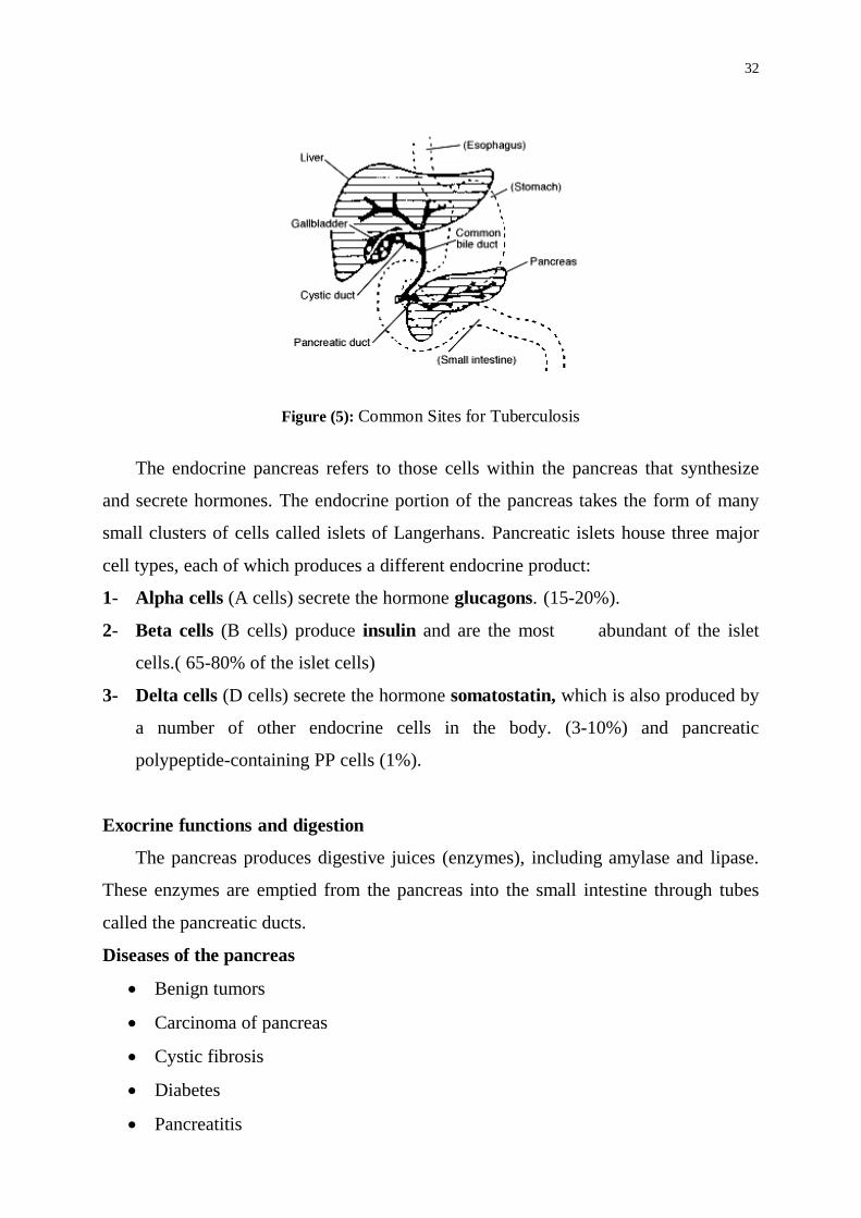
Diabetes Mellitus Introduction Functional Anatomy of the Endocrine Pancreas
The pancreas is an elongated organ nestled next to the first part of the small intestine ( figure 5).
32
Figure (5): Common Sites for Tuberculosis The endocrine pancreas refers to those cells within the pancreas that synthesize
and secrete hormones. The endocrine portion of the pancreas takes the form of many
small clusters of cells called islets of Langerhans. Pancreatic islets house three major
cell types, each of which produces a different endocrine product:
1- Alpha cells (A cells) secrete the hormone glucagons. (15-20%).
2- Beta cells (B cells) produce insulin and are the most abundant of the islet
cells.( 65-80% of the islet cells)
3- Delta cells (D cells) secrete the hormone somatostatin, which is also produced by
a number of other endocrine cells in the body. (3-10%) and pancreatic
polypeptide-containing PP cells (1%).
Exocrine functions and digestion
The pancreas produces digestive juices (enzymes), including amylase and lipase.
These enzymes are emptied from the pancreas into the small intestine through tubes
called the pancreatic ducts.
Diseases of the pancreas
• Benign tumors
• Carcinoma of pancreas
• Cystic fibrosis
• Diabetes
• Pancreatitis

33
Control of Insulin Secretion
Insulin is secreted in primarily in response to elevated blood concentrations of
glucose. This makes sense because insulin is "in charge" of facilitating glucose entry
into cells. Some neural stimuli (e.g. site and taste of food) and increased blood
concentrations of other fuel molecules, including amino acids and fatty acids, also
promote insulin secretion.
Physiologic Effects of Insulin
It has profound effects on both carbohydrate and lipid metabolism, and significant
influences on protein and mineral metabolism
Insulin and Carbohydrate Metabolism
Insulin acts on cells throughout the body to stimulate uptake, utilization and
storage of glucose. There are two important effects are:
1- Insulin facilitates entry of glucose into muscle, adipose and several other tissues.
It should be noted that: there are some tissues that do not require insulin for
efficient uptake of glucose: important examples are brain and the liver.
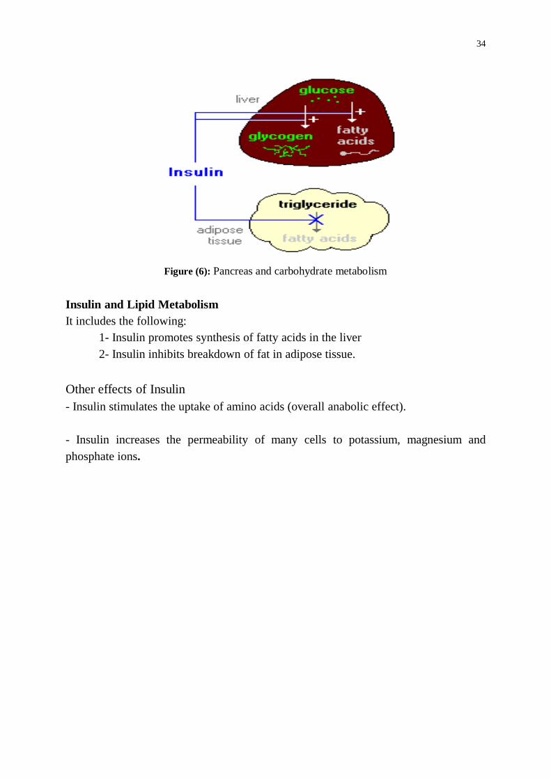
2- Insulin stimulates the liver to store glucose in the form of glycogen and it has
several effects in liver which stimulate glycogen synthesis (figure 6).
A well-known effect of insulin is to decrease the concentration of glucose in
blood. In the absence of insulin, a bulk of the cells in the body become unable to take
up glucose, and begin a switch to using alternative fuels like fatty acids for energy.
Neurons, however, require a constant supply of glucose, which in the short term, is
provided from glycogen reserves. Glycogen breakdown is stimulated not only by the
absence of insulin but by the presence of glucagons, which is secreted when blood
glucose levels fall below the normal range.
34
Figure (6): Pancreas and carbohydrate metabolism
Insulin and Lipid Metabolism It includes the following:
1- Insulin promotes synthesis of fatty acids in the liver 2- Insulin inhibits breakdown of fat in adipose tissue.
Other effects of Insulin - Insulin stimulates the uptake of amino acids (overall anabolic effect). - Insulin increases the permeability of many cells to potassium, magnesium and phosphate ions.

35
Diabetes Mellitus
Diabetes mellitus is a syndrome characterized by disturbance of metabolism of carbohydrates, protein, fats and vitamins due to absolute or relative deficiency of insulin. It may present with acute symptoms that include polydepsia (excessive thirst), Polyuria (excessive urination) and polyphagia (excessive hunger). Pathophysiology of diabetes mellitus:- 1- Decrease glucose utilization → hyperglycemia (↑blood glucose level), glucosuria
(above 180%) leading to: a) Osmotic diuresis causing Polyuria. b) Dehydration, decrease venous return, decreases cardiac out put and tissue
hypoxia. 2- Increase protein catabolism: leading to severe wasting of the muscles, delay of
wounds healing and osteoporosis. 3- Increase lipolysis leading to loss of body weight, ↑ fatty acids in blood and fatty
liver. Common Symptoms * Excessive fatigue. * Sudden weight loss. * Frequent urination * Excessive hunger. * Constant thirst * Numbness of hand or feet. * Vaginal infection. * Blurry vision * Impotence and infertility. * Prolonged wound healing. Types of diabetes
The three main types of diabetes are type 1, type 2 and gestational diabetes. 1- Type I or insulin-dependent diabetes mellitus:-
- There is little or endogenous insulin secretory capacity. - Formerly called juvenile diabetes (childhood onset). - It is due to destruction pancreatic B cells - Can be controlled by insulin replacement therapy.
2- Type II or non-insulin-dependent diabetes mellitus (90% of patients are obese): - There is a significant endogenous insulin secretory capacity.
- Formerly called adult-onset diabetes, is the most common form. People can develop it at any age, even during childhood. -Begins as a syndrome of insulin resistance. That is, target tissues fail to respond appropriately to insulin.

36
- Can be controlled may be decreased with dietary modification, weight loss and exercise and hypoglycemic agents.
3- Gestational diabetes develops in some women during the late stages of pregnancy. Although this form of diabetes usually goes away after the baby is born, a woman who has had it is more likely to develop type 2 diabetes later in life. Gestational diabetes is caused by the hormones of pregnancy or by a shortage of insulin. Diagnosis of diabetes The following tests are used for diagnosis: 1- A fasting plasma glucose test measures blood glucose after at least 8 hours without
eating. 2- An oral glucose tolerance test measures blood glucose after at least 8 hours
without eating and 2 hours after drinking a glucose-containing beverage. 3- In a random plasma glucose test, checks blood glucose without regard to when
subject ate his/her last meal.
Positive test results should be confirmed by repeating the fasting plasma glucose test or the oral glucose tolerance test on a different day.
Factors increase the risk for type 2 diabetes 1-Age 2-Weight 3- Sex 4-Race 5-Gestational diabetes, 6-Blood pressure is 140/90 or higher, 7-Cholesterol levels are not normal. HDL cholesterol ("good" cholesterol) is 35 or
lower, or triglyceride level is 250 or higher. 8-Lack in activity and exercise. Symptoms Commonly seen symptoms of a Diabetic patient are as follows: 1) Excessive urination 2) Excessive thirst 3) Excessive hunger 4) Loss of weight 5) Feeling of tiredness/Debility 6) Irritability, itching & frequent skin infections. Complications of diabetes Acute complications: 1- Hypoglycemia. 2- Ketoacidossis 3- Skin and mucosal infections

37
Chronic complications: 1- Osteomyelitis. 2- Diabetic nephropathy 3- Vascular disorders 4- Diabetic neuropathy. 5- Diabetic foot problems. 6- Diabetic eye disease 7-Diabetic kidney disease 8- Diabetic nerve damage 9- Gangrene 10- Gestational diabetes
Management of Diabetes The main principle of the treatment is as follows: 1) Drug 2) Diet 3) Exercise
1- Drugs 1- Oral hypoglycemic agents (OHA): They are taken orally to reduce the blood sugar. They are mainly used in NIDDM. 2-Insulin: Type I Diabetes Mellitus: - Requires Insulin only Type II Diabetes Mellitus: - Requires insulin when the OHA fail to control the blood sugar as in conditions like: 1) Infection, fever 2) Major surgery 3) Stressful condition 4) Pregnancy
2- Diet 1- The diabetic person can eat almost any food that other people normally eat provided the food is balanced and within the permissible caloric limits. 2- Facilitate variation in the diet without disturbing the caloric intake. 3- The diabetic diet must meet calorie requirements according to the needs of the patient (Thin, obese & underweight). 4- The proportion of energy derived from the food is as follows:
• Proteins - 15% • Fats - 30 - 35% • Carbohydrates - 55%
5-Diabetic people are asked to eat at short intervals i. e. not to keep long gaps between two meals to avoid lowering of blood sugar.

38
6-Fiber Supplement in diet helps in controlling blood sugar by slowing absorption of carbohydrates. In addition high fiber helps in satisfying hunger, reducing high cholesterol and preventing constipation. Keep the blood glucose at a healthy level by: 1-Eat about the same amount of food each day. 2-Eat your meals and snacks at about the same times each day. 3-Do not skip meals or snacks. 4-Take your medicines at the same times each day. 5-Exercise at about the same times each day. 6-Same dose of insulin. 7-Same level of activity. The Food Pyramid
Figure (7): The Food Pyramid
Eat a variety of food to get the vitamins and minerals you need. Eat more from the
groups at the bottom of the pyramid, and less from the groups at the top (figure 7). 3- Exercise Exercises have both benefits and risks. There are guidelines to assist patients with diabetes to exercise safely. Strategies to assist diabetic patients to exercise safely: 1-Adequate metabolic control before exercise program. If blood glucose -Less than 100 mg/dl before exercise, the person should eat a snake.

39
- 100-150 mg/dl, the person can go a head and exercise. - Over 250 mg/dl, the person takes insulin and delay exercise. 2-Self monitoring of blood glucose level before, during and after exercise. 3- Food intake may need to be increased to accommodate activity or exercise. 4-Strenuous exercise over an extended time may require reduction in insulin dosage. 5-After exercise test blood glucose to prevent post-exercise hypoglycemia. 6-Injection site of insulin is not a major concern unless the injection is given in a part of the body that will be exercising immediately. 7- Fluid intake during exercise is important. 8- Exercise should be done four times per week or every other day. 9- Exercise should not result in shortness of breath. 10- Warm up and cool down exercises are important. Stay healthy with diabetes
Follow the healthy eating plan that you and your doctor or dietitian have worked out.
Be active a total of 30 minutes most days. Ask your doctor what activities are best for you.
Take your diabetes medicines at the same times each day.
Check your blood glucose every day. Each time you check your blood glucose, write the number in your record book.
Check your feet every day for cuts, blisters, sores, swelling, redness, or sore toenails.
Brush and floss your teeth and gums every day.

40
Don't smoke.
Diabetic foot ulcer How can diabetes hurt my feet?
High blood glucose from diabetes causes two problems that can hurt your feet: 1. Nerve damage. Called diabetic neuropathy with damaged nerves, you might not
feel pain, heat, or cold in your legs and feet. It can lead to a large sore or infection. 2. Poor blood flow. Poor blood flow makes it hard for a sore or infection to heal.
Smoking when you have diabetes makes blood flow problems much worse.
Care of feet • Wash your feet in warm water every day. • Look at your feet every day to check for cuts, sores, blisters, redness, calluses, or
other problems. Inspect inside of your shoes daily for foreign objects. • If your skin is dry, rub lotion on your feet after you wash. • Cut your toenails once a week or when needed. • Always wear shoes or slippers to protect your feet from injuries. • Always wear socks or stockings to avoid blisters. • Wear shoes that fit well. • Avoid wearing open toed shoes. • Avoid pointed toes or high heal shoes.
Obesity Definition
Obesity is a condition characterized by excessive fat storage. It is obviously caused by excess energy input over energy output, and consequently deposition of excess fat in the body. Epidemiology of Obesity 1- Age
Obesity is often looked upon as a disease of middle age, but it can occur at any time of life. Obesity is now common in infants and young children as a result of changes in methods of feeding. Juvenile obesity sometimes followed by obesity in adult life.

41
2- Sex
Obesity may occur in either sex, but is usually more common in women, in whom it is liable to occur after pregnancy and at the menopause. A woman may be expected to gain 12.5 kg during pregnancy. 3- Social Class
There is an inverse correlation between social class and the prevalence of obesity. The only exceptions seem to be less affluent countries like India and Germany where there is usual negative relation between obesity and social class among women, but not among men. 4- Morbidity and Mortality
Excessive weight that associated with increased mortality Etiology of Obesity 1- Genetics versus Environment
When one parent is obese, the chances of a child's becoming obese are greater (40 percent) than when neither parent is obese (7 percent) if both parents are obese, the chances become 80 percent. Even though, the weight-for-height measures of both parents correlate with their children's measures, mother's measurements correlate more closely. 2- Endocrine factor
One of leptin's main effects may to inhibit the synthesis and release of hypothalamic neuropeptide Y, which increases food intake, decreases thermo genesis, and increases levels of insulin and corticosteroid in the plasma. 3- Inactivity People may be obese either because they eat too much, or because they spend too little energy. 4- Diet The composition of the diet and the frequency of eating is another etiologic factor in obesity. Eating several small meals /day is better than eating few large meals. 5- Drug Several drugs as glucocorticoids (cortisone) and birth control pills can lead to an increase in body weight. Smoking reduce food intake due to nicotine content.

42
6- Psychological factors Ingestion of food frequently had been used to reduce the feelings of emotional deprivation. Evaluation of Obesity 1- Measurements based on anthropometry A) Skin fold thickness:
Used by clinicians that depends on calipers to measure the fatty layer directly under the skin.
For greatest precision, the mean of the skin fold at four sites should be calculated. The following are example of caliper locations at different sites: 1) In the upper limb: * Subscapular: An oblique fold measured just below the interior angle of the scapula. * Triceps: A fold at the mid line half way between the olecranon and acromion with the arm
hanging freely at the side. * Over the biceps: Above the cubital fossa, at the same level as the triceps. 2) In the lower limb: * Thigh:
A fold in the anterior midline of the thigh, taken midway between the patel1a and the hip.
* Calf A fold measured in the leg at the level of the greatest calf girth.
3) In the trunk: * Chest:
A fold located one half of the distance between the anterior axillary line and nipple, for men, and one third of the distance, for women.
* Abdomen: A vertical fold measured 2 cm to the right of the umbilicus.
* Suprailiac: Suprailiac, on the mid-axillary line immediately superior to iliac crest.

43
The approximate desirable ranges of mean skin fold thickness are 3-1.0 mm in men and 1.0-22 mm in women.
(B) Waist to hip ratio:
-Measuring the circumference of the waist at its smallest point at level of umbilicus and the circumference of the hip at its widest point, and then calculating a ratio of the two can easily determine the site of fat in the body.
-A waist to hip ratio is recommended to be below 0.85 and 0.95 for women and men respectively. (C) Body Mass Index (BMI):
Weight (kg) BMI = ------------------
Height (m)2 Normal (average): BMI equal 20 -25Kg/m2
Over weight: BMI 25-30 Kg/m2
Obese: BMI > 30 Kg/m2
(D) Waist Circumference:
The waist circumference is a simple measure around a person's natural waist (just above the navel). A high-risk waist circumference is defined as 35 inches (88 cm) or more for women and 40 inches (102 cm) or more for men. Some well-trained people with dense muscle mass may have a high BMI score but very little body fat. For them the waist circumference may be a more useful measure. Other methods of evaluation
These are methods for estimation of body fat. These methods are: (a) Underwater weighting
For estimation of body fat, the subject exhales as much air as possible and then holds his breath and bends over at the waist. Once he is totally submerged, the underwater weight is recorded. Comparing a person's weight on a standard scale to his or her weight underwater can yield a very accurate estimate of total body fat. This works on the principle that' adipose tissue is less dense than lean tissue. The more adipose tissue there is in a body, the less it weights when submerged (the more it tends to float). Unfortunately, this method requires expensive equipment that is not widely available. (b) Bioelectrical Impedance

44
Bioelectrical impedance is a technique that uses low-energy electrical current to estimate total body fat. Researchers summarize that fat resists the low of electricity because it contains little water and few electrolytes such as potassium. Lean tissue, in comparison, has about 73% water and is rich in electrolytes. Thus the more fat a person has per inch of height, the more resistant he is. So although it is still unclear what aspect of body physiology bioelectrical impedance analyzers are actually measuring, they do provide a rapid and fairly accurate measurement of the percentage of body fat (body fat analyzer). Complications (1) Coronary heart disease (2) Hypertension (3) Cardiomyopathy (4) Diabetes Mellitus (5) Respiratory diseases (6) Reproductive disorders and decreased fertility (7) Gallbladder diseases as increases the risk of occurrence of gallstones (8) Psychological manifestation and reduced self-esteem (9) Arthritis of the hips and knees weight-bearing joints. (10) Varicose veins and hemorrhoids Treatment Strategies The aim of treatment is to: * Achieve weight loss and prevent weight gain if that is not possible, to preserve
weight at the present level. * Decrease medical risks and improve the quality of life. Lines of management of adult obesity
Include diet, exercise, behavioral, medication and surgical intervention.
(1) Diet Restriction of energy intake to low calorie (800 to 1200 KCal./day) or very low
calorie (less than 800 KCal./day) Balanced diet is a common treatment for obesity .A truly motivated individual will generally stay on a diet for a long time, initially for weight loss and then for weight maintenance.
There are three guiding principals in designing diet: a. The diet must supply less energy than the patient maintenance requirements. b. The diet must supply all nutrients to avoid malnutrition.

45
c. The required small decrease in energy intake can usually be achieved by reducing consumption of sweets, and substituting fruits and snacks for the usual potato crisps, biscuits and ice-cream.
(2) Physical activity (exercise) Exercise or increase physical activity should be used as a treatment modality for obesity as long as there is no contraindication to its use. Vigorous exercise should be avoided due to general lack of conditioning for most obese individuals. Regular aerobic activity promotes a basic good health and sense of well being. (3) Behavior modification: 1-Avoid simultaneous activities as watching television or reading during eating. 2- No eating between meals. 3- Watching portion of food eaten. 4- Eating slowly with concentration. 5- Increase physical activities as: * Taking stairs rather than elevators or escalators. * Park your car far from the store. Pharmacotherapy A- Appetite suppressants. B- Exogenous thyroid hormone. C- Drugs affecting the gastrointestinal tract. The use of these drugs has been popularized by the recent attention paid to obesity as well as by the development of new agents. Reported adverse effects such as loss of bone mineralization and cardiovascular complications have led to the withdrawal of certain drugs from the market. Surgical Treatment a. Selection of patient for surgical treatment:
Surgery done only to patients who weight more than 200% of their ideal body weight (BMI = 40 kg/m2) or, at a minimum have a BMI of at least 35 kg/m2 (weight-related comoribdties). In addition make sure that all candidates have shown repeated failure at controlling weight by medical means, including supervised dietary programs. b. Surgical procedures:
Surgical weight loss procedures generally fall into two main types, those that limit nutrient absorption (e.g. intestinal by pass) and those that limit intake (e.g. gastric by Pass) which is considered the operation of choice.

46
PHYSIOTHERAPY IN CARDIOTHORACIC SURGERY
FOR PHYSICAL THERAPY STUDENTS
Dr. Shehab M. Abd El-Kader Associate Prof. of Physical Therapy

47
Role of physiotherapy in cardiothoracic surgery
Aims of physiotherapy following cardiac surgery: 1. To preserve adequate ventilation. 2. To assist with removal of excess secretions in the airways. 3. To assist the circulation in the legs and thereby help to prevent post-operative
venous thrombosis. 4. To maintain mobility of the shoulders, shoulder girdle and spine. 5. To prevent postural defects. 6. To restore exercise tolerance. Pre-operative training 1) Explanation to the patient
Explanation by the physiotherapist, in order to gain the patient's confidence and co- operation, should be similar to that described for pulmonary surgery.
The importance of maintaining adequate ventilation of the lungs by breathing exercises and the clearance of excess secretions from the airways must be explained. Reassurance should be given that breathing exercises, huffing, coughing and moving around in bed will do no harm to the stitches, drainage tubes or operation site. 2) Removal of secretions
The majority of patients about to undergo cardiac surgery do not have excess bronchial secretions. There are, however, some patients with severe mitral valve disease or long- standing pulmonary hypertension that may have developed associated

48
chronic obstructive lung disease and assistance with removal of secretions is required. In the earlier stage of cardiac disease, the patient may have a persistent dry cough or expectorate frothy white sputum. This is not a problem that can be dealt with by physiotherapy. 3) Breathing exercises
(a) Diaphragmatic breathing 1- Diaphragm normally does the major action of breathing (about 70%).But
its action usually about 30%of the action of breathing in the first postoperative days.
2- Diaphragm improves ventilation in the lower lobes, which is the site of accumulation of secretions. (b) Unilateral lower thoracic expansion
Lower costal breathing exercises improve ventilation in lower lobes that is the site of secretion accumulation.
4) Effective huffing and coughing The physiotherapist should show the patient how she will support the chest over the incision and how he can support it himself (figure 8).
Figure (8): Median sternotomy supported by patient
5) Foot and leg exercises
All patients are taught simple foot exercises and knee flexion and extension in order to assist the circulation and help prevent post- operative venous thrombosis. 6) Posture, shoulder girdle and arm movements
Those patients having a median sternotomy are unlikely to have difficulty with shoulder movements after surgery, but the shoulder girdle may become stiff and many

49
patients tend to adopt a slightly kyphotic posture. Shoulder shrugging and 'shrug-circling' are useful exercises and can be practiced briefly pre-operatively.
Post-operative treatment
Day of operation If the patient is not on a ventilator, breathing exercises can be started on the day
of the operation (provided the cardiovascular system is stable) as soon as he is conscious enough to co-operate. After breathing exercises, attempts at huffing and coughing should be made. First and second day after operation
Physiotherapy will probably be necessary four times during the day. The length of treatment should be modified according to the patient's condition and should not cause fatigue. 1) Breathing exercises
If the patient is not being artificially ventilated, breathing exercises should be carried out. Those who have been ventilated should also start breathing exercises once the endotracheal tube has been removed. The patient should be sitting up in bed with the whole back supported by pillows, so that diaphragmatic and chest movements are not inhibited. Exercises should include: (a) Diaphragmatic breathing. (b) Unilateral lower thoracic expansion for both sides of the chest.
If pain is severely limiting .the respiratory excursion, the physiotherapist should
treat the patient after an analgesic has been administered. The patient should be reminded to practice breathing exercises at least every hour whilst awake.
2) Huffing and coughing
Effective huffing and coughing, as taught pre-operatively, must be encouraged with the chest firmly supported. 3) Foot and leg exercises
The exercises taught pre-operatively should be practiced and the patient should be reminded to do these movements 5-10 times every hour that he is awake. 4) Shoulder movements

50
With a lateral thoracotomy, it is important to start arm movements on the first post-operative day. With a median sternotomy, these need not be started until the second day. Third day onwards
The patient will start sitting out of bed from 24 hours after surgery according to his progress and the surgeon's instructions. Walking around the ward may be started as soon as the second or third post-operative day. Treatment should include: 1. Breathing exercises (as above). 2. Huffing and coughing, if secretions are present in the lungs. 3. Foot and leg exercises are given while the patient is confined to bed. These can be
discontinued when he is fully mobile. 4. Arm and shoulder girdle exercises, 5. Postural correction and gentle trunk exercises if necessary, 6. Walking up stairs can usually be started about 6 days from the time of operation.
This will depend on the instructions of the individual surgeon. After cardiac surgery, most patients find climbing stairs much less exhausting than pre-operatively. Treatment must be modified if any complications occur.
Before discharge
Thoracic expansion, shoulder mobility and posture should have returned to normal. The patient should be increasing his exercise tolerance. The patient should continue breathing exercises for about 3 weeks following the operation, although he will probably be discharged after 10-14 days.
Complications following cardiothoracic surgery
A) Factors that increase the postoperative complications: 1. General anesthesia: a- Decreases the normal ciliary action of the tracheobronchial tree. b- Depresses the respiratory center of the CNS, which causes a shallow respiratory pattern (decreased tidal volume). 2. Intubation (insertion of an endotracheal or nasogastric tube): a- Irritates the mucosal lining of the tracheobronchial tree which causes an increase in mucus production. b- Decreases the normal action of the cilia in the pulmonary tree, which leads to pooling of secretions.

51
3. Incisional pain: a- Causes the patient to take shallow breaths. Lung expansion is restricted and secretions are not adequately mobilized. b- Restricts a deep and effective cough. The patient usually has a deep shallow cough that does not effectively mobilize secretions. 4. Pain medication: Although pain medication administered postoperatively tend to diminish incisional pain it also: a- Depresses the respiratory center the CNS. b- Decreases the normal ciliary action in the bronchial tree. 5. General inactivity and bed rest postoperatively: It causes secretions to pool, particularly in the posterior basilar segments of the lower lobes. 6. General weakness and fatigue decreases the effectiveness of the cough. B) Complications following cardiothoracic surgery: 1. Respiratory problems. 2. Cardiac problems. 3. Thrombosis. 4. Hemorrhage. 5. Wound infections. 6. Pressure sores. 7. Muscle wasting and impairment of function.
I. Respiratory problems: a. Atelectasis Is incomplete expansion of the lung because of collapse of the alveoli. Hypoventilation is the most common postoperative cause b. Postoperative pneumonia: Due to infection of retained secretions. Present 2-3 days postoperative.

52
c. Pneumothorax: Is an accumulation of gas or air in the thoracic cavity. It can be therapeutic, spontaneous or traumatic. Chest tube inserted in the area of the 2nd intercostal space to measure the pressure and withdrawal the accumulated gas or air. i. Pulmonary embolism: Is obstruction of a pulmonary artery or one of its branches by a clot arises from a deep veins. k. Hypoxia: Is low oxygen content within the tissues of the body. It can result from ventilation-perfusion imbalance of underlying pulmonary disease or destruction of blood cells by the heart lung machine. Physiotherapy for the respiratory complications: Aim: Is to regain the normal vital capacity and to stimulate coughing and to encourage the full use of the lungs. Methods: 1. Breathing exercises: should be taught preoperatively while the patient is alert, pain free and fully cooperative. Emphasis is laid on diaphragmatic and lateral costal expansion with a good deep inspiration followed by relaxed expiration (diaphragm is normally responsible for 60% of normal respiratory movement, but in the first 24 hours after the operation, it's movement may be only 20% of the normal. 2. Effective coughing: Cough should be effective with less pain so, the patient should support the incisional area and lean his trunk toward the area of incision.
3. Mechanical assistance for the removal of secretions. The methods used are
percussion, deep breathing exercises with vibration and postural drainage .Nasopharyngeal suction may be necessary in some circumstances when the patient is unable to cough up secretions despite the assistance of physiotherapy. II. Cardiac complications: 1. Cardiac arrhythmias: Cardiac arrhythmias are variation from the normal rhythm
of the heart.

53
2. Cardiac tamponade: is a lin1itation of ventricular filling during diastole because of fluid collection within the pericardial sac. Physiotherapy may be contraindicated with this complication.
3. Cardiogenic shock: results from diminution of cardiac output. The cardiac output may fall very low immediately after cardiac damage. Physiotherapy may be contraindicated with this complication.
III. Deep venous thrombosis: Is a coagulation or clot of blood that remains at the site of origin, if it detaches the clot can travel to the right side of the heart and enter the lung called a pulmonary embolism. Physiotherapy: 1- Prevention: The preoperative instructions will include a program of active leg exercises and deep breathing exercises at least for five minutes in every hour and early postoperative leg mobilization. 2- If DVT developed: A-Physiotherapy is contra-indicated in acute cases.
B- In chronic cases: -Apply deep breathing exercise, -Active exercise and mobilization. -Elastic bandage to control swelling and aid venous return.
IV. Wound infection: Infected wound become hot, red and edematous the sutures tend to cut through
the tissues and the wound may gape either along the whole length or in between the sutures. Physiotherapy: 1- Clean wound can receive superficial heat (as infrared), if it is a superficial wound a
deep heat (as short wave), if the wound is deep. 2- Ultrasonic wave for the hard scars. 3- Paraffin wax to soften hard scars. V. Pressure sores: * Prevented by frequent changing of the patient posture. * Frequent check of the integrity of the skin and areas of redness. * Ultra violet is essential in its management.

54
VI. Neurological damage: During cardiac surgery, the brain may be damaged by embolism or anoxia. Physiotherapy must treat any form of paralysis that occurs. Obviously, the patient's cardiac state may limit the form of rehabilitation to some extent.

55
PHYSICAL THERAPY IN CARDIAC DISORDERS
FOR PHYSICAL THERAPY STUDENTS
Dr. Shehab M. Abd El-Kader Associate Professor of Physical Therapy
Physical Therapy in Cardiac Disorders

56
Congenital heart diseases Causes 1. Drugs 2. Hormones 3. Fever 4. X-ray 5. Uterine bleeding 6. Smoking 7. Repeated attack of abortion 8. Chromosomal abnormalities 9. Nutritional Classifications 1. Cyanotic or not cyanotic 2. With or without shunt 3. According to the direction of the shunt A. Right to left shunt B. Left to right shunt Specific congenital heart disease 1. Atrial septal defect (ASD) Types 1. Ostium secondum. 2. Ostium premium. 3. Sinus venous. 4. Patent foramen ovale.
Figure (8): Atrial septal defect (ASD)
Hemodynamics 1. Left to right to shunt. 2. Rt . Atrial dilatation and hypertrophy. 3. Rt. Vent. dilatation and hypertrophy.

57
4. Pulmonary hypertension. 5. Functional tricuspid regure. Manifestations 1. Repeated attacks of winter bronchitis. 2. Dyspnea on mild effort. 3. Underweight. 4. Central cyanosis in rare cases. Treatment Surgical by open heart technique and the defect is closed by direct sutures or by using synthetic material as tiphlon or darcon.
2. Ventricular septal defect (VSD) Types 1. Membranous. 2. Muscular.
Figure (9): Ventricular septal defect (VSD) Hemodynamics 1. Left to right shunt. 2. Right vent. Hypertrophy and dilatation. 3. Massive pulmonary hypertension and as result Rt to Lt shunt (Eisenmengers syndrome). Manifestations 1. Recurrent attack of winter bronchitis. 2. Dyspnea. 3. Neglected cases of cyanosis.

58
Treatment * Surgical by open heart technique and the defect is closed by direct sutures or by using synthetic material as tiphlon or darcon. * In 20% of cases there is happy transformation (spontaneous closure if it is small or in the muscular part of the septum).
3. Patent ductus arterioses (PDA) It is a duct between the arch of aorta and pulmonary artery.
Figure (10): Patent ductus arterioses (PDA) Hemodynamics 1. Oxygenated blood passes from the aorta to the left pulmonary artery. 2. Pulmonary hypertension in rare cases and reverse of shunt, and as a result differential cyanosis. Treatment Surgical by closed heart technique (excision and suture)
4. Coarcitation of aorta It is stenosis (constriction) of the aorta distal to the left subclavian artery. It is a cyanotic heart disease without a shunt.

59
Figure (11): Coarcitation of aorta Manifestations 1. Severe headache 2. Intermittent claudication. 3. Hypertension in upper part of the body. 4. Well developed upper half of the body and less developed lower half. 5. Abnormal delay between the femoral and radial pulsation. Treatment Surgical by closed heart technique (excision of the coarcitation segment and end to end anastomosis)
5. Fallot tetrology (F4) 1. Severe pulmonary stenosis. 2. Ventricular septal defect. 3. Rt. Ventricular hypertrophy. 4. Overriding of aorta. Hemodynamics 1. Severe pulmonary stenosis leads to Rt. vent. Hypertrophy. 2. VSD leads to overriding of aorta. 3. When Rt. Vent. Pressure exceeds that of Lt shunt will be reversed. Manifestations 1. Cyanosis since birth. 2. Prefer of squatting position. 3. Dyspnea on mild effort. 4. Clubbing of fingers and toes. 6. Hemoptysis.
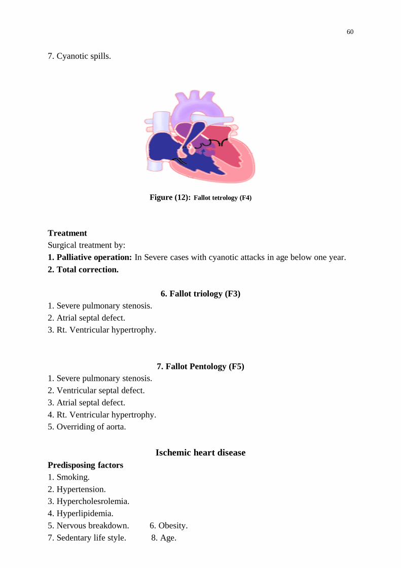
60
7. Cyanotic spills.
Figure (12): Fallot tetrology (F4)
Treatment Surgical treatment by: 1. Palliative operation: In Severe cases with cyanotic attacks in age below one year. 2. Total correction.
6. Fallot triology (F3) 1. Severe pulmonary stenosis. 2. Atrial septal defect. 3. Rt. Ventricular hypertrophy.
7. Fallot Pentology (F5) 1. Severe pulmonary stenosis. 2. Ventricular septal defect. 3. Atrial septal defect. 4. Rt. Ventricular hypertrophy. 5. Overriding of aorta.
Ischemic heart disease Predisposing factors 1. Smoking. 2. Hypertension. 3. Hypercholesrolemia. 4. Hyperlipidemia. 5. Nervous breakdown. 6. Obesity. 7. Sedentary life style. 8. Age.

61
9. Positive family history. 10. Male gender Pathogenesis 1. Intimal tear. 2. Precipitation of platelets, fibrin and lipoprotein. 3. Narrowing of the coronary vessels. 4. Rupture of atherosclerotic plaque. Clinical picture 1. Mild degree (angina pectoris) - It is due to coronary atherosclerosis. - Patient complains anginal pain( retrosternal referred to the left shoulder, arm and little finger and may be to the right arm, in rare cases to the back, side of the neck and lower jaw). - Pain is burning, stapping or compression (squeezing). - Pain relived by rest or coronary vasodilators. 2. Angina at rest It is a more severe stage of coronary atherosclerosis where anginal pain occurs at rest. 3. Unstable angina It is a more severe stage of coronary atherosclerosis where anginal pain is prolonged, not relieved by rest or coronary vasodilators (considered as pre infarction syndrome) this case is accompanied with severe sweating and pallor. 4. Acute myocardial infarction (acute M.I.) - There is coronary occlusion by thrombus or rupture of atherosclerotic plaque. - Anginal pain is severe accompanied with sweating and pallor. - Anginal pain can not be relived by rest or coronary vasodilators. - Patient is semi shocked (Hypotensive). - Treatment of acute M.I. and unstable angina: a. Transfer patient to coronary care unit. b. Oxygen inhalation. c. Morfia injections. Groups of drugs -Group (1): Nitroglycerine. -Group (2): Beta- blockers. -Group (3): Calcium channel blocker.

62
-Group (4): Anti-platelets. Investigations 1. Resting E.C.G. (if normal do Exercise stress test). 2. Blood lipid profile. 3. Blood sugar analysis. 4. Echocardiography. 5. Catheterization. Other treatment procedures 1. Balloon dilatation by coronary catheter. 2. Combination between balloon and stint. 3. Using laser technique. Surgical treatment(CABG) Take the graft from 1. Saphenous vein. 2. Internal mammary artery. 3. Superficial epigastric artery. 4. Radial artery. 5. Splenic artery.
Rheumatic fever It is a widespread disease in lack of hygiene, malnutrition and overcrowdness. It is caused by B-Hemolytic streptococci. Manifestations A. Major B. Minor 1. Fever. 1. Erythema margenatum 2. Carditis. 2. Subcutaneous nodules 3. Arthritis. 4. CNS chorea. Treatment

63
1. Rest. 2. Salt free diet. 3. Aspirin. Prophylactic treatment 1. Tonsillectomy. 2. Long acting penicillin. Complications 1. Rheumatic valvulitis. 2. Fibrosis of chorda tendinae and papillary muscles. 3. Fusion of commissures. 4. Shortening of papillary muscles. 5. Stenosis and/ or incompetence of cardiac valves. Hemodynamics of mitral stenosis 1. Increase in left atrial pressure leads to: A. Hypertrophy and dilation of left atrium. B. Pulmonary hypertension & hemoptysis. 2. Left atrial fibrillation &loss of contractile element leads to thrombosis and stroke. 3. Right ventricular hypertrophy and dilation. 4. Tricuspid incompetence (functional regurge). 5. Right atrial hypertrophy and dilation. 6. Congestive heart failure. 7. Small left ventricle. Hemodynamics of mitral regurge 1. Left ventricular hypertrophy and dilation. 2. Left atrial hypertrophy and dilation leads to pulmonary hypertension. 3. Tricuspid incompetence (functional regurge). 4. Right ventricular hypertrophy and dilation. 5. Congestive heart failure. Hemodynamics of Aortic stenosis 1. Left ventricular hypertrophy and dilation. 2. Chest pain. 3. Left ventricular failure. Hemodynamics of Aortic regurge 1. Left ventricular hypertrophy and dilation.

64
2. Diastolic blood pressure is low and pulse pressure is high. 3. Left ventricular failure. Hemodynamics of Tricuspid and pulmonary valve affection They are rare to be affected by rheumatic fever, bust in most cases the affection is functional and not organic & in the form of stenosis.
Heart failure Definition It is inability of the heart be perform its normal function. It may be 1. Right side heart failure. 2. Left side heart failure. 3. Congestive heart failure (both right and left side failure) Manifestations of right side heart failure 1. Congested pulstile neck veins. 2. Enlarged tender liver. 3. Edema in lower limbs. 4. Dyspnea. Manifestations of Left side heart failure 1. Dyspnea and /or orthopnea and paroxysmal nocturnal Dyspnea.

65
2. In some cases, Pulmonary edema and hemoptysis. Treatment 1. Complete rest. 2. Salt free diet. 3. Digitalis. 4. Diuretics. 5. Treatment of the cause.
Cardiac Rehabilitation Definition Rehabilitation is a therapeutic process designed to facilitate maximal restoration of function. Each patient must be individually assessed to determine diagnosis, associated injuries, responses, and achievable goals. Objectives The major goals of cardiac rehabilitative programs are:
• Reverse pathophysiologic and psychosocial effects of heart disease • Limit the risk for reinfarction or sudden death • Relieve cardiac symptoms, • Retard or reverse the atherosclerosis by instituting programs for exercise
training, education, counseling, and risk factors alteration • Reintegrate heart disease patients into successful functional status in their
families and the society

66
Indications • Recent myocardial infarction • Coronary bypass • Valve surgery • Coronary angioplasty • Cardiac transplantation • Angina • Compensated CHF
Exercise prescription depends on the results of exercise testing, which often includes cardiopulmonary exercise (CPX) testing. Contraindications
• Severe residual angina • Uncompensated heart failure • Uncontrolled arrhythmias • Severe ischemia, LV dysfunction, or arrhythmia during exercise testing • Poorly controlled hypertension • Hypertensive or any hypotensive systolic blood pressure response to exercise • Unstable concomitant medical problems (e.g. poorly controlled or "brittle"
diabetes, diabetes prone to hypoglycemia, ongoing febrile illness, active transplant rejection)
Rehabilitation Team Members and Their Roles: 1. Patients and his family: Patients and his family must never be overlooked as members of prescribing
team. The patient and his family must be made a ware of the program into which he is
about to enter, with all its implications. They must be oriented to the available types of mechanical aids and their
individual advantages and disadvantages related to the patient's personal and work needs.
2. Physician:
The physician is the leader and coordinator of the team; he attends to all medical aspects of the individual case. The physician in referring a patient should state the diagnosis the present condition of the patient', the limitations or the precautions to be observed the prognosis, the result to be achieved and the frequency and the length of treatment.
3. Therapists:
“Occupational and physical therapists”

67
In the treatment of physical disabilities the physical therapies have a similar ultimate goal, namely, to contribute to the restoration of the physical function of the patient.
4. Psychologist and psychiatrist: To provide information concerning the patient's mental abilities, emotional
adjustment, interest and vocational aptitudes mental abilities, emotional adjustment, interest and vocational aptitudes.
5. Social service: Social case work which helps the patient and his family to accept and adjust to
the problems resulting from his disability. 6. Nurses: The nurse is responsible for all patients under her care. 7. Dietician or nutritionist. The detection of patient’s nutritional requirements is the responsibility the dietician or nutritionist. 8. Vocational counselor.
The program frequently begins in a hospital setting and continues on an outpatient basis after the patient is discharged over a period of 6-12 months.
Phases of cardiac rehabilitation
Cardiac rehabilitation services are divided into 3 phases beginning with phase 1 that is initiated while the patient is still in the hospital, followed by phase 2 that is a supervised ambulatory outpatient program spanning 3-6 months, and subsequently continuing into phase 3, a lifetime maintenance phase, in which physical fitness, as well as additional risk factor reduction, are emphasized.
Basic Program Structure
Traditionally cardiac rehabilitation is divided into three phases with essential
medical, educational and exercise components being applied during each phase. Each patient rate of progression through these phases will vary depending on the nature and severity of illness, complications and rate of recovery. Phase I (Immediate inpatient phase)

68
It is the acute in hospital phase; it is usually 7-14 days in duration.
The goals of rehabilitation during Phase (I): 1-To initiate early physical therapy activities which allow: a- Return to activities of daily living. B-Decrease anxiety and depression. c-Determine the effects of medications. d-Prevent effects of prolonged bed rest. 2- To initate patient and family education to: a- Outline the course of cardiac rehabilitation. b- Modify the risk factors of atherosclerosis.
During phase I the rate of progression of people who have had a myocardial infarction is slightly slower than for those who have had coronary artery bypass grafts. Mobilization of surgical patients usually starts earlier and intensity and duration of ambulation are more accelerated. Table (2): Inpatient Rehabilitation: 7-Step Myocardial Infarction Program.
Step
Dat
e
Supervised Exercise
1 - Active and passive ROM all extremities, in bed. Teach patient ankle plantar and dorsiflexion-repeat hourly when awake.
2 - Active ROM all extremities, sitting on side of bed. 3 - Warm-up exercises: Stretching Calisthenics
Walk 50 ft and back at slow pace. 4 - ROM and calisthenics.
Walk length of hall (75 ft) and back, average pace. 5 - ROM and calisthenics.
Practice walking few stair steps & Walk 300 ft bid. 6 - Continue above activities.
Walk down/flight of steps (return by elevator) & Walk 500 ft bid. 7 - Continue above activities , Walk up /light of steps & Walk 500 ft bid.

69
Figure (13): A patient walking in the hallway with a physical therapist following bypass surgery. Table (3): Criteria for Termination of an Inpatient Exercise Session. 1. Fatigue 2. Failure of monitoring equipment 3. Light-headedness, confusion, cyanosis, dyspnea, nausea. 4. Onset of angina with exercise. 5. ST displacement (3 mm) horizontal or downsloping from rest 6. Ventricular tachycardia (3 or more consecutive PVCs) 7. Exercise-induced left bundle branch block 8. Onset of 2° and/or 3° A-V block. 9. Exercise hypotension (>20 mmHg drop in systolic blood pressure during exercise) 10. Excessive blood pressure rise: systolic ≥ 220 mmHg or diastolic ≥ 110 mmHg. 11. Inappropriate brachycardia (drop in heart rate greater than 10 bpm) with increase or no change in work
load
Phase II: The term "Phase II" refers to that part of the cardiac rehabilitation program
conducted on an outpatient basis immediately after hospitalization, It is the early convalescent phase (8-12 weeks in duration), during this phase myocardial and/or post operative healing is taking place. By 6-8 weeks the myocardial scar formation has taken place and the sternum is healed following surgery. The goals of rehabilitation during Phase (II): 1- Increase exercise capacity and endurance in a safe and progressive manner. 2- Educate the patient on proper technique of exercises. 3- Work with the patient and family to establish healthy life style. 4- Prepare the patient to return to work. 5- Enhance psychological status. 6- To provide the patient with guideline of long term exercises. Training Program: 1-Conditioning exercises: Rhythmic aerobic exercises as walking, jogging, swimming and rowing. Lower extremity aerobic exercise is accomplished with stationary equipment such as treadmills and bicycle ergometers. Upper extremity training is done with arm ergometer units and rowing machines. This type of equipment can improve both endurance and physical work capacity of post-myocardial infarction and post-bypass patients during phase II. 2- Calisthenics exercises: Active free exercises for upper limbs, lower limbs and trunk.
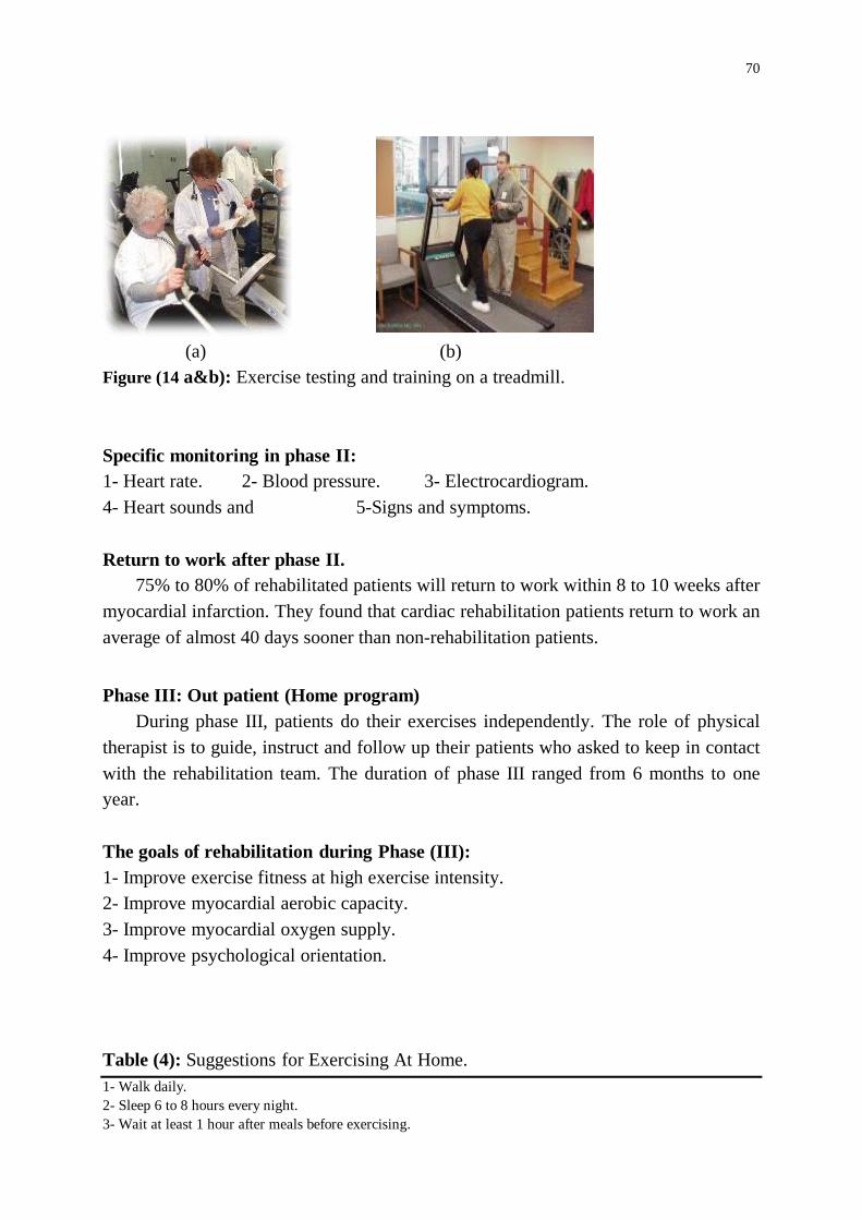
70
(a) (b) Figure (14 a&b): Exercise testing and training on a treadmill. Specific monitoring in phase II: 1- Heart rate. 2- Blood pressure. 3- Electrocardiogram. 4- Heart sounds and 5-Signs and symptoms. Return to work after phase II.
75% to 80% of rehabilitated patients will return to work within 8 to 10 weeks after myocardial infarction. They found that cardiac rehabilitation patients return to work an average of almost 40 days sooner than non-rehabilitation patients.
Phase III: Out patient (Home program)
During phase III, patients do their exercises independently. The role of physical therapist is to guide, instruct and follow up their patients who asked to keep in contact with the rehabilitation team. The duration of phase III ranged from 6 months to one year.
The goals of rehabilitation during Phase (III): 1- Improve exercise fitness at high exercise intensity. 2- Improve myocardial aerobic capacity. 3- Improve myocardial oxygen supply. 4- Improve psychological orientation. Table (4): Suggestions for Exercising At Home. 1- Walk daily. 2- Sleep 6 to 8 hours every night. 3- Wait at least 1 hour after meals before exercising.

71
4- Avoid extremes in weather: In the winter, exercise during the warmer parts of the day; in the summer, exercise in the early morning or evening.
5- Avoid vigorous arm and shoulder activities, especially overhead arm activity (arm activity requires more energy than leg activity).
6- Avoid lifting heavy weights or objects (isometric exercise). 7- Avoid situations and people who make you anxious or angry. 8- If you have chest pain, dizziness, excessive fatigue, unusual palpitation or shortness of breath stop what you
are doing and Call your physician. 9- Take your medications as ordered. 10- Don't exercise if you have an acute illness.
Table (5): Contraindications for Entry into Inpatient and Outpatient Exercise Programs. 1. Unstable angina 2. Resting systolic blood pressure >200 mmHg or resting diastolic blood pressure> 100 mmHg 3. Orthostatic blood pressure drop of ≥ 20 mmHg 4. Moderate to severe aortic stenosis 5. Acute systemic illness or fever 6. Uncontrolled atrial or ventricular dysrhythmias 7. Uncontrolled sinus tachycardia (>120 beats.min-1) 8. Uncontrolled congestive heart failure 9. 3° A-V heart block. 10. Active pericarditis or myocarditis 11. Recent embolism 12. Thrombophlebitis 13. Resting ST displacement (> 3 mm) 14. Uncontrolled diabetes 15. Orthopedic problems that would prohibit exercise
Outcomes of Cardiac Rehabilitation Training
1. Improved exercise tolerance
Cardiac rehabilitation exercise training for patients with coronary heart disease or CHF leads to objectively verifiable improvement in exercise capacity in men and women, regardless of age. This beneficial effect does not persist long-term after completion of cardiac rehabilitation without a long-term maintenance program. Therefore, exercise training must be maintained long term to sustain the improvement in exercise capacity. 2. Control of symptoms

72
In patients with coronary heart disease, angina significantly improves during the cardiac rehabilitation exercise program and patients with LV failure or dysfunction show improvement in the symptoms of heart failure. 3. Improvement in the blood levels of lipids
Improvements in lipid and lipoprotein levels are observed in patients undergoing cardiac rehabilitation exercise training and education. Exercise must be combined with dietary and medical interventions for required lipid control. 4. Effect on body weight
Optimal management of obesity requires multifactorial rehabilitation, including nutritional education and counseling, behavioral modification and exercise training. 5. Effect on blood pressure
Rehabilitative exercise training as a sole intervention has minimal effect; however, multifactorial intervention has been shown to have beneficial effects. 6. Reduction in smoking
Cardiac rehabilitation services with well-designed educational, counseling and behavioral modification programs result in cessation of smoking in a significant number of patients. 7. Improved psychosocial well being
Cardiac rehabilitation exercise and education services enhance measures of psychological and social functioning. 8. Enhanced social adjustment and functioning
Cardiac rehabilitation exercise training improves social adjustment and functioning. Exercise training is recommended to improve these social outcomes. 9. Return to work
Cardiac rehabilitation exercise training exerts less influence on rates of return to work than on other aspects of life. Many non exercise variables also affect this outcome (eg, prior employment status, employer attitude, economic incentives). 10. Reduced mortality
Scientific data suggest a survival benefit for patients who participate in cardiac rehabilitation exercise training, but it is not attributable to exercise alone.

73
Exercise prescription for cardiac patients
I. Mode
Aerobic exercise training includes walking, jogging, running, swimming and stationary bicycling or any combination of these activities.
II. Frequency
- Individuals with a less than 3-MET capacity should engage in multiple short sessions each day. - Individuals with a 3- to 5-MET capacity should engage in 1-2 sessions per day. - Individuals a greater than 5-MET capacity should engage in 3-5 sessions per week.
III. Duration Patients usually need to allow 30-60 minutes for each session, which includes a warm-up of at least 10 minutes
IV. Intensity The intensity prescribed according to:
1-Target heart rate (training heart rate) which determined according to Karvonen formula as following:
Target heart rate = Resting heart rate + 60%-80 %( Maximum heart rate –resting heart rate) Maximum heart rate = 220- age.
2- Based on the results of the exercise stress test (prescribed in METs)
N.B. One MET (metabolic equivalent) is the amount of oxygen consumed by the myocardium each minute is about 3.5 ml. O2 /Kg of body weight/ minute.
3- The Borg scale of Rate of Perceived Exertion (RPE). The RPE scale is used widely in exercise science and sports medicine to monitor or prescribe levels of exercise intensity. Borg's original intention was to construct a category scale from 6-20.During the exercise you are to rate your perception of exertion. Use this scale where 6 mean no exertion at all and 20 means a totally maximum effort.
• Table (2): Borg scale of perceived exertion
o 6

74
o 7 - Very, very light o 8 o 9 - Very light o 10 o 11 - Light o 12 o 13 - Somewhat hard o 14 o 15 - Hard o 16 o 17 - Very hard o 18 o 19 - Very, very hard o 20 – Exhaustion
RPE values should be rated as follows:
• Less than 12 - Perceived as fairly light (light intensity), 40-60% of HR max • From 12-13 - Perceived as somewhat hard (moderate intensity), 60-75% of HR
max • From 14-16 - Perceived as hard (high intensity), 75-90% of HR max
Benefits of exercise
• Routine exercise improves tissue oxygen uptake. • Improves insulin sensitivity and glycemic control in patients with diabetes. • Decreases blood pressure. • Increases high-density lipoprotein levels. • Decreases low-density lipoprotein and triglyceride levels. • Exercise may decrease mortality.
Exercise session consists of:
A- Warming-up: Applied for about 10 minutes in the form of light calisthenics and muscular stretching are performed to: 1- Avoid muscle injury 2-Prepare cardiopulmonary system to exercise. 3-Reduce incidence of arrhythmias. B- Aerobic exercise: Applied for about 40 minutes in the form of walking, jogging and bicycling.
D- Cooling down: Applied for 10 minutes in the same form of the applied aerobic exercises used during training.

75
The cool-down period is very important to: 1- Prevent ventricular arrhythmias. 2-Prevent pooling down of blood in lower limbs.
Investigations of cardiac patients
1. Blood analysis A- Complete blood picture
- Hemoglobin Male: 14-18 mg/dl
Female: 14-16 mg/dl
- RBCs and WBCs (Requested in rheumatic fever, RBCs & WBCs).
- ESR (less than 10 in males, less than 20 in female in the 2nd hours).
- Antistreptolycin – O – titer (N = zero).
- C – Reactive protein (Normally is negative, changes +, ++, +++).
- Serum cholesterol level.
- Triglyceride.
- Total lipids requested in Ischemic Heart Disease.
- Low-density lipids.
- High-density lipids.
- Blood sugar requested in Rheumatic Fever.
- Blood urea nitrogen (BUN) 8-23 mg/dl
- Serum creatinine less than 1.5 mg/dl
- Serum enzymes
The enzymes that are diagnostic of cardiac injury include:
-Creatine phosphokinase (CPK) 55-71 IU.
-Lactic dehydrogenase (LDH) 127 IU.
-Aspartic aminotransferase (AST) 24 IU,
(Formerly called SGOT).
B-Blood gases
1. PH 7.35 - 7.45
2. PaO2 80 - 100 mmHg
3. PaCO2 35 -45 mmHg

76
4. SaO2 (arterial oxygen saturation) 98% (>95%)
II- Catheters
Catheters can measure pressure in each cardiac chamber and in the great vessels
also to obtain blood sample for oxygen saturation analysis. Patient should be well
sedated, shaving pubic hair.
A-Right sided catheter and angiography
Right sided catheter should be passed from femoral vein or anticubital vein to
the interior vena cava or superior vena cava respectively to right atrium through the
tricuspid valve to the right ventricle and to the pulmonary artery through the
pulmonary valve in each chamber (Read the pressure and take blood sample for
oxygen saturation analysis) at the end of the cauterization inject radio opaque dye to
visualize big vessels and cardiac chambers (Cardiac angiography).
B-Left sided catheter and angiography Left sided catheter should be passed from the femoral artery to the common iliac
artery to the abdominal aorta to the thoracic aorta then to the arch of the aorta to the
ascending aorta then to the left atrium through the aorta valve to the left ventricle
through the mitral valve (Read the pressure and take blood sample for oxygen
saturation analysis) at the end of the catheterization inject radio opaque dye to
visualize the big vessels and the cardiac chambers (Cardiac angiography).
C-Coronary angiography
1-Selective left coronary angiography
Pass the catheter (as in left sided catheter) but from aorta to the left coronary
ostium and inject dye to see anatomy of left coronary artery and its branches.
2- Selective right coronary angiography
Pass the catheter (as in left sided catheter but from aorta to the right coronary
ostium and inject dye to see anatomy of right coronary artery and its branches.

77
III. Chest and heart x-ray I. How to read chest and heart x-ray
1) Determine the type of ray: May be plain X-ray, CT scan or MRI
2) Determine view: (postero-anterior or dead lateral views).
3) Determine the examined organ: (chest or heart).
4) Determine the side: (Right and Left): in right side right copula of diaphragm is
higher.
5) Determine the centralization of the patient:
a- lateral ends of clavicles must be at the same level.
b- Tracheal air shadow must be not vertical with any horizontal.
6) Determine the size of the heart:
Distance from the cardiophrenic angles to the costophrenic angles must equal to
the transverse diameter of the heart.
7) Lung zone: (P-A and lateral views).
8) Cardiac zone: (P-A and lateral views).
II. X- ray findings in common chest diseases
1. Pleural effusion: obliteration of costophrenic angle and obliteration of lung and
obliteration of lung zone, in massive pleural effusion there is shift of the mediastinum
2. Pneumonia: obliteration of lung zone with no shift of the mediastinum.
3. Neoplasm: single nodular opacity in the lung zone.
4. Pulmonary tuberculosis:
a) Chronic pulmonary T.B: lung zone shows scattered white opacities with cotton
wool appearance.
b) Miliary T.B.: lung zone shows multiple widely scattered diffuse opacities affecting
both lung fields.
5. Pulmonary edema: Lung zone shows fluffy, homogenous shadows in the inner two
thirds of both lung fields giving bat's wings appearance
6. Emphysema: (Barrel chest appearance).
* The lung fields on either side look darker than normal
* The peripheral vessels are markedly narrowed the ribs are horizontal.

78
*The heart looks smaller in size and assumes a vertical position.
* Both copula of the diaphragm are low placed and flattened.
7- Lung abscess: Lung zone shows thick walled cavity with a lower opacified area
and upper dark zone.
* The opacifiation suggests fluid while the dark area represents air.
8- Bronchiectasis: Lung zone shows multiple oval and circular black zones in the
lower parts of both lung fields giving honey combed appearance.
9- Poly cystic lung: Both lungs show thin walled cavities giving soap bubble
appearance.
10- Asthma: Mild cases: normal chest X-ray
* Severe chronic cases: chest X-ray shows marked bronchovascular marking.
III. X- ray findings in common cardiac diseases
1- Left atrial enlargement
Straightening of the left border of the heart with enlargement the left atrial appendage
(mitralization of the heart).
2- Right atrial enlargement
There is enlargement towards the right and there is no obliteration of the waist of the
heart.
3- Right ventricular enlargement
The apex is displaced upwards; the left cardio-phrenic angle is acute instead of being
obtuse in normal subjects.
4- Left ventricular enlargement
The apex becomes rounded and descends towards the diaphragm.
5- Enlargement of pulmonary artery
X-ray shows an evident prominence in the region of the pulmonary artery.
6- Pericardial effusion
1- Heart is grossly enlarged
2- Enlargement is symmetrical, the borders are smooth
3- The heart looks like a flask or onion
7- Fallot tetrology, hypertension and aortic regurge
* Prominent aortic knuckle

79
*Exaggerated waist
*Enlargement left ventricle
* The heart simulates a boot called Coeur en sabot.
IV. Electrocardiography (ECG) Electrocardiogram or electrocardiograph (ECG) provides evidence to support
diagnosis; it’s essential for diagnosis of abnormal cardiac rhythm.
Uses of ECG
1-Detect abnormal cardiac rhythm.
2-Diagnosis of the causes of heart rate abnormality.
3-Proper use of thrombolysis in treating myocardial infarction.
4-Diagnosis of the causes of breathlessness.
Rhythm of the heart
The part of the heart, which controls the activation sequence, is SA node (Sinus
rhythm).
Figure (15): Nervous conduction system of the heart. Position of the six chest electrodes

80
Figure (16): Position of the six chest electrodes .
Their names and places Of E.C.G. Leads are
A. The chest leads are
V1: right 4th intercostal space;
V2: left 4th intercostal space;
V3: between V2 and V4;
V4: mid-clavicular line, 5th space;
V5: anterior axillary line, horizontally in line with V4;
V6: mid-axillary line, horizontally in line with V4.
B.The limb leads are
I: from the right upper limb and the left upper limb.
II: from the right upper limb and the right foot.
III: from the left upper limb and the right foot.
C.The augmented leads are
AVR: The right upper limb.
AVL: The left upper limb.
AVF: The right foot.
The normal ECG

81
Figure (17): Normal ECG waves.
Heart Rate
In regular rhythm, the heart rate is calculated by counting the number of large square
between two consecutive R waves, and dividing it into 300. Alternatively, the number
of small squares between two consecutive R waves may be divided into 1500.
The heart rate in irregular rhythm
The heart rate per minute can be calculated by counting the number of intervals
between QRS complexes in 10 seconds [namely, 25 cm of recording paper] and
multiplying by six.
V. Exercise Stress Test Definition

82
It is a safe relatively non-invasive and sensitive method of measuring
cardiovascular and pulmonary responses to increased activity.
Indications
1-Evaluation of chest pain suggested to be related to a coronary disease.
2- Determination of prognosis and severity of coronary disease.
3- Evaluation of the effects of medical or surgical treatment.
4- Evaluation of arrhythmias and hypertension with exercises.
5- Assessment of functional capacity.
Preparation
Patients are usually instructed not to eat or smoke for several hours before the test.
They should also tell the doctor about any medications they are taking. They should
wear comfortable sneakers and exercise clothing.
Description
The technician runs resting ECG tests while the patient is lying down, then
standing up, and then breathing heavily for half a minute. These tests can later be
compared with the ECG tests performed while the patient is exercising. The patient's
blood pressure is taken and the blood pressure cuff is left in place, so that blood
pressure can be measured periodically throughout the test.
The patient begins riding a stationary bicycle or walking on a treadmill. Gradually
the intensity of the exercise is increased. For example, if the patient is walking on a
treadmill, the speed of the treadmill increases and the treadmill is tilted upward to
simulate an incline. If the patient is on an exercise bicycle, the resistance or "drag" is
gradually increased. The patient continues exercising at increasing intensity until he or
she reaches his or her target heart rate (generally set at a minimum of 85% of the
maximal predicted heart rate based on the patient's age) or experiences severe fatigue,
dizziness, or chest pain. During this time, the patient's heart rate, ECG pattern, and
blood pressure are continually monitored.

83
Aftercare
After the test, the patient should rest until blood pressure and heart rate return to
normal. If all goes well, and there are no signs of distress, the patient may return to his
or her normal daily activities.
Figure (18): Cardiopulmonary exercise testing to determine objectively functional capacity in
patients with congestive heart failure prior to beginning a rehabilitation exercise program. Risks
There is a very slight risk of a heart attack from the exercise, as well as cardiac
arrhythmia (irregular heart beats), angina, or cardiac arrest (about 1 in 100,000).
Normal results
A normal result of an exercise stress test shows normal electrocardiogram tracings
and heart rate, blood pressure within the normal range, and no angina, unusual
dizziness, or shortness of breath.
Abnormal results
1. An abnormal electrocardiogram (ECG) may indicate deprivation of oxygen-rich
blood to the heart muscle (e.g. ST wave segment depression)
2. Heart rhythm disturbances.
3. Structural abnormalities of the heart, such as overgrowth of muscle (hypertrophy).

84
4. If the blood pressure rises too high or the patient experiences distressing symptoms
during the test, the heart may be unable to handle the increased workload.
Table (6): Contraindications to Exercise Testing Absolute Contraindications 1. A recent significant change in the resting ECG suggesting infarction or other acute
cardiac events 2. Recent complicated myocardial infarction 3. Unstable angina 4. Uncontrolled ventricular dysrhythmia 5. Uncontrolled atrial dysrhythmia that compromises cardiac function 6. 3° A-V block 7. Acute congestive heart failure 8. Severe aortic stenosis 9. Active or suspected myocarditis or pericarditis 10. Thrombophlebitis or intracardiac thrombi 11. Recent systemic or pulmonary embolus 12. Acute infection Relative Contraindications 1. Resting diastolic blood pressure > 120 mmHg or resting systolic blood pressure >
200 mmHg 2. Moderate valvular heart disease 3. Known electrolyte abnormalities (hypokalemia, hypomagnesemia) 4. Frequent or complex ventricular ectopy 5. Cardiomyopathy, including hypertrophic cardiomyopathy 6. Uncontrolled metabolic disease (e.g., diabetes, thyrotoxicosis, or myxedema) 7. Chronic infectious disease (e.g., mononucleosis, hepatitis, AIDS) 8. Neuromuscular, musculoskeletal, or rheumatoid disorders that are exacerbated by
exercise 9. Advanced or complicated pregnancy.
PHYSIOTHERAPY IN INTENSIVE CARE UNIT
FOR PHYSICAL THERAPY STUDENTS

85
Dr. Shehab M. Abd El-Kader Associate Prof. of Physical Therapy
Intensive Care Unit Patients are admitted to an intensive care unit if they require: 1-Intensive therapy and/or
2- Intensive monitoring.
ICU may be under a variety of names according to its specific purpose as respiratory care unit (R.C.U.), coronary care unit (C.C.U.), cardiac surgery units (C.S.U) and pediatric care unit (P.C.U.).

86
Aims of chest physiotherapy inside I.C.U.: 1- Assist in the removal of secretions. 2- Improve ventilation of all areas of the lungs. 3- Maintain mobility and good posture of the patients. 4- Decrease patient’s hospital stay. The most common techniques used by physiotherapists in the intensive care unit are: 1- Postural drainage, percussion and vibrations. 2- Tracheal suctioning, Lavage and coughing. 3- Mobilization techniques. 4- Breathing exercises and incentive spirometer.
I. Methods of Airway Clearance
1-Ciliary Action The Ciliary action causes the mucus blanket which covers the tracheobronchial
tree epithelium to be mobilized in a continuous motion toward the hilum of the lung,
and to the Larynx where the mucus is moved into the pharynx and may be coughed a
swallowed. The mucous blanket moves at a reasonably rapid rate 16 mm/min and can
completely clean the normal adult lung in less than 20 minutes.
Ciliary activity may be impaired by a history of smoking; surgery anesthesia,
trauma, or pre-existing lung disease, pain and mobility accompany recovery and
further impede secretion clearance and reduce lung volumes - consequently secondary
techniques for airway clearance such as coughing and suctioning become increasingly
important in the prevention of atelectasis and pulmonary infection.
2- Coughing Coughing is considered an extremely important mechanism for the removal of lung
secretions. In addition, Coughing is a major defense against retained secretions.
The cough mechanism
I . Adequate inspiratory volume:
Effective cough must be preceded by an adequate inspiration.
87
II. Inspiratory pause:
The inspiratory is a significant part of an effective cough a deep inspiration, a
breath - holding maneuver.
III. Glottic closure:
After maximal peripheral distribution of air, the glottis must close tightly.
Adequate glottic function is important a part of an effective cough mechanisms.
IV. Increased intrathoracic pressure:
With the glottis closed, the prime mechanism for increasing intrathoracic
pressures is to increase intro - abdominal pressure. Increased intra - abdominal
pressure will push the diaphragm upward, therapy decreasing the volume of the
thoracic cavity.
V. Glottic Opening:
After intra - alveolar pressures are increased; the glottis suddenly opens and
allows high - velocity airflow from the lungs. Peak flow rotes may be as high as
300 liter/minute.
Cough Suppression:
1. Involuntary Cough Suppression:
It can be result from the following:
A. Decreased inspiratory effect, as in patients with quadriplegia.
B. Inability to close and then open the glottis as in patients with having recurrent
laryngeal palsy.
C. Diminished expiratory effect as in patients with quadriplegia and paraplegia.
2. Voluntary Cough Suppression:
Controlled suppression of cough reflex is common in patients following
surgery. This is usually a result of fear or pain. Fear can be minimized by instruction
preoperative. This should include a general explanation of expected surgery, and the
importance of coughing. Pain after surgery cannot be eliminated but can be alleviated
with analgesics, and instruct the patients to compress or support the operated part.
Methods of Cough Stimulation:
When voluntary control of coughing is absent. This methods well be used:

88
1. Huffing: It is a forced expiratory effort is made but the glottis is remains open and
the intrathoracic pressure dues not rise to such high levels as in cough, cause
rapid changes in airflow may oscillate the secretions and hence mechanically
stimulate a normal cough.
2. Vibration: It reported to stimulate a spontaneous cough as much the same reason as
huffing. Vibration over the chest during expiration causes increased expiratory
force and may increase cough effectiveness.
3. Deep breathing exercise: The increased lung volume achieved by accumulating
inspiratory effect may make a more effective cough.
4. External tracheal stimulation: This achieved by applying manual pressure to the
trachea above the manubrial notch. This creates partial tracheal compression,
which often causes mechanical stimulation to cough mechanism.
5. Stimulation of oropharynx with a suction catheter: When none of the above
methods of cough stimulation is successful.
6. Suctioning.
Complications of Cough:
* Respiratory:
Bronchoconstriction Trauma to airways and Larynx. Barotrauma, pneumothorax,
interstitial emphysema.
* Haemodynamic:
Decreased venous return, Arrhythmia, Transient systemic hypertension and
hypotension
* Cerebral:
Syncope
* Chest wall
Rib fractures Ruptured rectus abdominus muscles
* Miscellaneous
Urinary incontinence pulmonary emboli.
3- Tracheal Suction

89
Suctioning is performed routinely on intubated patients to aid in secretion removal
and cough stimulation. The frequency of suctioning is determined by the quantity of
secretions.
Oropharyngeal airways:
Oropharyngeal airways are used in unconscious patients who are unable to
maintain a patent airway. These airways should not be used in conscious and
semiconscious patients in whom they may induce vomiting and subsequent aspiration.
The proper size of an oropharyngeal airway is estimated by placing it along the cheek
and measuring the distance from the corner of the mouth to the ear.
Nasopharyngeal airways:
Nasopharyngeal airways are used to maintain a potent airway and for frequent
nasotracheal suctioning. The advantage of the nasal airway over the oral airway is that
the conscious and semiconscious patient better tolerates it. The proper distance for
insertion of the nasopharyngeal airway is estimated by measuring from the tip of the
nose to the ear and adding l inch. Before insertion, the airway should be lubricated
with water - soluble lubricant.
Endotracheal tubes:
The endotracheal tube is used to prevent airway obstruction, to facilitate
suctioning, to provide mechanical ventilation and to protect the lower airway from
foreign objects. The endotracheal tubes are usually constructed of polyvinyl chloride
(PVC) or silicone. PVC is rigid to facilitate insertion of the tube becomes softer at
body temperature. PVC does not react with tissues and is smooth to facilitate passage
of suction catheters. The tube contains marking for inside diameter (ID) and outside
diameter (OD) in millimeters. The cuff present in the endotracheal tube can be inflated
with air using a syringe. The cuff provided fixation of the endotracheal tube prevents
air leak from the trachea and produces minimal pressure on the tracheal mucosa and
thus minimal ischemic injury to the tracheal wall.

90
Tracheostomy tubes:
A tracheostomy tube has several advantages over oral or nasal endotracheal tubes
Suctioning is facilitated, it is better tolerated by the conscious patient, fixation of the
tube is easier, eating and even speaking are possible, and changing the tube is easier. A
tracheostomy is also used when a long-term airway is needed and it is usually
considered after 10 to 14 days of intubation.
Basic steps of the suctioning procedure: 1. Provide the patient with supplemental oxygen before suctioning to increase arterial
oxygenation - patient receiving mechanical ventilation may not require this step.
2. Check the amount of negative pressure produced by the suction apparatus and, if
necessary, adjusts to 100 - 160 mmHg.
3. Put a sterile glove on the dominant hand. Gloves should be worn on both bands to
protect the clinician from contamination.
4. Expose the vent end of the catheter and connect it to the suction tubing. Any part of
the catheter that may contact the patent’s trachea must be kept sterile.
5. Slide the catheter out of its packaging, taking care not to cause contamination.
6. Disconnect the patient from the ventilator or oxygen source.
7. Gently insert the catheter into the tracheal tube. No suction is applied during
insertion of the catheter.
8. If resistance to the catheter is present, pull the catheter back slightly and attempt to
reinsert.
9. Apply suction by placing a finger over the vent. Turn the catheter slowly while
withdrawing it, so that the side holes of the catheter are exposed to a greater
surface area.
10. Reconnect the patient to the ventilator or oxygen source.
Difficulty cannulating the main stems bronchus:
It is more difficult to pass a suction catheter into the left than the right main
bronchus. In adults; the right main stem bronchus usually comes off at an angle of
about 200 from a midline sagittal plane, whereas the left main stem bronchus has a
more marked angle of about 35o (making the left more difficult to successfully

91
cannulate) similar angles of bifurcation are noted in the neonate (24o for the right and
44o for the left). It is suggested that turning the head to the right or tilting the body to
the left increases the chances of successful cannulation of the left bronchus. Curved tip
(crude) catheters are thought to improve the chances of entering the left lung during
suctioning.
Suction catheters:
1. Tip design: Straight or curved.
2. Material: Polyvinyl chloride (PVC) or rubber.
3. Number of side holes: one or more.
4. Size.
5. Length.
6. Packaging: straight or coiled.
N.B.:
* In addition to tip design where curved is better than straight in order to facilitate
it’s entrance in left main stem bronchus.
* Catheters are typically made of polyvinyl chloride or rubber, polyvinyl chloride
catheters are preferred as:
1. They are less likely to cause irritation.
2. Allow visualization of suctioned secretion because they are clear.
3. Easier in insertion and can be directed more easily.
* Lubricating the PVC catheter before suctioning is usually, not necessary and only
increases the possibility for contamination.
* Suction catheters with more than one side hole or “eye” are preferable because
secretion removal is more effective and results in less mucosal damage as only
one cause more mechanical damage to the trachea because they become adherent
to the tracheal wall.

92
* The size of suction catheters should not be greater than half of the inside diameter
of the tube. This allows an adequate flow of air into the lungs around the catheter
during suctioning. French size 10 - 14 catheters are most commonly used in
adults. If a catheter is too small, secretion removal is less effective.
* A suction catheter should be of sufficient length to be advanced several inches
beyond the end of the tracheal tube. This allows entry into one of the main stem
bronchi.
* Catheters are packaged either coiled or straight, coiled catheters take up less
storage space but are more difficult to handle, increasing the likelihood of
contamination. Catheters packaged in a straight position may be more effective at
entering the left main stem bronchus.
Complications of tracheal suctioning:
1. Hypoxemia:
Pre - oxygenation is useful in avoiding hypoxemia during suctioning and each
suctioning procedure is limited to a total of 15 seconds.
2. Arrhythmia:
Arrhythmia may occur during the suctioning process from two sources:
a. Arterial hypoxemia leading to myocardial hypoxia.
b. Vagal stimulation secondary to tracheal irritation.
These complications can best be avoided by appropriate technique.
3. Hypotension:
Hypotension may occur from either of two circumstances:
a. Profound bradycardia resulting from vagal stimulation; or
b. Prolonged coughing maneuvers during the suctioning process.
Hypoxemia, arrhythmia and hypotension are bet avoided by suctioning technique
that: (1) Include pre and intermittent oxygenation with high inspired oxygen
concentrations; (2) Limit the suctioning process to 10 - 15 seconds or less; and (3)
Close cardiac monitoring.
4. Lung collapse:

93
The insertion of a large suction catheter into a small diameter artificial airway
results in inadequate space for air to present around the catheter. Thus, when a vacuum
is applied the lung may collapse. This is avoided by using a catheter whose diameter is
smaller than one-half the internal diameter of the tube being suctioned.
5. Bacterial contamination:
The user should wear gloves for traditional self-protection.
6. Nasotracheal suctioning complications:
They include oxygen desaturation, hypoxemia, sever cardiac arrhythmias, and
Laryngeal spasm or bronchospasm.
Other adjuncts to coughing and suctioning:
A. Lavage (Lung wash) The infusion of sterile saline into the lungs with the intent of washing out secretions
or mucus plugs is used in some centers.
Types of Lavage:
1- Small amount Lavage: usually use < 10 ml of sterile saline are instilled directly
into the tracheal tube before suctioning.
2-large amount Lavage: usually 50-100 ml of sterile saline are instilled using a
flexible bronchoscope.
The role of bronchoalveolar Lavage remains experimental in most diseases and plays
a more important role in diagnosis than in the therapeutic management of lung
pathology.
B. Bagging:
Bagging is a means of providing artificial ventilation by use of a manual
resuscitator bag, which is usually connected to an oxygen supply. If the patient is not
intubated a mask may be attached to the bag and placed over the patient’s face,
covering the nose and mouth. For the intubated patient, the mask is removed and the
bag is connected directly to the tracheal tube. Bagging is performed by squeezing the
bag rhythmically, to deliver a volume of gas to the patient. Expiration is passive.

94
Bagging is most frequently used for resuscitation, transportation of a patient requiring
mechanical ventilation and in conjunction with suctioning of spontaneously breathing
patients.
4- Postural drainage • They are positions that promote gravity-assisted drainage of secretions.
• Lung segments receiving drainage are positioned uppermost.
• The majority of I.C.U. treatment is for lower lobes.
Problems associated with obtaining the ideal postural drainage inside I.C.U.: 1-Turning the patient with multiple injuries:
- Obtain the patient history and diagnosis.
- Identify the presence of fractures, injuries, catheters and monitoring equipments.
- Move the patient to the side of the bed before turning.
- Move lines and E.C.G. wires away from the side into which the patient is turning.
- Place one hand over the hip and the other on the shoulder to rotate the patient.
- Place a roll behind the patient to prevent him from rolling supine. 2- Turning the patient into the prone position:

95
- Put the patient head on a pillow to prevent kinking of the tracheal and/or ventilator tubes. - Patient with lower limb traction or external fixation an assistant is needed to position the affected limb. - Put a roll under the chest to prevent occlusion or obstruction of the Tracheostomy tube. - After turning check and reconnect ventilator tubes and monitoring equipment.
3- Turning patients with intravenous lines: - Central intravenous subclavin line: Suture it to be parallel to the patient thorax.
- Peripheral intravenous line: Usually not interfere with turning and should not cross the joints.
- Arterial line: Require good position with secure dressing or splint.
4- Turning the patient with: A- Chest tube: Careful patient positioning prevent kinking and compression of the chest tube.
B- Tracheal tube: Place a large roll under the chest in prone lying position.
C- Feeding tube: -Chest physical therapy before or 30 minutes after feeding to prevent vomiting. -Continuous feeding should be stopped for physical therapy and resumed when the patient is no positioned with head down.
D- Sump drains: -Usually sump drain not interferes with patient positioning. -Try not to pull or disconnect intra abdominal tubes. -Dislodging the sump leads to hemorrhage and peritonitis.
5- Turning patients with a urinary catheter:
- Clamp the tube between the collection bag and the patient before turning.
- The collection bag is moved to the side of the bed that the patient faces after turning.

96
- Keep the collection bag in a dependent position.
Percussion and vibration
- They are maneuvers used in a combination with postural drainage.
- They facilitate large and small airway clearance by advancing secretions centrally so that it can be expectorated or suctioned.
Percussion
* It is a rhythmic clapping with cupped hands over the involved lung segment. * It aids in separation of secretions from the wall of airways.
* It is applied during inspiration and expiration.
* Should not cause pain to the patient.
* Applied over the bare area of the thorax.
* Obesity and bulky dressing decrease percussion effects.
For application of percussion in children use:
- Bell end of the stethoscope.
- 30 ml medicine cup with padded rim.
- Rubber nipple.
- Tenting hand (overlapping of the 2nd finger on 1st and 3rd fingers).

97
Vibration
* It is intermittent chest wall compression. * Applied during expiration over the affected area of the lung.
* Applied in the direction of the chest motion.
* Used in combination with postural drainage.
* Separate secretions from the walls of large and small airways.
* Frequency of vibration is 12-20Hz.
* Shaking is a gentle form of vibration.
Mechanical Vibrators and precursors
Produce vertical or rotatory movement or a combination of them. Recently introduced into the I.C.U. Also, used in-home care of patients with chronic chest diseases. Manual vibration and percussion have the following advantages over mechanical devices:
• Can be modified for cases with rib fractures not detected with x-ray.
• Therapist can monitor patient responses by manual palpation and visual
inspection.
• Can be adjusted to suit patient needs.
• Many benefits can be obtained from manual techniques.
• There is no advantage for mechanical devices over manual techniques.

98
II. Methods of improving ventilation A-Breathing exercises Goals: 1-Assist in removal of secretions. 2-Improve respiratory muscles strength and endurance. 3-Increase thoracic cage mobility and expansion. 4-Promote relaxation.
Types: 1-Deep breathing includes diaphragmatic breathing exercise, pursed lips breathing and nose exercise. 2-Segmental (localized) breathing exercise includes upper costal breathing, lower costal breathing, apical breathing and sternal breathing exercise.
3-Exercise connected with breathing.
4- Belt exercise.
B-Incentive spirometer (I.S.)
* It is a therapeutic modality relies on the patient own effort to perform a hyperinflation maneuver and provides biofeedback to the patient .When the patient observe his/her inspired volume he/she can be encouraged to work by inhaling more and more.
*It is an effective method in prevention and reversal of lung collapse and promotion of cough in postoperative cases.
Types 1-Volume –oriented incentive spirometers
Voldyne Volurex 2-Flow-oriented incentive spirometers
Triflow (Triflo)

99
3-Photoelectric–oriented incentive spirometers Spirocare
Clinical application of incentive spirometers
1- Evaluation and diagnosis of respiratory disorders. 2-Prevention and treatment of atelectasis and promotion of cough in postoperative cases. 3-Improve patient exercise performance.
III. Immobilization and methods of mobilization inside I.C.U.
I-Immobilization
Factors lead to immobilization inside I.C.U.:
1-Admininistration of anesthesia, sedation and neuromuscular blockers.
2-Skeletal traction, casting and splinting.
3-Neurological disorders as paralysis.
4-Pain.
5-General weakness and malnutrition.
6-Use of monitoring equipment.
Effects of immobilization
1- Cardiovascular system:
1- ↓Blood volume, ↓plasma volume and ↓Hb concentration.
2-Physical deconditioning as ↓ aerobic work capacity and endurance.
3-Postural hypotension.
4-Venous thrombosis and pulmonary emboli.
2-Respiratory system:
1- ↓ Vital capacity and total lung capacity.
2- ↓ anter-posterior diameter and lateral diameter of the chest.
3-Secretion retention → small airway closure → atelectasis (collapse).
3-Metabolic system:
1-Osteoporosis.
2-Formation of kidneys and uretheral stones.
4- Musculoskeletal system:
1-Muscle weakness and atrophy.
2-Joint contractures.

100
3-Pressure ulcers.
5-Central nervous system:
Emotional, behavioral changes (child like behavior), anxiety and depression.
Methods of mobilization inside I.C.U.
* Patient mobility should be initiated in I.C.U. in order to prevent complications of
bed rest and immobilization.
* As the patient progress, activities can be modified accordingly.
* Passive movement is always possible inspite of numerous intravascular lines, life
sustaining and monitoring equipment.
A- Bedridden patient:
* Passive movement should be done for bed-ridden patient.
* Once there is active participation in the desired motions, active exercises become
possible.
* Continuous passive motion (C.P.M.): can be used in I.C.U. to improve range of
motion and tissue healing as well as decrease pain and edema following joint
surgery.
* Adding resistance to movement may improve strength by the effect of gravity,
manual resistance, weights and pulleys.
* Endurance can be improved by increasing the number of repetitions of any given
exercises.
B- Standing and ambulation:
* Sitting balance is a prerequisite for standing.
* Standing balance is a prerequisite for walking.
* Walker and crutches may be used during walking.
* During ambulation, intravenous lines may be attached to a rolling I.V. pole.
* E.C.G., arterial, central venous pressure lines, chest tubes and abdominal sump can
be disconnected temporary with permission.
* Collection bag of the urinary catheter may be fixed to the base of the rolling pole
during ambulation.

101
* Oxygen tank with added humidification can be secured to a standard walkers or
I.V. pole.
* Early mobilization often diminishes the need for long and vigorous chest physical
therapy.
Equipment used for mobilization:
1- Pulley system with overhead traction units, ropes, weights and pulleys.
2- Safety belt for patient transferring.
3- Adjustable walker and crutches.
4- I.V. rolling pole and a source of supplemental oxygen.