phylogenetic distribution of an endogenous strain of
TRANSCRIPT

Fort Hays State UniversityFHSU Scholars Repository
Master's Theses Graduate School
Spring 2017
Phylogenetic Distribution Of An EndogenousStrain Of Dahlia Mosaic Virus In Members OfAsteraceaeKeri L. CaudleFort Hays State University, [email protected]
Follow this and additional works at: https://scholars.fhsu.edu/theses
Part of the Biology Commons
This Thesis is brought to you for free and open access by the Graduate School at FHSU Scholars Repository. It has been accepted for inclusion inMaster's Theses by an authorized administrator of FHSU Scholars Repository.
Recommended CitationCaudle, Keri L., "Phylogenetic Distribution Of An Endogenous Strain Of Dahlia Mosaic Virus In Members Of Asteraceae" (2017).Master's Theses. 2.https://scholars.fhsu.edu/theses/2
PHYLOGENETIC DISTRIBUTION OF AN ENDOGENOUS STRAIN
OF DAHLIA MOSAIC VIRUS IN MEMBERS OF ASTERACEAE
being
A Thesis Presented to the Graduate Faculty
of the Fort Hays State University in
Partial Fulfillment of the Requirements for
the Degree of Master of Science
by
Keri L. Caudle
B.S., Fort Hays State University
Date: Approved:
Major Advisor
Approved:
Chair, Graduate Council
i
This thesis is for
the Master of Science Degree
by
Keri L. Caudle
has been approved by:
Chair, Supervisory Committee
Supervisory Committee
Supervisory Committee
Supervisory Committee
Chair, Department of Biological Sciences

ii
ABSTRACT
A newly discovered strain of Dahlia mosaic virus (DMV) called DMV-D10 was
first observed in Dahlia variabilis in 2008. DMV-D10 does not induce visible symptoms
of infection in the host plant, and is classified as an endogenous virus. Endogenous
viruses like DMV-D10 have the ability to integrate their viral sequences into the host
plant genome, which can be transmitted to offspring. No studies have examined the host
range of DMV-D10 outside of the Dahlia genus. Because DMV-D10 has only been
observed in Dahlia, the objective for this study was to determine if presence of DMV-
D10 follows an evolutionary relationship among species closely related to Dahlia. It was
hypothesized species in the same tribe (Coreopsideae) as Dahlia were more likely to be
infected with DMV-D10 compared to species in other Asteraceae tribes. Ten tribes
consisting of thirty-five species were collected and DNA was extracted to determine
DMV-D10 infection. Polymerase chain reaction (PCR) results for a movement protein
gene indicate DMV-D10 is widely spread across Asteraceae. Fragments of the DMV-D10
genome were present in thirteen species across seven tribes. Thirty-seven percent of
species in this study contained DMV-D10 viral sequences. Additionally, six species
across five tribes contained Dahlia common mosaic virus sequences, and three species
across two tribes contained Dahlia mosaic virus sequences. Phylogenetic relationship of
host plants does not necessarily determine DMV-D10 infection. This leads to questions
of how this virus can move to species in other Asteraceae tribes. Some potential
hypotheses include pollen transmission or possible plant-virus coevolution.
iii
ACKNOWLEDGMENTS
Permission for collection at botanical gardens was provided by Tyler Mason at the
Cheyenne Botanic Gardens (Cheyenne, WY), Cindy Newlander at the Denver Botanic
Gardens (Denver, CO), Lisa Clark and Victoria Schoell-Schafer at the Lauritzen Gardens
(Omaha, NE), Alan Branhagan at Powell Gardens (Kingsville, MO), and Rebecca Sucher
at the Missouri Botanical Gardens (St. Louis, MO). In particular, I especially thank Tyler
Mason and Cindy Newlander. It was a delight to interact with such pleasant botanists.
Funding for this research was generously provided by a K-INBRE Student-
Faculty Mini-Grant for the 2016-2017 academic year. A special thanks goes to those who
continue to promote student research by providing funds to students.
I thank my graduate advisor Dr. Eric Gillock. He accepted me into his lab and
took the time to explain new concepts. I greatly appreciate the opportunity to explore new
areas of science. Thank you, Dr. Gillock, for helping me to broaden my horizon as a
scientist. I also thank my other thesis committee members Dr. James Balthazor, Dr.
Mitch Greer, and Dr. Brian Maricle. I am in debt to them for their advice and
encouragement through the construction of this thesis.
I thank the Fort Hays State University Department of Biological Sciences for their
use of lab space and equipment. I express my deepest appreciation to the faculty, staff,
and students in the department that were there to answer questions and assist me during
the past two years as a graduate student (and the many years before that as an
undergraduate in the department). Their words of encouragement and support made all
the difference, and I would not be where I am today without all of them.

iv
I thank Dr. Hanu Pappu at Washington State University for his correspondence
throughout this research and willingness to share his vast knowledge about Dahlia mosaic
virus and DMV-D10. His collaboration with Dr. Eric Gillock was the initial step in me
constructing a thesis project, and I am forever grateful for his contribution.
This research would not have been possible without the help of many brilliant
scientists I am honored to know in my life. I thank graduate students Anuja Paudyal and
Tej Man Tamang for their instruction on lab techniques and patience in answering my
multiple questions surrounding the field of molecular biology. They are excellent role
models for future graduate students. I thank undergraduate students Morgan Ambrosier,
Ryan Engel, Diedre Kramer, and Georgie Tauber for their help extracting DNA and
running PCR in the lab. I also thank Dr. Brian Maricle for his help traveling and
collecting samples in the field.
I thank my family and friends for their support. In particular, I thank Renae and
Patrick Schmidt. They accepted me as family without hesitation and supported me
through my graduate career. Additionally, I thank my friend Diedre Kramer for always
being there for me at the drop of a hat and helping me through some tough times. I am
fortunate to call her my friend. Most importantly, thank you, Diedre, for always being
available to go grab a beer after a long week. Cheers, homeskillet!
My most special thanks in these acknowledgments have been saved for here.
Three individuals in my life have been there for me continually throughout my pursuit of
higher education. First, I thank my mother for being an excellent role model. She
provided me with a wonderful childhood that formed the foundation to my success. I

v
know my career choice was vastly different than she had predicted, but she made the
effort to understand and relate nonetheless. As a daughter, I am blessed to have such a
brilliant and supportive mother. Second, I thank Brian Maricle. Words cannot express my
appreciation for his help throughout the years, and patience the past two years listening to
my exasperations with research, teaching, and all the other facets of graduate school. He
helped me realized there is a lot more to life than just work, and I am thrilled to be
associated with such a wonderful botanist. Thank you, Brian, for always supporting me.
Last, I thank my furry feline friend named Hope. She has been my study buddy through
my undergraduate and graduate years, and has listened to (actually, mostly sleeping
through) many rehearsals of scientific presentations at home. Thank you, Hope, for
laying on my computer keyboard and helping me make time to play.

vi
TABLE OF CONTENTS
Page
GRADUATE COMMITTEE APPROVAL ......................................................................... i
ABSTRACT ........................................................................................................................ ii
ACKNOWLEDGMENTS ................................................................................................. iii
TABLE OF CONTENTS ................................................................................................... vi
LIST OF TABLES ........................................................................................................... viii
LIST OF FIGURES ........................................................................................................... ix
LIST OF APPENDICES .................................................................................................... xi
PREFACE ........................................................................................................................ xiii
INTRODUCTION ...............................................................................................................1
MATERIALS AND METHODS .........................................................................................7
Greenhouse plant material and growing conditions.................................................7
Field collection.........................................................................................................7
DNA isolation and extraction ..................................................................................8
DNA quantification and analysis .............................................................................8
DNA sequencing ....................................................................................................10
RESULTS ..........................................................................................................................12
DISCUSSION ....................................................................................................................14
Horticultural cultivation practices ........................................................................15
Unknown insect vector .........................................................................................15
Pollen transmission ...............................................................................................17
vii
Plant-virus coevolution .........................................................................................18
Evolutionary incorporation of viral sequences .....................................................19
Conclusions ..........................................................................................................21
LITERATURE CITED ......................................................................................................22
viii
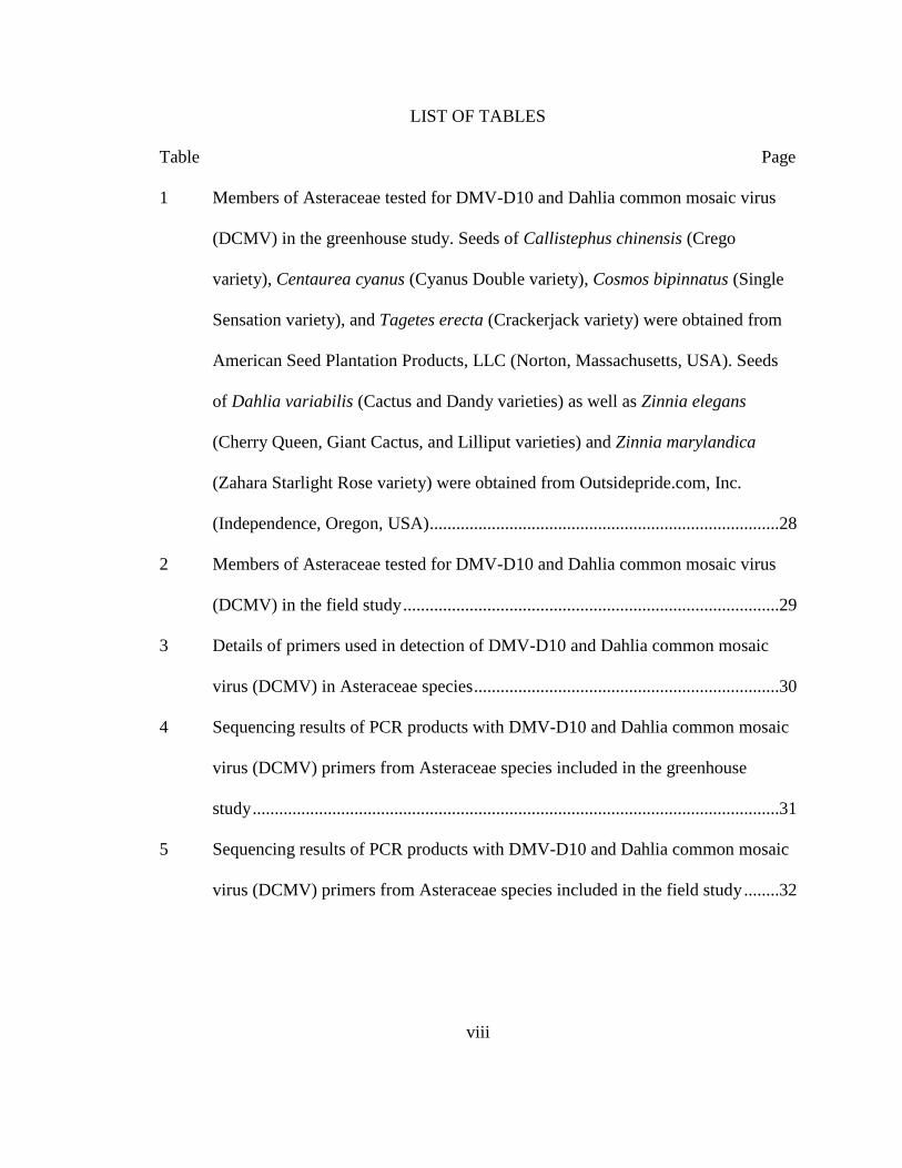
LIST OF TABLES
Table Page
1 Members of Asteraceae tested for DMV-D10 and Dahlia common mosaic virus
(DCMV) in the greenhouse study. Seeds of Callistephus chinensis (Crego
variety), Centaurea cyanus (Cyanus Double variety), Cosmos bipinnatus (Single
Sensation variety), and Tagetes erecta (Crackerjack variety) were obtained from
American Seed Plantation Products, LLC (Norton, Massachusetts, USA). Seeds
of Dahlia variabilis (Cactus and Dandy varieties) as well as Zinnia elegans
(Cherry Queen, Giant Cactus, and Lilliput varieties) and Zinnia marylandica
(Zahara Starlight Rose variety) were obtained from Outsidepride.com, Inc.
(Independence, Oregon, USA) ...............................................................................28
2 Members of Asteraceae tested for DMV-D10 and Dahlia common mosaic virus
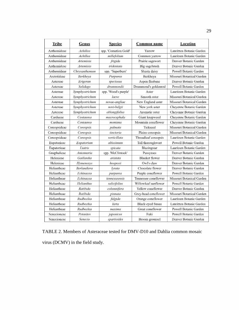
(DCMV) in the field study .....................................................................................29
3 Details of primers used in detection of DMV-D10 and Dahlia common mosaic
virus (DCMV) in Asteraceae species .....................................................................30
4 Sequencing results of PCR products with DMV-D10 and Dahlia common mosaic
virus (DCMV) primers from Asteraceae species included in the greenhouse
study .......................................................................................................................31
5 Sequencing results of PCR products with DMV-D10 and Dahlia common mosaic
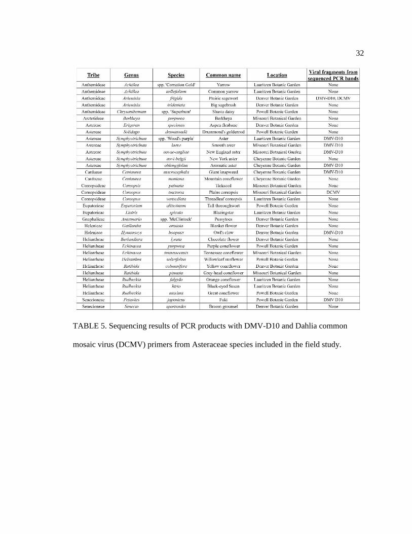
virus (DCMV) primers from Asteraceae species included in the field study ........32
ix
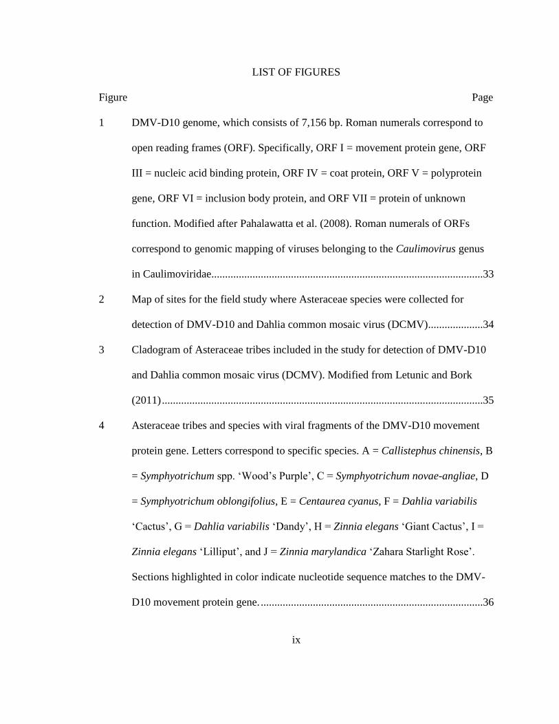
LIST OF FIGURES
Figure Page
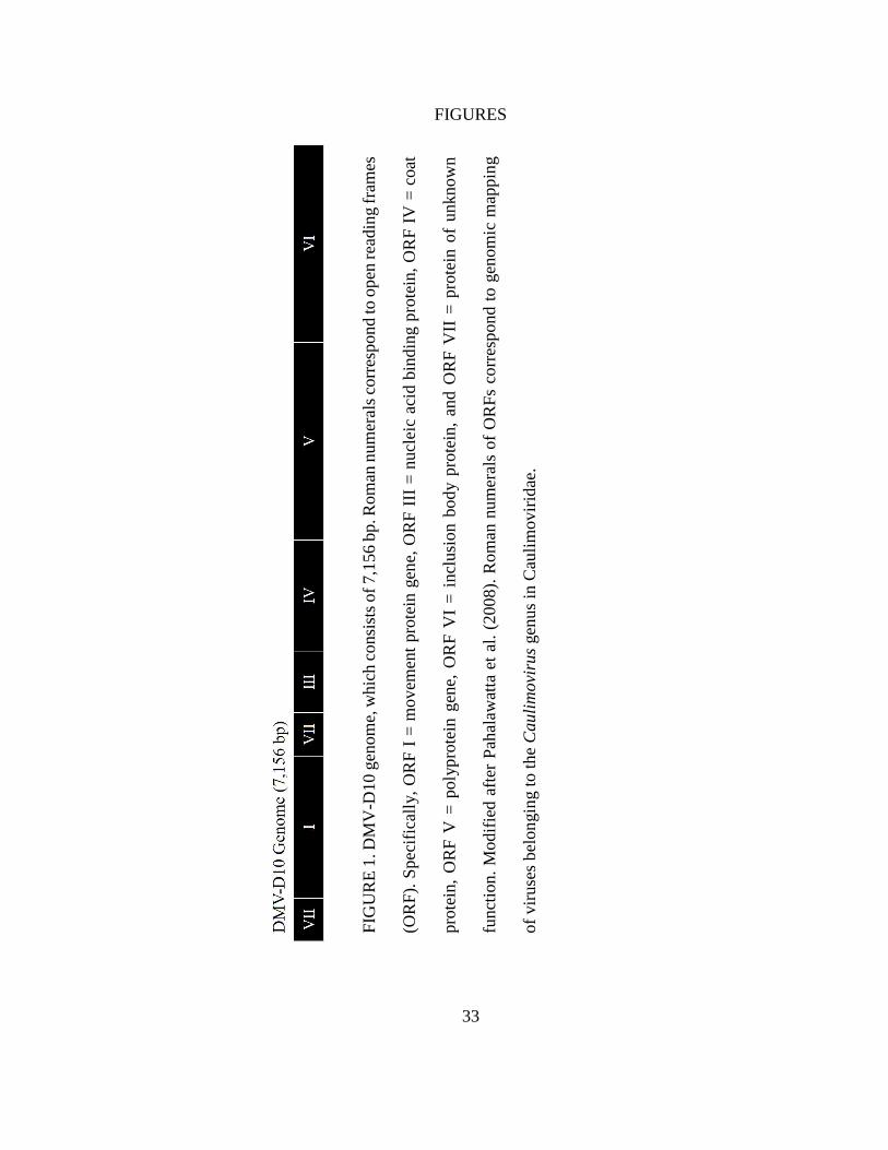
1 DMV-D10 genome, which consists of 7,156 bp. Roman numerals correspond to
open reading frames (ORF). Specifically, ORF I = movement protein gene, ORF
III = nucleic acid binding protein, ORF IV = coat protein, ORF V = polyprotein
gene, ORF VI = inclusion body protein, and ORF VII = protein of unknown
function. Modified after Pahalawatta et al. (2008). Roman numerals of ORFs
correspond to genomic mapping of viruses belonging to the Caulimovirus genus
in Caulimoviridae...................................................................................................33
2 Map of sites for the field study where Asteraceae species were collected for
detection of DMV-D10 and Dahlia common mosaic virus (DCMV)....................34
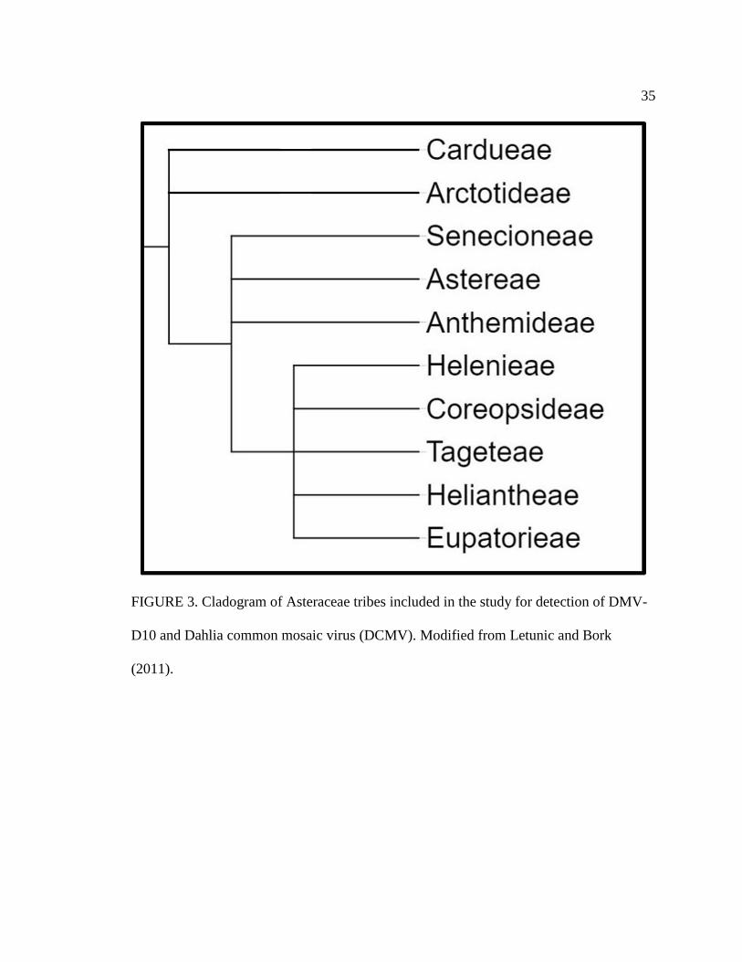
3 Cladogram of Asteraceae tribes included in the study for detection of DMV-D10
and Dahlia common mosaic virus (DCMV). Modified from Letunic and Bork
(2011) .....................................................................................................................35
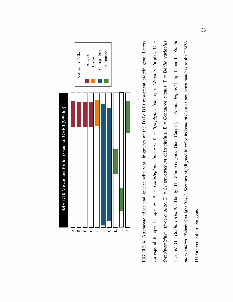
4 Asteraceae tribes and species with viral fragments of the DMV-D10 movement
protein gene. Letters correspond to specific species. A = Callistephus chinensis, B
= Symphyotrichum spp. ‘Wood’s Purple’, C = Symphyotrichum novae-angliae, D
= Symphyotrichum oblongifolius, E = Centaurea cyanus, F = Dahlia variabilis
‘Cactus’, G = Dahlia variabilis ‘Dandy’, H = Zinnia elegans ‘Giant Cactus’, I =
Zinnia elegans ‘Lilliput’, and J = Zinnia marylandica ‘Zahara Starlight Rose’.
Sections highlighted in color indicate nucleotide sequence matches to the DMV-
D10 movement protein gene. .................................................................................36
x
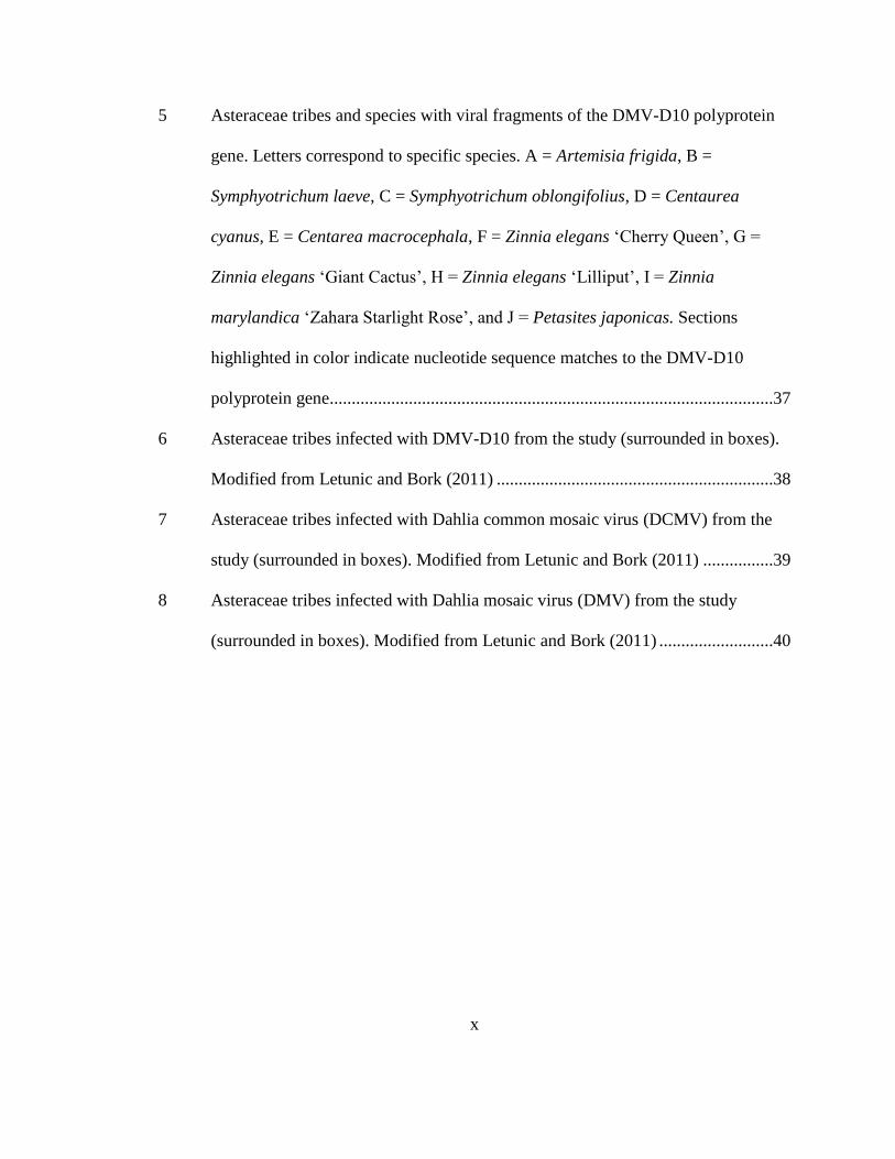
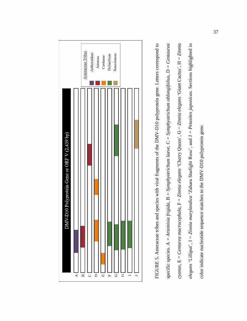
5 Asteraceae tribes and species with viral fragments of the DMV-D10 polyprotein
gene. Letters correspond to specific species. A = Artemisia frigida, B =
Symphyotrichum laeve, C = Symphyotrichum oblongifolius, D = Centaurea
cyanus, E = Centarea macrocephala, F = Zinnia elegans ‘Cherry Queen’, G =
Zinnia elegans ‘Giant Cactus’, H = Zinnia elegans ‘Lilliput’, I = Zinnia
marylandica ‘Zahara Starlight Rose’, and J = Petasites japonicas. Sections
highlighted in color indicate nucleotide sequence matches to the DMV-D10
polyprotein gene.....................................................................................................37
6 Asteraceae tribes infected with DMV-D10 from the study (surrounded in boxes).
Modified from Letunic and Bork (2011) ...............................................................38
7 Asteraceae tribes infected with Dahlia common mosaic virus (DCMV) from the
study (surrounded in boxes). Modified from Letunic and Bork (2011) ................39
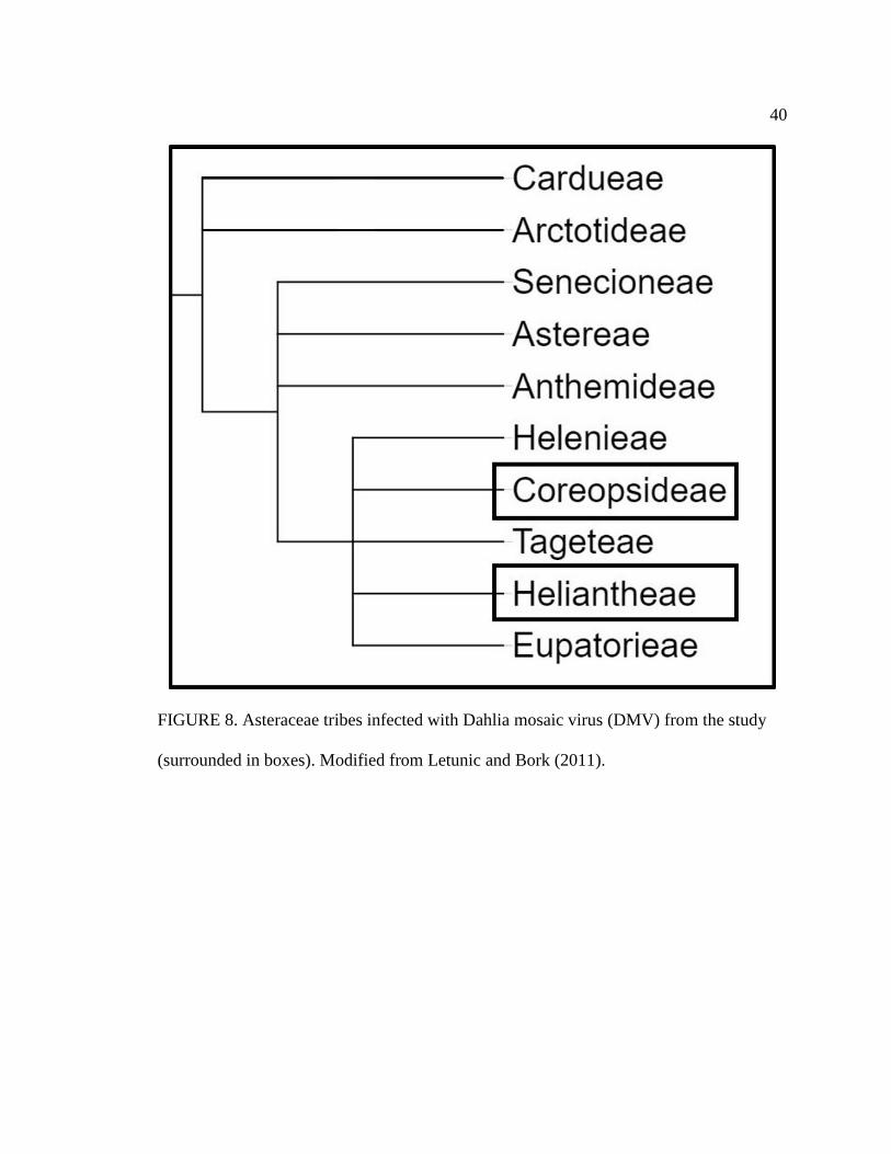
8 Asteraceae tribes infected with Dahlia mosaic virus (DMV) from the study
(surrounded in boxes). Modified from Letunic and Bork (2011) ..........................40
xi
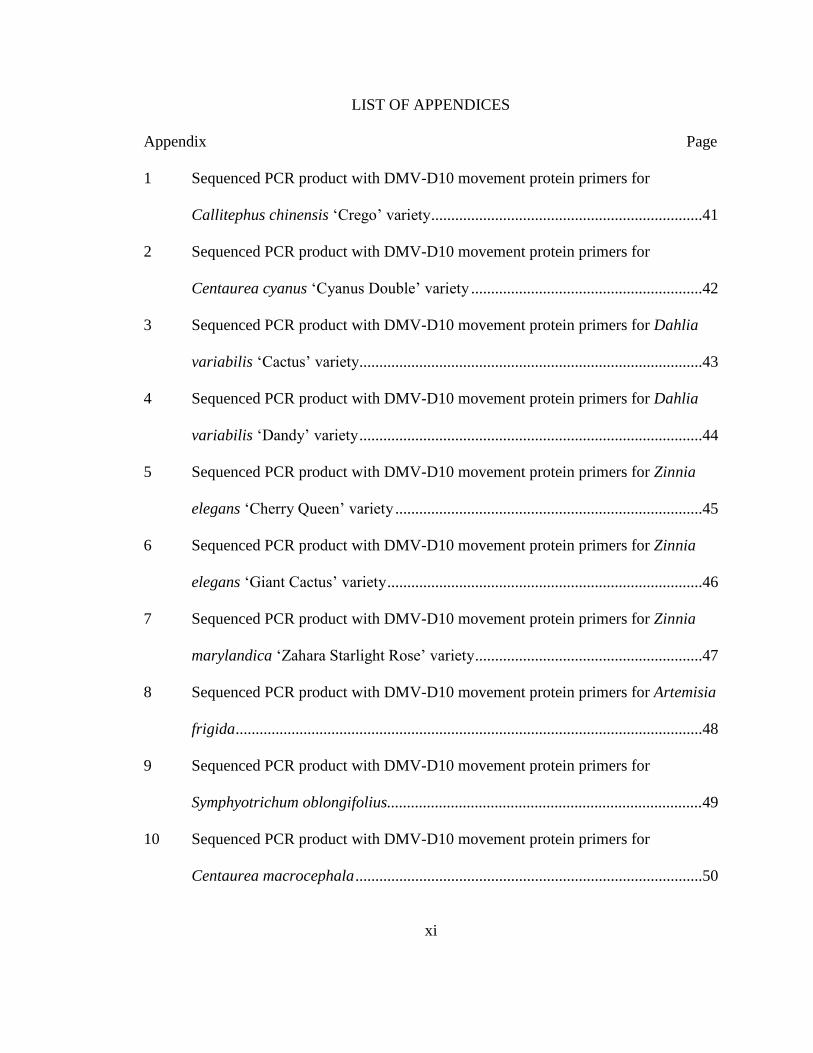
LIST OF APPENDICES
Appendix Page
1 Sequenced PCR product with DMV-D10 movement protein primers for
Callitephus chinensis ‘Crego’ variety ....................................................................41
2 Sequenced PCR product with DMV-D10 movement protein primers for
Centaurea cyanus ‘Cyanus Double’ variety ..........................................................42
3 Sequenced PCR product with DMV-D10 movement protein primers for Dahlia
variabilis ‘Cactus’ variety......................................................................................43
4 Sequenced PCR product with DMV-D10 movement protein primers for Dahlia
variabilis ‘Dandy’ variety ......................................................................................44
5 Sequenced PCR product with DMV-D10 movement protein primers for Zinnia
elegans ‘Cherry Queen’ variety .............................................................................45
6 Sequenced PCR product with DMV-D10 movement protein primers for Zinnia
elegans ‘Giant Cactus’ variety ...............................................................................46
7 Sequenced PCR product with DMV-D10 movement protein primers for Zinnia
marylandica ‘Zahara Starlight Rose’ variety .........................................................47
8 Sequenced PCR product with DMV-D10 movement protein primers for Artemisia
frigida .....................................................................................................................48
9 Sequenced PCR product with DMV-D10 movement protein primers for
Symphyotrichum oblongifolius...............................................................................49
10 Sequenced PCR product with DMV-D10 movement protein primers for
Centaurea macrocephala .......................................................................................50
xii
11 Sequenced PCR product with DMV-D10 movement protein primers for
Hymenoxys hoopesii...............................................................................................51
12 Sequenced PCR product with DMV-D10 movement protein primers for Petasites
japonicas ................................................................................................................52

xiii
PREFACE
This thesis follows the journal style for the American Journal of Botany.

1
INTRODUCTION
Evolution of plant detection and defense against pathogenic microorganisms is
one of the most notable developmental successes of modern plants (Chisholm et al.,
2006). The first terrestrial plants likely evolved under the presence of microbes several
hundred million years ago (Dangl et al., 2013); therefore, land plants have continually
been exposed to microorganisms throughout their evolutionary history. During this
period, there has been a continual arms race between pathogenic microbes and plants,
well explained by the concept of gene-for-gene resistance introduced in the early 1940’s
by Harold Henry Flor (Flor, 1942, 1947). However, even with progressive research in
plant evolutionary virology, new viruses are continually emerging (Hull, 2009).
Therefore, many areas of plant virology have not yet been explored.
For a microbe to become a pathogenic threat to a plant, the microorganism must
first breach the external barriers of the plant body (Chisholm, et al., 2006; Jones and
Dangl, 2006). Initial infection can be accomplished in several ways. Stomates, or
modified stomates known as hydathodes, have been suggested as a primary entry for viral
particles (Jones and Dangl, 2006). Pathogens and parasitic organisms can enter the
wounds of a plant (Esau, 1977), which takes on a similar method of penetration into the
plant body as in viral invasion of stomates. Insects and herbivores can also play a large
role in the plant-virus relationship (Stout et al., 2006). For example, aphids can introduce
viral particles directly into the plant vasculature via their stylet when feeding (Esau,
1961). Additionally, herbivores cause damage to the external plant body when eating,
which results in a wound and potential entry for pathogenic invasion (Stout et al., 2006).
2
After infecting the plant, a virus can spread rapidly throughout the plant body
(Curtis, 1935). Viral movement in a plant can be accomplished by cell-to-cell movement
and long-distance systemic transport (Esau, 1961). Plant viruses can move cell-to-cell
symplastically through plasmodesmata (Leisner and Howell, 1993). However, the size
and shape of plasmodesmata can present a challenge to some viruses to access other plant
cells (Wolf et al., 1989). Therefore, some successful invaders possess movement proteins,
which can either alter the size exclusion limit of plasmodesmata (Leisner and Howell,
1993) or induce the removal of desmotubules and replace them with viral tubules to
shuttle the virus directly to other cells (Hull, 2009). In the case of either strategy, these
movement proteins allow the virus to move throughout the plant body at a rate of microns
per hour (Dawson and Hilf, 1992). A plant’s immune system can respond to these
methods of infection by apoptosis, which hinders further viral infection by localized
death of virus-infected cells as well as surrounding healthy cells (Coll et al., 2011). It has
been suggested that relative susceptibility of plant species to particular viruses can be
linked to the interaction of viral movement proteins with plasmodesmata characteristics
of the host plant (Dawson and Hilf, 1992; Hull, 2009). Additionally, some viruses have
the ability to move directly through cell walls of the host plant (Chisholm et al., 2006).
Some plant viruses rely on transport by vasculature to be distributed throughout
the plant body (Hipper et al., 2013). This can include dispersal of viral particles via
xylem or phloem (Esau, 1961). In particular, phloem can serve as a form of systemic
transport for a virus to access all portions of the plant (Curtis, 1935). A virus can cross
several cellular barriers (e.g., bundle sheaths, vascular parenchyma, companion cells) into

3
the sieve elements of phloem (Hull, 2009). Therefore, a virus can be passively
transported with photoassimilates from one plant organ to another during source-to-sink
flow (Hipper et al., 2013). This dispersal method can be successful in transporting the
virus to areas of the plant to replicate and be transmitted to other hosts (Esau, 1961).
Once viral infection of the plant body is complete, a host plant may start to show
visible symptoms of infection (Lucy et al., 1996). Some symptoms largely depend on
whether the host was infected by a source-pathogen (i.e., pathogens which infect above-
ground biomass, such as leaves) or a sink-pathogen (i.e., pathogens which infect below-
ground biomass, such as roots) (Berger et al., 2007). For instance, infection of above-
ground biomass, such as leaves, can result in an increased demand for assimilates in the
plant, but also develop chlorotic or necrotic areas on leaves that decrease photosynthetic
assimilate production (Berger et al., 2007). As a result, photosynthesis decreases along
with a concomitant increased demand for assimilates, leads to source tissue being
transformed into sink tissue. However, if the virus is able to use long-distance transport to
infect the whole plant body, symptoms could vary throughout the plant (Hull, 2009).
Much emphasis in plant virology has been placed on the negative forms of plant-
viral relationships, mostly due to the large accumulation of data on plant viruses that
induce disease (Wren et al., 2006). Particular attention is given to viral symptoms in
cultivated plants, such as corn, that can negatively influence crop yield (Muthukumar et
al., 2009). However, there is importance in emphasizing mutualistic plant-viral
relationships. Plant viruses can be credited with giving plants the ability to tolerate
abiotic stresses (Hull, 2009). For instance, panic grass (Dichanthelium lanuginosum) has
4
the ability to grow in geothermal soils of Yellowstone National Park due to a three-way
mutualism between the plant, its associated fungal endophyte, and a thermal-tolerant
virus that infects the endophyte (Roossinck, 2011). Additionally, it has been hypothesized
that some plants, which harbor endogenous viral sequences, can be immune to other plant
pathogens (Roossinck, 2011). Given the high abundance and diversity of viruses, it is
possible many viral species can be attributed to mutualistic, commensal, or neutral
relationships with plants.
According to the International Committee for the Taxonomy of Viruses, known
plant viruses make up about 73 genera belonging to 49 families (ICTV, 2016). The viral
family Caulimoviridae is composed of eight genera (ICTV, 2016). Caulimoviridae is the
only family of plant viruses with double-stranded DNA genomes (Hull, 2009), with some
endogenous viruses in the family (Geering et al., 2010). Viruses belonging to
Caulimoviridae are plant pararetroviruses, which have a reverse transcription step during
viral replication (Stavolone et al., 2003; Abdel-Salam et al., 2010; Geering, 2014). Plant
pararetroviruses are similar to retroviruses in mammals, such as HIV. However, several
differences set them apart, such as circular double-stranded DNA in pararetroviruses
compared to linear single-stranded RNA in retroviruses (Hull, 2009). Caulimoviridae
viruses can be transmitted between plants in a variety of ways, such as aphid transmission
(Abdel-Salam et al., 2010). Once a plant is infected, a Caulimoviridae virus uses
movement proteins to replace the desmotubules of plasmodesmata with its own viral
tubules to move viral particles from cell to cell (Hull, 2009). Notable pathogens in the
Caulimoviridae family include Banana streak virus (Badnavirus), Cauliflower mosaic

5
virus (Caulimovirus), Petunia vein clearing virus (Petuvirus), as well as Dahlia mosaic
virus (Caulimovirus) (Pappu and Druffel, 2009; Abdel-Salam et al., 2010).
Dahlia mosaic virus (DMV) is a viral pathogen belonging to the genus
Caulimovirus in the family Caulimoviridae (Pappu and Druffel, 2009). Host plant
symptoms associated with DMV include vein clearing in the leaves, flower-breaking, and
stunted growth (Abdel-Salam et al., 2010). DMV is most commonly observed in
horticultural varieties of Dahlia variabilis (Pappu et al., 2005; Eid, et al., 2011), but
DMV can also occur in other members of the Asteraceae family, including Zinnia
(Kitajima and Lauritis, 1969). A newly discovered strain of this virus called DMV-D10
(Figure 1) was first observed in Dahlia, and is one of the few endogenous viruses to be
discovered in Caulimoviridae (Pahalawatta, et al., 2008). Endogenous viruses have the
ability to integrate their viral sequences into the host plant genome, which can be
inherited from parent to offspring (Geering et al., 2010). DMV-D10 is detected in every
part of the plant (e.g., leaves, stems, roots, flower petals, seeds, pollen), which has caused
additional concern about its method of transmission, especially with respect to clonal
propagation of Dahlia (Pahalawatta et al., 2008). Additionally, it has been suggested
pollen transmission could be a potential risk of infection in horticultural settings
(Pahalawatta et al., 2008). Currently, no studies have examined the full extent of DMV-
D10 host range and what effects, if any, infection may have on the host plant.
Given the limited availability of data concerning DMV-D10, the purpose of this
study was to determine its host range within Asteraceae. Since DMV-D10 and closely
related viruses have only been observed in Dahlia, an objective of this study was to

6
determine if there was a relationship among other infected plant species compared to
Dahlia within the Asteraceae family. It was hypothesized if a phylogenetic relationship
were present with respect to DMV-D10 infection, the virus would be observed in Dahlia
varieties, as well as in Asteraceae tribes more closely related to the Coreopsideae tribe,
which contains Dahlia. Furthermore, potential host plants were examined for presence or
absence of the DMV-D10 movement protein as well as Dahlia common mosaic virus.
The objective was not only to determine if host species were infected with DMV-D10,
but also to examine if plants may be infected with a closely related virus. This was tested
with a two-part study consisting of a greenhouse and field study encompassing 35 species
from 22 genera representing 10 Asteraceae tribes.
7
MATERIALS AND METHODS
Greenhouse plant material and growing conditions—
Ten varieties of seven species belonging to six genera within Asteraceae were
grown under greenhouse conditions for five weeks (Table 1). Seeds of Callistephus
chinensis (Crego variety), Centaurea cyanus (Cyanus Double variety), Cosmos
bipinnatus (Single Sensation variety), and Tagetes erecta (Crackerjack variety) were
obtained from American Seed Plantation Products, LLC (Norton, MA). Seeds of Dahlia
variabilis (Cactus and Dandy varieties) as well as Zinnia elegans (Cherry Queen, Giant
Cactus, and Lilliput varieties) and Zinnia marylandica (Zahara Starlight Rose variety)
were obtained from Outsidepride.com, Inc. (Independence, OR). All seeds were planted
in 11 x 11 x 12 centimeter pots, with five to six seeds per pot. Plants were allowed to
grow for five weeks from November to December 2015. The greenhouse received natural
lighting. Greenhouse relative humidity ranged from 17 to 68% during daytime and 26 to
75% during nighttime hours.
Field collection—
Five regional botanical gardens were selected for this study (Figure 2). Samples
from two to nine Asteraceae species mostly native to each region, as well as some
horticultural species, were selected at each site. Altogether, 28 species representing 10
Asteraceae tribes were collected (Table 2). Two to three leaves were collected from three
individuals of each species. Specifically, leaves were collected from individual plants that
were spread across each flowerbed to prevent collection of identical offspring from

8
maternal plants. Leaf samples were placed in labeled coin envelopes on site, and kept in a
cool location. Samples were then transported to Fort Hays State University (Hays, KS,
USA), and transferred to a drying oven to dry for 24-72 hours at 50 °C. Duration of the
drying process depended on individual factors of each sample, including thickness of
leaves and leaf moisture content. Following this, samples were preserved in a desiccator
until DNA extraction and analysis.
DNA isolation and extraction—
DNA was isolated from greenhouse and field samples with a Qiagen DNeasy
Plant Mini Kit (Catalog #69104; Hilden, Germany). With greenhouse samples, one pot
per variety was transferred from the greenhouse to the lab. Fresh tissue from three whole,
healthy plants (i.e., roots, stem, and leaves) was harvested from each variety. Lysis of
cells was accomplished by adding liquid nitrogen to the whole plant tissue in a mortar,
and tissue was ground thoroughly into a fine powder with a pestle. With field samples,
dried leaves from three individuals per species were ground into a powder with a mortar
and pestle. Protocol for isolating DNA from plant tissue followed the Quick-Start guide
included in the Qiagen kit. Following isolation, samples were frozen at -20 °C until
further analysis.
DNA quantification and analysis—
Polymerase chain reaction (PCR) was conducted to quantify DNA samples for
presence or absence of a DMV-D10 movement protein with an expected size of 900 base
pairs (bp). Primers and PCR cycling conditions followed Abdel-Salam et al. (2010)
(Table 3). PCR was conducted with a Phusion High-Fidelity PCR Kit (New England
9
Biolabs, Ipswich, Massachusetts, USA), and PTC-100 Programmable Thermal
Controller. DMV-D10 movement protein primers and PCR program followed Abdel-
Salam et al. (2011) (Table 3) Specifically, a program was designed to have a four minute
denaturation period at 94 °C, 20 second annealing period at 50 °C, and one minute
extension period at 72 °C for 50 cycles, followed by a seven minute extension period at
72 °C. Samples were then kept at 4 °C until electrophoresis.
Agarose gel electrophoresis was performed to separate DNA fragments from
samples following PCR. A 1% agarose gel solution was prepared with TAE (i.e., Tris
base, acetic acid, and Ethylenediaminetetraacetic acid) and SYBER Safe DNA Gel Stain
(Thermo Fisher Scientific, Waltham, Massachusetts, USA), as well as a 1 kb DNA
Ladder (Promega, Madison, Wisconsin, USA). After electrophoresis, the gel was
transferred to a Kodak Gel Logic 100 Imaging System to determine presence or absence
of bands at 900 bp for the DMV-D10 movement protein. Samples that indicated a
positive result at 900 bp as well as those samples that had strong bands amplified by
DMV-D10 movement protein primers that could be a related virus at another base pair
size were prepared for DNA sequencing.
For those samples that had a positive result using the DMV-D10 movement
protein primers, primers to detect Dahlia common mosaic virus (DCMV) were used to
aid in further analysis of infection. DCMV coat protein primers were used to determine
potential presence of DCMV in samples. Primers and PCR program for the DCMV coat
protein followed Eid et al. (2009) (Table 3). Specifically, a PCR program was designed to
have a four minute denaturation period at 94 °C, 20 second annealing period at 59 °C,

10
and 50 second extension period at 72 °C for 50 cycles, followed by a seven minute
extension period at 72 °C. A gel was run with PCR products and bands around 1,517 bp
were examined to indicate presence of DCMV. Samples that indicated a positive result at
1,517 bp as well as those samples that had strong bands that could be a related virus
amplified by DCMV coat protein primers at another base pair size were prepared for
DNA sequencing.
DNA Sequencing—
Once it was determined which samples could potentially possess DMV-D10 or an
associated virus based on electrophoresis results, PCR products with positive results were
cleaned with a Qiagen PCR clean-up kit (Hilden, Germany). Following this, 2 µL of each
sample was measured for DNA concentration with a NanoDrop 2000 Spectrophotometer
(Thermo Fisher Scientific, Waltham, Massachusetts, USA).
Samples for this study were sent to GENEWIZ (South Plainfield, New Jersey,
USA) for DNA sequencing. For sequencing, PCR samples were prepared at a template
concentration of 2 ng µL-1 x kb with a total volume of 10 µL. Custom primers were also
sent with samples. Primers were chosen according to the specifications of GENEWIZ,
including 18-24 bases in length, Tm between 50 and 60 °C, and G or C nucelotide at the
3ʹ end. The DMV-D10 movement protein reverse primer was chosen with a Tm of 59.4
°C and initial concentration of 100 µM. The DCMV reverse primer was chosen with a Tm
of 68.0 °C and initial concentration of 100 µM. The GENEWIZ Premixed option was
chosen for preparing samples before shipping to the facility for sequencing. This included
addition of the 5 µL of custom primer to the corresponding 10 µL of prepared sample.
11
DNA sequences from the resulting chromatograms were inspected and then
analyzed in the program NIH Nucleotide BLAST program to determine whether DMV-
D10 or other related viruses were present. Specifically, sequenced products from each
plant species was analyzed for presence of DMV-D10, DCMV, and Dahlia mosaic virus
(DMV) viral sequences using the alignment function through the NIH Nucleotide
BLAST program. Additionally, results were compared to determine whether there was a
correlation between the various viruses and members of the Asteraceae tribes (Figure 3).
12
RESULTS
There was no phylogenetic relationship among host plants infected with DMV-
D10 viral fragments; furthermore, DMV-D10 had a wider host range than expected
(Tables 4 and 5). Gel electrophoresis results with DMV-D10 specific primers suggested
DMV-D10 was present in samples belonging to tribes other than Coreopsideae.
Concerning the DMV-D10 movement protein, gel electrophoresis indicated positive
results in the Anthemideae, Astereae, Cardueae, Helenieae, Heliantheae, Senecioneae
tribes, as well as the Coreopsideae tribe. In contrast, use of primers to detect Dahlia
common mosaic virus (DCMV) yielded mixed results. Overall, this indicated DMV-D10,
a related virus (e.g., DCMV), or a different endogenous virus with similar sequences
enhanced by the primers used in this study could have been present in the samples.
Clarification of these observations came from DNA sequencing of PCR products
with the positive gel results (Tables 4 and 5), which confirmed fragments of the DMV-
D10 movement protein (Figure 4) and other pieces of the DMV-D10 genome (Figure 5)
were in species sampled from the Anthemideae, Astereae, Cardueae, Helenieae,
Heliantheae, Senecioneae tribes, as well as the Coreopsideae tribe. In particular, the
genus Symphotrichum, which belongs to the tribe Astereae, had fragments of DMV-D10
viral sequences from the movement protein gene and polyprotein gene, as well as other
viral fragments from the DMV-D10 complete genome. It was initially hypothesized that
Asteraceae tribes more closely related to Coreopsideae would be infected if DMV-D10
infection could be related to phylogeny of its host plants. However, DNA sequencing

13
results from this study suggest there may be a wider host range of DMV-D10, but with no
clear phylogenetic relationship of infected hosts (Figure 6).
Fragments of the DCMV and Dahlia mosaic virus (DMV) genomes matched
sequenced PCR products from species in several of the same tribes, but to an apparent
narrower host range than DMV-D10. For instance, DCMV was detected in species
representing the Anthemideae, Astereae, Cardueae, Coreopsideae, and Heliantheae tribes
(Figure 7) whereas DMV was only in species representing the Coreopsideae and
Heliantheae tribes of Asteraceae (Figure 8). As seen with results from potential DMV-
D10 infection, there was no obvious phylogenetic relationship between Asteraceae tribes
infected with DCMV or DMV.
14
DISCUSSION
Viral fragments of DMV-D10 were in thirteen out of thirty-five, or thirty-seven
percent, of plant species sampled in this study. Furthermore, DMV-D10 viral fragments
were widely distributed across Asteraceae species and tribes. Therefore, there was no
phylogenetic relationship relative to DMV-D10 infection of Asteraceae members. This
suggests either another mode of viral transmission or evidence of long-term coevolution
between DMV-D10 and members of Asteraceae. In addition, viral fragments of Dahlia
common mosaic virus (DCMV) and Dahlia mosaic virus (DMV) were in a few plant
species sampled in this study, but to a lesser extent compared to DMV-D10.
Past studies indicate DMV-D10 spreads via vertical transmission from parent to
offspring in Dahlia species (Pahalawatta et al., 2008). Therefore, the inconsistency of
viral infection with relation to phylogeny and indications of a potentially wider host
range of DMV-D10 in this study leads to speculations about how this virus could be
transmitted other than from parent to progeny. Furthermore, questions are raised, given
sequencing results indicated only fragments of DMV-D10 viral sequences were detected
in DNA of plant species. The exception to this was observed in Dahlia samples, which
contained longer and continuous sequences of the DMV-D10 viral genome compared to
other species in this study. There are a few possibilities on how fragments of DMV-D10,
DCMV, and DMV could have been transmitted to other tribes of Asteraceae, including
horticultural cultivation practices, pollen transmission, or an unknown insect vector.
15
Additionally, results from this research indicate the possibility of plant-virus coevolution
and evolutionary incorporation of viral sequences into Asteraceae species.
Horticultural cultivation practices—
Cultivation practices in horticultural and agricultural systems can be a source of
viral spread to other plant hosts (Hull, 2009; Sastry, 2013). In particular, vegetative
propagation of tubers, corms, bulbs, and cuttings, as well as grafting methods, can
transmit viruses (Hull, 2009). Several viruses that infect agricultural crops are spread
easily through vegetative propagation (Sastry, 2013). Studies indicate DMV can easily be
transmitted during Dahlia cultivation and propagation, whereas DMV-D10 also can be
spread by seed (Eid and Pappu, 2013). Dahlia variabilis varieties in this study were
infected with DMV, DCMV, and DMV-D10. This could be due to propagation
techniques in horticultural systems (Pappu et al., 2005), since varieties of Dahlia
variabilis in this study were grown from seed. Since it has been documented in previous
research, cultivation practices are a strong possibility for how DMV, DCMV, and DMV-
D10 were transmitted to Dahlia variabilis in this study. However, cultivation practices do
not explain how other species in this study became infected, but there are other possible
transmission methods.
Unknown insect vector—
Most plant viruses are not able to enter their host directly without the assistance of
vectors or other methods of infection (Power, 2000). For instance, many plant viruses
rely on insects as vectors (Power, 2000). Given that fragments of DMV-D10 viral
sequences were detected in host genomes of several Asteraceae species with no
16
phylogenetic relationship, this could be evidence of possible insect transmission. DMV-
D10 has been documented in every part of the plant (Pahalawatta et al., 2008). Therefore,
it is possible that an insect feeding on one plant infected with DMV-D10 could transmit
to an uninfected plant. In particular, insects with sucking mouthparts that feed on phloem
sap could inadvertently carry plant materials containing DMV-D10 viral sequences from
an infected plant to an uninfected plant by mechanical introduction (Esau, 1961).
Approximately 90% of plant pathogens are spread solely by insects with sucking
mouthparts (Power, 1987). For instance, many viruses can be transmitted by aphids
during feeding (Esau, 1961), but DMV-D10 is known to lack an aphid transmission
factor (Pahlawatta et al., 2008). Given the seemingly wide distribution of DMV-D10 viral
sequences in several species, it is possible DMV-D10 can be mechanically introduced to
other species of plants by an insect vector that has yet to be discovered. However, given
DMV-D10 lacks an aphid transmission factor, this seems less likely (Pahalawatta et al.,
2008).
It has been suggested that mode of insect transmission is a stable evolutionary
trait when comparing viral genera (Nault, 1997). In other words, there is great specificity
which insect vectors are able to transmit particular viruses (Power, 2000). DMV, a
relative of DMV-D10, is transmitted by several species of aphids, allowing DMV to have
a wider range of infection than the Dahlia host genus (Pappu et al., 2005). For this
reason, it is understandable why some species in this study were infected with DMV. All
varieties of Zinnia elegans and Zinnia marylandica, as well as varieties of Dahlia
variabilis, were infected with DMV. These results are consistent with previous studies
17
that have shown DMV to infect species in the genera Zinnia (Kitajima and Lauritis, 1969;
Hull, 2009) and Dahlia (Eid et al., 2009). Therefore, it is possible DMV could have been
transmitted by aphids between these two species.
DCMV is another distinct virus belonging to Caulimovirus, but related to DMV
and DMV-D10 (Almeyda et al., 2015). Much like DMV-D10, the host range of DCMV
or possible modes of transmission are not well studied. A mixed infection of DMV-D10,
DMV, and DCMV in Dahlia is common (Eid et al., 2009), and reflects results of this
study. However, Artemisia frigida, Callistephus chinensis, Centaurea cyanus, Coreopsis
tinctoria, and Zinnia elegans also had fragments of DCMV in their DNA. Due to the lack
of information about DCMV, it is possible this virus does have an aphid transmission
factor, such as that of DMV, or there is an unknown insect factor transmitting DCMV to
other species of plant hosts. Since several species of virus belonging to Caulimovirus are
spread by insects with sucking mouthparts (Nault, 1997): either of these hypotheses are
possible explanations of viral transmission.
Pollen transmission—
Pollination can serve as transportation for viruses to infect plants (Card et al.,
2007). Viruses contained in pollen can either infect the embryo and, thus, the seedling
that grows from the seed or can infect the maternal plant through the fertilized flower
(Hull, 2009). Pollen transmission of viruses has been shown to occur between plants of
different species, where the pollen tube from one species germinates through the stigma
and penetrates the style tissue of another plant species to transmit the virus to the
maternal tissue (Isogai et al., 2014). DMV-D10 has been detected in all parts of Dahlia,

18
including the pollen grains (Pahalawatta et al., 2008). Therefore, it is possible DMV-D10
could be transmitted to other plant species during pollination by transmitting directly to
the maternal tissue.
Furthermore, it is possible the floral anatomy of the family Asteraceae encourages
fertilization by different pollen donors. The head inflorescence of Asteraceae is made of
several hundred individual flowers that mature at different times over a period of days
and can be pollinated by pollen from different species in Asteraceae during this time. It is
hypothesized this characteristic gave rise to the great diversity of the family (Barreda et
al., 2015). Therefore, it is possible a combination of the reproductive biology of
Asteraceae and DMV-D10’s ability to infect every part of a plant shaped the genetic
makeup of Asteraceae. Although, even if pollen may contain viral particles, it does not
necessarily mean the virus is pollen transmitted; for instance, Tobacco mosaic virus is
contained in pollen, but not pollen transmitted (Card et al., 2007). Therefore, further
research is needed to determine if DMV-D10 is a virus that can be pollen transmitted and
may help to understand the potential host range of this virus.
Plant-virus coevolution—
Perhaps one of the more perplexing questions regarding DMV-D10 is what
evolutionary events were involved in integrating these viral sequences into the plant
genome. It is possible the integration of this virus into the plant genome was a process of
coevolution and can be aged based on distribution in related species (Geering et al, 2010).
For instance, one study suggested the integration events of Badnavirus (Caulimoviridae)
into the plant genome occurred more than 4.6 million years ago when two species of
19
Musa were derived from a common ancestor (Duroy et al., 2016). Similarly, results of
this study hint a wider distribution of DMV-D10, which may be evidence of long-term
coevolution between DMV-D10 and members of Asteraceae.
Even though there was no phylogenetic relationship between DMV-D10 viral
infection and Asteraceae tribes in this study, it is possible that particular species harbored
viral sequences due to an evolutionary advantage. For example, studies suggest the
exchange and integration of genetic information between cyanophages (i.e., viruses that
infect cyanobacteria) and their hosts have led to higher photosynthetic efficiency of
cyanobacteria (Sullivan et al., 2006). Specifically, cyanobacteria appear to have obtained
genes from cyanophages that code for components of photosynthetic proteins used in
photosystem II (Sullivan et al., 2006). Considering cyanobacteria are the smallest and
most numerous photosynthetic cells in marine systems (Sullivan et al., 2006), the
coevolution of this particular virus and host was an important event. Similarly, the
evolutionary benefit of possessing DMV-D10 viral sequences in particular Asteraceae
species may be due to a significant evolutionary advantage. Conversely, it is possible
presence of DMV-D10 viral sequences in members of Asteraceae is a neutral
relationship.
Evolutionary incorporation of viral sequences—
As with any biological entity, genetic variation gives rise to viral diversity
(Garcia-Arenal et al., 2001). Specifically, mutation, recombination, and reassortment are
the variants that natural selection acts upon for evolution (Roossinck, 1997). Viral studies
suggest natural selection favors plant viruses that have a wider host range and possess the
20
ability to use several different vectors for transmission (Roossinck, 1997). The
heterogeneity of viral populations due to genetic variation allows mechanisms of
evolution to shape the specificity of plant-viral relationships (Garcia-Arenal et al., 2001).
Furthermore, evidence has supported the claim by scientists that viruses have played a
larger role in shaping the evolution of biological organisms than previously thought
(Hendrix, et al., 2000).
Longer and continuous sequences of DMV-D10, DMV, and DCMV were
localized in both varieties of Dahlia variabilis. However, shorter fragments of these
viruses were detected in other species of Asteraceae. Given the possible modes of viral
transmission (e.g., pollen transmission, unknown insect vector, long-term coevolution), it
is possible to hypothesize further that integration of DMV-D10 viral fragments, as
observed in many species of this study, could be treated as potentially new viruses.
DMV-D10, as with any endogenous virus, has the ability to integrate its viral sequences
into the host genome (Eid and Pappu, 2013). Studies have shown viral fragments of other
Caulimoviridae endogenous viruses became either rearranged or decayed when integrated
into genomes of differing plant species (Geering et al., 2010). Therefore, it is possible
that during transmission to other species, only fragments of DMV-D10 were compatible
and integrated differently into the associated host genome of other plant species.
Additionally, because studies have suggested DMV-D10 does not induce any physical
symptoms of disease, integration of particular DMV-D10 viral fragments could have
been considered advantageous to the host plant (e.g., the mutualistic relationship between
cyanophages and cyanobacteria). It is possible mutation and recombination also

21
attributed to how these pieces of DMV-D10 viral sequences were integrated into the host
genome, which could have influenced the evolution of Asteraceae.
Conclusions—
Many of the transmission strategies mentioned previously, including pollen
transmission, an unknown insect vector, or coevolution between DMV-D10 and
Asteraceae members, could be single or a combination of possibilities for how DMV-
D10 was able to infect the thirteen species of Asteraceae in this study. Given these
possibilities for viral transmission and previous knowledge of DMV-D10, this study
suggests pollen transmission and plant-virus coevolution are perhaps the most plausible
ways in which DMV-D10 is transmitted to host plant species outside the Dahlia genus.
However, further research is needed to determine how DMV-D10 is spread to Asteraceae
members. Studying this could provide us with a better understanding of the biology of
this virus in relation to their host plants. Furthermore, given these results indicate only
fragments of DMV-D10 (and DCMV and DMV) were present in some species other than
Dahlia, more support is given to the idea that viral fragments observed in several plant
species in this study are evidence of long-term coevolution between an ancestral virus
and members of Asteraceae.

22
LITERATURE CITED
Abdel-Salam, A.M., M.M. Al Khazindar, S.G. Eld, and H.R. Pappu. 2010. Caulimoviral
sequences in Dahlia variabilis in Egypt. African Journal of Biotechnology 9: 6835-
6839.
Almeyda, C.V., G. Raikby, and H.R. Pappu. 2015. Characterization and comparative
analysis of promoters from three plant pararetroviruses associated with Dahlia
(Dahlia variabilis). Virus Genes 51: 96-104.
Barreda, V., L. Palazzesi, M.C. Telleria, E.B. Olivero, J.I. Raine, and F. Forest. 2015. Early
evolution of the angiosperm clade Asteraceae in the Cretaceous of Antarctica.
Proceedings of the National Academy of Sciences 112: 10989-10994.
Bent, A.F. 1996. Plant disease resistance genes: function meets structure. Plant Cell 8:
1757-1771.
Berger, S., A.K. Sinha, and T. Roitsch. 2007. Plant physiology meets phytopathology: plant
primary metabolism and plant-pathogen interactions. Journal of Experimental
Botany 58: 4019-4026.
Card, S.D., M.N. Pearson, and G.R.G. Clover. 2007. Plant pathogens transmitted by pollen.
Australasian Plant Pathology 36: 455-461.
Chisholm, S.T., G. Coaker, B. Day, and B.J. Staskawicz. 2006. Host-microbe interactions:
shaping the evolution of the plant immune response. Cell 124: 803-814.
Coll, N.S., P. Epple, and J.L. Dangl. 2011. Programmed cell death in the plant immune
system. Cell Death and Differentiation 18: 1247-1256.

23
Curtis, O.F. 1935. The Translocation of Solutes in Plants. McGraw-Hill Book Company,
Inc., New York, New York, USA.
Dangl, J.L., D.M. Horvath, and B.J. Staskawicz. 2013. Pivoting the plant immune system
from dissection to deployment. Science 341: 746-751.
Dawson, W.O. and M.E. Hilf. 1992. Host-range determinants of plant viruses. Annual
Review of Plant Physiology and Plant Molecular Biology 43: 527-555.
Duroy, P., X. Perrier, N. Laboureau, J. Jacquemoud-Collet, and M. Iskra-Caruana. 2016.
How endogenous plant pararetroviruses shed light on Musa evolution. Annals of
Botany 117: 625-641.
Eid, S., K.L. Druffel, D.E. Saar, and H.R. Pappu. 2009. Incidence of multiple and distinct
species of Caulimoviruses in Dahlia (Dahlia variabilis). HortScience 44: 1498-
1500.
Eid, S., and H.R. Pappu. 2013. Expression of endogenous para-retroviral genes and
molecular analysis of the integration events in its plant host Dahlia variabilis. Virus
Genes 47: 998-1008.
Eid, S., D.E. Saar, K.L. Druffel, and H.R. Pappu. 2011. Plant pararetroviral sequences in
wild Dahlia species in their natural habitats in Mexican mountain ranges. Plant
Pathology 60: 378-383.
Esau, K. 1961. Plants, Viruses, and Insects. Harvard University Press, Cambridge,
Massachusetts, USA.
Esau, K. 1977. Anatomy of Seed Plants. 2nd edition. John and Wiley, Inc, San Francisco,
California, USA.

24
Flor, H.H. 1942. Inheritance of pathogenicity in Melampsora lini. Phytopathology 32: 653-
669.
Flor, H.H. 1947. Inheritance of reaction to rust in flax. Journal of Agricultural Research
74: 241-262.
Garcia-Arenal, F., A. Fraile, and J.M. Malpica. 2001. Variability and genetic structure of
plant virus populations. Annual Review of Phytopathology 39: 157-186.
Geering, A.D.W., T. Scharaschkin, and P. Teycheney. 2010. The classification and
nomenclature of endogenous viruses of the family Caulimoviridae. Archives of
Virology 155: 123-131.
Geering, A.D.W. 2014. Caulimoviridae (plant pararetroviruses). eLA.
Hammond-Kosack, K.E. and J.D.G. Jones. 1997. Plant disease resistance genes. Annual
Review of Plant Physiology and Plant Molecular Biology 48: 575-607.
Hendrix, R.W., J.G. Lawrence, G.F. Hatfull, and S. Casjens. 2010. The origins and ongoing
evolution of viruses. Trends in Microbiology 8: 504-507.
Hipper, C., V. Brault, V. Ziegler-Graff, and F. Revers. 2013. Viral and cellular factors
involved in phloem transport of plant viruses. Frontiers in Plant Science 4: 1-24.
Hull, R. 2009. Comparative Plant Virology. 2nd edition. Elsevier Academic Press,
Burlington, Massachusetts, USA.
International Committee on Taxonomy of Viruses (ICTV). 2016. Website
http://www.ictvonline.org.
Isogai, M., T. Yoshida, C. Nakanowatari, and N. Yoshikawa. 2014. Penetration of pollen
tubes with accumulated Raspberry bushy dwarf virus into stigmas is involved in
25
initial infection of maternal tissue and horizontal transmission. Virology 452-453:
247-253.
Jones, J.D.G. and J.L. Dangl. 2006. The plant immune system. Nature 444: 323-329.
Kitajima, E.W. and J.A. Lauritis. 1969. Plant virions in plasmodesmata. Virology 37: 681-
685.
Leisner, S.M. and S.H. Howell. 1993. Long-distance movement of viruses in plants. Trends
in Microbiology 1: 314-317.
Letunic, I. and P. Bork. 2011. Interactive Tree of Life v2: Online annotation and display
of phylogenetic trees made easy. Nucleic Acids Research 39: 475-478.
Lucy, A.P., M.I. Boulton, J.W. Davies, and A.J. Maule. 1996. Tissue specificity of Zea
mays infection by maize streak virus. Molecular Plant-Microbe Interactions 9: 22-
31.
Melotto, M., W. Underwood, J. Koczan, K. Nomura, and S. Yang He. 2006. Plant stomata
function in innate immunity against bacterial invasion. Cell 126: 969-980.
Mukhtar, M.S. A.R. Carvunis, M. Dreze, P. Epple, J. Steinbrenner, J. Moore, M. Tasan, et
al. 2011. Independently evolved virulence effectors converge onto hubs in a plant
immune system network. Science 333: 596-601.
Muthukumar, V., U. Melcher, M. Pierce, G.B. Wiley, B.A. Roe, M.W. Palmer, V. Thapa,
et al. 2009. Non-cultivated plants of the Tallgrass Prairie Preserve of northeastern
Oklahoma frequently contain virus-like sequences in particulate fractions. Virus
Research 141: 169-173.
26
Nault, L.R. 1997. Anthropod transmission of plant viruses: a new synthesis. Annals of the
Entomological Society of America 87: 521-541.
Pahalawatta, V., K. Druffel, and H. Pappu. 2008. A new and distinct species in the genus
Caulimovirus exists as an endogenous plant pararetroviral sequence in its host,
Dahlia variabilis. Virology 376: 253-257.
Pappu, H.R., S.D. Wyatt, and K.L. Druffel. 2005. Dahlia mosaic virus: molecular detection
and distribution in Dahlia in the United States. HortScience 40: 697-699.
Pappu, H.R. and K.L. Druffel. 2009. Use of conserved genomic regions and degenerate
primers in a PCR-based assay for the detection of members of the genus
Caulimovirus. Journal of Virological Methods 157: 102-104.
Power, A.G. 2000. Insect transmission of plant viruses: a constraint on virus variability.
Current Opinion in Plant Biology 3: 336-340.
Power, A.G. 1987. Plant community diversity, herbivore movement, and an insect-
transmitted disease of maize. Ecology 68: 1658-1669.
Roossinck, M.J. 1997. Mechanisms of plant virus evolution. Annual Review of
Phyotpathology 35: 191-209.
Roossinck, M.J. 2011. The good viruses: viral mutualistic symbioses. Nature Reviews:
Microbiology 9: 99-108.
Sastry, K. 2013. Seed-Borne Plant Virus Diseases: Plant virus transmission through
vegetative propagules (asexual reproduction). Springer, USA.
27
Stavolone, L., A. Ragozzino, and T. Hohn. 2003. Characterization of Cestrum yellow leaf
curling virus: a new member of the family Caulimoviridae. Journal of General
Virology 84: 3459-3464.
Stout, M.J., J.S. Thaler, and B.P.H.J. Thomma. 2006. Plant-mediated interactions between
pathogenic microorganisms and herbivorous arthropods. Annual Review in
Entomology 51: 663:689.
Sullivan, M.B., D. Lindell, J.A. Lee, L.R. Thompson, J.P. Bielawski, and S.W. Chisholm.
2006. Prevalence and evolution of core photosystem II genes in marine
cyanobacterial viruses and their hosts. PLoS Biology 4: 1344-1357.
Wolf, S., W.J. Lucas, C.M. Deom, and R.N. Beachy. 1989. Movement protein of tobacco
mosaic virus modified plasmodesmatal size exclusion limit. Science 246: 377-379.
Wren, J.D., M.J. Roossinck, R.S. Nelson, K. Scheets, M.W. Palmer, and U. Melcher. 2006.
Plant virus biodiversity and ecology. PLoS Biology 4: e80.

28
TABLES
TABLE 1. Members of Asteraceae tested for DMV-D10 and Dahlia common mosaic
virus (DCMV) in the greenhouse study. Seeds of Callistephus chinensis (Crego variety),
Centaurea cyanus (Cyanus Double variety), Cosmos bipinnatus (Single Sensation
variety), and Tagetes erecta (Crackerjack variety) were obtained from American Seed
Plantation Products, LLC (Norton, Massachusetts, USA). Seeds of Dahlia variabilis
(Cactus and Dandy varieties) as well as Zinnia elegans (Cherry Queen, Giant Cactus, and
Lilliput varieties) and Zinnia marylandica (Zahara Starlight Rose variety) were obtained
from Outsidepride.com, Inc. (Independence, Oregon, USA).
29
TABLE 2. Members of Asteraceae tested for DMV-D10 and Dahlia common mosaic
virus (DCMV) in the field study.

30
TA
BL
E 3
. Det
ails
of
pri
mer
s use
d in d
etec
tion o
f D
MV
-D10 a
nd D
ahli
a co
mm
on m
osa
ic v
irus
(DC
MV
) in
Ast
erac
eae
spec
ies.

31
TABLE 4. Sequencing results of PCR products with DMV-D10 and Dahlia common
mosaic virus (DCMV) primers from Asteraceae species included in the greenhouse study.
32
TABLE 5. Sequencing results of PCR products with DMV-D10 and Dahlia common
mosaic virus (DCMV) primers from Asteraceae species included in the field study.
33
FIGURES
FIG
UR
E 1
. D
MV
-D10
gen
om
e, w
hic
h c
onsi
sts
of
7,1
56 b
p. R
om
an n
um
eral
s co
rres
po
nd to
op
en r
ead
ing f
ram
es
(OR
F).
Spec
ific
ally
, O
RF
I =
mov
emen
t pro
tein
gen
e, O
RF
III
= n
ucl
eic
acid
bin
din
g p
rote
in,
OR
F I
V =
coat
pro
tein
, O
RF
V =
poly
pro
tein
gen
e, O
RF
VI
= i
ncl
usi
on b
od
y p
rote
in,
and O
RF
VII
= p
rote
in o
f unknow
n
funct
ion.
Modif
ied a
fter
Pah
alaw
atta
et
al.
(2008).
Rom
an n
um
eral
s of
OR
Fs
corr
espond
to g
enom
ic m
appin
g
of
vir
use
s bel
on
gin
g t
o t
he
Cauli
movi
rus
gen
us
in C
auli
movir
idae
.

34
FIGURE 2. Map of sites for the field study where Asteraceae species were collected for
detection of DMV-D10 and Dahlia common mosaic virus (DCMV).
35
FIGURE 3. Cladogram of Asteraceae tribes included in the study for detection of DMV-
D10 and Dahlia common mosaic virus (DCMV). Modified from Letunic and Bork
(2011).
36
FIG
UR
E 4.
Ast
erac
eae
trib
es an
d sp
ecie
s w
ith vir
al fr
agm
ents
of
the
DM
V-D
10 m
ovem
ent
pro
tein
gen
e. L
ette
rs
corr
espond
to
spec
ific
sp
ecie
s.
A
=
Call
iste
phus
chin
ensi
s,
B
=
Sym
phyo
tric
hum
sp
p.
‘Wood’s
P
urp
le’,
C
=
Sym
phyo
tric
hum
no
vae-
angli
ae,
D
=
Sym
ph
yotr
ichum
oblo
ngif
oli
us,
E
= C
enta
ure
a cy
anus,
F
=
D
ahli
a va
riab
ilis
‘Cac
tus’
, G
= D
ahli
a v
ari
abil
is ‘
Dan
dy’,
H =
Zin
nia
ele
gans
‘Gia
nt
Cac
tus’
, I
= Z
innia
ele
gans
‘Lil
liput’
, an
d J
= Z
innia
mary
landic
a ‘
Zah
ara
Sta
rlig
ht
Rose
’. S
ecti
ons
hig
hli
ghte
d i
n c
olo
r in
dic
ate
nucl
eoti
de
sequen
ce m
atch
es t
o t
he
DM
V-
D10 m
ovem
ent
pro
tein
gen
e.
37
FIG
UR
E 5
. A
ster
acea
e tr
ibes
and s
pec
ies
wit
h v
iral
fra
gm
ents
of
the
DM
V-D
10 p
oly
pro
tein
gen
e. L
ette
rs c
orr
espond t
o
spec
ific
spec
ies.
A =
Art
emis
ia f
rigid
a,
B =
Sym
phyo
tric
hum
laev
e, C
= S
ymphyo
tric
hum
oblo
ngif
oli
us,
D =
Cen
taure
a
cyanus,
E =
Cen
tare
a m
acr
oce
phala
, F
= Z
innia
ele
gans
‘Cher
ry Q
uee
n’,
G =
Zin
nia
ele
gans
‘Gia
nt
Cac
tus’
, H
= Z
innia
eleg
ans
‘Lil
liput’
, I
= Z
innia
mary
landic
a ‘
Zah
ara
Sta
rlig
ht
Rose
’, a
nd J
= P
etasi
tes
japonic
as.
Sec
tions
hig
hli
ghte
d i
n
colo
r in
dic
ate
nucl
eoti
de
sequen
ce m
atch
es t
o t
he
DM
V-D
10 p
oly
pro
tein
gen
e.

38
FIGURE 6. Asteraceae tribes infected with DMV-D10 from the study (surrounded in
boxes). Modified from Letunic and Bork (2011).

39
FIGURE 7. Asteraceae tribes infected with Dahlia common mosaic virus (DCMV) from
the study (surrounded in boxes). Modified from Letunic and Bork (2011).
40
FIGURE 8. Asteraceae tribes infected with Dahlia mosaic virus (DMV) from the study
(surrounded in boxes). Modified from Letunic and Bork (2011).
41
APPENDICES
NNNNNNNNNNNNTNNTNNNTNNTACTNNNTNNNNNNNGAGAGNNCATGTTG
GCGCTTTTGATTCCCACCCCCAGGTGAAANNGGNCTTACNTTGAAAATGCTG
ACCATGTTCAANGACTTGNATGATTTGACTTATAACAATTTGAGTNNNGTNA
AATCAGTACAAGTTACTGTTTCCCTTNNGGANGATGATGAAGACTACAATAC
AGACGTCACCGANNACGCCGATGTGGATCCCGATGATGACGAGTCTGATGAG
GGTAATGATGAACCAGTTCAANGAATCATGAACAATAATATCAAGATCCATG
AACAAGGCTCGNAGTTGNATGANGNNNCCATTTATGGCAAANATTTTTTTCT
ANTGCATATTTGCCTTTNTGNGNATNTTGNNACNANTNTTGTTGTCATTTGTT
GCCNTTTCCCCNTGNNATCTNGGCGNANNNACCATGAANACGNAGGTTGCAT
CTTGCTTTGANTTGGGAANTGATCACAACTTTTCTGATATGATGAATGTCTNC
AATTCATACCTANCCACCTTAATCATCTTCANATCTAAATACCTTTTTGTCACT
CCCGGATAAGTACAATATTAGGATTTATGCGCCTCTTACAGTAGGATATTCTT
AACTTCACTCCGGATAGGTACCCAGATTCTACCATTTCNAANCAAAATATCTC
GAGTATCCTTGGTGANNCTTTAACGTGNCCTTTTATCCTCTCTTTCTTCTCATC
ATTCTGGANAGCTACTCACTGANCTTCACAAATTTTATCAAAGNATTCGGTAG
AGACCACCATGGCTAACGATTTCACGATAAGAGGATAATGATGAACAAGTTC
NAN
APPENDIX 1. Sequenced PCR product with DMV-D10 movement protein primers for
Callitephus chinensis ‘Crego’ variety.

42
NNNNNNNNNNNNNNNNNANNNNNNNNNNTCAGTGNNNCNNGGNTTGNTGA
TGGTGAGATGATGNNNAACATGTTCTANTTGCCCNAGANAACNGGTGGGTGC
AGTTGGTGTTATGCAAAACTTGATTACTGGTTCAATTGGATAATGATGACCAA
ATTCAAAANTGNTTCTGTATTGCCCACAAAACCCCTTGAAATCTTGGCTTAAA
TTCAGCAAACCCCACNAGTNTCCCCGATTTNGCNTTCGTGATANGNCAATTA
AAAGTCNNGNTCCCNCTNAGAGTGGGTTAATCTCTAACAAAATCACGAGGAC
AATCGAGCGGCAATNNATGCTTATCAACCGATAATGANGAACAATTTCAANG
ATGGAAGATTCTAAGGTAATAAAGAGGCGTATTTTGAGCAGGGTACCGATCG
AAAGCCGGAAAAACCTCATCAAGAATAGTACGGACAACGAGTTCTATGAGGT
GGTGGAGCACACCATGCACAAAGATGATAATTTTAAGAGAAAGGTGACCGTC
ATTGAAGATCTACNTGAAATGACACGTANGGTTCTGCATTACTACAAAGGGG
ATTTCTTGGATGAACTACCCGGACTTGTGGACTTCTCGGTCTTTCTCAACCTC
NGCCCCAAGCAAAAACACGAGGTTTCAGAATTGAGAAAGTTATCAAAGAAA
TTCAAGATCAGTTCTGATGGAAGTGCAATTTATGTGCACCCATGGCTAANGTC
CCTCACAAAGAATACGGCTTCTANAGACAAAAACGATGACAACAGTAACAA
GATTGATGAGCTGCTCGAGAANCTGGATGANAGGNATGGAGTGAAAGCCAA
GTTTTTTCTGAATATGCTTCGGCTATGTGAATCTGGAGGAGAAAGGCTTTTAG
TCTTTNNNCAGTATCTGTTACCCCTAAANTTCCTGATGANATTGGCGATGAAA
GTTNNAANGNNNNNTCCNACNAGGANATTTTTATGANAACAGGAGATCATTG
ATATGATGAACNAGTTCAAA
APPENDIX 2. Sequenced PCR product with DMV-D10 movement protein primers for
Centaurea cyanus ‘Cyanus Double’ variety.
43
TAATATGGTTGCGTGCGGTAGTATCTTCGATCGGTAAACCGCTGAAGTAATA
AACTTCTAGTGAAACAAACAAAGGATTTGTGTCAGACTACATGATTAATTCT
GATTATTTAGAAAAAATCATGAAGCTTAAGCTAAAGCTTGATACAAAACAGG
TTTTTAATCAACCTAGTAATTTACAGAGATTAGTTTCAAAAGCTTTCTCTAGA
AAAAATAATATCTTTTATTGCTTTAATACTGAAGAATTGTCAGTAGATATAAA
AGATACTACAGGTGAAGTGTATTTACCACTTCTAACAAAAGGAGAAATAGCC
CGAAGACTTCTGACTATTAAACCAGAATTAAGAAAAACCATGAATATGGTGC
ACATCGGAGCAGTAAAAATCCTTCTGAAGGCACAGTTCAGAGATGGAATTAA
CTTCCCGATAAAAATGGCTTTAGTTGATAACAGAACTATCAACAGGCAAGAC
GCTCTACTCGGAGCAGTTCAAGGAAATTTAGCATACGGTAAATTTATGTTTAC
TGTTTATCCTAAATTTGCATTACATCGAGATTCAAAAGATTTCGATAAAACCT
TAAGTTTCATACATCAGTGCGAAAGGACTGACCTCATGGGAACCAGGTAACA
AAGTATTTACGATTAATTATTTAATTTCGTATGCTTTGACAAATAGTACTCATT
CAATTGAGTATAAAGAAAAGGAGAGTATAACACTTGATGATGTATTCTCAGG
AATAGGTACTGTCGAAAGAAGCAAGTTCGCTGAACCCTTCTCAGATACAGGA
AAATTGGCGATTGACTATTGCTCGAGAGAAAACAACTCTAGGATTTCAACCC
TAGACATAGTTTTACAGGATCCTTTACAAATAGGGCGAGTCCAGTAGAAACA
CAGANANCAGA
APPENDIX 3. Sequenced PCR product with DMV-D10 movement protein primers for
Dahlia variabilis ‘Cactus’ variety.
44
NNACATCGCTGACGTGCGGTACATCTTCGATCAGGAATCTCACTGAAGTGAT
AAACTTTCAGTGAAACATGACAAAGGCATTTGTATCAGACTACATGATTAAT
TCTGATTATTTAGAAAAAATCATGAAGCTTAAGCTAAAGCTTGATACAAAAC
AGGTTTTTAATCAACCTAGTAATTTACAGAGATTAGTTTCAAAAACTTTCTCT
AGAAAAAATAATATCTTTTATTGCTTTAATACTGAAGAATTGTCAGTAGATAT
AAAAGATACTACAGGTGAAGTGTATTTACCACTTCTAACAAAAGGAGAAATA
GCCCGAAGACTTCTGACTGTTAAACCTGAATTAAGAAAAACCATGAATATGG
TGCACATCGGAGCAGTAAAAATCCTTCTGAAGGCACAGTTCAGAGATGGAAT
TAACTTCCCGATAAAAATGGCTTTAGTTGATAACAGAATTATCAACAGGCAA
GATGCTCTACTCGGAGCAGTTCAAGGAAATTTAGCATACGGTAAATTTATGTT
TACTGTTTATCCTAAATTTGCATTACATCGAGATTCAAAAGATTTCGATAAAA
CCTTAAGTTTCATACATCAGTGCGAAAGGACTGACCTCATGGAACCAGGTAA
CAAAGTATTTACGATTAATTATTTAATTTCGTATGCTTTGACAAATAGTACTC
ATTCAATTGAGTATAAAGAAAAAGAGAATATAACACTTGATGATGTATTCTC
AGAAATAGGTAACTGTCGAAGGAAGCAAGTTCGCTGAACCTTCTCAGATACA
GGAAAATTGGGCGATTGACATTGCTCGAGAAAAACAAAACTCTAGGATTTCA
ACCTAGAAAATAGTTTTACAGGAATCCCTTTACAAAATAAGGTGACTCCAGT
AGAAACACAGGAAAAAAACAGA
APPENDIX 4. Sequenced PCR product with DMV-D10 movement protein primers for
Dahlia variabilis ‘Dandy’ variety.

45
NNNNNNNNNNNNNNANNNNNNGNGCTGTTTTTCTGTGTTTCTACTGGCTGTT
TTTCTGTGTTTCTACTGGCTGTTTTTCTGTGTTTCTACTGGCTGTTTTTCTGTGT
TTCTACTGGCTGTTTTTCTGTGTTTCTACTGGCTGTTTTTCTGTGTTTCTACTGG
CTGTTTTTCTGTGTTTCAACTGCATAAAAACAGCCAGAAAAAAAACACAAAA
ACAGCCAGTAAAAACACAAAAAAACAGCCAGTAAAAAAACAGAAAAACAG
CCAGTAAAAAAACAAAAAAACAGCCAGTAAAAAAACAAAAAAACAGCCAGT
AAAAAAACAAAAAAACAGCCAGTAAAAAAACAAAAAAACAGCCAGTAAAA
AAACAAAAAAACAGCCAGTAAAAAAACAAAAAAACAGCCAGTAAAAACAC
AAAAAAACAGCCAGTAAAAAAACAAAAAAACAGCCAGTAAAAAAACAAAA
AAACAGACAGTAAAAAAACAAAAAAAAAGCCCGTAAAAACACAAAAAAAC
AGCCAGTAAAAACACANAAAAACNGCCAAAAAAAAAACAAAAAAACCGCC
AAAANAAACACAAAAAAACAGCCNGTAAAAACACAGAAAAACCNCCCNTAA
AAAAACAGAAAAACCGCCNGTAAAAANACANNANAACCGCCCTTTNAAANA
CNNNAAAACCCGCCNNTAAAAAAACCAGAAAAAACNNNNNNTNNAAACCCN
CNNAN
APPENDIX 5. Sequenced PCR product with DMV-D10 movement protein primers for
Zinnia elegans ‘Cherry Queen’ variety.

46
GNANNNNNNNNNNNNNNNNNTGNNCNNNNGATNNAGNNGCTTGTANCTTGG
GTGNNNCGTGTNATGCAGTAACTACGATTGNCGTCCGCAGCATCGCAGTAGT
GTCTTAGATTATTCGTGAGAGTGTGGATGCCGCACGGGGGAGCTAGGTTCCC
CTGAATCGTTCATAACAGTNATGTCCTTTTGCGGTGCTTTTGNNNTTCNTACG
CTTCTGAATTTTGNTGCCTTTTCTGACGCTCGCGTACCCCCTCCTTGATNGCTA
ATGTCATTGGACTCCCAGCCTTTTCNACATACCGACAAACAGAGACACAATT
GCANTCCCTATCTCGTCCNACACGCACTACTCAAAATCGACCCTCACGGCCCC
AATACGTCTACCAACATCNTCTACACCAAGCNNATGTGGTATTATGGTCCTAC
CAATCACGGTTCACGAAGCCATAAGAAACATCGNAAAACTGACGGCTACAA
CTNCTAACACAAGCCAACAACCCGAACCTCGCGAACAANCCGCACACCAACC
AGTCCAAACACANACAAACAGAATCAGACCATGAACAACGNACAACAGGAA
CAATATACGTTGCGGCCCAGTCATCGCATCATATTTATCATTATAAACACAGA
ANANCAGANAAAAAGCACCCCCCTTTTTTTTTCTTTGGNGAAACCATTAAAA
ATTTTTTTTTTAGGNAGTGAAGATNCATGGNNCNGCGACCCCCNNGGNCCCC
CCCNNGNAAANNANTTTNTTTTTGGNAACNNNNNNNACCNNCACCCCANATA
ANNAAAACNTTGNNNNNNTGGGCNCNGGGNTGGCTCCAGCCNTAANTNTTCC
NNGCNNNGGNANTNTNNNNTNTTTTTCCNNAAAAAATCGGATTTTNNNNNNA
NNTTTTANANNAAAATTTNGCCAATNNNNGNAGNGNCTTNNCNAANCNNCTN
NNGNGATNNNNCCCNNAAAGGGGAATTTTGTNNCAANANNNGNGCCCCNCA
CCCCCNNNNNNGGAGNGNGNNNNNAAGTNNANTATNNNTTANNCCNNGNN
NNNATTNNGNGNANNNNNAAAAAATTGNGGGGGNNNNNNCTN
APPENDIX 6. Sequenced PCR product with DMV-D10 movement protein primers for
Zinnia elegans ‘Giant Cactus’ variety.

47
NNNNNNNNNNNNNNNANNNNNGGCTGTTTTTCTGTGTTTNCTACTGGCTGTT
TTTCTGTGTTTATACTGGCTGTTTTTCTGTGTTTCNACTGGCTGTTTTTCTGTGT
TTCTACTGGCTGTTTTTCTGTGTTTCNACTGGCTGCTTNTCNGTGNGGNCACG
ATCNTTTTATGCNGGNCNNNNCNNNNCACNNTNNNAAAANNNGAGTTTGCN
ATNNNGATGTCCTCTACTGCGCCGGGNNNNTNCAAANTNTGTACCNGNNNGG
CNTAGNNTGAGNAANTTNNCATTCCAANCCTTNTTAGAGNNANANNCNCTAN
ACAATNNNNNCAANCGAGAAGGAAAATNAAGGTGNGTCGNGATANNCNTNA
TAGGGAAGACNCANTATGGANNANTNNACTANAATAAGGGCNTGAANNGNA
AANTNCCAANCANNCGTNNANNNACCTNGTCCGGNATTNNCTTTGAAAATGT
GNANATTCCNNTNATNTNTNGAATCTTGNANNNTACTGTATCNTTCNAANAC
ANNAAATCTGNNNNNCTCCATANGTAATCGNNNCTNNTAAANATACGNNGC
GGNNCAGTATNNATCNGNTNANNTTTTGAATCTNGAAANACAGANTATGGTA
TTCCNTTTTTCNNTTTTTGATTCTTATTTTNTNNTTTTNTTATTTTNTTATTTNN
TNNTTTTATTTCAAATTTNTCTTATNTAATTTATTTTTTTTCATTNNTATCTTNA
CTCTTTTTTNATTTTTTCTNNNTTTTTTTTTNTTTTTTNCTNNNNTTCTNCTTTTT
CCTTNTNNATTTTNTANAACTCNNTNTTTTCTNTTTANTTATTTTCATCTCTCTT
TGNTNNTATCATCNTTGTGGTCAGAATCTCTTAATGTCTATTGTTTTAANTGA
NCNCCCTCNNNTNTCTNTGTGTNCTCCNTTTTTTNTTNTNNTNTNNNNTNTNN
TCTNANTTTNTNTCTNNNNNNTNCTNNNNTNN
APPENDIX 7. Sequenced PCR product with DMV-D10 movement protein primers for
Zinnia marylandica ‘Zahara Starlight Rose’ variety.
48
GNNNNNNNNNNNNNNNNNNNNNANGGTNCCNANTGCNNNGTNNNNNANNA
TGNNNGCACCTGGTGAGTCTCCTCTTTGATTNGAATACNTGTNTTGTTCACCT
ACTTGTATATCTTACCTCCTTTTGCTNTGAACTGCCTTAAANTGCTTCACTNNT
CACTCTTTTGNATGAACCGGATGATGACACAGGTGTTAATGTTTCGACCACTA
GAACTGATGAAGGACCCTTNNNCCCCTCNCCNTNTGCTTCTNAAGAGAATGA
CCCTATNACTCTTACCTTCCTGTATGACTTACGTCTCACGTACTCACGGGCCG
TTGATGGTCTCTNGAAAGACCTTGCGCATACAAAAGTCTCTGTTTCAACTAAN
ATTGTCCAACTGCCGGGGAACGTTCAGGAGTTCCAAACNCANCTGGGACANA
GGTAATNNGNNGTTAGTGATTGTCNCCTATGACCACCAATTGNTCTGGCAAC
TGGGCCCCCTTGGTGTTCTGGTTGAGGTACCCCTGNNNACNNANGTNATTTCT
CCTATGATCNNGATGCTGACCCNTNATGGAAATCGAGATGACCCACGTTCCA
ACGAAGAAGAGAAGATCTTATCTGGACAAGGATGGGGCTGCTCCTTCGCATG
ATCATGCTCATGATGATGCTCATGATGAGGAGATGGACTGTGATTAGGAGTT
TCATGNNCNTCTTGANNNTNNTTGCGGANGNNNTTCNCATGANACGCANNNN
AANNANGTNNANGACCCTCACCCNGANCNNCTGAAGGATTNCCCTCANAGN
ACNCANNANANNAGGANNNGAGANGNNNGNNGATGGAANTACTNNNNNGG
GTNNNTNNNTNNTACCAANNTGGGNTGNGACGAGGAANTAANNNCGTTGTT
ATTCCNTCGANNNNAAAANAAAANAAGNGATCNGTGGNTCNCTTATCGTTNN
GNCAAGCCTCTCTGCTCNNCANCACCTNNTCNTCACCANATNNGNNNNNTCN
TCNGATANNANANNTNNNTNNNNTTGGANNAAGAAGGNNNNNGTNTNNCCT
TCNNNANATGNNNNNNTCNNNNNANGGNNNNN
APPENDIX 8. Sequenced PCR product with DMV-D10 movement protein primers for
Artemisia frigida.
49
NNNNNNNNNNNNNNNNNNNNNGCTGTTTTTCTGTGTTTCTACTGGCTGTTTTT
CTGTGTTTCTACTGGCTGCTTTTCTGTGTTTCNNCTGGCTGTTTTTCTGTGTTTC
TACTGGCCTTTCTTCNNACAGAAAAACTTTTAGANCCCTCAGGATTCCCTTT
CTTTTTTNATATGACTTTCGCCTCCCGTTCTTACTTCNNATTCGTCCNGCTCCA
TCGCTCCCCTGGNTTGCAGTACTATCCGTTCNAAAGTCTCTTGNCGTCCTTTTA
NTTCTTAAATCATTGAGAAAACCANGTTCTAAGTTTCATCTAACCATGAATG
TAATATAAGGCAGATTANTCAGAANAACATTCTTCTGTGGGTTATTTATCCCT
TTAATAAANCTTTGTANATTNNAAATTTCTTTGTATTATNAAATATGATAATG
TGTTGAAATGCTTTTNNTATTCCANTGTTCANNTNAATTGTNAGGTGGACTGA
ACNACACTAGANAAATGTATAGTATCAATTGTAANTGTAATGACNAATGNCA
AATAGTNTAATGTACNATGNTTAATTAGTATTCACTATTTCTGCACAGTAGAA
ACACCGACAAACAGANCCAAATCAATTAACCCATCTATTTTTTTGAATTTGAA
CCTACTTCTCANCCTAGGTGACTTTGGTAGCCTAATGAGTGGTGGCTTCTGAG
TTGGTTGTGATGATTGGTTTCTTTTTGGTCTAGGAGTAAAAACACAGAAAAAA
CAGAATTCCTTTGCNNNACNNNGGGAATATCGGTCGGATATAATTAAAAACA
ATTTANTGTAATATAATCCACTTTTTTGGNAATGTANTTGNAAAANTGGNTGN
NAAANGNNACTNAAACTNCNNGTGAANCNAAATTTTGTGGANGNTGNNANT
GAAANTNNNNGTTTNNCTNCTACTTTACCNGACACNGTNNTGGNAGCGTATN
APPENDIX 9. Sequenced PCR product with DMV-D10 movement protein primers for
Symphyotrichum oblongifolius.
50
NNNNNNNNNNNNNNNNNNNNANNNNNGATGNTCGTCTCANGATTGNGGAAA
TCTCATTTTCACCATTATAACCTGCCNCTCCATGGCATATATNACGAGTATTG
AGAAGCTTATGCATTGGGCTAGAGGAAACTAGGAAAGTCCCTGATTGCTCAA
ATTCNCAAGTNAAATTTCTGAAAAAGAANATCATGCTTCNNGGNNNTTTTGA
NNGCAGATNNTNAGGTGCTGATTTGTTCAGAAGATGCCCATAAAAGATAATN
TGATTGGCTTGCAANANATTCTCAGCAAATTTGACCACAGAAAAACNGANGG
CTTGCACAGCCCCTTAAATAGGATCTAACGGATGCACCTTAGTCAATTTATAC
NGTATNNCAAACTAGAAACTTGGATTCTNTTGAATATGAGTTTCTTGTTGATC
TGCGTTAGAAACAGAAATCAAAAGACAAAATGTGGCCTCTCAAGAAGACAG
AGTATGCCACGATGATGATTACTAANCTTATCNATGAATCTGATGCACTTTTT
GAGCCGGTTATTCAACCACCAAAAGGAAGACCGCCNAAACCAAAAAAGAAA
AGAGGAATAACTTCGTCAAGAAGAGACCCATCGAGGTTTGAGCATGTAGAAT
CATCACAAACACAAAACTCGTCAACATCTACTTGTGTCGAAAGAAACAGTGG
AACAACTAATGAATTTAGTTATATCTTTCATGACAACAATTCACTTGATTTAA
ATCTGTACTCAGATTTTTCAAGTGATTATATGTTGTTAGAGTAATAAGGTAGC
CTTGACTCTTGAACAATGAATTTTTGTATATTCNACAGGCACTTTTTGGAAAC
TTGTACGCTTTTAGCTTTGGATCGTATGTTTTGTACTCGTGTCCTAATCAAGTA
TAGATATANGCCTGTTGTGTGCTTTTGAAAGCTTGTTTTATGTAATAGCTTTTG
TTAATGTATTGAAACGGATGCATGTATGCACTTGAATTTTTATTTTATGGGNA
TTATTCNGCTAGTAATTNGACTGTAGNNNNANGTGTNANTTTTANTNANAAA
GAAAAATTGAANTATTANANNTAACNNNNTTTNNTNGNNGTTGNTTNNNANN
N
APPENDIX 10. Sequenced PCR product with DMV-D10 movement protein primers for
Centaurea macrocephala.
51
NNNNNNNNNNNNNNNNNNNNNNNNGNGNGNNCGTTCCTGCATNNGGNGGTC
TNAATCNTGACAGGTGGTTCACTTGACCTNCTCTTAAGTGTGCGATTCANTAT
CCACAGACTCGACTGATAGTGAAATNTATGAACTTTTCTAATGACCTGGATG
AAACTTNTGATTGCAGGAAAAATGTGGCTGAACTGCAGNAGATTACTCGCGN
GANNGTCTATCTNTGNNAATCTNAGAAATGAANATTTTTCAATTTANTTCTTT
CANCATATCTCTCNAATTTNNTGGNGANATTTTTTCNTNCAATAACCGCCNTT
NCATGGTTATTCTAAATTTTCAAATACTACNNCTGAGCTTCGGAGATTTTGAC
TGAAAGAGTGGGANANTGGAATATTCCCACCANACTCAAGTGTAAACCGCAT
TCTTGGATAAACCCAAGGAACTAAGTCCTTCTATTTCACTTCCTCAATCTATN
GNTGATAATGATGAGCACCCTTGGGAGGAAGNANTGTGTCTTGNNTGNNNAG
NNNNNNCCNCCATCTACANGNATTATCATCNTCGTATCGAAAGATGTTTGAG
ATGATNCTCCCANNAAACACACAAAAAAACNGAGAACANGAACNNCNTACA
NNNGNTNCAAAAANNTGNACNNNTGTCNNNNCGNAANNNATTANAACCGTA
ATGCTANCGNNNNCACNNNNNNNNCTNCCACCNANATCNCTTTGATGATGCA
TCGCAACTTANNAANNTNNGTANCGTTNNNCCTGTNTNNNACTCTCTGAANT
ANNNCAATNNNGTCCNNAAGTTCGNTNTTNNNNGAAAAACTCTNNAAANCCT
TCATAGAGTATCCTGAAANAGGTGATCNNAACGTCTNNNNCGCNGNNCNNN
NANCAAANGCNNTTNNNNCNNANNTANNGNNNNGANNTNNNCNANCCCNCC
NATGGCNTNGCG
APPENDIX 11. Sequenced PCR product with DMV-D10 movement protein primers for
Hymenoxys hoopesii.
52
NNNNNNNNNNNNNNNNNNCNTGNTACNNNCTTCTGATGATTTCTTTAGATAC
TTATACTGACTTATAAAGATACCAGTTGGCTTTTGCTTGACTTGTAACCCAAG
AAAGAATGTCAATTCTCCCATTGAACTCATTTTGAACTTTAAATGCATTAAAG
CTTCCAACTCTCGACATAACTCCTCTCTTGGAGAACCAAAAATGATATCATCT
ACATGTATTTGAACCAGTAGAATCTCANTCTAANCNANAAAGATTNTAAAGT
CTTATTAATTGCTCCTCTTTTGAANTTGTTTTCCAGTNGATTNCGNAAAANNG
NANTATCCTTGATCTCNTAAATNANCAAGGCTTTGTTATCTTCAAGGACAATA
AAAAAGGCCCTTTCAAAAATTTTAACTTCCGCCTGANTTTTCAAAAATTCATT
NGGAGGCTTTTGGGGGAAAAAAATNATGGCCTCCAAACTTTNNGNACCTTTT
TGTGTTTANTTTTTAANNNTCCCTGAAACCCACCAAATTAACAAAACCAGTAT
TTTTTGACTTTTTTCNACTAATGCCCTGATATANCCCAAACTTAATCCAACAC
ACGAATTTTTTATGCATCCTCGTTGGTTTTCAACATGAAAACTTAAATTTCCA
GATCCAGAGCCNATGGNNCTGATACCAGTTGTGATCANCNGATANGGNTCGA
TGANTGNNTCGCCATGAGNNACTCATCAGTANAAACTGCTGCGGAGCNNTAC
NCNNNTTGNNCANNGTTTANCTGTTAAACNNNNNGNANNNANGNAAANNGG
NNNANGANGGGNNNTTGTTTTTCACGCNCCNCCNTCAAGCTGAAATATTGAT
NAATCNNAGNATGTAACAATNTTCANAANGTCCTATTATTTNNTCCTTGNGG
CTAACAANCCTNGTTTTCCANNNNAAAAGATTAANATGATNACNNNNTCANA
CTATCTTAATNNGNNCCCTNNNANCNNNGAATGNGGTACTACCATATTTACA
NGAACNGTANNGNNNAATTTCNCNANNANTNNNTGAN
APPENDIX 12. Sequenced PCR product with DMV-D10 movement protein primers for
Petasites japonicus.