photothermal and photochemical uses of the fel for biomedical
TRANSCRIPT

• Biomedical research and treatments require selective interactions at the cellular and molecular levels. FEL is a unique tool to achieve these. For example --
• Selective photothermal targeting– Lipid-rich tissues– nanoparticles
• Multiphoton excitation for – microscopy – photodynamic therapy
Photothermal and Photochemical Uses of the FEL for Biomedical Problems
R. Rox Anderson MD, DirectorWellman Center for Photomedicine
Harvard Medical School

Blood vessels Pigmented Cells
Tattoos Hair removal
Fat and Acne (two problems of fatty tissue)?
“Selective Photothermolysis”RR Anderson, JA Parrish Science 220:524-527 (1983)
Selective Absorption WavelengthThermal Confinement Pulse duration
~ 1 million treatments / month for skin, eye, larynx, GI diseases

**

Selective light absorptionvery local heating
epidermis
Blood vesseldermis

Selective light absorptionvery local heating
epidermis
Blood vesseldermis

Selective light absorptionvery local heatingselective repair
epidermis
Blood vesseldermis

Subcutaneous Fat
Sebaceous glandStemcells
Epidermis
1-4 mm
Three fatty tissue targets:• Sebaceous Glands – cause of acne• Subcutaneous fat – liposuction is #1 plastic surgery in the US• Atherosclerotic plaque – lipid-rich plaques kill 40% of the
US population by stroke or heart attacks.

ΔT: Temperature riseE: Energy density
μa: Absorptionρ: Density c: Heat capacity
ΔT = (Eμa/ρc)
ρfat = 0.85 g cm-3
ρdermis= 1.08 g cm-3
cfat = 2.3 J g-1 K-1
cdermis= 3.5 J g-1 K-1
Because of lower ρc, fat should be prone to photothermal heating.
Photo-thermal Excitation

0 .1
1
10
10 0
Hum an FatW ater
915
*1205
Absorp tion coefficientsα[cm -1]
λ[nm]
1715
2305
*
*
Fat and Water have nice “colors” in the NIR

Ideally, ratio of photothermalheating for fat vs. water
Ratio of the temperature rises
01
23
45
850 1000 1150 1300 1450 1600 1750 1900 2050 2200 2350
Wavelength, nm
Subc
utan
eous
fat /
de
rmis

Subcutaneous Fat
Sebaceous glandStemcells
Epidermis
1-4 mm
Cold Window
CH-selective LaserCan we ‘target’ sebaceous glands and fat?

Selective Fatty Tissue TargetingMonte Carlo Simulations :Sebaceous gland (depth = 2.5 mm, radius = 1.0 mmn=1.45, μa=0.17 /mm, μs’= 0.58 /mm) below epidermis (n=1.4,μa=0.039 /mm, μs’= 0.79 /mm) and capillary layers (n=1.37,
μa=0.04 /mm, μs’= 0.3 /mm) within 3.8 mm thick dermis (n=1.4, μa=0.035 /mm, μs’= 0.2 /mm) irradiated by focused beam (λ = 1200 nm r= 2 mm, focusing depth = 3.5 mm )
Subcutaneous Fat
Dermis
Blood
Epidermis
Sebaceous Gland

3-5 μm thermal camera ΔT
Sample
JLab FEL
Energymeter E

3-5 μm thermal camera ΔT
SampleCold window
JLab FEL
Energymeter E

Photothermal excitation spectrum taken with J-Lab FELshows “where” to build a fat-seeking laser
0
0.5
1
1.5
2
2.5
3
1670 1680 1690 1700 1720
Wavelength
ΔΤ
/Ε fatdermis

Nanoscale Particle Targeting
Thermal Confinement• Optical pulse < Thermal relaxation time (τt)• τt ≅ d2/4κ (κ is thermal diffusivity)• 1 μm object cools in ~ 1 μs
Inertial Confinement• Optical pulse < Acoustic relaxation time (τa)• τa ≅ d/v (v is sound velocity)• 1 μm object relaxes in ~ 1 nanosecond• FEL is an ideal source for nanoparticle targeting

Laser pulse targeting at the single cell level:Laser pulse targeting at the single cell level:
Cytoplasmic cavitationCytoplasmic cavitation
10 μm
melanin granules
0.5 μsafter 5 nspulse

10 μm
melanin granules
0.5 μsafter 5 nspulse
Laser pulse targeting at the single cell level:Laser pulse targeting at the single cell level:
Cytoplasmic cavitationCytoplasmic cavitation

10 μm
melanin granules
0.5 μsafter 5 nspulse
Laser pulse targeting at the single cell level:Laser pulse targeting at the single cell level:
Cytoplasmic cavitationCytoplasmic cavitation

Laser pulse targeting at the single cell level:Laser pulse targeting at the single cell level:
New tNew treatment of glaucomareatment of glaucoma
10 μm
melanin granules
0.5 μsafter 5 nspulse

Nanoparticle “laser knock out” of immune effector cells. Selective Targetinging of CD8+ T Cells
(30 nm gold Nanoparticles)
0
20
40
60
80
100
100 250 500
Au particles per cell
CD8- CD8+
A B
C D
•(A) T lymphocytes labeled with 30 nm gold particles •(B) Cells double-labeled with anti-CD8 phycoerythrin (PE) fluorescent probe •(C) and (D) Cells are irradiated with 20 nsec, 565 nm laser pulses at a fluence of 0.5 J/cm2. Calcein-AM fluorescence before and after irradiation indicates loss of viability in CD8+ cells.•(E) Results of selective killing of human lymphocytes using 30 nm gold particles directed against the CD8 membrane receptor.
E

Multiphoton excitation for microscopy and drug activation
(“photodynamic therapy”
• Requires high power, femtosecond, tunable pulses.
• Current work is limited by Ti:sapphire and similar laser technology

One photon fluorescence
Two photon fluorescence
Multiphoton Fluorescence Microscopy
Emission ~ Ifrom allLayers
Emission ~ I2
from a point
Excitation source
fs, IR laser

A recent example of 2-photon in-vivomicroscopy at WellmanThis image (published in Nature) is the live bone marrow imaged through a mouse’s skull, intact. The green GFP-labelled cells are metastatic cancer. We found that cancer metastasizes by using the same microdomains employed by normal bone marrow stem cells. This has major implications for developing new cancer therapy.
Sipkins DA, Wei X, Wu JW, Runnels JM, Cote D, Means TK, Luster AD, Scadden DT, Lin CP. In vivo imaging of specialized bone marrow endothelial microdomains for tumor engraftment. Nature 2005;435:969-73.

Why use the FEL for femtosecond laser microscopy?
• Deeper imaging will be possible• Wider range of probe dyes• Can take spectra, which reveal chemical
changes and micro-environments in vivo• Spectroscopy and microscopies can be
combined by tuning and wavelength combinations, e.g. Coherent Anti-Stokes Raman Spectroscopy (CARS) microscope
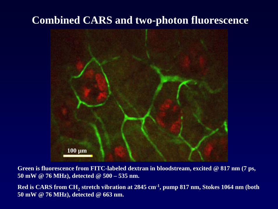
Combined CARS and two-photon fluorescence
Green is fluorescence from FITC-labeled dextran in bloodstream, excited @ 817 nm (7 ps, 50 mW @ 76 MHz), detected @ 500 – 535 nm.
Red is CARS from CH2 stretch vibration at 2845 cm-1, pump 817 nm, Stokes 1064 nm (both 50 mW @ 76 MHz), detected @ 663 nm.
100 µm

Multiphoton excitation forPhotodynamic Therapy
• Activate drugs, e.g. for cancer treatment, and natural pathways by multiphoton absorption
• Minimizes collateral damage because the rate of photochemistry ∝ I2 , which is confined
• Demonstrated in cultured cells, but not in vivo due to lack of high average power, tunable fs lasers
• Requires on-site biomedical facilities not yet available at JLab
• A multiphoton excitation source with the power and spectrum of JLab FEL is unprecedented for photomedicine and photobiology research.

Thank you, Fred Dylla and
the JLab FEL team