photography in veterinary medicine - medirabbit€¦ · photography in veterinary medicine dr...
TRANSCRIPT
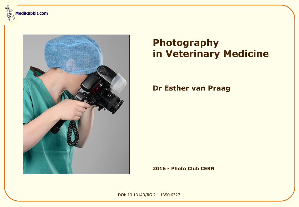
Photography in Veterinary Medicine Dr Esther van Praag
2016 - Photo Club CERN
DOI: 10.13140/RG.2.1.1350.6327
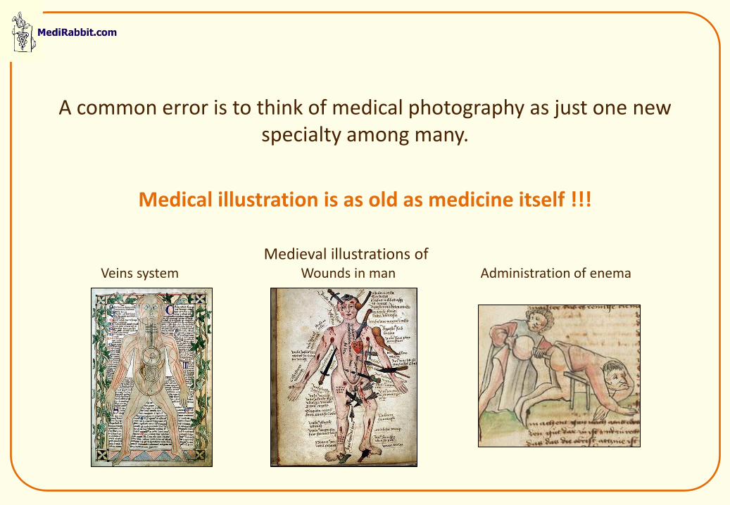
A common error is to think of medical photography as just one new specialty among many.
Medical illustration is as old as medicine itself !!!
Medieval illustrations of Veins system Wounds in man Administration of enema
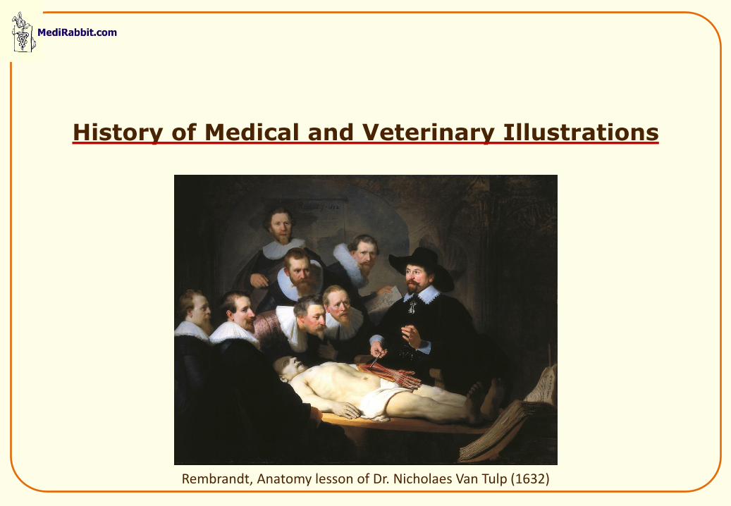
History of Medical and Veterinary Illustrations
Rembrandt, Anatomy lesson of Dr. Nicholaes Van Tulp (1632)
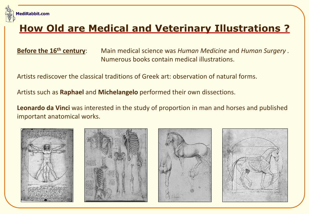
Before the 16th century: Main medical science was Human Medicine and Human Surgery . Numerous books contain medical illustrations. Artists rediscover the classical traditions of Greek art: observation of natural forms.
Artists such as Raphael and Michelangelo performed their own dissections.
Leonardo da Vinci was interested in the study of proportion in man and horses and published important anatomical works.
How Old are Medical and Veterinary Illustrations ?
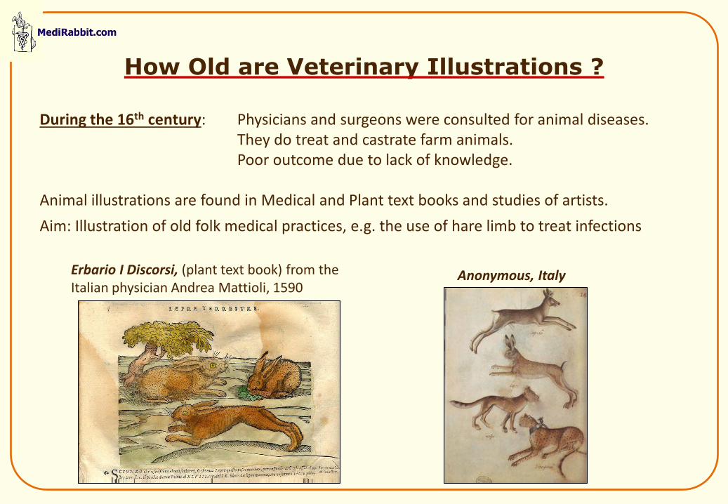
During the 16th century: Physicians and surgeons were consulted for animal diseases. They do treat and castrate farm animals. Poor outcome due to lack of knowledge. Animal illustrations are found in Medical and Plant text books and studies of artists.
Aim: Illustration of old folk medical practices, e.g. the use of hare limb to treat infections
Erbario I Discorsi, (plant text book) from the Italian physician Andrea Mattioli, 1590
Anonymous, Italy
How Old are Veterinary Illustrations ?
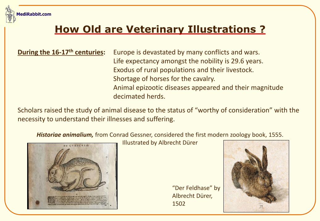
During the 16-17th centuries: Europe is devastated by many conflicts and wars. Life expectancy amongst the nobility is 29.6 years. Exodus of rural populations and their livestock. Shortage of horses for the cavalry. Animal epizootic diseases appeared and their magnitude decimated herds.
Scholars raised the study of animal disease to the status of “worthy of consideration” with the necessity to understand their illnesses and suffering.
Historiae animalium, from Conrad Gessner, considered the first modern zoology book, 1555. Illustrated by Albrecht Dürer
“Der Feldhase” by Albrecht Dürer, 1502
How Old are Veterinary Illustrations ?
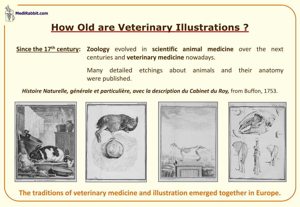
Since the 17th century: Zoology evolved in scientific animal medicine over the next centuries and veterinary medicine nowadays.
Many detailed etchings about animals and their anatomy were published.
Histoire Naturelle, générale et particulière, avec la description du Cabinet du Roy, from Buffon, 1753.
How Old are Veterinary Illustrations ?
The traditions of veterinary medicine and illustration emerged together in Europe.
Macro Veterinary Illustrations
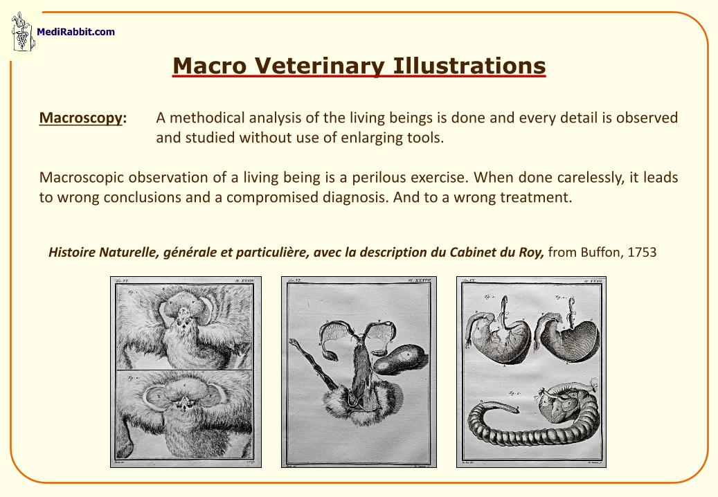
Histoire Naturelle, générale et particulière, avec la description du Cabinet du Roy, from Buffon, 1753
Macroscopy: A methodical analysis of the living beings is done and every detail is observed and studied without use of enlarging tools. Macroscopic observation of a living being is a perilous exercise. When done carelessly, it leads to wrong conclusions and a compromised diagnosis. And to a wrong treatment.
Micro Veterinary Illustrations
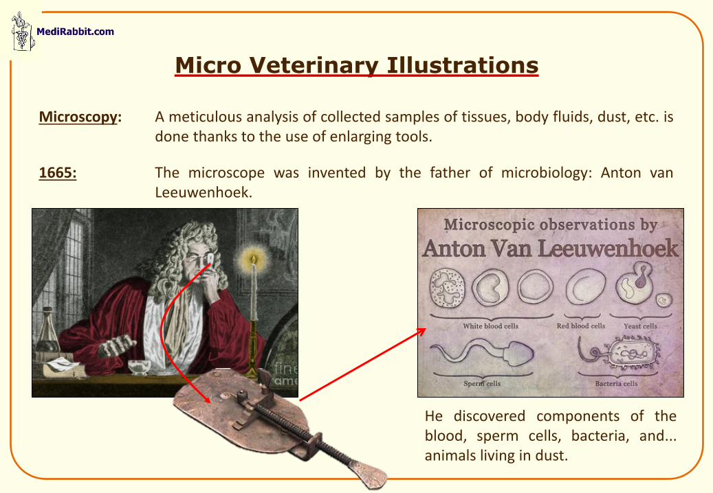
Microscopy: A meticulous analysis of collected samples of tissues, body fluids, dust, etc. is done thanks to the use of enlarging tools.
1665: The microscope was invented by the father of microbiology: Anton van Leeuwenhoek.
He discovered components of the blood, sperm cells, bacteria, and... animals living in dust.
Problem of Veterinary Antique Prints
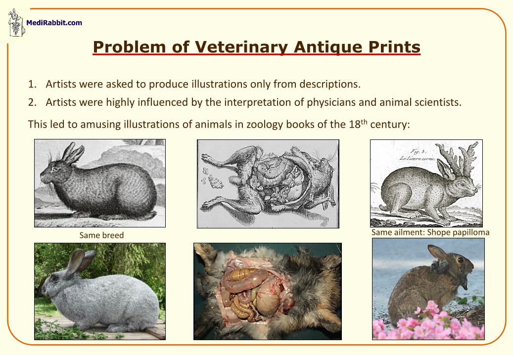
1. Artists were asked to produce illustrations only from descriptions.
2. Artists were highly influenced by the interpretation of physicians and animal scientists.
This led to amusing illustrations of animals in zoology books of the 18th century:
Antique prints 18th century
Same ailment: Shope papilloma Same breed
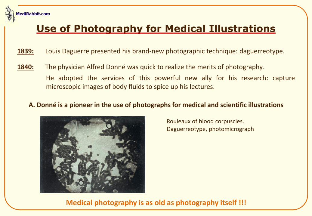
1839: Louis Daguerre presented his brand-new photographic technique: daguerreotype.
1840: The physician Alfred Donné was quick to realize the merits of photography.
He adopted the services of this powerful new ally for his research: capture microscopic images of body fluids to spice up his lectures.
A. Donné is a pioneer in the use of photographs for medical and scientific illustrations
Use of Photography for Medical Illustrations
Rouleaux of blood corpuscles. Daguerreotype, photomicrograph
Medical photography is as old as photography itself !!!
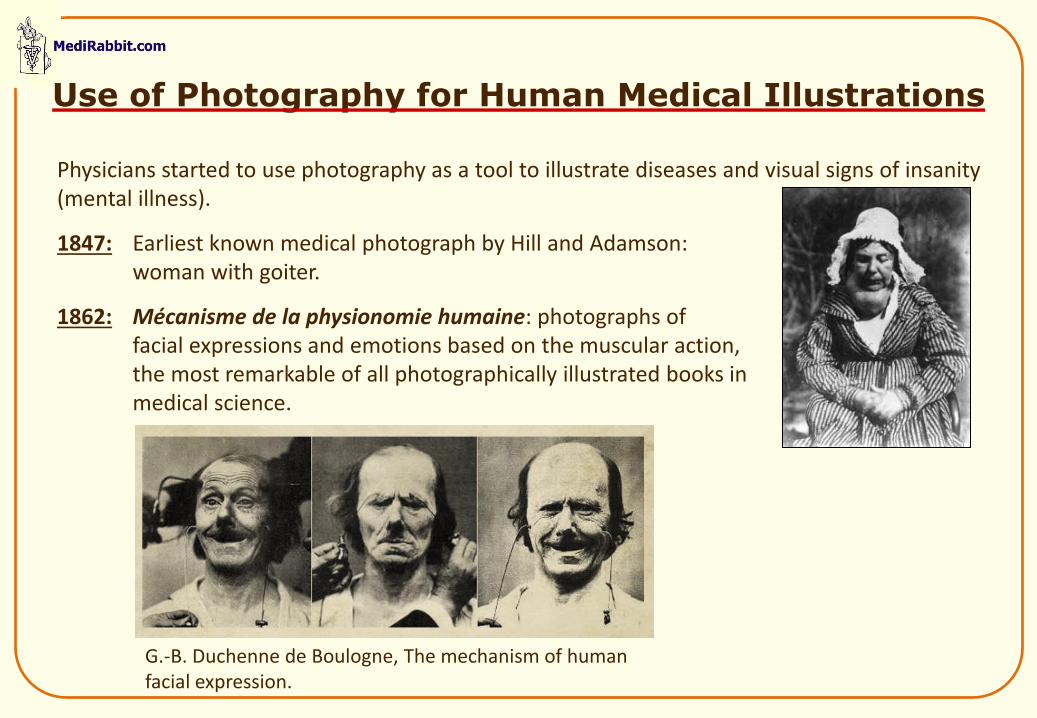
Physicians started to use photography as a tool to illustrate diseases and visual signs of insanity (mental illness).
1847: Earliest known medical photograph by Hill and Adamson: woman with goiter.
1862: Mécanisme de la physionomie humaine: photographs of facial expressions and emotions based on the muscular action, the most remarkable of all photographically illustrated books in medical science.
Use of Photography for Human Medical Illustrations
G.-B. Duchenne de Boulogne, The mechanism of human facial expression.
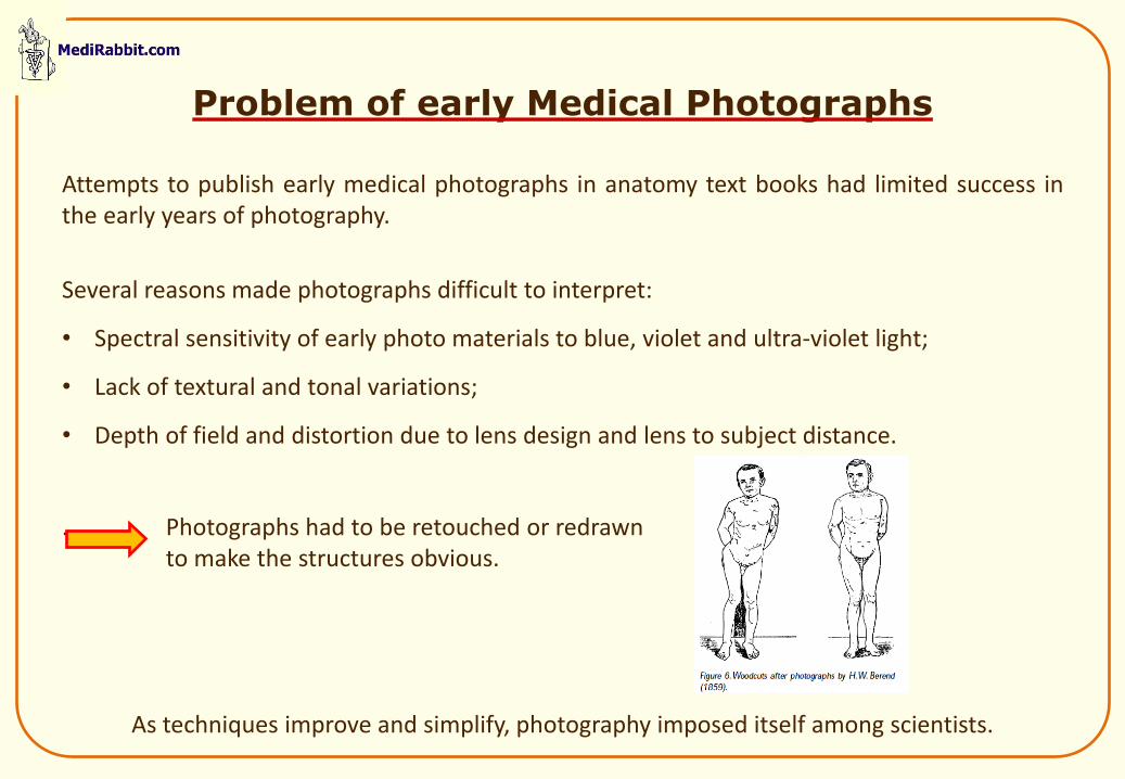
Attempts to publish early medical photographs in anatomy text books had limited success in the early years of photography.
Several reasons made photographs difficult to interpret:
• Spectral sensitivity of early photo materials to blue, violet and ultra-violet light;
• Lack of textural and tonal variations;
• Depth of field and distortion due to lens design and lens to subject distance.
. Photographs had to be retouched or redrawn to make the structures obvious.
As techniques improve and simplify, photography imposed itself among scientists.
Problem of early Medical Photographs
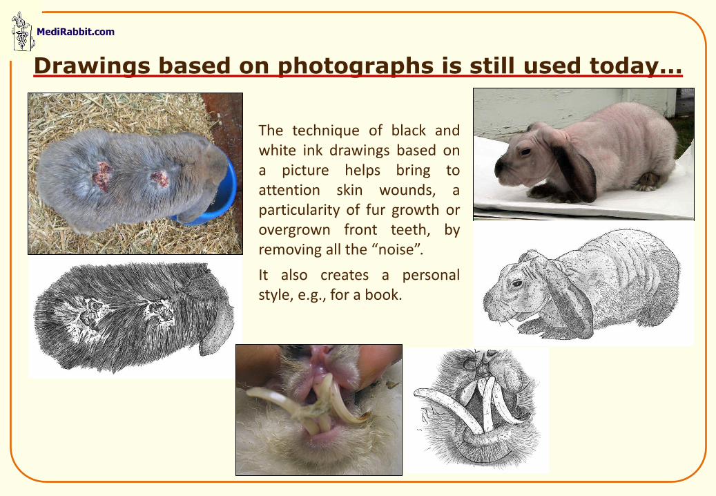
Drawings based on photographs is still used today...
The technique of black and white ink drawings based on a picture helps bring to attention skin wounds, a particularity of fur growth or overgrown front teeth, by removing all the “noise”.
It also creates a personal style, e.g., for a book.
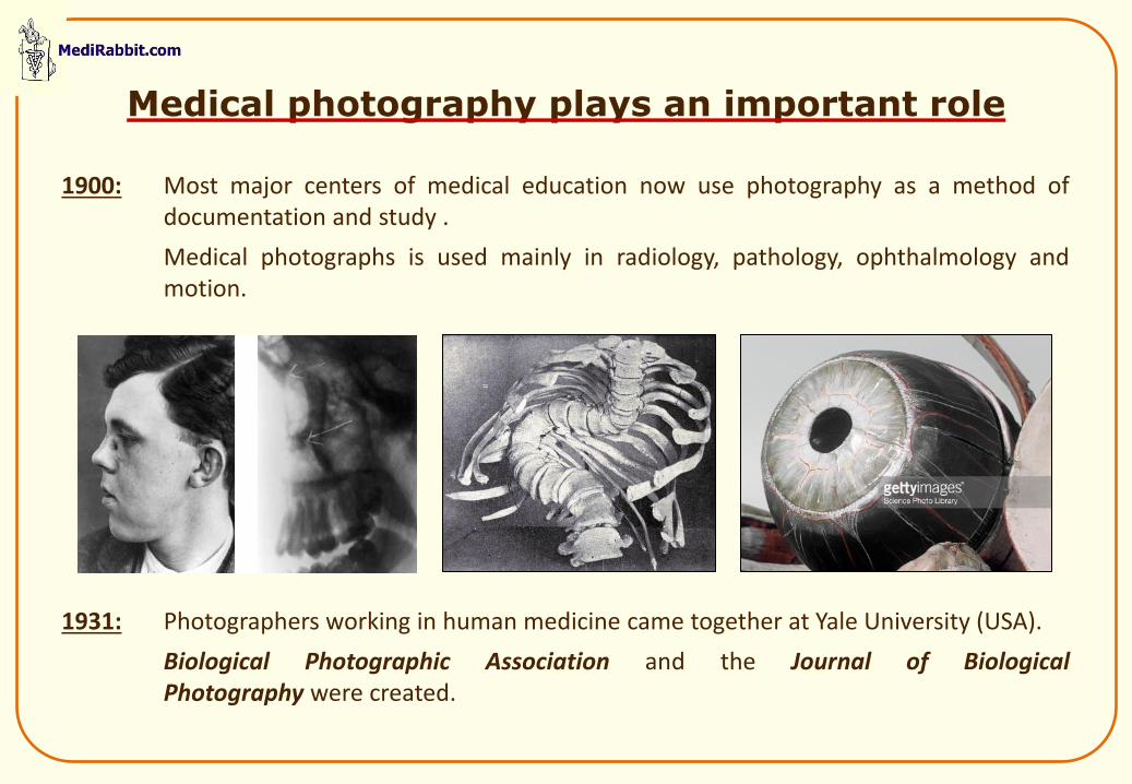
1900: Most major centers of medical education now use photography as a method of documentation and study .
Medical photographs is used mainly in radiology, pathology, ophthalmology and motion.
1931: Photographers working in human medicine came together at Yale University (USA).
Biological Photographic Association and the Journal of Biological Photography were created.
Medical photography plays an important role
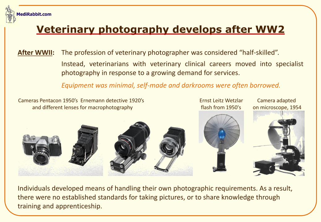
After WWII: The profession of veterinary photographer was considered “half-skilled”.
Instead, veterinarians with veterinary clinical careers moved into specialist photography in response to a growing demand for services.
Equipment was minimal, self-made and darkrooms were often borrowed.
Individuals developed means of handling their own photographic requirements. As a result, there were no established standards for taking pictures, or to share knowledge through training and apprenticeship.
Veterinary photography develops after WW2
Cameras Pentacon 1950’s Ernemann detective 1920’s Ernst Leitz Wetzlar Camera adapted and different lenses for macrophotography flash from 1950's on microscope, 1954
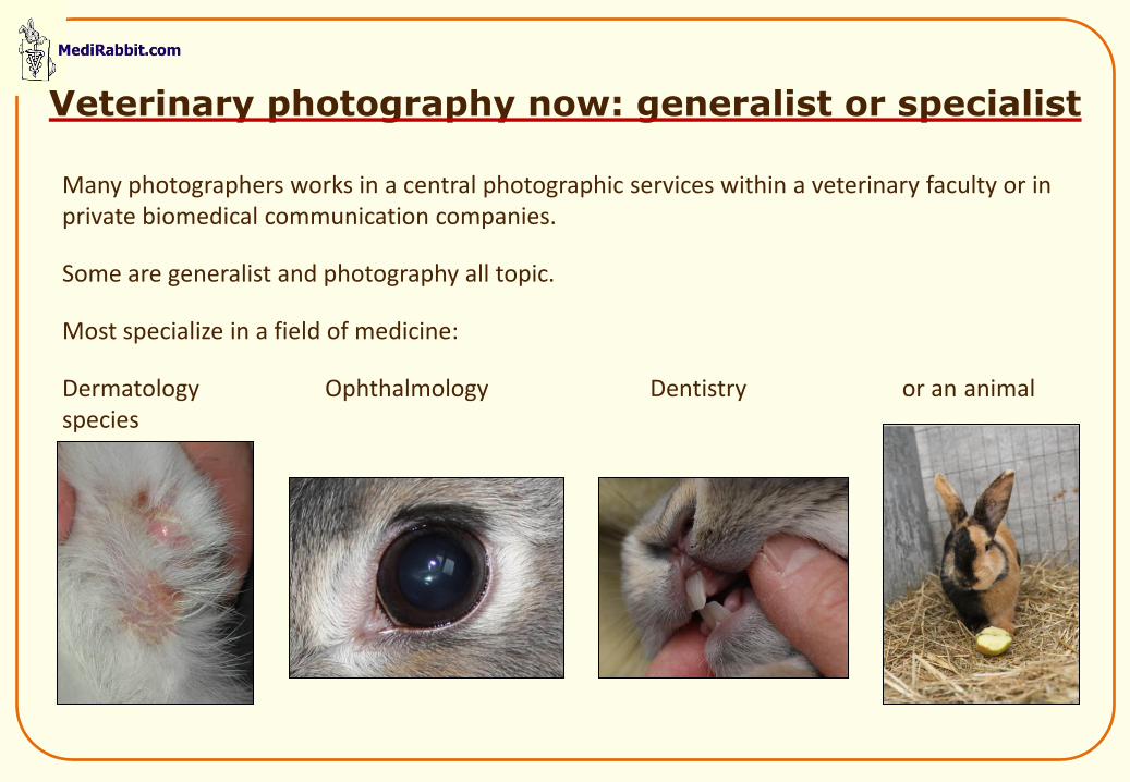
Veterinary photography now: generalist or specialist
Many photographers works in a central photographic services within a veterinary faculty or in private biomedical communication companies.
Some are generalist and photography all topic.
Most specialize in a field of medicine:
Dermatology Ophthalmology Dentistry or an animal species
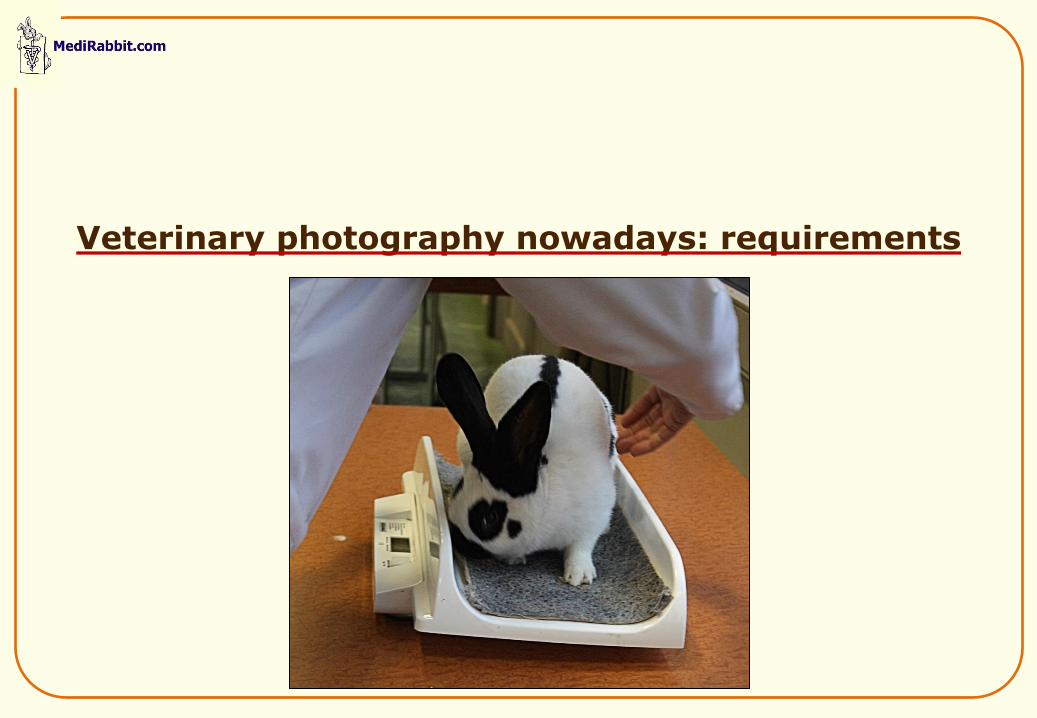
Veterinary photography nowadays: requirements
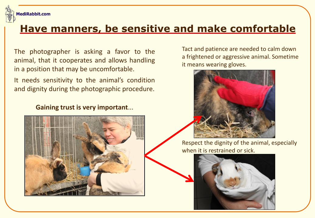
Tact and patience are needed to calm down a frightened or aggressive animal. Sometime it means wearing gloves.
Respect the dignity of the animal, especially when it is restrained or sick.
Have manners, be sensitive and make comfortable
The photographer is asking a favor to the animal, that it cooperates and allows handling in a position that may be uncomfortable.
It needs sensitivity to the animal’s condition and dignity during the photographic procedure.
Gaining trust is very important...
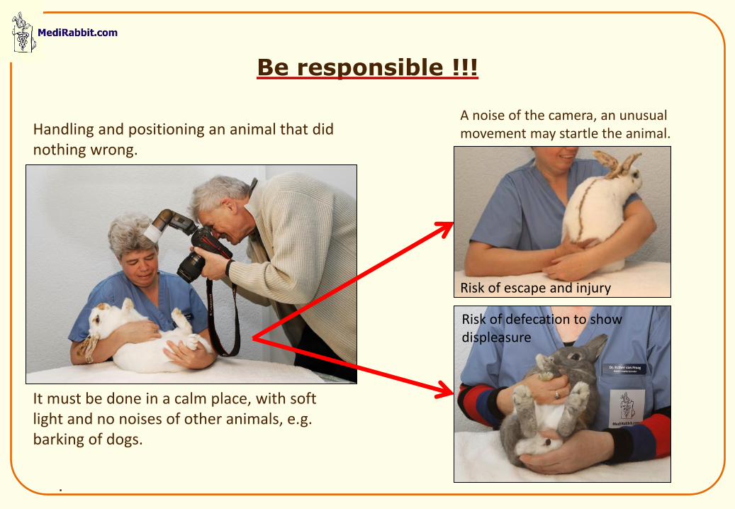
Be responsible !!!
Handling and positioning an animal that did nothing wrong.
It must be done in a calm place, with soft light and no noises of other animals, e.g. barking of dogs.
.
A noise of the camera, an unusual movement may startle the animal.
Risk of escape and injury
Risk of defecation to show displeasure
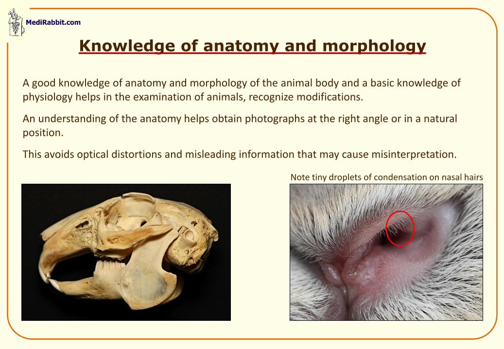
Knowledge of anatomy and morphology
A good knowledge of anatomy and morphology of the animal body and a basic knowledge of physiology helps in the examination of animals, recognize modifications.
An understanding of the anatomy helps obtain photographs at the right angle or in a natural position.
This avoids optical distortions and misleading information that may cause misinterpretation.
Note tiny droplets of condensation on nasal hairs
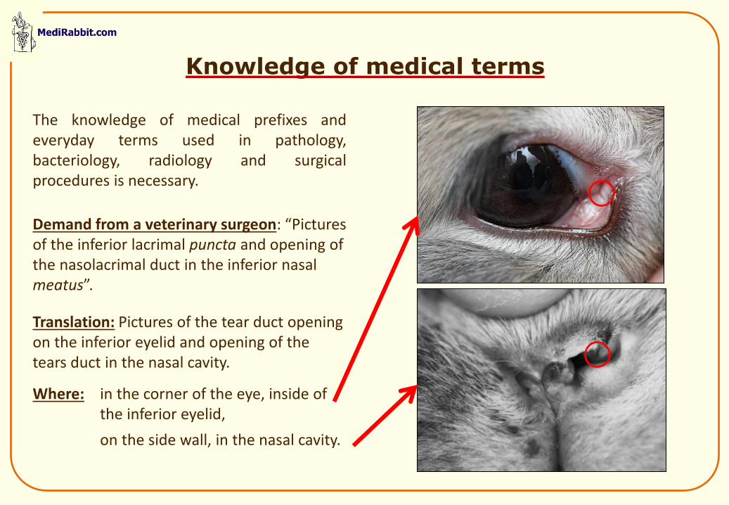
Knowledge of medical terms
The knowledge of medical prefixes and everyday terms used in pathology, bacteriology, radiology and surgical procedures is necessary.
Demand from a veterinary surgeon: “Pictures of the inferior lacrimal puncta and opening of the nasolacrimal duct in the inferior nasal meatus”.
Translation: Pictures of the tear duct opening on the inferior eyelid and opening of the tears duct in the nasal cavity.
Where: in the corner of the eye, inside of the inferior eyelid,
on the side wall, in the nasal cavity.
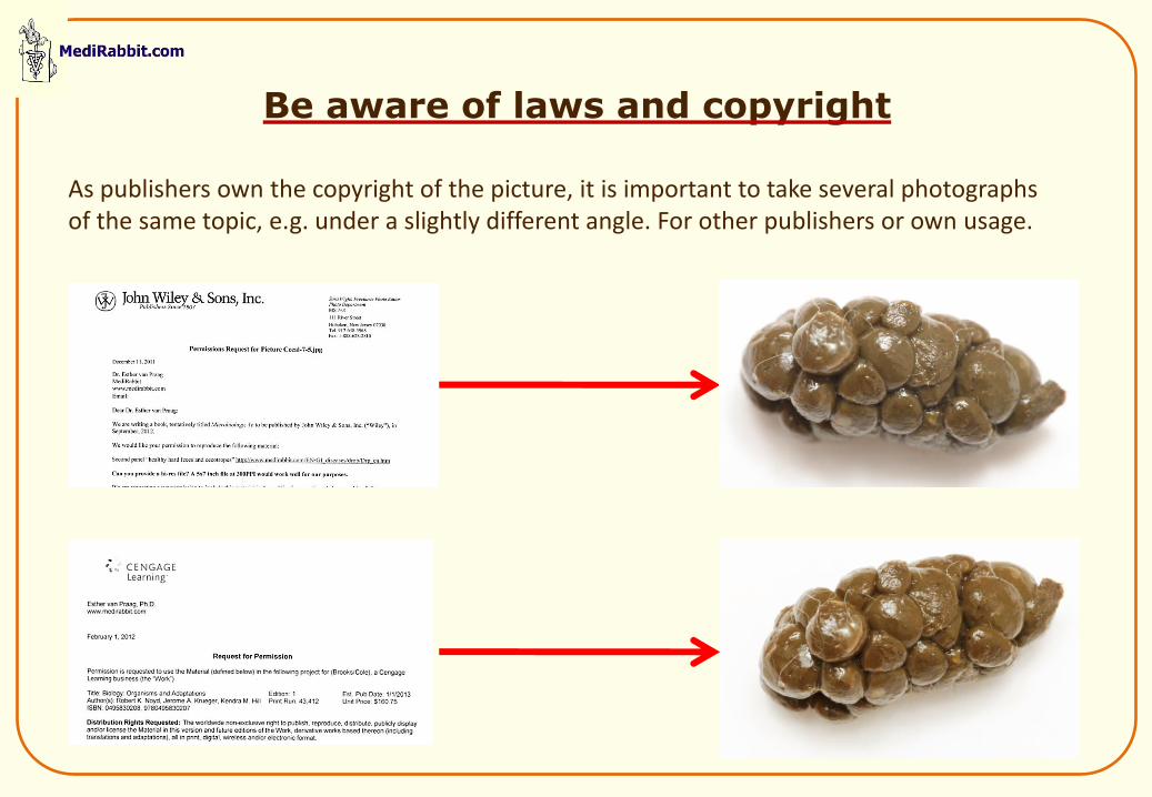
Be aware of laws and copyright
As publishers own the copyright of the picture, it is important to take several photographs of the same topic, e.g. under a slightly different angle. For other publishers or own usage.
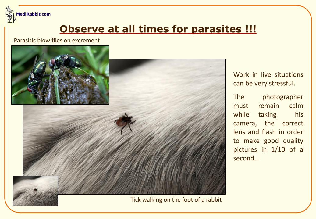
Observe at all times for parasites !!!
Work in live situations can be very stressful.
The photographer must remain calm while taking his camera, the correct lens and flash in order to make good quality pictures in 1/10 of a second...
Tick walking on the foot of a rabbit
Parasitic blow flies on excrement
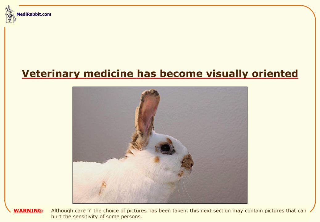
Veterinary medicine has become visually oriented
WARNING: Although care in the choice of pictures has been taken, this next section may contain pictures that can hurt the sensitivity of some persons.
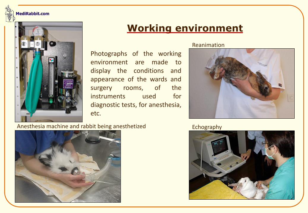
Working environment
Photographs of the working environment are made to display the conditions and appearance of the wards and surgery rooms, of the instruments used for diagnostic tests, for anesthesia, etc.
Anesthesia machine and rabbit being anesthetized
Reanimation
Echography
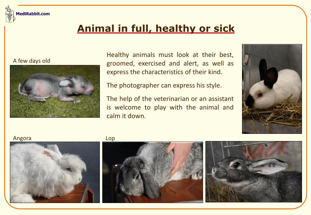
Animal in full, healthy or sick
Healthy animals must look at their best, groomed, exercised and alert, as well as express the characteristics of their kind.
The photographer can express his style.
The help of the veterinarian or an assistant is welcome to play with the animal and calm it down.
A few days old
Angora Lop
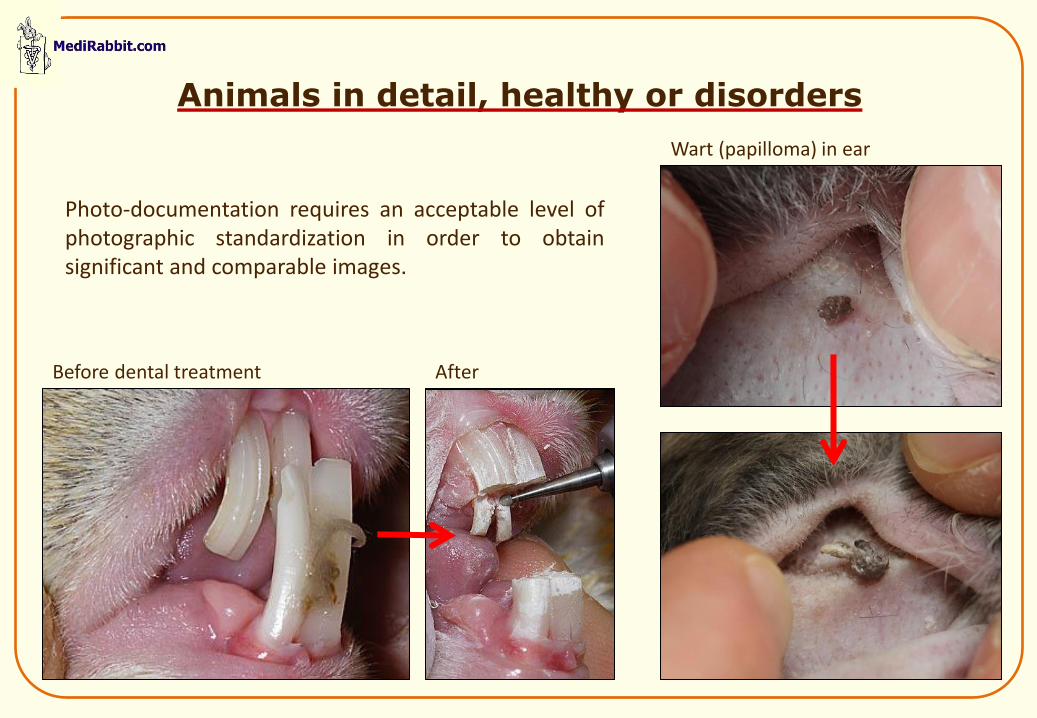
Animals in detail, healthy or disorders
Photo-documentation requires an acceptable level of photographic standardization in order to obtain significant and comparable images.
After Before dental treatment
Wart (papilloma) in ear

Infectious diseases
The veterinary photographer must remain serene and calm during any emergency or surgery.
He must also be aware that some disease have an infectious nature. He must take measures to avoid contact and protect colleagues from hazardous circumstances.
Fungal infection that is highly contagious to man
Dental abscess, with pus
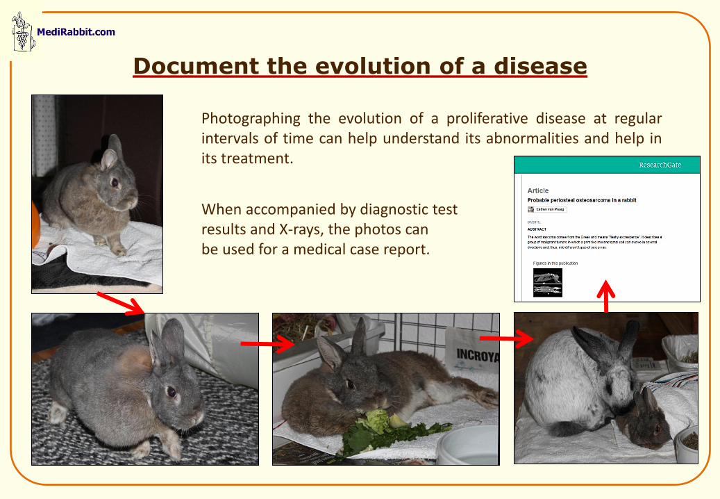
Document the evolution of a disease
Photographing the evolution of a proliferative disease at regular intervals of time can help understand its abnormalities and help in its treatment.
When accompanied by diagnostic test results and X-rays, the photos can be used for a medical case report.
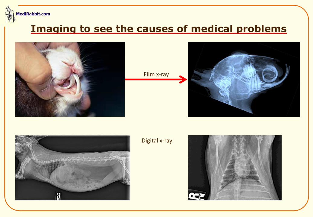
Imaging to see the causes of medical problems
Film x-ray
Digital x-ray
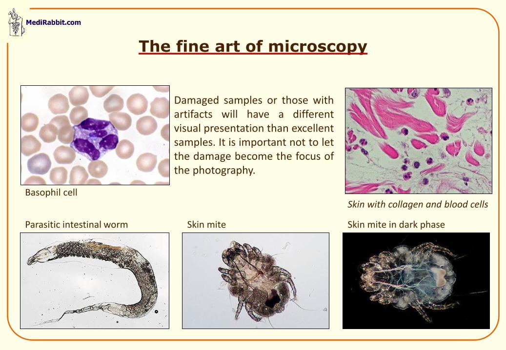
Damaged samples or those with artifacts will have a different visual presentation than excellent samples. It is important not to let the damage become the focus of the photography.
The fine art of microscopy
Basophil cell
Parasitic intestinal worm Skin mite Skin mite in dark phase
Skin with collagen and blood cells
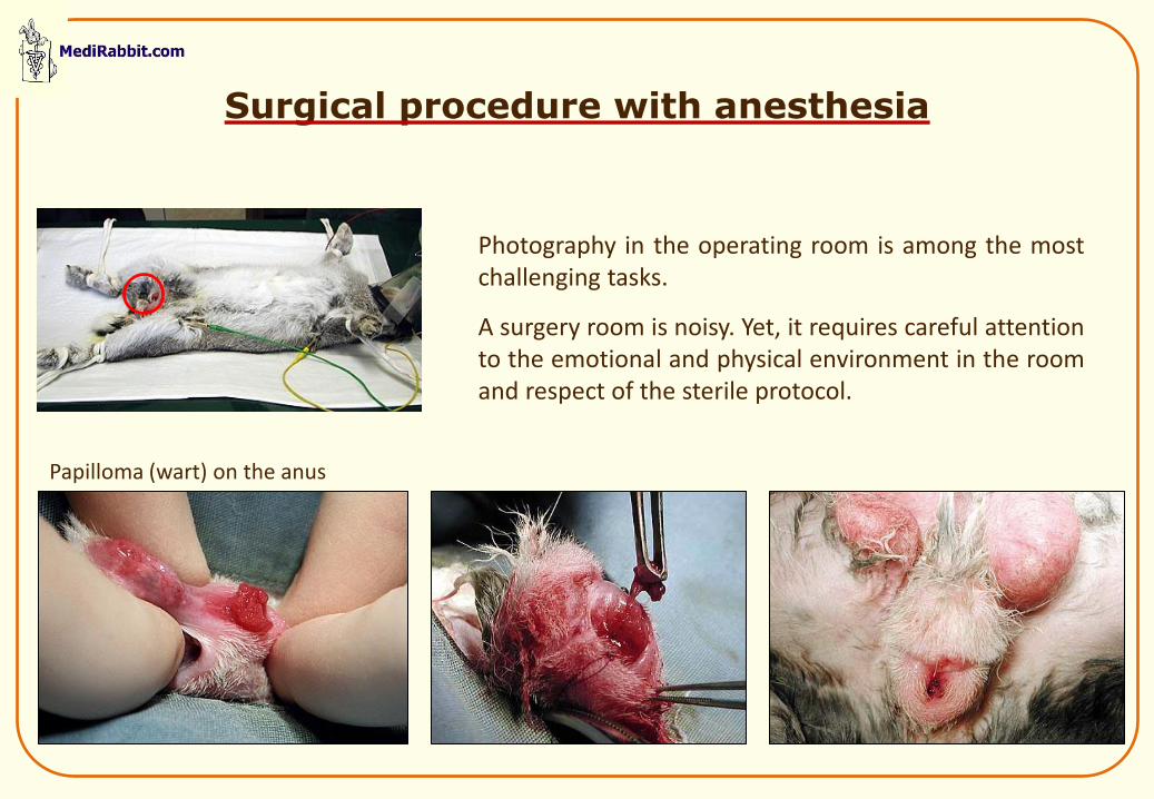
Surgical procedure with anesthesia
Photography in the operating room is among the most challenging tasks.
A surgery room is noisy. Yet, it requires careful attention to the emotional and physical environment in the room and respect of the sterile protocol.
Papilloma (wart) on the anus
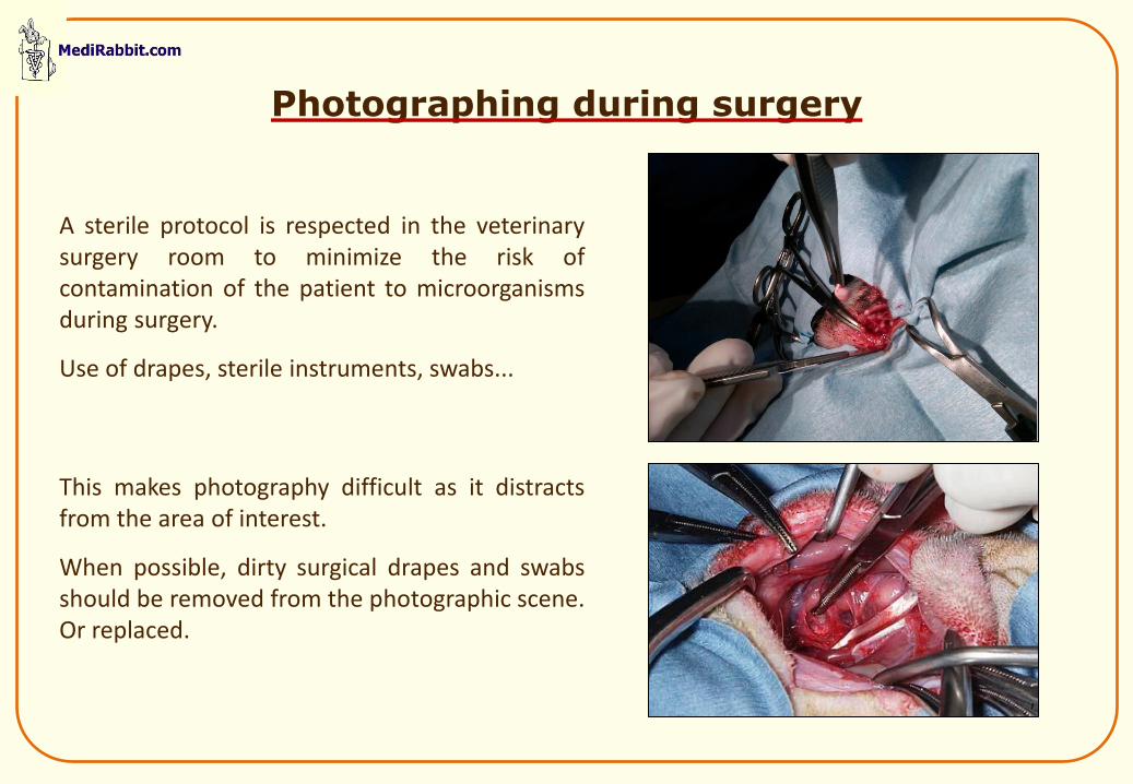
Photographing during surgery
A sterile protocol is respected in the veterinary surgery room to minimize the risk of contamination of the patient to microorganisms during surgery.
Use of drapes, sterile instruments, swabs...
This makes photography difficult as it distracts from the area of interest.
When possible, dirty surgical drapes and swabs should be removed from the photographic scene. Or replaced.
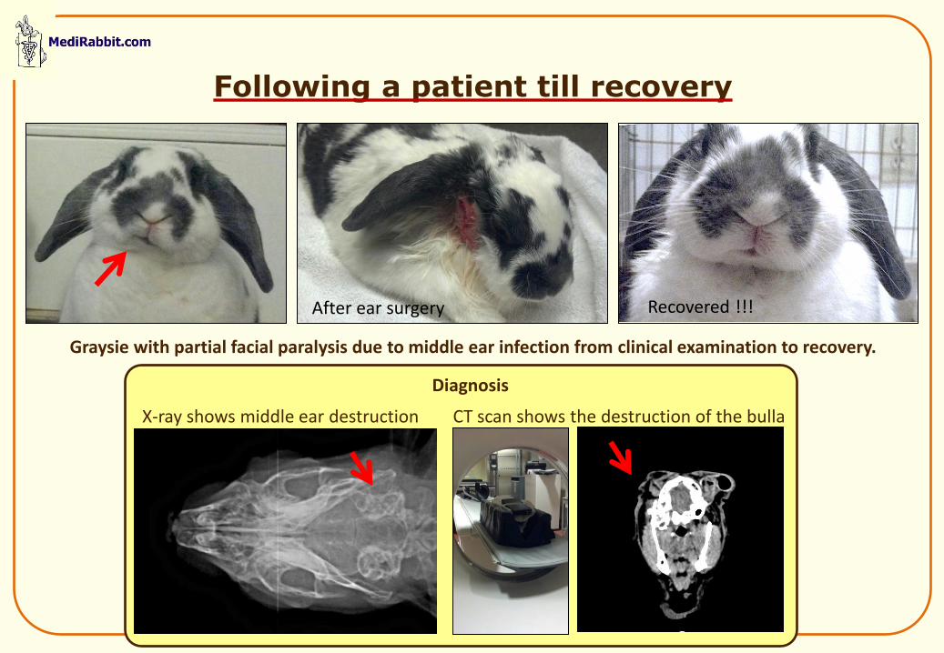
Following a patient till recovery
Graysie with partial facial paralysis due to middle ear infection from clinical examination to recovery.
X-ray shows middle ear destruction CT scan shows the destruction of the bulla
Diagnosis
After ear surgery Recovered !!!
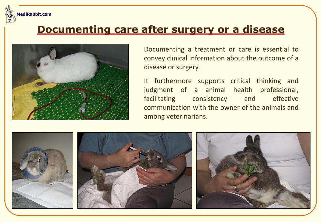
Documenting care after surgery or a disease
Documenting a treatment or care is essential to convey clinical information about the outcome of a disease or surgery.
It furthermore supports critical thinking and judgment of a animal health professional, facilitating consistency and effective communication with the owner of the animals and among veterinarians.
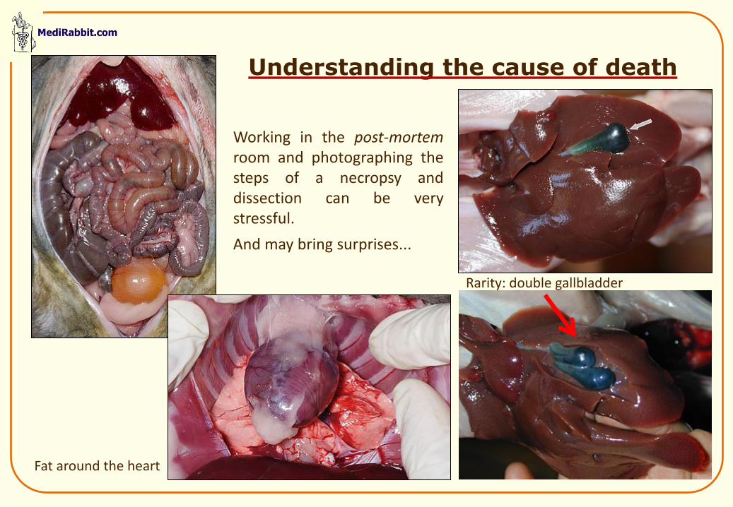
Understanding the cause of death
Working in the post-mortem room and photographing the steps of a necropsy and dissection can be very stressful.
And may bring surprises...
Rarity: double gallbladder
Fat around the heart
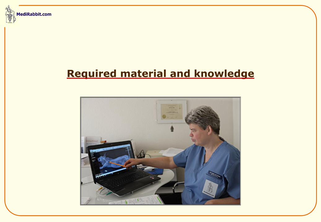
Required material and knowledge
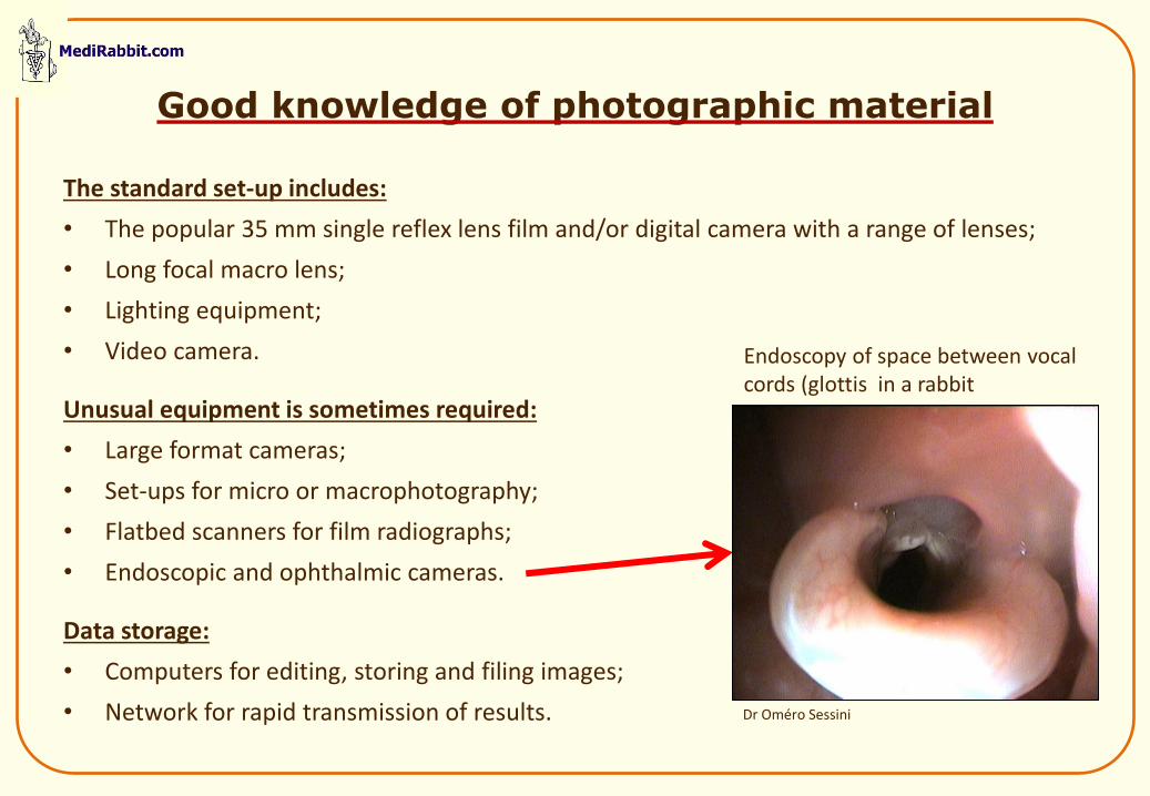
Good knowledge of photographic material
The standard set-up includes:
• The popular 35 mm single reflex lens film and/or digital camera with a range of lenses;
• Long focal macro lens;
• Lighting equipment;
• Video camera.
Unusual equipment is sometimes required:
• Large format cameras;
• Set-ups for micro or macrophotography;
• Flatbed scanners for film radiographs;
• Endoscopic and ophthalmic cameras.
Data storage:
• Computers for editing, storing and filing images;
• Network for rapid transmission of results.
Endoscopy of space between vocal cords (glottis in a rabbit
Dr Oméro Sessini
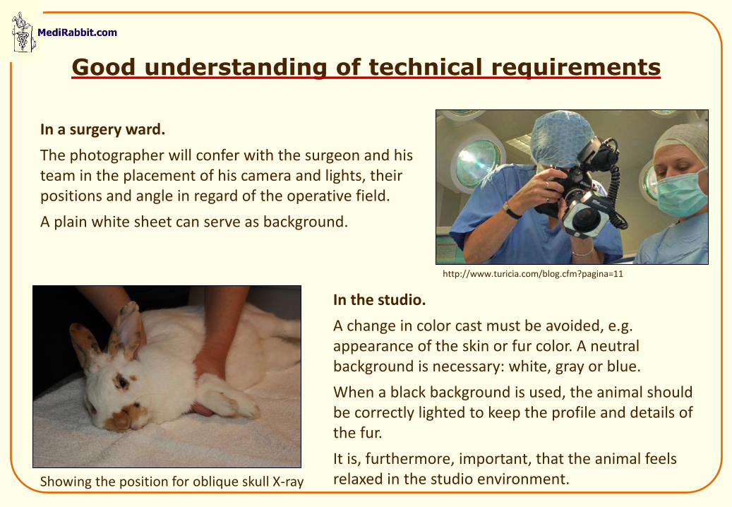
Good understanding of technical requirements
In a surgery ward.
The photographer will confer with the surgeon and his team in the placement of his camera and lights, their positions and angle in regard of the operative field.
A plain white sheet can serve as background.
In the studio.
A change in color cast must be avoided, e.g. appearance of the skin or fur color. A neutral background is necessary: white, gray or blue.
When a black background is used, the animal should be correctly lighted to keep the profile and details of the fur.
It is, furthermore, important, that the animal feels relaxed in the studio environment.
http://www.turicia.com/blog.cfm?pagina=11
Showing the position for oblique skull X-ray
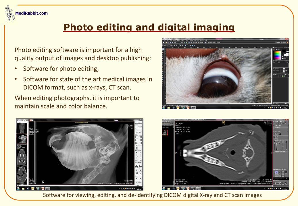
Photo editing and digital imaging
Photo editing software is important for a high quality output of images and desktop publishing:
• Software for photo editing;
• Software for state of the art medical images in DICOM format, such as x-rays, CT scan.
When editing photographs, it is important to maintain scale and color balance.
Software for viewing, editing, and de-identifying DICOM digital X-ray and CT scan images
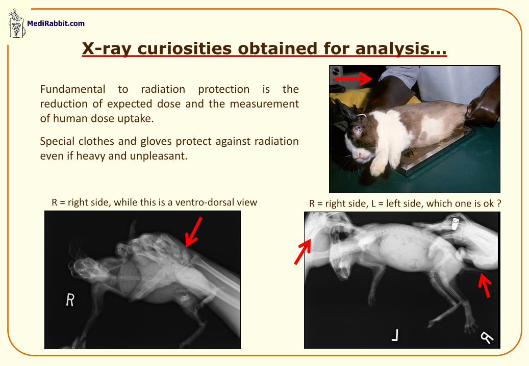
X-ray curiosities obtained for analysis...
Fundamental to radiation protection is the reduction of expected dose and the measurement of human dose uptake.
Special clothes and gloves protect against radiation even if heavy and unpleasant.
R = right side, while this is a ventro-dorsal view R = right side, L = left side, which one is ok ?
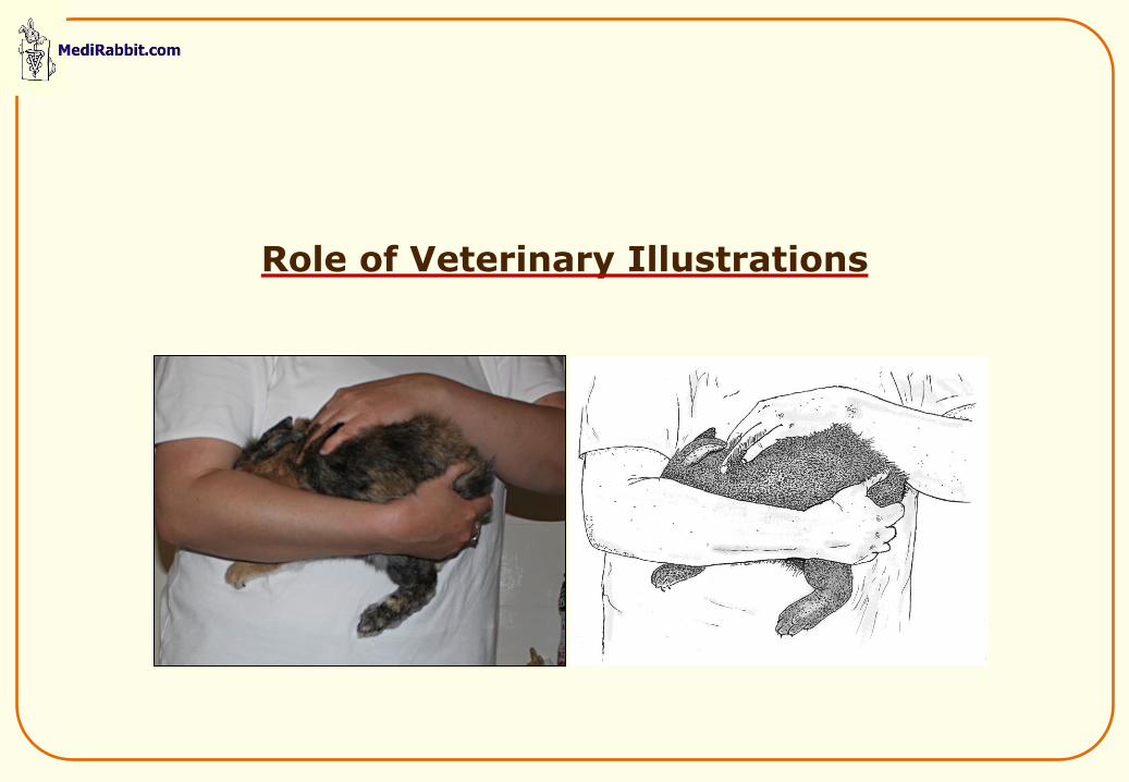
Role of Veterinary Illustrations
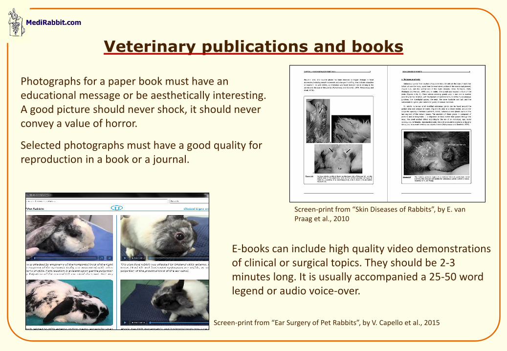
Veterinary publications and books
Photographs for a paper book must have an educational message or be aesthetically interesting. A good picture should never shock, it should never convey a value of horror.
Selected photographs must have a good quality for reproduction in a book or a journal.
E-books can include high quality video demonstrations of clinical or surgical topics. They should be 2-3 minutes long. It is usually accompanied a 25-50 word legend or audio voice-over.
Screen-print from “Ear Surgery of Pet Rabbits”, by V. Capello et al., 2015
Screen-print from “Skin Diseases of Rabbits”, by E. van Praag et al., 2010
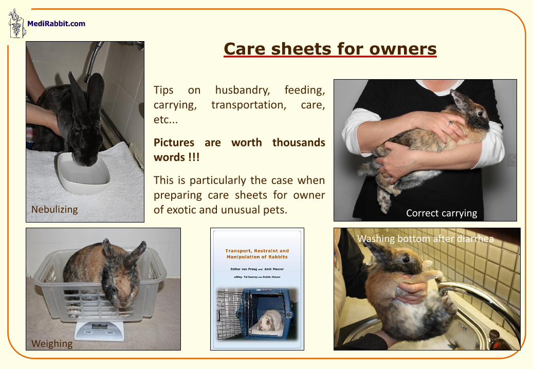
Care sheets for owners
Nebulizing
Weighing
Washing bottom after diarrhea
Tips on husbandry, feeding, carrying, transportation, care, etc...
Pictures are worth thousands words !!!
This is particularly the case when preparing care sheets for owner of exotic and unusual pets. Correct carrying
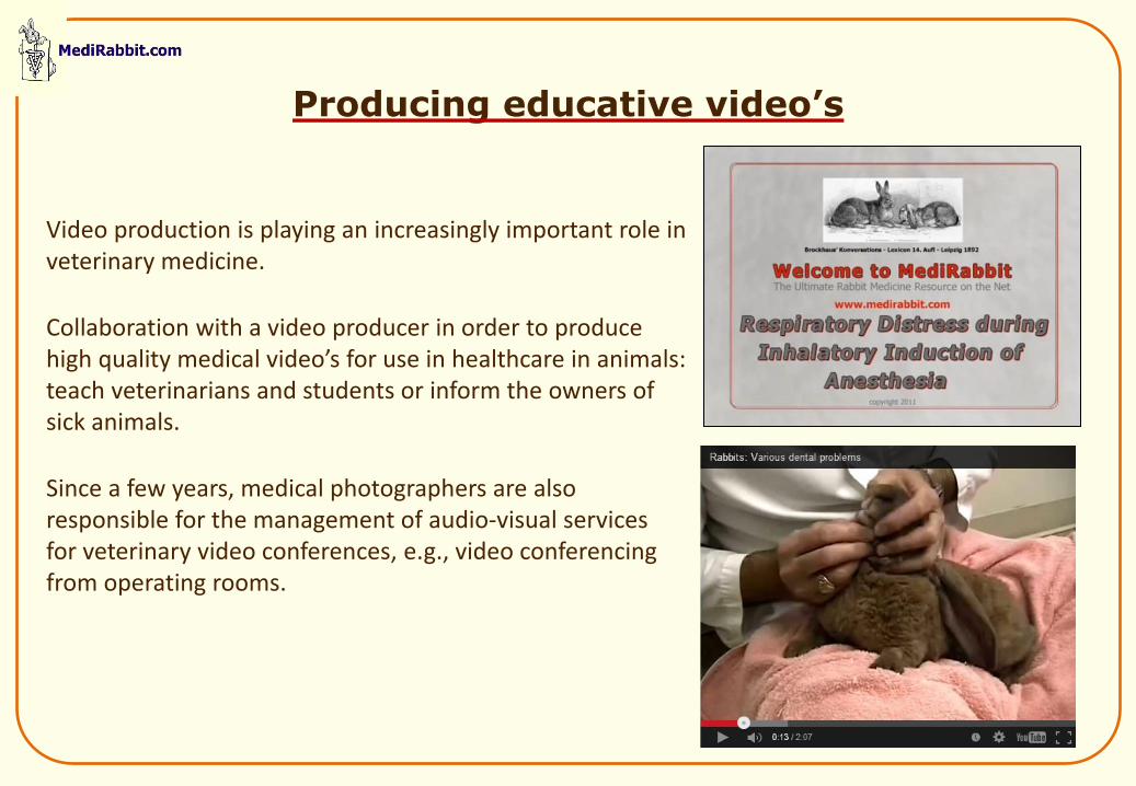
Video production is playing an increasingly important role in veterinary medicine.
Collaboration with a video producer in order to produce high quality medical video’s for use in healthcare in animals: teach veterinarians and students or inform the owners of sick animals.
Since a few years, medical photographers are also responsible for the management of audio-visual services for veterinary video conferences, e.g., video conferencing from operating rooms.
Producing educative video’s
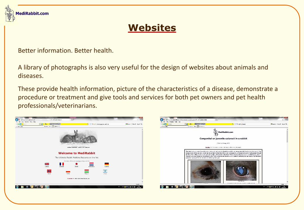
Better information. Better health. A library of photographs is also very useful for the design of websites about animals and diseases.
These provide health information, picture of the characteristics of a disease, demonstrate a procedure or treatment and give tools and services for both pet owners and pet health professionals/veterinarians.
Websites
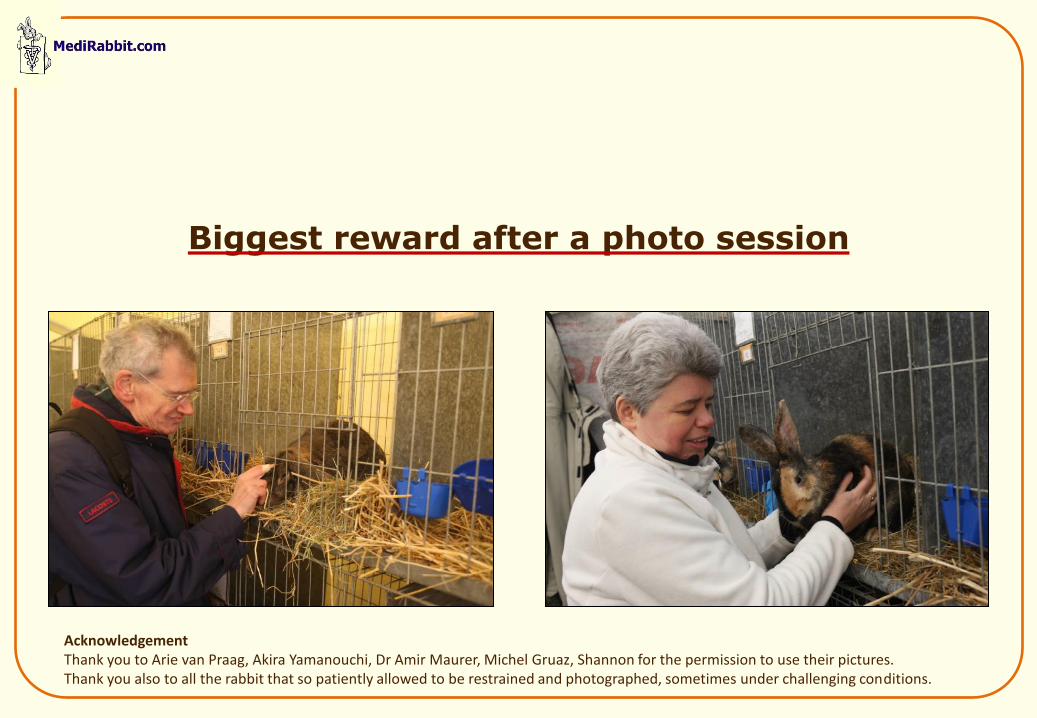
Acknowledgement Thank you to Arie van Praag, Akira Yamanouchi, Dr Amir Maurer, Michel Gruaz, Shannon for the permission to use their pictures. Thank you also to all the rabbit that so patiently allowed to be restrained and photographed, sometimes under challenging conditions.
Biggest reward after a photo session
Thank you