photodegradation of hyaluronic acid and of the … · gen. physiol. biophys. (1990). 9, 419 429 419...
TRANSCRIPT
Gen. Physiol. Biophys. (1990). 9, 419 429 419
Short communication
Photodegradation of Hyaluronic Acid and of the Vitreous Body
Ľ . L A P Č Í K . J R . 1 . L . O M I I K A \ K . K U B K N A \ A . G A I . A T Í K 2 , V . K Ľ L L Ô 1
1 Centre far Chemical Research. Slovak Academy of Science.''. Dúbravská eesla 9. X4105 Bratislava. Czechoslovakia
2 Faculty of Chemical Technology. Slovak Technical University, Radlinského 9. H1237 Bratislava. Czechoslovakia
3 State Hospital. 761)00 /.I'm. Czechoslovakia
Hyaluronic acid is a mucopolysaccharide. The chemical structure and properties of this group have been extensively studied (Brimacombe and Webber 1964; Schubert and Hamerman 1968; Mathews 1975; Laurent 1987).
Hyaluronic acid is present in the connective tissue. It was discovered by Meyer and Palmer (1934) in the vitreous body tissue where its concentration ranges from 140 to 338 mg/dm3 (Laurent 1987). It has a number of physiological functions, including influence on the polymer-solvent interactions and on the diffusion processes (Wik 1979). Many of these functions have been proved to be closely related to the physicochemical properties of the compound (Laurent 1966, 1968. 1970).
Recently, pathological processes in the eye due to luminous radation have come to the foreground of scientific interests (Lawwill 1982). X-rays and ra-diolysis of hyaluronic acid cause vigorous depolymerization and liquefaction (Caputo 1957; Ragan et al. 1947: Lai 1985).
The vitreous body is a loose connective tissue built up of a collagen network and tied up with a molecularly dispersed glycosaminoglycan system to form an organized composite proteoglycan structure. It contains an aqueous solution of different species, including sodium chloride, metabolities and serum proteins.
From the physiological point of view the vitreous body seems to be a large interstitial pre-lymphatic space inserted between the venous system of the ciliary body, which produces the chamber fluid, and the drainage system of the lamina cribrosa space.
The present work examines the influence of UV-radiation on the photode-
Reprint requests to : Ľ. Lapčik. Jr.. Department of Physical Chemistry, Faculty of Chemical Technology. Slovak Technical University. Radlinského 9. Bratislava. CS-812 37. Czechoslovakia
420 Lapčík Jr. et al.
Table I. List of samples studied
Sample labelling Mode of preparation and slorage
A B C n i i
freshly excised porcine vitreous body sample A after 7 days of storage at 4°C sample A after 14 da>s of storage al 4°C sodium hyaluronate solution freshly excised bovine vitreous body freshly excised human vitreous bod>
I
250 350 450 550 Wavelength (nm)
650
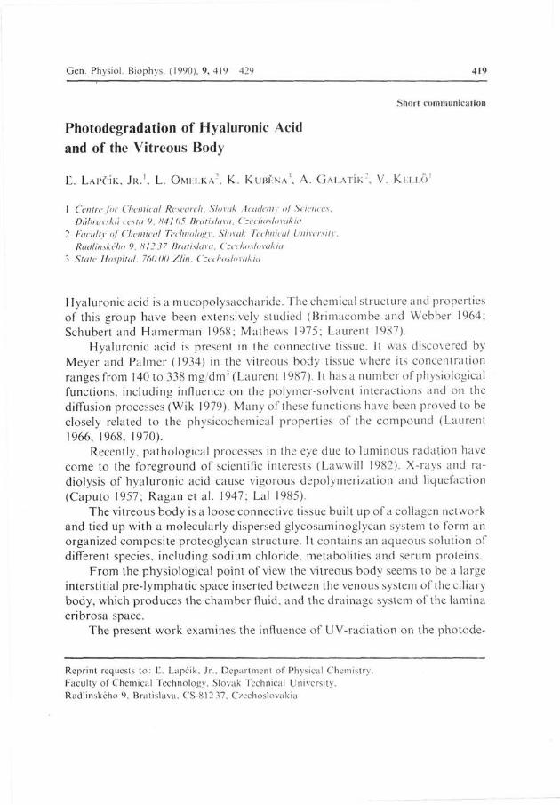
Fig. 1. Spectral eharacleiistic of the 500W high pressure mercury lamp used for sample irradiation (Lapčik et al. 1989).
gradation processes of both the vitreous body and hyaluronic acid. UV and IR absorption spectra, changes in molecular weight as well as photo-induced paramagnetism (determined by EPR measurements) are analyzed.
A sample of unfractionated hyaluronic acid (sodium salt) of pharmacological purity was obtained from MOVIS Co. (extract from rooster combs). The sample was purified according to the Czechoslovakian patent (Galatik et al. 1989). Table 1 presents a list of investigated probes. The samples of the vitreous

Photodegradation of Hyaluronic Acid 421
230 280 330 ran
Fig. 2. L'V spectra of collagen, samples D (2) and F (3)
230 280 330 nm 190 240 290 nm 340
Fig. 3. a) UV spectra of sample E before (I), and after 60 min (2) and 120 min irradiation, b) UV spectra of sample D before (1) and after 180 min irradiation.
422 Lapčík Jr. et al
1.0-
ABS-
0 4 ^ 1 1 1 1 1 1 1 1 1 1 1800 1780 1760 1740 1720 e m - 1 1700
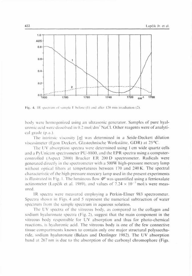
Fig. 4. IR spectrum of sample F before (I) and after 120 min irradiation (2).
body were homogenized using an ultrasonic generator. Samples of pure hyaluronic acid were dissolved in 0.2 mol dm' NaCl. Other reagents were of analytical grade (p.a.).
The intrinsic viscosity [;;] was determined in a Seide-Deckert dilution viscosimeter (Egon Deckert, Glastechnische Werkstätte, GDR) al 25°C.
The UV absorption spectra were determined using l cm wide quartz cells and a PyUnicam spectrometer PU-8800, and the EPR spectra using a computer-controlled (Aspect 2000) Brucker ER 200 D spectrometer. Radicals were generated directly in the spectrometer with a 500W high-pressure mercury lamp without optical filters at temperatures between 170 and 240 K. The spectral characteristic of the high pressure mercury lamp used in the present experiments is illustrated in Fig. I. The luminous flow (IP was quantified using a ferrioxalate actinometer (Lapčík et al. 1989). and values of 7.24 x 10~7mol/s were measured.
IR spectra were measured employing a Perkin-Elmer 983 spectrometer. Spectra shown in Figs. 4 and 5 represent the numerical subtraction of water spectrum from the sample spectrum in aqueous solution.
The UV spectra of the vitreous body, as compared to the collagen and sodium hyaluronate spectra (Fig. 2), suggest that the main component in the vitreous body responsible for UV absorption and thus for photo-chemical reactions, is hyaluronic acid. The vitreous body is one of the few connective tissue compartments known to contain only one major structural polysaccharide, sodium hyaluronate (Balazs and Denlinger 1982). The UV absorption band at 267 nm is due to the absorption of the carbonyl chromophore (Figs.

Photodegradation of Hyaluronic Acid 423
1 1000
1400 1200 cm-1 1000
Fig. 5. a) IR spectrum of sample F before (l) and after 20 min irradiation (2). b) IR spectrum of sample D before (!) and after 60 min irradiation (2).
3a, b) (Balazs 1954). The process of photodegradation after exposure to UV radiation is assumed to start at this chromophore. This agrees with the IR spectrum (Fig. 4): after irradiation the absorption band of the valence vibrations of carbonyl groups disappears. This is associated with a decrease of pH value (Balazs et al. 1959), probably due to the formation of carbonic acid from the liberated carbon dioxide. Simultaneously, the molecular weight decreases (see Fig. 6) suggesting that irradiation in the presence of oxygen leads to breaking of bonds of the hyaluronic acid main chain. It may be concluded from

424 Lapčík Jr. et al.
2500
0.15 0.25 1000.C (g/ml)
0.35
Fíg. 6. Plols of i;M, < = /(<•) o\~ Huggin's equation. I: before irradiation, [//] = I 594 ml g: 2: after 60 min.. [;/] = 678 ml/g; and 3: after 120 min. ii radiation. [/;] = 447 ml g. Hyaluronate solution in 0.2 mol I NaCI at 25°C. The molecular weight calculated by the Mark-Howink equation [//] = K . A/'' using the constants reported by Cleland and Wang (1970) decreases from 8.7 x 10* g mol (I) lo 1.8 x lO'gmol (3).
IR spectra shown in Figs. 5a, b that as a result of energy-consuming formation of hydroperoxides after UV exposure, qualitatively similar changes occur in the region which corresponds to the stretching vibrations of C O groups. The spectrum of pure hyaluronic acid (Fib. 5b) shows a remarkable decrease in intensity of the absorption band in the 1150 cm"' region, corresponding to the stretching vibrations of the interglycosidic bond (Pigman and Horton 1980). This confirms the occurrence of photodegradation connected with breaking off the main chain, and is in agreement with the results described by Phillips (1980).
Two types of EPR spectra, singlet (jip - 0.9 m) and doublet (j)p = 0.9 mT, aH = 1.6 mT) with approximately the same g-factor (g = 2.0045) were obtained. All experiments were carried out at temperatures below 240 K as at higher temperatures the radicals disappeared. EPR spectra of the radicals were obser-

Photodegradation of Hyaluronic Acid 425
5mT
Fig. 7. The EPR spectra of photochcmically generated radicals: porcine vitreous body, ai sample A. measured al 200K. «„ = 1.61 mT. />/> = 0.9 mT: bi sample B. measured at 200K. /;/; = 0.9 mT: c I sample C. measured at 200K: and di sample D (sodium hyaluronate). measured at I70K. /;/; = 0.9 mT; CF = 335 mT. SW = 20 mT. /)/;:peak to peak value.
ved only during irradiation. The radicals decay, within several minutes of turning off the radiation source, suggesting a very limited stability.
The EPR spectrum of the radical generated in sample A is shown in Fig. la. (measured at 200 K). The dominating signal here is a doublet with a splitting constant of 1.6 mT. Considering the structure of hyaluronic acid (I) (see Fig. 8) and the probability of photochemical reactions, one may assume that the generated radical originates from the —CH 2 —OH group which loses one hydrogen atom. Consequently, the formation of a R—ČH—OH type radical (III) is possible, where R is the hyaluronic acid skeleton. The splitting constant

426 Lapčík Jr. et al
Fig. 8. Possible photochemical reactions of hyaluronic acid induced by UV light. (+ see Khan et al. (1981): Hon (1981): 4= see Pigman and Horton (1980)).
obtained is in good agreement with the data reported for the R— ČH—OH type radical (Berndt et al. 1977). Hyperfine splitting of further hydrogens is assumed to be smaller than the peak to peak width (0.9 mT), and therefore not observable in the EPR spectrum. Additional minor bands probably originate from other radical products generated at substantially lower concentrations (not identified as yet). If sample A (freshly excised vitreous body) was irradiated after seven (Fig. lb) or fourteen days (Fig. 7c) (samples B, C), the initially observed doublet disappeared. The predominating EPR signal was a singlet (Fig. lb) or superposition of two singlets (Fig. 7c).
Irradiated pure sodium hyaluronate gave a singlet EPR spectrum (Fig. Id), the g-factor of which was similar to the value measured for sample B. This

Photodegradation of Hyaluronic Acid 427
suggests that the radical products are identical or similar. Similar spectra were measured also by Balazs et al. (1967) during irradiation of hyaluronic acid in the presence of photosensitizers. Hill et al. (1986) in studying the photochemical behavior of N-acetyl amino acids, obtained EPR spectra resulting from superpositions of several radical structures. In view of the analogy to the structures examined (the presence of the —NH—CO—CH, group in the hyaluronic acid molecule) the EPR spectrum (Fig. Id) obtained in our measurements can be interpreted as reflecting the R—NH—CO radical formation (IV). Since the singlet EPR spectrum cannot unambiguously be ascribed to a defined structure, also structure (II) shown in Fig. 8 can be considered.
The results obtained in the present study show that UV radiation induces significant processes in the vitreous body matrix. The nature of the EPR spectra was shown to depend on the storage time of samples; therefore may be concluded that gradual enzymic degradation and hydrolysis also occurs. As stated by Balazs et al. (1983). degradation is normal in stored samples of hyaluronate and is probably due to slow hydrolysis of glycosidic links. UV-radiation breaks off the interglycosidic bond and, consequently, changes the molecular weight of the polysaccharide; it also induces photochemical oxidative reactions involving the lateral sequences and the end macromolecular chain groups. A possible sequence of light-induced chemical reactions is shown on Fig. 8. It can be supposed that these radical processes are initiated by reactive'OH radicals. They are formed during photochemical dissociation of water, under the catalytic effect of trace amonts of the transition metals present in the solution, and the simultaneous production of hydroperoxides which undergo decomposition after a certain accumulation. Roberts et al. (1989) who studied the bulk-phase generation of hydroxyl radicals in the presence of H : 0 : and oxigenous metal ions in the aqueous solutions, came to similar conclusions. These radicals are responsible for random degradation of the aggregates of proteoglycan with hyaluronic acid to small fragments.
Hyaluronic acid together with collagen, forms the fibrillar structure with a characteristic tertiary and quarternary structure (Smith and Serafini-Fracasini 1967; Parry 1988). The precise nature of the link between hyaluronate and the collagen tissue in the vitreous body remains open. It can be proposed based on the present results that an increased number of molecules within the final volume of the vitreous body brings about an increase of the internal pressure; this in turn triggers successive changes in the optical parameters of the vitreous body and causes the disappearance of the quarternary and tertiary configurations of collagen adducts with hyaluronic acid. Disintegration of microfibrillar structures is another feature accompanying the process of "amorphization" in the complex system of the vitreous body (Kubéna et al. 1989).

428 Lapčík Jr. et al
References
Balazs E. A. (1954): The structure of the vitreous body. I. The absorption of ultraviolet light. Amer. J. Ophthalmol. 38, 21 28
Balazs E. A.. Laurent T. C . Howe A P.. Varga L. (1959): Irradiation of mucopolysaccharides with ultraviolet light and electrons. Radiat. Res. 11, 149 64
Balazs E A.. Phyllips G O . Young M D. (1967): Polyanions and their complexes. II. Light-induced paramagnetism in solid glycosaminoglycan-dye complexes. Biochim. Biophys Acta 141, 382 90
Balazs E A.. Denlinger J. L. (1982): Aging Changes in the Vitreous. In: Aging and Human Visual Function, p. 49. Alan R. Liss Inc.. New Yoik
Balazs E. A.. Cowman M. K.. Briller S O . (1983): On the limiting viscosity number of hyaluronate in potassium phosphate buffers between pH 6.5 and 8.Biopolymers 22, 589 591
Berndt A . Fischer H . Paul H. (1977): Landolf-Bornstein Numerical Data and Functional Relationships in Science and Technology. 9, P a n B Magnetic Properties of Free Radicals, p. 135. Springer Verlag. Berlin
Brimacomhe J. S . Webber J. W. (1964): Mucopolysaccharides. Elsevier. Amsterdam Caputo A. (1957): Depolymerization of hyaluronic acid by X-rays. Nature 179, 1133 34 Cleland R. L . WangJ. L. (1970): Ionic polysaccharides III Dilute solution properties of hyaluronic
acid fractions. Biopolymers 9, 799 810 Galatik A.. Kubeňa K.. Blažej \. (1989): Pharmacological preparation on the ba-sis ofhyaluronic
acid. CS-Patent 264719 Hill D.J .T. . O-Donnel J. H.. Pomery P.J. (1986): The UV photolysis of N acetyl amino acids
studied by ESR as a model for biological polypeptides. Polymer Photoehem. 1986, 13 25 Hon D N. S. (1981): Photochemical degradation oflignocellulosis materials. In: Developments in
Polymer Degradation-3. (Ed. N Grassie. pp.229 281. Applied Science Publishers Ltd.. Barking. Essex
Khan K. A.. Parsons B J.. Philips G. O. (1981): A comparison of the ultra-violet radiation stabilities ofhyaluronic acid an chondtoitin-4 sulphate in aqueous solution. Polymer Photoehem. 1981, 33 — č I
Kubeňa K.. Galatik A.. Lapčik Ľ.. Jr.. Smečka Z (1989): A fluid transport mechanism through the normal and pathological vitreous body. Proc. Glaucoma Meeting. Budapest
Lai M. (1985): Radiation induced depolymerization ofhyaluronic acid (HA) in aqueous solutions at pH 7.4. J. Radioanal. Nucl. Chem. 92, 105 12
Lapčik C . Pelikán P.. Čeppan M. (1989): Photochemical Processes. Alfa. Bratislava Laurent T. C. (1966): In vitro transport of macromolecules through the connective tissue. Fed. Proc
25, 1128 34 Laurent T. C. (1968): The exclusioacromolecules from polysaccharide media. In.: The Chemical
Physiology of Mucopolysaccharides G. (Ed. Quintarelli. pp. 153 70. Little Brown and Co . Boston Laurent T. C. (1970): Structure ofhyaluronic acid. In: Chemistry and Molecular Biology of the Intracellular Matrix (Ed. E A. Balazs). pp. 703 32. Academic Press. London
Laurent T. C (1987): Biochemistry of hyaluronan. Acta Oto-laryngol. (Stockholm) 442, 7 24 Lawwill T. (1982): Three major pathologic processes caused by light in primate retina: a search for
mechanism. Trans. Amer. Ophth. Soc 80, 517 579 Mathews M. B. (1975): Connective Tissue. Macromolecular Structure and Evolution. Springer,
Berlin Meyer K . Palmer J. W. (1934): The polysaccharide of the vitreous humor. J. Biol. Chem. 107,
629 34

Photodegradation of Hyaluronic Acid 429
Parry D A D . (1988): Collagen and its relationship to the mechanical properties of connective tissue Biophys Chem 29, 195 209
Phillips G. O. (1980): The Effects of Radiation on Carbohydrates. Chemistry and Biochemistry. IB (Eds. W. Pigman and D. Horton). pp. 1217 -87 . Academic Press. New York
Pigman W.. Horton D. (1980): The Carbohydrates. Chemistry and Biochemistry. IB, Academic Press. New York
Ragan Ch.. Donlan Ch. P.. Loss J. A. Jr.. Grubin A F. (1947): Effects of X-ray irradiation on viscosity of synovial fluid. Proc Soc. Exp. Biol. Med. 66, 170 2
Roberts C R.. Roughley P. J.. Mori J. S. (1989): Degradation of human proteoglycan aggregate induced by hydrogen peroxide. Biochem. J. 259, 805 11
Schubert M.. Hamerman D A . (1968): A Prirnar on Connective Tissue Biochemistry Lea and Fcbigei. Philadelphia
Smith J. W., Serafini-Fracasini A. (1967): The relation of hyaluronate and collagen in the bovine vitreous bod>. J. Anat. 101, 99 112
Wik K O (1979): Physicochemical studies on hyaluronate. Acta Univ. Upsaliensis 334, 5 - 3 8
Final version accepted January 26. 1990