phosphorylation of a wrky transcription factor by two ... · phosphorylation of a wrky...
TRANSCRIPT

Phosphorylation of a WRKY Transcription Factor by TwoPathogen-Responsive MAPKs Drives Phytoalexin Biosynthesisin Arabidopsis C W
Guohong Mao,a,1 Xiangzong Meng,a Yidong Liu,a Zuyu Zheng,b,2 Zhixiang Chen,b and Shuqun Zhanga,3
a Department of Biochemistry, Interdisciplinary Plant Group, and Bond Life Sciences Center, University of Missouri, Columbia,
Missouri 65211b Department of Botany and Plant Pathology, Purdue University, West Lafayette, Indiana 47907
Plant sensing of invading pathogens triggers massive metabolic reprogramming, including the induction of secondary
antimicrobial compounds known as phytoalexins. We recently reported that MPK3 and MPK6, two pathogen-responsive
mitogen-activated protein kinases, play essential roles in the induction of camalexin, the major phytoalexin in Arabidopsis
thaliana. In search of the transcription factors downstream of MPK3/MPK6, we found that WRKY33 is required for MPK3/
MPK6-induced camalexin biosynthesis. In wrky33 mutants, both gain-of-function MPK3/MPK6- and pathogen-induced
camalexin production are compromised, which is associated with the loss of camalexin biosynthetic gene activation.
WRKY33 is a pathogen-inducible transcription factor, whose expression is regulated by the MPK3/MPK6 cascade. Chromatin
immunoprecipitation assays reveal that WRKY33 binds to its own promoter in vivo, suggesting a potential positive feedback
regulatory loop. Furthermore, WRKY33 is a substrate of MPK3/MPK6. Mutation of MPK3/MPK6 phosphorylation sites in
WRKY33 compromises its ability to complement the camalexin induction in the wrky33 mutant. Using a phospho-protein
mobility shift assay, we demonstrate that WRKY33 is phosphorylated by MPK3/MPK6 in vivo in response to Botrytis cinerea
infection. Based on these data, we conclude that WRKY33 functions downstream of MPK3/MPK6 in reprogramming
the expression of camalexin biosynthetic genes, which drives the metabolic flow to camalexin production in Arabidopsis
challenged by pathogens.
INTRODUCTION
Plant recognition of pathogen-associated molecular patterns
(PAMPs) or pathogen-derived effector proteins triggers massive
changes in gene expression, cellular metabolism, and eventually
induced resistance (Staskawicz et al., 1995; Dangl and Jones,
2001; Nurnberger and Scheel, 2001; Martin et al., 2003; Ausubel,
2005; Boller, 2005). One of the earliest signaling events after
plant sensing of invading pathogens is the activation of mitogen-
activated protein kinases (MAPKs) (Tena et al., 2001; Zhang and
Klessig, 2001; Ichimura et al., 2002; Nakagami et al., 2005).
Arabidopsis thaliana has three stress/pathogen-responsive
MAPKs: MPK3, MPK6, and MPK4. MPK3 and MPK6 function
together in a single MAPK cascade because they share common
upstream kinases, are coactivated, and are functionally redun-
dant (Asai et al., 2002; Ren et al., 2002, 2008; Wang et al., 2008).
MPK3 and MPK6 are orthologous to tobacco (Nicotiana taba-
cum) WIPK and SIPK, respectively (Zhang and Klessig, 2001;
Ichimura et al., 2002; Ren et al., 2002). In tobacco, SIPK and
WIPK share a common upstreamMAPKK, Nt MEK2 (Yang et al.,
2001). There are two Nt MEK2 orthologs in Arabidopsis, MKK4
and MKK5 (Ren et al., 2002). Arabidopsis MPK4 forms another
independent MAPK cascade with upstream MKK1/MKK2 and
MEKK1 (Petersen et al., 2000; Suarez-Rodriguez et al., 2007; Qiu
et al., 2008a).
Loss- and gain-of-function studies provide genetic evidence
supporting a positive role of the MPK3/MPK6 cascade in signal-
ing plant disease resistance (Yang et al., 2001; Asai et al., 2002;
Jin et al., 2003; Kroj et al., 2003; del Pozo et al., 2004; Menke
et al., 2004; Beckers et al., 2009). Identification of the first plant
MAPK substrate revealed that MPK3/MPK6 regulate ethylene
production by phosphorylating a subset of ACC synthase (ACS)
isoforms (Liu and Zhang, 2004; Joo et al., 2008; Han et al., 2010).
Ethylene plays important roles in plant defense (Broekaert et al.,
2006; van Loon et al., 2006). Recently, ERF104, an ethylene
response factor, was shown to be a MPK6 substrate that plays
important roles in plant resistance to a nonadapted bacterial
pathogen (Bethke et al., 2009). TheMPK3/MPK6 cascade is also
involved in defense gene activation, reactive oxygen species
generation, and hypersensitive response–like cell death (Ren
et al., 2002; Kroj et al., 2003; Kim and Zhang, 2004; Liu et al.,
2007). The importance of MAPK signaling in plant–pathogen
1Current address: Donald Danforth Plant Science Center, 975 NorthWarson Road, St. Louis, MO 63132.2 Current address: Salk Institute for Biological Studies, 10010 NorthTorrey Pines Road, La Jolla, CA 92036.3 Address correspondence to [email protected] author responsible for distribution of materials integral to thefindings presented in this article in accordance with the policy describedin the Instructions for Authors (www.plantcell.org) is: Shuqun Zhang([email protected]).CSome figures in this article are displayed in color online but in blackand white in the print edition.WOnline version contains Web-only data.www.plantcell.org/cgi/doi/10.1105/tpc.111.084996
The Plant Cell, Vol. 23: 1639–1653, April 2011, www.plantcell.org ã 2011 American Society of Plant Biologists

interactions is also supported by studies of bacterial effectors,
several of which target plantMAPK cascades (Zhang et al., 2007;
Cui et al., 2010).
Induction of antimicrobial phytoalexins is an integral part of
plant disease resistance (VanEtten et al., 1989; Hammerschmidt,
1999; Dixon, 2001). Evidence supporting a positive role of
phytoalexins in plant disease resistance comes from studies of
both pathogens and plants. Disruption of pathogen genes that
encode enzymes known to detoxify phytoalexins can lead to loss
of pathogenicity, and the virulence of a pathogen on a specific
host sometimes coevolves with the generation of enzymes that
are capable of degrading plant phytoalexins (VanEtten et al.,
1989; Morrissey and Osbourn, 1999). In addition, mutations of
plant genes in the phytoalexin biosynthetic and regulatory path-
ways, which result in reduced phytoalexin biosynthesis, can lead
to increased susceptibility of plants to pathogens (Thommaet al.,
1999; Ferrari et al., 2003, 2007; Nafisi et al., 2007; Ren et al.,
2008). In recent years, the biosynthetic pathways of a number of
phytoalexins have been fully elucidated, and it has been dem-
onstrated that phytoalexin induction is associated with the
activation of genes encoding enzymes in the biosynthetic path-
ways (Hammerschmidt, 1999; Dixon, 2001). However, the signal
transduction pathway(s) leading to the activation of these genes
are mostly unclear.
We previously reported that the pathogen-responsive MPK3/
MPK6 cascade plays a positive role in regulating the biosynthe-
sis of camalexin (3-thiazol-2’-yl-indole; Tsuji et al., 1992), the
major phytoalexin in Arabidopsis (Ren et al., 2008). Activation of
the MPK3/MPK6 cascade leads to coordinated upregulation of
multiple genes encoding enzymes in the camalexin biosynthetic
pathway, including CYP71A13, which converts indole-3-acetal-
doxime to indole-3-acetonitrile, and PAD3, which encodes an-
other P450 enzyme (CYP71B15) that carries out the last step of
camalexin biosynthesis (Zhou et al., 1999; Schuhegger et al.,
2006; Nafisi et al., 2007; Bottcher et al., 2009). We also hypoth-
esized thatMPK3/MPK6are likely to phosphorylate a transcription
factor or factors, which is/are directly responsible for activating the
expression of camalexin biosynthetic genes (Ren et al., 2008).
In our search for the transcription factor(s) downstream of
MPK3/MPK6 in Arabidopsis or their orthologous WIPK/SIPK in
tobacco, we identified WRKY transcription factors, including
Arabidopsis WRKY33, as potential downstream targets based
on their gene activation in the gain-of-function GVG-Nt-MEK2DD
plants (Kim and Zhang, 2004; Wan et al., 2004). Later, it was
shown thatWRKY33 expression is highly induced in Arabidopsis
treated with PAMPs or infected by pathogens and that wrky33
mutants are more susceptible to Botrytis cinerea and to Alter-
naria brassicicola (Zheng et al., 2006; Lippok et al., 2007). In the
same studies, it was also demonstrated that WRKY33 is nuclear
localized and that it binds to the W-box cis-element. More
recently,WRKY33was shown to be essential for the induction of
camalexin biosynthesis in Arabidopsis infected with Pseudomo-
nas syringae, and WRKY33 directly binds to the PAD3 promoter
(Qiu et al., 2008b).
In this report, we demonstrate that the WRKY33 transcription
factor functions downstream of MPK3/MPK6 in activating the
expression of camalexin biosynthetic genes. In the wrky33
mutant background, both the gain-of-function MPK3/MPK6-
and B. cinerea–induced camalexin production are compro-
mised, which is associated with the loss of activation of cama-
lexin biosynthetic genes. WRKY33 is a pathogen-inducible
transcription factor, whose expression is regulated by theMPK3/
MPK6 cascade. In addition, WRKY33 is a substrate of MPK3/
MPK6. Using a phospho-protein mobility shift assay, we show
that WRKY33 is phosphorylated by MPK3/MPK6 in vivo
in response to B. cinerea infection. Furthermore, mutation of
MPK3/MPK6 phosphorylation sites in WRKY33 compromises its
ability to complement the deficiency of camalexin induction in the
wrky33 mutant. These results demonstrate that WRKY33 acts
downstream of MPK3/MPK6 in reprogramming the expression of
camalexin biosynthetic genes, which drives the metabolic flow to
camalexin production in Arabidopsis infected by pathogens.
RESULTS
WRKY33 Is Essential for Gain-of-Function
GVG-Nt-MEK2DD– and B. cinerea–Induced
Camalexin Biosynthesis
Using a gel mobility shift assay, we identifiedWRKY transcription
factors as potential targets of SIPK/WIPK in tobacco defense
response (Kim and Zhang, 2004). To identify the specificWRKY(s)
involved, we took a genetic approach in Arabidopsis by crossing
the dexamethasone (DEX)-inducible promoter-driven constitu-
tively activeNtMEK2DD transgene (GVG-Nt-MEK2DD, abbreviated
as DD) (Yang et al., 2001; Ren et al., 2002) into different wrky
mutant backgrounds. Our initial efforts were focused on WRKY
members, including WRKY6, WRKY33, WRKY40, and WRKY53,
whose expressions are induced in the DD plants after DEX
treatment (Wan et al., 2004) (Y. Liu and S. Zhang, unpublished
data). Known MPK3/MPK6-regulated defense responses, includ-
ing defense gene activation, ethylene induction, and camalexin
production (Kim and Zhang, 2004; Liu and Zhang, 2004; Ren et al.,
2008), were monitored in the DD/wrky double mutants. As shown
in Figure 1A, DD-induced camalexin production was blocked in
the wrky33, but not wrky6, wrky40, or wrky53, background,
suggesting that WRKY33 is downstream of MPK3/MPK6 in reg-
ulating camalexin biosynthesis. DD expression and MPK3/MPK6
activation after DEX treatment were not affected in theDD/wrky33
double mutant (Figure 1B). Compromised camalexin induction
was associated with the loss of activation of camalexin biosyn-
thetic genes, including CYP71A13 and PAD3 (Figures 1C and
1D), consistent with our previous report that gene activation is
involved in MPK3/MPK6-induced camalexin biosynthesis (Ren
et al., 2008).
Two wrky33 mutant alleles were analyzed, which gave similar
results. In addition, we transformed a WRKY33 native promoter
driven tandem-affinity purification (TAP)-tagged WRKY33 con-
struct (WRKY33-TAP) into the DD/wrky33 background. As
shown in Figure 1E, this construct fully rescued camalexin
induction in the DD/wrky33 plants. Again, the induction of the
DD protein and the MPK3/MPK6 activation were the same in
plants with different genotypes (Figure 1F, top and middle).
Previously, we reported that WRKY33 expression is highly in-
duced by MPK3/MPK6 activation (Wan et al., 2004). With the
1640 The Plant Cell

TAP tag, we examined the WRKY33 protein levels before and
afterMPK3/MPK6 activation. As shown in Figure 1F (bottom), the
WRKY33 protein was undetectable before MPK3/MPK6 activa-
tion and accumulated to high levels afterMPK3/MPK6 activation,
consistent with the activation ofWRKY33 gene expression in DD
plants after DEX treatment (Wan et al., 2004).
WRKY33 is also essential for B. cinerea–induced camalexin
biosynthesis. Camalexin induction in the wrky33 mutants was
compromised (Figure 2A), which was associated with the greatly
reduced activation of CYP71A13 and PAD3 gene expression
(Figures 2C and 2D). The activation of MPK3/MPK6 was not
affected in the wrky33 mutants (Figure 2B), consistent with a
function of WRKY33 downstream of MPK3/MPK6. TheWRKY33-
TAP transgene fully rescued the deficiency of wrky33 at 24 h af-
ter B. cinerea inoculation (Figure 2E). Again, the WRKY33-TAP
protein was absent before pathogen infection and was induced
by B. cinerea infection (Figure 2F), which was associated with the
activation WRKY33 at transcriptional level (see Supplemental
Figure 1 online).
The Pathogen-Responsive MPK3/MPK6 Cascade Is
Involved inWRKY33 Gene Activation
WRKY33 is a PAMP/pathogen-responsive WRKY transcription
factor that is essential to B. cinerea resistance (Zheng et al.,
2006; Lippok et al., 2007). Gain-of-function activation of MPK3/
MPK6 is sufficient to activate WRKY33 expression (Wan et al.,
2004), suggesting that the MPK3/MPK6 cascade might be
involved in PAMP/pathogen-induced WRKY33 gene activation.
To provide loss-of-function evidence, we examined WRKY33
expression in B. cinerea–infected wild type (Columbia-0 [Col-0]),
single mpk3 or mpk6 mutants, and the rescued mpk3 mpk6
double mutant (Wang et al., 2007; Ren et al., 2008). The condi-
tionally rescued mpk3 mpk6 double mutant was obtained
by transforming a DEX-inducible promoter-driven MPK6 (GVG-
MPK6) into mpk32/2/mpk6+/2 plants. When the T3 mpk32/2/
mpk6+/2/GVG-MPK6+/+ plants began to flower, DEX was
sprayed every other day to rescue the embryo lethality of
the mpk32/2/mpk62/2/GVG-MPK6+/+ zygotes. Progenies with
mpk32/2/mpk62/2/GVG-MPK6+/+ genotype, which still show
developmental defects (Wang et al., 2007), were called rescued
mpk3 mpk6 double mutants and were used for experiments.
As shown in Figure 3A,WRKY33 activation was not affected in
the singlempk3 ormpk6mutants. However, in the rescuedmpk3
mpk6 double mutant,WRKY33 induction was compromised and
much delayed, suggesting that the MPK3/MPK6 cascade is
required for full induction of WRKY33 expression. The residual
activation ofWRKY33 in thempk3mpk6 doublemutant suggests
that other pathways are also involved in B. cinerea–induced
WRKY33 expression. It is also possible that the basal level
Figure 1. Induction of Camalexin Biosynthesis in the Gain-of-Function GVG-Nt-MEK2DD Transgenic Plants (DD) Is Dependent on the WRKY33
Transcription Factor.
(A) Mutation of WRKY33 inhibited camalexin biosynthesis in DD seedlings. Two-week-old DD and DD/wrky33 seedlings were treated with DEX (1 mM
final concentration). Camalexin accumulation was measured at indicated times. Error bars indicate SE (n = 3). FW, fresh weight.
(B) Normal DD induction (bottom) and MPK3/MPK6 activation (top) in DD/wrky33 seedlings. Flag-tagged DD protein was detected by immunoblot
analysis using an anti-Flag antibody. MPK6 and MPK3 activation were determined by an in-gel kinase assay using MBP as a substrate.
(C) and (D) Activation of camalexin biosynthetic genes, including CYP71A13 (C) and PAD3 (D), was compromised in thewrky33 background. Transcript
levels were determined by real-time qPCR. Error bars indicate SE (n = 3).
(E) Complementation of wrky33 mutation by a native WRKY33 promoter-driven WRKY33-TAP construct. Error bars indicate SE (n = 3).
(F) Induction of WRKY33-TAP protein in DD/WRKY33-TAP/wrky33 plants after MPK3/MPK6 activation. Total protein extracts prepared from seedlings
shown in (E)were subjected to an in-gel kinase assay using MBP as a substrate (top), and immunoblot analyses using anti-Flag antibody to detect Flag-
tagged DD protein (middle) and anti-IgG-HRP conjugate to detect the TAP-tagged WRKY33 (bottom). Statistically different data groups at a specific
time point (P value < 0.05) are indicated using different numbers of asterisks (0 to 2) vertically placed above the columns in the graphs.
A MAPK Substrate in Camalexin Induction 1641

expression of the GVG-MPK6 transgene might be able to com-
pensate the mutant to a certain extent, although this is unlikely
since we detected little/no activity from the transgenic MPK6 by
the in-gel kinase activity assay in the rescuedmpk3 mpk6 double
mutant afterB. cinerea infection (Ren et al., 2008; Han et al., 2010).
In Vitro Phosphorylation of WRKY33 by MPK3 and MPK6
Despite a partial blockage of WRKY33 gene activation, cama-
lexin induction in the mpk3 mpk6 double mutant was almost
completely inhibited (Ren et al., 2008). Associated with it, induc-
tion of CYP71A13 and PAD3 expression was also abolished
(Figures 3Band 3C). This result suggests thatMPK3/MPK6might
regulate WRKY33 at additional levels. It is possible that phos-
phorylation by MPK3/MPK6 is required to fully activate the de
novo synthesized WRKY33 protein. In the absence of MPK3 and
MPK6, residual induction of WRKY33 is unable to fully activate
the expression of downstream camalexin biosynthetic genes,
resulting in compromised camalexin induction. An examination
of WRKY33 protein sequence revealed a cluster of five potential
MAPK phosphorylation sites (Ser-54, Ser-59, Ser-65, Ser-72,
and Ser-85) in the N terminus of theWRKY33 protein (Figure 4A).
As a result, we prepared a His-tagged recombinant WRKY33
protein for in vitro MAPK phosphorylation assays.
As shown in Figure 4B (top), activated recombinant MPK3 and
MPK6 strongly phosphorylatedWRKY33. By contrast, MPK10, a
closely related homolog of MPK3 and MPK6, failed to do so. All
three MAPKs were able to phosphorylate myelin basic protein
(MBP), demonstrating that all were active (Figure 4B, bottom).
Without activation by the constitutively active MKK4DD/MKK5DD,
neither MPK3 nor MPK6 was able to phosphorylate WRKY33
(Figure 4B), confirming the importance of phosphorylation acti-
vation of MPK3/MPK6 by its upstream MKK4/MKK5. In the
autoradiogram, the phosphorylation labeling of MAPKs by
MKK4DD/MKK5DD was evident (Figure 4B, top). When all five
Ser residues were mutated to Ala (WRKY33SA), the protein could
no longer be phosphorylated by MPK3/MPK6 (Figure 4C).
In addition to recombinant MAPKs, we also analyzed the
phosphorylation of WRKY33 by the native MAPKs. In this assay,
recombinant WRKY33WT or WRKY33SA protein was embedded
in an SDS-PAGE gel instead of MBP. Phosphorylation of the
embedded WRKY33 was determined by an in-gel kinase assay
using total protein extracts from Col-0, mpk3, mpk6, and mpk3
mpk6 seedlings treated with B. cinerea, which activates MPK3/
MPK6 andMPK4 cascades (Ren et al., 2008; Han et al., 2010). As
shown in Figure 4D, identical kinase activity patterns were
observed when WRKY33WT and MBP were used as the sub-
strates. By contrast, no kinase activity was detected when
WRKY33SA was embedded in the gel. The loss of kinase bands
in their respective mutants confirmed the MAPK identities. In
addition to MPK3 and MPK6, we also detected the activity of
MPK4 in assays usingWRKY33WT andMBP (but notWRKY33SA)
Figure 2. WRKY33 Is Essential to Camalexin Induction in Arabidopsis after B. cinerea Infection.
(A) Mutation of WRKY33 compromised B. cinerea–induced camalexin biosynthesis. Two-week-old wild-type (Col-0) and wrky33 seedlings were
inoculated with B. cinerea spores, and camalexin accumulation was measured at indicated times. Error bars indicate SE (n = 3). FW, fresh weight.
(B)MPK3/MPK6 activation in the wrky33mutant was not affected. MAPK activation in these seedlings was determined by an in-gel kinase assay using
MBP as a substrate.
(C) and (D) Activation of camalexin biosynthetic genes, including CYP71A13 (C) and PAD3 (D), was compromised in the wrky33 mutant. Transcript
levels were determined by real-time qPCR. Error bars indicate SE (n = 3).
(E) Complementation of wrky33 mutation by a native WRKY33 promoter-driven WRKY33-TAP construct. Error bars indicate SE (n = 3).
(F) Induction of WRKY33-TAP protein in WRKY33-TAP/wrky33 plants by B. cinerea. Total protein extracts prepared from seedlings shown in (E) were
subjected to immunoblot analyses using a goat anti-IgG-HRP conjugate to detect the TAP-taggedWRKY33 (top). Equal amounts (10 mg) were loaded to
each lane and were confirmed by Ponceau S staining (bottom). Statistically different data groups at a specific time point (P value < 0.05) are indicated
using different numbers of asterisks (0 to 2) vertically placed above the columns in the graphs.
1642 The Plant Cell

as substrates, suggesting that MPK4 is able to phosphorylate
WRKY33 in vitro on the same Ser residues. Similar to our
previous reports (Ren et al., 2008; Han et al., 2010), higher levels
ofMPK4 activity were observed in thempk3mpk6 doublemutant
after B. cinerea infection (Figure 4D). This could be a result of
higher MPK4 protein levels in thempk3mpk6 double mutant (Han
et al., 2010). It is also possible that MPK4 cascade and MPK3/
MPK6 cascade share common upstream components after the
sensing ofB. cinerea. The loss ofMPK3/MPK6 cascade leads to a
higher signaling strength feeding into the MPK4 cascade.
MPK3/MPK6 Phosphorylation Sites in WRKY33 Are
Essential for Its Full Activity in Vivo
To determine the importance of WRKY33 phosphorylation by
MPK3/MPK6 in vivo, we investigated the ability of WRKY33SA in
complementing the wrky33 mutant phenotype. In this experi-
ment, we used the constitutive 35S promoter–drivenWRKY33WT
and WRKY33SA constructs so that the regulation of WRKY33 at
the phosphorylation level by MPK3/MPK6 could be studied
separately from its regulation at the transcriptional level. A
four-copy myc tag (4myc) was added to the N terminus for
easy detection of the WRKY33 protein. More than 30 indepen-
dent lines of each construct were analyzed to determine the
induction of camalexin production after B. cinerea infection. We
found thatWRKY33SA lines had lower levels of camalexin induc-
tion than WRKY33WT lines with similar levels of expression, and
none of the WRKY33SA lines showed full rescue of the wrky33
Figure 3. WRKY33 Induction after B. cinerea Infection Is Dependent on
the MPK3/MPK6 Cascade.
(A) Induction of WRKY33 gene expression was compromised in the
rescued mpk3 mpk6 double mutant. Wild-type (Col-0),mpk3,mpk6, and
mpk3 mpk6 seedlings were inoculated with B. cinerea. Samples were
collected at indicated times. Induction of the WRKY33 transcript was
quantified by real-time qPCR. Error bars indicate SE (n = 3).
(B) and (C) Activation of camalexin biosynthetic genes, including
CYP71A13 (B) and PAD3 (C), was compromised in the mpk3 mpk6
mutant. Error bars indicate SE (n = 3). Asterisks above the columns
indicate the data sets that are statistically different from those without
asterisk at a specific time point (P value < 0.05).Figure 4. In Vitro Phosphorylation of WRKY33 by MPK3 and MPK6.
(A) Putative MAPK phosphorylation sites in the N terminus of WRKY33
and the loss-of-phosphorylation WRKY33 mutant with all five Ser mu-
tated to Ala (WRKY33SA).
(B) Phosphorylation of WRKY33 in vitro by the activated MPK3 and
MPK6 but not MPK10. Reactions with various components omitted (�)
were used as controls. The asterisks in the top panel indicate the
phosphorylation of MAPKs by MKK4DD/MKK5DD.
(C) Mutation of MAPK phosphorylation sites abolished the phosphory-
lation of WRKY33 by MPK3 and MPK6. Recombinant WRKY33WT (WT)
and WRKY33SA (SA) were incubated with activated MPK3 and MPK6 as
in (B). Phosphorylated WRKY33 was visualized by autoradiography after
gel electrophoresis.
(D) Phosphorylation of WRKY33WT, but not WRKY33SA, by the native
MAPKs extracted from seedlings treated with B. cinerea. Total extracts
were prepared from wild-type (Col-0), mpk3, mpk6, and mpk3 mpk6
seedlings infected with B. cinerea. MAPK activities were detected by an
in-gel kinase assay using recombinant WRKY33WT (top), recombinant
WRKY33SA (middle), and MBP (bottom) as substrates.
A MAPK Substrate in Camalexin Induction 1643

mutant. The highest level of rescue was ;50% at 24 h after
inoculation (Figure 5A). By contrast, many WRKY33WT-rescued
lines were obtained. Examination of WRKY33 protein levels
revealed that even the lines with partial complementation ex-
pressed WRKY33SA at a higher level than the WRKY33WT line
with full rescue (Figures 5A and 5B). This result revealed that
WRKY33SA was less efficient in complementing the wrky33
mutant. The higher WRKY33SA protein level was associated
with a higher level of gene expression (Figure 5C). In thewild-type
(Col-0) control, induction ofWRKY33 expression was evident. In
both transgenic lines, transcripts were constitutively expressed
because of the 35S promoter. In the vector/wrky33 control, no
WRKY33 transcript was detectable, demonstrating the specific-
ity of the RT-PCR reaction. RT- PCRwith a primer pair that spans
the whole open reading frame was used to examine WRKY33
transgene expression in thewrky33 background becausewrky33
mutant alleles still produce nonfunctional transcripts (Zheng
et al., 2006). We tried several pairs of quantitative PCR (qPCR)
primers, and all of them amplified the cDNAs from the mutated
gene transcripts. We found that the native promoter driven
WRKY33SA construct also failed to fully complement the cama-
lexin induction in the wrky33 mutant background (see Supple-
mental Figure 2 online).
Loss-of-phosphorylation mutant WRKY33SA was also less
efficient in complementing the activation of camalexin biosyn-
thetic genes in the wrky33 mutant (Figures 5D and 5E). The
induction of CYP71A13 and PAD3 gene expression was much
lower at 12 h after B. cinerea inoculation in the WRKY33SA/
wrky33 seedlings in comparison to that in the wild-type control
(Col-0) and WRKY33WT/wrky33 seedlings. The much-delayed
induction of camalexin biosynthetic genes is likely to hamper the
accumulation enzyme activities, resulting in the lower camalexin
production inWRKY33SA/wrky33 seedlings (Figure 5A). Once the
cell death sets in at the later stage of the infection process, the
cell will eventually have a reduced metabolic capacity and may
lose the ability to produce camalexin.
To determine the importance of WRKY33 phosphorylation in
camalexin induction in the gain-of-function DD plants, we
crossed the transgenic lines shown in Figure 5A (homozygous
vector/wrky33, WRKY33WT/wrky33, and WRKY33SA/wrky33) with
DD/wrky33 to generate DD/vector/wrky33, DD/WRKY33WT/
wrky33, and DD/WRKY33SA/wrky33. Large numbers of crosses
were performed to obtain enough F1 seeds for experiments. They
were homozygous for wrky33 and heterozygous for DD and
WRKY33WT or WRKY33SA. Camalexin accumulation after DEX
treatment in these lines was compared. As shown in Figure 6A,
WRKY33WTwas able to fully complement the loss of endogenous
WRKY33. However, WRKY33SA could only partially rescue
wrky33. Immunoblot analysis using an anti-myc antibody revealed
that WRKY33SA expressed at a higher level than WRKY33WT
(Figure 6B), ruling out the possibility that the partial complemen-
tation by the WRKY33SA transgene was a result of lower expres-
sion. Again, the lower efficiency of WRKY33SA in complementing
the DD-induced camalexin production in the wrky33 mutant
(Figure 6A) was associated with the compromised induction of
camalexin biosynthetic genes, including CYP71A13 and PAD3
(Figures 6C and 6D). Based on these data, we can conclude that
WRKY33SA, a loss-of-phosphorylationmutant, cannot achieve the
full activity ofWRKY33WT in activating the expression of camalexin
biosynthetic genes, highlighting the importance ofWRKY33 phos-
phorylation by MPK3/MPK6 in camalexin induction.
Phosphorylation of WRKY33 by MPK3/MPK6 in Vivo
The genetic evidence above demonstrates that MPK3/MPK6
phosphorylation sites in theN terminus ofWRKY33 are important
for the full induction of camalexin biosynthesis in plants chal-
lenged byB. cinerea or in the gain-of-functionDD plants (Figures
5 and 6). To provide direct evidence that WRKY33 is phosphor-
ylated by MPK3/MPK6 in vivo, we used the Phos-tag mobility
shift assay, in which the binding of phospho-proteins to the
Phos-tag reagent in the SDS-PAGE gel matrix slows down
their movement (Bethke et al., 2009). Protein extracts from
WRKY33WT/wrky33 and WRKY33SA/wrky33 plants treated with
B. cinereawere first separated in a Phos-tag SDS-PAGE gel, and
4myc-tagged WRKY33 was detected by immunoblot analysis.
Extracts from the wild type (Col-0) were used as a negative
control to determine the specificity of the anti-myc immunoblot
analysis. As shown in Figure 7A, upshift of 4myc-tagged
WRKY33WT was observed after B. cinerea infection, which was
associated with a decrease in unphosphorylated WRKY33WT
protein. Such upshift was absent in the extracts fromWRKY33SA/
wrky33 plants, demonstrating the phosphorylation of WRKY33
on the fiveMAPK phosphorylation sites afterB. cinerea infection.
Total 4myc-tagged WRKY33 proteins were determined by reg-
ular immunoblot (Figure 7A,middle). Equal loading of proteinwas
double confirmed by staining of nitrocellulose membrane with
Ponceau S (Figure 7A, bottom).
To demonstrate the phosphorylation of WRKY33 by MPK3/
MPK6,we analyzed the phosphorylation status ofWRKY33 in the
DD background. Protein extracts from DD, DD/WRKY33WT/
wrky33, and DD/WRKY33SA/wrky33 plants treated with DEX for
different times were subjected to Phos-tag mobility shift assays.
Within 6 h after DEX treatment, the majority of the WRKY33WT
protein was phosphorylated, as indicated by the upshift of the
4myc-tagged WRKY33. Associated with this, the amount of
unphosphorylated protein decreased (Figure 7B, top). By con-
trast, no such upshift of WRKY33SA was observed, demonstrating
again that the phosphorylation was on the MAPK phosphorylation
sites. Based on these data, we conclude that WRKY33 is phos-
phorylated after MPK3/MPK6 activation and that the phosphory-
lation is dependent on the MAPK phosphorylation sites in the N
terminus of WRKY33. Combined with the genetic evidence that
the MPK3/MPK6 phosphorylation sites are required for the com-
plementation of the wrky33 mutant phenotype, we can conclude
that MPK3/MPK6 phosphorylation of WRKY33 is important to the
activation of camalexin biosynthetic genes.
MPK3/MPK6Phosphorylation ofWRKY33DoesNot Alter Its
DNA Binding Activity
Phosphorylation of a transcription factor by a kinasemay change
the DNA binding activity of the transcription factor. To determine
whether phosphorylation of WRKY33 by MPK3/MPK6 alter its
W-box binding activity, we performed an electrophoresis mobil-
ity shift assay (EMSA). As shown in Figure 8, wild-type WRKY33
1644 The Plant Cell

(WRKY33WT) and loss-of-phosphorylation WRKY33 mutant
(WRKY33SA) had similar DNA binding activity toW-box. Inclusion
of unlabeledW-box, but not GCC-box or as-1 box, in the binding
reaction effectively competed the binding ofWRKY33 to the 32P-
labeled W-box probe, demonstrating the specificity of W-box
binding activity of WRKY33 protein. This experiment also dem-
onstrated that the mutation of the five Ser residues in the MPK3/
MPK6 phosphorylation sites to Ala residues does not interfere
with the DNA binding activity of WRKY33.
To determine the effect of MPK3/MPK6 phosphorylation on
the DNA binding activity of WRKY33, we first phosphorylated
WRKY33 using the activated MPK3/MPK6. Since both MPK3
and MPK6 phosphorylate WRKY33 on the same sites (Figure 4),
we used an equal mixture of MPK3 and MPK6 as the enzyme. A
control reaction without the addition of MPK3/MPK6 was set
side-by-side, which was used as unphosphorylated WRKY33.
EMSA revealed no difference in the W-box binding activity be-
tween the phosphorylated WRKY33 and unphosphorylated
WRKY33 (Figure 8B). This finding suggests that the phosphory-
lation ofWRKY33 byMPK3/MPK6might affect the transactivation
activity rather than the W-box binding activity of WRKY33. This is
consistent with the fact that MPK3/MPK6 phosphorylation sites in
WRKY33 are far away from the DNA binding domain (Zheng et al.,
2006), making it unlikely that the phosphorylation will change
its DNA binding activity. In addition, we found that WRKY33 is
constitutively associated with the chromatin since extraction
buffer without SDS was unable to extract WRKY33 from cells.
Binding ofWRKY33 to Its OwnPromoter Suggests Potential
Self-Activation of Transcription
Increase in the levels of WRKY33 protein, which is associated
withWRKY33 gene activation (Figures 1F, 2F, and 3A), is likely to
play an important role in the activation of downstream genes
during plant defense response. It was demonstrated that a
cluster of W-box in the promoter of WRKY33 is involved in the
WRKY33 gene activation in plants infected by pathogens or after
PAMP treatment (Lippok et al., 2007). Furthermore, it was shown
in the same report that WRKY transcription factors interact with
these W-boxes in vivo based on chromatin immunoprecipitation
(ChIP) assay. However, since an antibody against all/mostWRKY
Figure 5. MPK3/MPK6 Phosphorylation Sites in WRKY33 Are Important
for Its Function in Vivo.
(A) Loss-of-phosphorylation WRKY33 can only partially complement the
wrky33 mutant. Myc epitope-tagged WRKY33WT and WRKY33SA under
the control of the constitutive 35S promoter were transformed into the
wrky33 mutant. An empty vector was used as a negative control.
Camalexin accumulation was determined at indicated times after B.
cinerea inoculation, and seedlings were collected for protein and RNA
preparations. Error bars indicate SE (n = 3). FW, fresh weight.
(B) Expression of WRKY33SA at a higher level despite a lower level of
complementation. Levels of WRKY33 protein in samples collected in (A)
were determined by immunoblot analysis using an anti-myc epitope
antibody (top). Equal loading was confirmed by Ponceau S staining
(bottom).
(C) Higher WRKY33SA protein levels were associated with higher tran-
script levels. Levels of WRKY33 transcript in samples collected in (A)
were examined by RT-PCR using a primer pair that did not amplify
WRKY33 with the T-DNA insertion (top). EF1a was used to show equal
inputs of cDNA templates (bottom). Twenty-five cycles of PCR were
performed.
(D) and (E) WRKY33SA is less efficient in supporting the B. cinerea–
induced activation of camalexin biosynthetic genes, including
CYP71A13 (D) and PAD3 (E). Transcript levels were determined by
real-time qPCR. Error bars indicate SE (n = 3). Statistically different data
groups at a specific time point (P value < 0.05) are indicated using
different numbers of asterisks (0 to 3) vertically placed above the
columns in the graphs.
A MAPK Substrate in Camalexin Induction 1645

transcription factors was used in the ChIP assay, the identity of
the WRKY remains unknown. To determine whether WRKY33 is
involved in regulating its own expression, we performed ChIP-
qPCR assay to see whether WRKY33 binds to its own promoter.
As shown in Figure 9A, WRKY33 promoter was greatly enriched
with an anti-myc antibody that immunoprecipitates the 4myc-
tagged WRKY33 transgene product. By contrast, IgG control
failed to pull down WRKY33 promoter DNA.
Figure 6. MPK3/MPK6 Phosphorylation Sites in WRKY33 Are Also
Required for Camalexin Induction in the Gain-of-Function DD Seedlings.
(A) Loss-of-phosphorylation WRKY33 can only partially complement the
camalexin induction in DD/wrky33 mutant. The same transgenic lines
shown in Figure 5 were crossed to DD/wrky33 lines to generate DD/
wrky33/4myc-WRKY33WT, DD/wrky33/4myc-WRKY33SA, and vector
control lines. Camalexin accumulation was monitored at indicated times
after DEX (1 mM) treatment, and seedlings were collected for protein
preparations. Error bars indicate SE (n = 3).
(B) Partial complementation by WRKY33SA was not a result of a lower
expression level. Levels of WRKY33 protein in the transgenic lines were
determined in samples collected in (A) using an anti-myc antibody (top).
Equal loading was confirmed by Ponceau S staining (bottom).
(C) and (D) WRKY33SA is less efficient in supporting the MPK3/MPK6-
induced activation of camalexin biosynthetic genes, including CYP71A13
(C) and PAD3 (D). Transcript levels were determined by real-time qPCR.
Error bars indicate SE (n = 3). Statistically different data groups at a spe-
cific time point (P value < 0.05) are indicated using different numbers
of asterisks (0 to 2) vertically placed above the columns in the graphs.
Figure 7. In Vivo Phosphorylation of WRKY33 by MPK3/MPK6.
(A) WRKY33 becomes phosphorylated in seedlings infected with B.
cinerea. Protein extracts from wild-type (Col-0), wrky33/4myc-
WRKY33WT, and wrky33/4myc-WRKY33SA seedlings treated with B.
cinerea for different times were separated in an SDS-PAGE gel with
Phos-tag reagent. After being transferred to a nitrocellulose membrane,
myc-tagged WRKY33WT and WRKY33SA proteins were detected by an
anti-myc antibody (top). A regular immunoblot (IB) was done at the same
time to detect total WRKY33 protein (middle). Equal loading was con-
firmed by Ponceau S staining (bottom).
(B) WRKY33 phosphorylation in gain-of-function DD seedlings after DEX
treatment. Protein extracts from DD, DD/wrky33/4myc-WRKY33WT, and
DD/wrky33/4myc-WRKY33SA seedlings treated with DEX (1 mM) at
various times were separated in a Phos-tag SDS-PAGE gel. After being
transferred to nitrocellulose membranes, WRKY33WT and WRKY33SA
proteins were detected by an anti-myc antibody (top). A regular immu-
noblot was done at the same time to detect the total WRKY33 protein
(middle). Equal loading was confirmed by Ponceau S staining (bottom).
1646 The Plant Cell

To further validate the ChIP-qPCR experiment, we quantified
the enrichment of the PAD3 promoter. Our results indicated that
WRKY33 is likely to target PAD3 genes directly and is involved in
the upregulation of PAD3 expression (Figures 1D and 2D). As
shown in Figure 9B, anti-myc antibody effectively enriched the
PAD3 promoter DNA, while the control IgG failed to do so. This
result is consistent with previous finding that WRKY33 directly
interacts with PAD3 promoter (Qiu et al., 2008b).
DISCUSSION
Induction of phytoalexins in plants after pathogen invasion is an
integral part of induced plant disease resistance (VanEtten et al.,
1989; Glazebrook et al., 1997; Hammerschmidt, 1999; Morrissey
and Osbourn, 1999; Dixon, 2001). The biosynthetic pathways of
a number of phytoalexins have been fully defined. However, the
signaling pathways regulating their biosynthesis are largely un-
clear. Our previous study demonstrated that the Arabidopsis
MPK3/MPK6 cascade is an important regulatory pathway con-
trolling camalexin biosynthesis in Arabidopsis (Ren et al., 2008).
The activation of MPK3/MPK6 leads to the upregulation of the
expression of camalexin biosynthetic genes, implicating the
involvement of downstream transcription factors. In this report,
we demonstrate that WRKY33 is a key component downstream
of MPK3/MPK6 in the pathogen-induced camalexin biosynthesis.
In wrky33 mutants, both the gain-of-function MPK3/MPK6- and
the pathogen-induced camalexin productions are compromised,
which is associated with the loss of activation of camalexin
biosynthetic genes. Genetic analysis revealed that the MAPK
phosphorylation sites in WRKY33 are important for its full func-
tion/activity in vivo. Phospho-protein mobility shift assays al-
lowed us to demonstrate the in vivo phosphorylation of WRKY33
by MPK3/MPK6 after B. cinerea infection. Taken together, we
can conclude that WRKY33, a novel MPK3/MPK6 substrate,
plays an essential role in the transcriptional activation of cama-
lexin biosynthetic genes and camalexin induction in Arabidopsis
in response to pathogen infection.
Dual-Level Regulation of WRKY33 by the MPK3/MPK6
Cascade in Plant Defense Response
Expression of many WRKY genes is highly induced by stresses,
especially pathogen-related stimuli (Dong et al., 2003; Pandey
and Somssich, 2009; Rushton et al., 2010). However, the sig-
naling pathways and downstream transcription factors are
unknown. Gain-of-function activation of MPK3/MPK6 was suf-
ficient to induce WRKY33 expression and WRKY33 protein
accumulation (Figure 1F) (Wan et al., 2004), suggesting that
the MPK3/MPK6 cascade might be involved in pathogen-
induced WRKY33 expression. In this report, we provide loss-of-
function evidence to support this conclusion. In the mpk3 mpk6
double mutant, B. cinerea–induced WRKY33 induction was
Figure 8. Phosphorylation of WRKY33 Does Not Alter Its DNA Binding
Ability to the W-Box cis-Element.
(A) EMSA was performed using freshly prepared recombinant
WRKY33WT or WRKY33SA protein and 32P-labeled W-box probe. The
specificity of W-box binding activity was demonstrated by competition
assay using 250-fold excess unlabeled W-box, GCC-box, or as1-box
DNAs.
(B) Phosphorylation of WRKY33 does not enhance its W-box binding
activity. Freshly prepared recombinant WRKY33WT was phosphorylated
using the activated MPK3 and MPK6 (equal mixture). A control reaction
without MPK3/MPK6 was processed side-by-side. The W-box binding
activity of the phosphorylated and unphosphorylated (from the control
reaction) was determined by EMSA as in (A).
Figure 9. WRKY33 Binds to Its Own Promoter and the Promoter of
PAD3 in Vivo.
ChIP-qPCR analysis was performed using DD/4myc-WRKY33WT plants
generated from the cross of wrky33/4myc-WRKY33WT with DD lines.
Input chromatin was isolated from 2-week-old seedlings 12 h after DEX
treatment. Epitope-tagged WRKY33-chromatin complex was immuno-
precipitated with an anti-myc antibody. A control reaction was pro-
cessed side-by-side using mouse IgG. ChIP- and input-DNA samples
were quantified by real-time qPCR using primers specific to the pro-
moters of WRKY33 (A) and PAD3 (B) genes. The ChIP results are
presented as percentage of input DNA. Error bars indicate SE (n = 3).
A MAPK Substrate in Camalexin Induction 1647
compromised (Figure 3A). However, the induction of WRKY33
was not completely inhibited but rather reduced greatly, espe-
cially at earlier time points. This delayed induction of WRKY33
(Figure 3A) was associated with the blockage of camalexin
induction (Ren et al., 2008). Based on these results, we conclude
that, although the MPK3/MPK6 cascade plays important roles in
regulating WRKY33 expression, other signaling pathways are
also involved. It is also possible that the downstream signaling
process involved in the induction ofWRKY33 expression can still
be triggered in the absence of MPK3/MPK6. Another scenario is
that, in the rescued mpk3 mpk6 double mutant, the basal level
expression of MPK6 gene from the leaky DEX-inducible pro-
moter might be able to partially compensate the mutant, al-
though this is unlikely since we detected little/no activity from the
transgenic MPK6 by the in-gel kinase activity assay in the
rescued mpk3 mpk6 double mutant after B. cinerea infection
(Ren et al., 2008; Han et al., 2010).
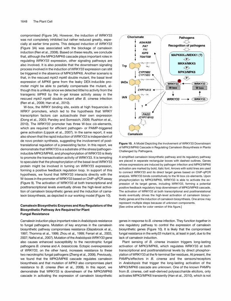
W-box, the WRKY binding site, exists at high frequencies in
WRKY promoters, which led to the hypothesis that WRKY
transcription factors can autoactivate their own expression
(Dong et al., 2003; Pandey and Somssich, 2009; Rushton et al.,
2010). The WRKY33 promoter has three W-box cis-elements,
which are required for efficient pathogen- or PAMP-triggered
gene activation (Lippok et al., 2007). In the same report, it was
also shown that the rapid induction ofWRKY33 is independent of
de novo protein synthesis, suggesting the involvement of post-
translational regulation of a preexisting factor. In this report, we
demonstrate thatWRKY33 is a substrate of the stress/pathogen-
inducible MPK3/MPK6, and phosphorylation of WRKY33 is likely
to promote the transactivation activity of WRKY33. It is tempting
to speculate that the phosphorylation of the basal-level WRKY33
protein might be involved in turning on WRKY33 expression,
forming a positive feedback regulation loop. In support of this
hypothesis, we found that WRKY33 interacts directly with the
W-boxes in the promoter ofWRKY33 based onChIP-qPCR assay
(Figure 9). The activation of WRKY33 at both transcriptional and
posttranscriptional levels eventually drives the high-level activa-
tion of camalexin biosynthetic genes and the induction of cama-
lexin biosynthesis, as depicted in our working model (Figure 10).
CamalexinBiosynthetic Enzymes andKeyRegulators of the
Biosynthetic Pathway Are Required for Plant
Fungal Resistance
Camalexin induction plays important roles inArabidopsis resistance
to fungal pathogens. Mutation of key enzymes in the camalexin
biosynthetic pathway compromises resistance (Glazebrook et al.,
1997; Thomma et al., 1999; Zhou et al., 1999; Ferrari et al., 2003,
2007;Nafisi et al., 2007).Mutation of theArabidopsisWRKY33gene
also causes enhanced susceptibility to the necrotrophic fungal
pathogens B. cinerea and A. brassicicola. Ectopic overexpression
of WRKY33, on the other hand, increases resistance to these
two necrotrophic fungal pathogens (Zheng et al., 2006). Previously,
we found that the MPK3/MPK6 cascade regulates camalexin
biosynthesis and that mutation of MPK3 also compromises plant
resistance to B. cinerea (Ren et al., 2008). In this report, we
demonstrate that WRKY33 is downstream of the MPK3/MPK6
cascade in activating the expression of camalexin biosynthetic
genes in response to B. cinerea infection. They function together in
one regulatory pathway to control the expression of camalexin
biosynthetic genes (Figure 10). It is likely that the compromised
fungal resistance in thewrky33mutant is, at least in part, due to the
lack of camalexin induction.
Plant sensing of B. cinerea invasion triggers long-lasting
activation of MPK3/MPK6, which regulates WRKY33 at both
transcriptional and posttranslational levels by direct phosphor-
ylation ofWRKY33 at the N-terminal Ser residues. At present, the
PAMPs/effectors in B. cinerea and the sensors/receptors
in Arabidopsis that trigger the long-lasting activation of the
MPK3/MPK6 cascade are unknown. One of the known PAMPs
from B. cinerea, cell wall–derived polysaccharide elicitors, only
activates MPK3/MPK6 transiently (Han et al., 2010), which is not
Figure 10. A Model Depicting the Involvement of WRKY33 Downstream
of MPK3/MPK6 Cascade in Regulating Camalexin Biosynthesis in Plants
Challenged by Pathogens.
A simplified camalexin biosynthetic pathway and its regulatory pathway
are placed in separate rectangular boxes with dashed outlines. Genes
whose expressions are induced by pathogen infection and MPK3/MPK6
activation are marked by bold, italic font. Arrows with solid lines are used
to connect WRKY33 and its direct target genes based on ChIP-qPCR
analysis. WRKY33 binds constitutively to the W-box cis-elements. Upon
phosphorylation by MPK3/MPK6, WRKY33 is able to activate the ex-
pression of its target genes, including WRKY33, forming a potential
positive feedback regulatory loop downstream of MPK3/MPK6 cascade.
The activation of WRKY33 at both transcriptional and posttranslational
levels eventually drives the high-level activation of camalexin biosyn-
thetic genes and the induction of camalexin biosynthesis. One arrowmay
represent multiple steps because of unknown components.
[See online article for color version of this figure.]
1648 The Plant Cell

associated with the induction of camalexin (data not shown).
Prolonged activation of MPK3/MPK6 leads to the coordinated
high-level induction of multiple genes in the camalexin biosyn-
thetic pathway (Ren et al., 2008), which drives themetabolic flow
from primary metabolism to the formation of camalexin, a sec-
ondary metabolite.
Is MPK4 Involved in Camalexin Induction in Arabidopsis
Challenged by B. cinerea?
Our finding that WRKY33 is essential to camalexin biosynthesis
is consistent with a previous report (Qiu et al., 2008b). However,
more research is needed to reconcile howWRKY33 is regulated.
Qiu et al. (2008b) conclude that MPK4 regulates WRKY33 by
sequestering it in the MPK4/MKS1 complex in the absence of
pathogens. After sensing an invading pathogen, the activation of
MPK4 phosphorylates MKS1 (but not WRKY33), which releases
WRKY33 from the complex so it can activate gene expression
(Qiu et al., 2008b). In the conditional gain-of-function DD Arabi-
dopsis plants, no MPK4 activation was detectable (Figure 1B)
(Liu and Zhang, 2004; Ren et al., 2008). However, WRKY33-
dependent camalexin induction was normal, suggesting that
MPK4 activation is not essential to the WRKY33-dependent
activation of camalexin induction. Furthermore, in the rescued
mpk3 mpk6 double mutant, high levels of MPK4 protein and
activation was detected (Figure 4D) (Ren et al., 2008; Han et al.,
2010), but camalexin induction by B. cinerea infection was
compromised (Ren et al., 2008), suggesting thatMPK4 activation
is not sufficient to support the camalexin induction. Finally, we
analyzed camalexin induction in the mpk4 mutant and found no
difference in the camalexin induction between the mpk4 mutant
and its wild-type control after B. cinerea infection (see Supple-
mental Figure 3 online). Based on these results, we conclude that
MPK4 is not required for camalexin induction inArabidopsis after
B. cinerea infection. It is possible that MPK4 has differential roles
in camalexin induction in response to different pathogens; for
example, MPK4 is not required for the camalexin induction by a
fungal pathogen (this study) but is involved in camalexin induc-
tion by a bacterial pathogen (Qiu et al., 2008b).
The Signaling Specificity of Multifunctional MPK3/MPK6 Is
Conferred by Their Diverse Substrates
MPK3/MPK6 are involved in many different processes, including
induction of ethylene biosynthesis in plants under stress (Kim
et al., 2003; Liu and Zhang, 2004; Joo et al., 2008; Han et al.,
2010), camalexin induction (Ren et al., 2008; this report), stoma-
tal development (Wang et al., 2007; Lampard et al., 2008), flower
petal abscission (Cho et al., 2008), and ovule development
(Wang et al., 2008). It appears that their multifunctionality and
signaling specificity are conferred by their ability to phosphorylate
different substrates. Four MPK3/MPK6 substrates have been
reported with functional data (Liu and Zhang, 2004; Lampard
et al., 2008; Bethke et al., 2009; this report). A subset of ACS
isoforms, the rate-limiting enzyme in the ethylene biosynthetic
pathway, can be directly phosphorylated by MPK3 and MPK6,
which stabilize the ACS protein and lead to ethylene induction
(Liu and Zhang, 2004; Joo et al., 2008; Han et al., 2010). In the
stomatal pathway, phosphorylation of SPEECHLESS, a basic
helix-loop-helix transcription factor involved in stomatal initiation,
negatively regulates stomatal development (Lampard et al., 2008).
ERF104, a member of the ethylene response factor (ERF) tran-
scription factor family, forms a complex with MPK6. Upon MPK6
activation by flg22 PAMP treatment, ERF104 is released from the
complex so it can access its target genes (Bethke et al., 2009).
Phosphorylation of WRKY33 by MPK3/MPK6 enhances its
activity in promoting the expression of downstream camalexin
biosynthetic genes. Different from ACS2/ACS6, accumulation
of WRKY33 protein in plants after B. cinerea infection or in gain-
of-function DD plants after DEX treatment is a result of tran-
scriptional activation (Figures 1F and 2F) but not of protein
stabilization. WRKY33 expressed under the 35S promoter
showed no change in protein levels after B. cinerea infection or
in DD background after DEX treatment (Figures 5B and 6B). As a
result, MPK3/MPK6 are capable of regulating their substrates at
different levels, including transcriptional activity, protein stability,
and protein complex formation. In addition, the expression
pattern of MPK3/MPK6 substrates can also affect signaling
specificity. ACS2/ACS6, ERF104, andWRKY33 are expressed in
most tissues, which is consistent with the general stress/defense
responses. By contrast, SPEECHLESS is expressed only in cells
about to enter the stomatal lineage, which confers the specific
role of MPK3/MPK6 in plant stomatal development (Wang et al.,
2007; Lampard et al., 2008). Research aimed at identifying addi-
tional MPK3/MPK6 substrates will reveal the molecular mecha-
nismsunderlying the complex rolesofMPK3/MPK6 in plant growth,
development, and response to environment and/or pathogens.
METHODS
Plant Growth, Treatments, Camalexin Measurement, and
Statistical Analysis
Arabidopsis thaliana plants were grown under a 14-h light cycle (100 mE
m22 s21) at 228C. Seedlings were grown in 20-mL gas chromatography
vials with 6 mL of half-strength Murashige and Skoog liquid medium in a
growth chamber under continuous light as described before (Ren et al.,
2008). Two-week-old seedlings were used for experiments. Seedlings
were collected at various time points after the addition of DEX or
inoculation of Botrytis cinerea spores (4.0 3 105 spores per vial). Proce-
dures for B. cinerea maintenance and spore preparation were as previ-
ously described (Ren et al., 2008; Han et al., 2010).
Camalexin production byArabidopsis seedlings was determined using a
previously described method (Tsuji et al., 1992; Glazebrook and Ausubel,
1994) with slight modification (Ren et al., 2008). Briefly, camalexin accu-
mulation in the culture medium, which reflects its production in the
seedlings, was quantified by fluorospectrometry with a standard curve
established using known concentrations of camalexin.
At least two independent repetitions were performed for experiments
with multiple time points. For single time point experiments, at least three
independent repetitions were done. Results from one of the independent
repeats that gave similar results were shown. n = 3 indicates independent
biological samples from one of the repeats. Student’s t test was used
to determine whether the difference between two groups of data at
a specific time point is statistically significant (P < 0.05). Statistically
A MAPK Substrate in Camalexin Induction 1649

different data groups are indicated using different number of asterisks
(0 to 3) vertically placed above the columns in the graphs.
Mutant Lines and Generation of Transgenic Plants
Mutant alleles ofmpk3-1 (SALK_151594) andmpk6-2 (Salk_073907)were
used for experiments (Liu and Zhang, 2004; Wang et al., 2007). The
generation of rescued mpk3 mpk6 double mutant was detailed by Wang
et al. (2007). Allmutants used in this study are in theCol-0 background. Two
T-DNA insertion mutant alleles, wrky33-1 (SALK_006603) and wrky33-2
(GABI_324B11), were described previously (Zheng et al., 2006). Both
wrky33-1 and wrky33-2 alleles were used for experiments demonstrating
the requirement of WRKY33 in B. cinerea– and DD-induced camalexin
production (Figures 1A to 1D and 2A to 2D). Similar results were obtained
and results using wrky33-1 were shown. Complementation experiments
usingWRKY33-TAP,WRKY33WT, andWRKY33SA transgenes (Figures 1E,
2E, and 5 to 7) were done in onlywrky33-2 background becausewrky33-1
acquired kanamycin resistance from the T-DNA insertion. For crosses,
wrky33mutationswere followedbyPCRgenotyping, and theDD transgene
was followed by hygromycin resistance. F3 double homozygous seedlings
were used for experiments unless stated otherwise.
For the generation of the native promoter-driven WRKY33-TAP
(PWRKY33:WRKY33-TAP) construct, an ;1.3-kb DNA fragment of the
WRKY33 promoter was PCR amplified from Arabidopsis genomic DNA
using primers 59-ATCAAGCTTCCACATATCGTGCAATAAGAAACT-39
and 59-ATCGAGCTCACGAAAAATGGAAGTTTGTTTTATAAAAGA-39. The
promoter sequence was digested with HindIII/SacI and inserted into the
same restriction sites to replace the cauliflower mosaic virus 35S promoter
of plant transformation/expression vector pOCA30 (Chen and Chen,
2002). The full-length WRKY33 cDNA was PCR amplified using primers
59-ATCGAGCTCTATATGGACAATAGCAGAACCAGACA-39 and 59-ATC-
GGATCCGGGCATAAACGAATCGAAAAATG-39 and fused with the TAP
tag as previously described (Xing and Chen, 2006). The WRKY33-TAP
construct was then subcloned behind theWRKY33 promoter in pOCA30.
The constructs were verified by DNA sequencing.
To generate loss-of-phosphorylation WRKY33 mutant (WRKY33SA),
we PCR amplified the wild-type WRKY33 cDNA and cloned it into
pBluescript II KS vector. Mutations were introduced by QuickChange
site-directed mutagenesis (Stratagene) and confirmed by sequencing.
Primers used for mutagenesis were as follows: 12AAF1, 59-ctccttcttca-
atctctatcGctccttctcttgtcGctccttccacttgtttc-39; 34AAF1, 59-ctccttccactt-
gtttcGCtccctctctttttctcgatGcccctgcttttgtctcc-39; 5AF1, 59-ctctgctaacg-
ttctagctGctccaaccacaggagc-39, and their complementary primers.
Mutated nucleotides are marked with uppercase letters. WRKY33SA
with all five Ser residues mutated to Ala residues was generated by three
successive mutagenesis steps.
To generate the 35S:4myc-WRKY33WT and 35S:4myc-WRKY33SA
constructs, we amplified the wild-type and WRKY33SA cDNA fragments
using primers 59-GGAATTCCATATGGCTGCTTCTTTTCTTACAATGGA-
CAATAGCAGAACCAGACA-39 and 59-GACTAGTTCAGGGCATAAAC-
GAATCG-39 and cloned the PCR fragment into a modified pBlueScript
II KS vector with a 4myc epitope tag coding sequence at the 59-end. The
WRKY33 cDNA with a 4myc epitope tag coding sequence was then
moved into the SpeI/XhoI sites of a modified pBI121 vector.
All generated binary vectors were transformed into Agrobacterium
trumefaciens strain GV3101. Arabidopsis transformation was performed
by the floral dip procedure (Clough and Bent, 1998), and transformants
were identified by screening for kanamycin or hygromycin resistance.
Independent lines with expression of tagged WRKY33 were identified
based on RNA gel blots and/or immunoblotting analyses. From these
transformants, those with a single copy of T-DNA insertion (based on the
3:1 segregation of antibiotic/herbicide resistance in T2 progeny) were
isolated, and homozygote transgenic plants were further identified in the
progeny based on segregation of antibiotic resistance.
Preparation of Recombinant WRKY33 Proteins and in Vitro
Phosphorylation Assay
WRKY33WT and WRKY33SA cDNA was PCR amplified using primers
(59-GGAATTCATGGCTGCTTCTTTTCTTACAATG-39 and 59-CCGCTC-
GAGTCAGGGCATAAACGAATCG-39) and ligated into pET32a (+) vector
in frame. The constructs were transformed into Escherichia coli strain
BL21 (DE3). Recombinant protein expression was induced with 0.25 mM
isopropylthio-b-galactoside for 3 h at 288C. His-tagged proteins were
purified using nickel columns and dialysis to 20 mM Tris-HCl, pH 7.5,
overnight at 48C.
The in vitro phosphorylation assay was performed as previously
described (Liu and Zhang, 2004; Han et al., 2010). In brief, recombinant
WRKY33 proteins were mixed with activated MPK3, MPK6, MPK4, and
MPK10 (20:1 substrate enzyme ratio) in the kinase reaction buffer (20 mM
HEPES, pH7.5, 10mMMgCl2, and 1mMDTT)with 25mMATP and [g-32P]-
ATP (1mCiper reaction). The reactionswere stoppedby theaddition ofSDS
sample buffer after 30 min. Phosphorylated WRKY33 was visualized by
autoradiography after being resolved in a 10% SDS-PAGE gel.
DNA-Protein EMSA
Synthetic DNA oligonucleotide (59-CGTTGACCGTTGACCGAGTTGACT-
TTTTA-39) with three W-boxes (underlined) was used as probe. DNA
probe labeling and gel mobility shift assays were performed as previously
described (Kim and Zhang, 2004). Briefly, two complementary strands of
the oligonucleotides were annealed and then labeled at the 59-end using
T4 polynucleotide kinase. The 32P-labeled DNA probe was purified using
Bio-Spin column (Bio-Rad). Freshly prepared recombinantWRKY33WT or
WRKY33SA protein (1 mg) was incubated with 20,000 to 50,000 cpm of
DNA probe (2 pmole) for 30 min at room temperature in binding buffer
(20 mM HEPES, pH 7.9, 0.1 mg/mL herring sperm DNA, 0.5 mM DTT,
0.1 mM EDTA, and 50 mM KCl) in the presence or absence of unlabeled
competitor DNA. The resulting protein-DNA complexes were resolved in
5% nondenaturing polyacrylamide gel in half-strength TBE buffer. Fol-
lowing electrophoresis, the gel was dried onto 3MM paper and exposed
to x-ray film. For testing the effect of MAPK phosphorylation on the DNA
binding activity of WRKY33, recombinant WRKY33WT was first incubated
with the activated MPK3 and MPK6 (equal mix) in the kinase reaction
buffer with 50 mMATP for 60 min at room temperature before performing
the EMSA assay.
Protein Extraction, Immunoblot Analysis, and in-Gel Kinase Assay
Proteins for in-gel kinase assay and immunoblot detection of Flag-tagged
DD were extracted as previously described (Zhang and Klessig, 1997).
For detection of tagged WRKY33 proteins, total proteins were extracted
using 3 volumes (v/w) of SDS-loading buffer without bromophenol blue
dye (Joo et al., 2008). The concentration of protein was determined using
the Bio-Rad protein assay kit with BSA as the standard. Immunoblot
detection of tagged transgene products was performed as previously
described (Liu and Zhang, 2004). Antibody against the myc-epitope tag
was purchased from Millipore, and the anti-IgG-horseradish peroxidase
(HRP) conjugate used to detect the TAP-taggedWRKY33was purchased
from Sigma-Aldrich. MBP, WRKY33, or WRKY33SA recombinant protein
was used as the substrate for the in gel-kinase assay (Zhang and Klessig,
1997; Liu and Zhang, 2004).
Mobility Shift Assay to Detect in Vivo Phosphorylated Proteins
Phos-tag reagent (NARD Institute) was used for the phospho-protein
mobility shift assay to detect in vivo phosphorylated WRKY33 protein.
Proteins (10 mg) were separated in a 10% SDS-PAGE gel containing
100 mmol/L Phos-tag and 200 mMMnCl2. After proteins were transferred
1650 The Plant Cell

to a nitrocellulosemembrane, 4myc-taggedWRKY33wasdetected using
the anti-myc antibody (Millipore).
qPCR Analysis
Total RNA was extracted using TRIzol reagent (Invitrogen). After DNase
treatment, 1 mg of total RNA was used for reverse transcription. qPCR
analysis was performed using an Optican 2 real-time PCR machine (MJ
Research) as previously described (Ren et al., 2008). After normalization
to an EF-1a control, the relative levels of gene expression were calcu-
lated. The primer pairs (forward and backward) used for qRT-PCR
were EF1a (At5g60390, 59-TGAGCACGCTCTTCTTGCTTTCA-39 and
59-GGTGGTGGCATCCATCTTGTTACA-39), CYP71A13 (At2g30770,
59-GGGTAGAGGCTGGACCAAAT-39 and 59-ACAACCGAAGATGGA-
AATGC-39), CYP71B15 (PAD3, At3g26830, 59-GGTACGGGATAAAT-
CTCTATGA-39 and 59-AGATACAGTCGATGAACCTAC-39), WRKY33 (At
2g38470, 59-GTGATATTGACATTCTTGACGA-39 and 59-GATGGTTGTG-
CACTTGTAGTA-39), and WRKY33-TAP transgene (59-AACAACGAAA-
CGCCTTCATC-39 and 59-CGGAATTCGCGTCTACTTTC-39).
WRKY33 transgene expression in the wrky33 mutant background was
examined by RT-PCR using a primer pair (59-TTCAGTCCCTCTC-
TTTTTCTCGAT-39 and 59-GGTCTCCTCGTTTGGTTCTTC-39) that spans
the whole open reading frame because nonfunctional transcripts are still
produced from themutatedwkry33 gene (Zheng et al., 2006). Equal cDNA
input was confirmed by PCR using EF1a control (59-GATGGTCAGA-
CCCGTGAGCACG-39 and 59-CAGTCTCAACACGTCCCACTGGC-39).
Twenty-five cycles of PCR were performed.
ChIP-qPCR Analysis
F1 plants generated from the cross of wrky33/4myc-WRKY33 and DD
lines were used for ChIP assay. Two-week-old seedlings treated with
1 mMDEX for 12 h were processed as described (Kaufmann et al., 2010).
Chromatin was isolated from 0.8 g of frozen tissue and sonicated with a
Bioruptor sonicator (15 s on and 15 s off cycles, medium-energy settings)
for 6 min. Immunoprecipitation was performed by incubating chromatin
with 2 mg of anti-myc antibody (Millipore) or mouse IgG (negative control)
for 1 h at 48C. The protein-chromatin immunocomplexes were captured
using Protein G-Dynal magnetic beads (Invitrogen). After Proteinase K
digestion, the immunoprecipitated DNA was purified using ChIP DNA
Clean and Concentrator kit (Zymo Research). Immunoprecipitated DNA
and input DNA were analyzed by qPCR using primers specific for
the promoter regions of PAD3 and WRKY33. The primer pairs (forward
and backward) used for ChIP-qPCR were PAD3 (59-TGTTCATG-
CACTTCGTCTCG-39 and 59-CTTCACTGACCGAGCTAACAAA-39) and
WRKY33 (59-TTTTTGAGCAAGAGCCAAGAAT-39 and 59-GGCTCAATG-
CTTTCATCATCTT-39) that flank the W-boxes in the promoters. The ChIP
results are presented as percentage of input DNA.
Accession Numbers
Sequence data from this article can be found in the Arabidopsis Genome
Initiative or GenBank/EMBL databases under the following accession
numbers: MPK3 (At3g45640), MPK6 (At2g43790), MKK4 (At1g51660),
MKK5 (At3g21220), EF1a (At5g60390),CYP71A13 (At2g30770),CYP71B15
(PAD3, At3g26830), andWRKY33 (At 2g38470).
Supplemental Data
The following materials are available in the online version of this article.
Supplemental Figure 1. Induction of WRKY-TAP Expression in
WRKY33-TAP/wrky33 Plants Infected by B. cinerea.
Supplemental Figure 2. MPK3/MPK6 Phosphorylation Sites in
WRKY33 Are Required for Full Complementation of wrky33 Mutation.
Supplemental Figure 3. B. cinerea–Induced Camalexin Biosynthesis
Is Not Affected by MPK4 Mutation.
ACKNOWLEDGMENTS
We thank Melody Kroll for proofreading the manuscript. This work was
supported by National Science Foundation Grants MCB-0543109 and
IOS-0743957 to S.Z. and IOS-0958066 to Z.C.
Received March 9, 2011; revised March 9, 2011; accepted March 29,
2011; published April 15, 2011.
REFERENCES
Asai, T., Tena, G., Plotnikova, J., Willmann, M.R., Chiu, W.-L.,
Gomez-Gomez, L., Boller, T., Ausubel, F.M., and Sheen, J.
(2002). MAP kinase signalling cascade in Arabidopsis innate immu-
nity. Nature 415: 977–983.
Ausubel, F.M. (2005). Are innate immune signaling pathways in plants
and animals conserved? Nat. Immunol. 6: 973–979.
Beckers, G.J.M., Jaskiewicz, M., Liu, Y., Underwood, W.R., He, S.Y.,
Zhang, S., and Conrath, U. (2009). Mitogen-activated protein kinases
3 and 6 are required for full priming of stress responses in Arabidopsis
thaliana. Plant Cell 21: 944–953.
Bethke, G., Unthan, T., Uhrig, J.F., Poschl, Y., Gust, A.A., Scheel, D.,
and Lee, J. (2009). Flg22 regulates the release of an ethylene
response factor substrate from MAP kinase 6 in Arabidopsis thaliana
via ethylene signaling. Proc. Natl. Acad. Sci. USA 106: 8067–8072.
Boller, T. (2005). Peptide signalling in plant development and self/non-
self perception. Curr. Opin. Cell Biol. 17: 116–122.
Bottcher, C., Westphal, L., Schmotz, C., Prade, E., Scheel, D., and
Glawischnig, E. (2009). The multifunctional enzyme CYP71B15 (PHY-
TOALEXIN DEFICIENT3) converts cysteine-indole-3-acetonitrile to
camalexin in the indole-3-acetonitrile metabolic network of Arabidop-
sis thaliana. Plant Cell 21: 1830–1845.
Broekaert, W.F., Delaure, S.L., De Bolle, M.F.C., and Cammue, B.P.
A. (2006). The role of ethylene in host-pathogen interactions. Annu.
Rev. Phytopathol. 44: 393–416.
Chen, C., and Chen, Z. (2002). Potentiation of developmentally regu-
lated plant defense response by AtWRKY18, a pathogen-induced
Arabidopsis transcription factor. Plant Physiol. 129: 706–716.
Cho, S.K., Larue, C.T., Chevalier, D., Wang, H., Jinn, T.-L., Zhang, S.,
and Walker, J.C. (2008). Regulation of floral organ abscission in
Arabidopsis thaliana. Proc. Natl. Acad. Sci. USA 105: 15629–15634.
Clough, S.J., and Bent, A.F. (1998). Floral dip: A simplified method for
Agrobacterium-mediated transformation of Arabidopsis thaliana. Plant
J. 16: 735–743.
Cui, H., Wang, Y., Xue, L., Chu, J., Yan, C., Fu, J., Chen, M., Innes,
R.W., and Zhou, J.-M. (2010). Pseudomonas syringae effector protein
AvrB perturbs Arabidopsis hormone signaling by activating MAP
kinase 4. Cell Host Microbe 7: 164–175.
Dangl, J.L., and Jones, J.D.G. (2001). Plant pathogens and integrated
defence responses to infection. Nature 411: 826–833.
del Pozo, O., Pedley, K.F., and Martin, G.B. (2004). MAPKKKalpha is a
positive regulator of cell death associated with both plant immunity
and disease. EMBO J. 23: 3072–3082.
Dixon, R.A. (2001). Natural products and plant disease resistance.
Nature 411: 843–847.
Dong, J., Chen, C., and Chen, Z. (2003). Expression profiles of the
Arabidopsis WRKY gene superfamily during plant defense response.
Plant Mol. Biol. 51: 21–37.
A MAPK Substrate in Camalexin Induction 1651

Ferrari, S., Galletti, R., Denoux, C., De Lorenzo, G., Ausubel, F.M.,
and Dewdney, J. (2007). Resistance to Botrytis cinerea induced in
Arabidopsis by elicitors is independent of salicylic acid, ethylene, or
jasmonate signaling but requires PHYTOALEXIN DEFICIENT3. Plant
Physiol. 144: 367–379.
Ferrari, S., Plotnikova, J.M., De Lorenzo, G., and Ausubel, F.M.
(2003). Arabidopsis local resistance to Botrytis cinerea involves
salicylic acid and camalexin and requires EDS4 and PAD2, but not
SID2, EDS5 or PAD4. Plant J. 35: 193–205.
Glazebrook, J., and Ausubel, F.M. (1994). Isolation of phytoalexin-
deficient mutants of Arabidopsis thaliana and characterization of their
interactions with bacterial pathogens. Proc. Natl. Acad. Sci. USA 91:
8955–8959.
Glazebrook, J., Zook, M., Mert, F., Kagan, I., Rogers, E.E., Crute,
I.R., Holub, E.B., Hammerschmidt, R., and Ausubel, F.M. (1997).
Phytoalexin-deficient mutants of Arabidopsis reveal that PAD4 en-
codes a regulatory factor and that four PAD genes contribute to
downy mildew resistance. Genetics 146: 381–392.
Hammerschmidt, R. (1999). Phytoalexins: What have we learned after
60 years? Annu. Rev. Phytopathol. 37: 285–306.
Han, L., Li, G.J., Yang, K.Y., Mao, G., Wang, R., Liu, Y., and Zhang, S.
(2010). Mitogen-activated protein kinase 3 and 6 regulate Botrytis cine-
rea-induced ethylene production in Arabidopsis. Plant J. 64: 114–127.
Ichimura, K., et al; MAPK Group. (2002). Mitogen-activated protein
kinase cascades in plants: A new nomenclature. Trends Plant Sci. 7:
301–308.
Jin, H., Liu, Y., Yang, K.-Y., Kim, C.Y., Baker, B., and Zhang, S.
(2003). Function of a mitogen-activated protein kinase pathway in N
gene-mediated resistance in tobacco. Plant J. 33: 719–731.
Joo, S., Liu, Y., Lueth, A., and Zhang, S. (2008). MAPK phosphory-
lation-induced stabilization of ACS6 protein is mediated by the non-
catalytic C-terminal domain, which also contains the cis-determinant
for rapid degradation by the 26S proteasome pathway. Plant J. 54:
129–140.
Kaufmann, K., Muino, J.M., Østeras, M., Farinelli, L., Krajewski, P., and
Angenent, G.C. (2010). Chromatin immunoprecipitation (ChIP) of plant
transcription factors followed by sequencing (ChIP-SEQ) or hybridization
to whole genome arrays (ChIP-CHIP). Nat. Protoc. 5: 457–472.
Kim, C.Y., Liu, Y., Thorne, E.T., Yang, H., Fukushige, H., Gassmann,
W., Hildebrand, D., Sharp, R.E., and Zhang, S. (2003). Activation of
a stress-responsive mitogen-activated protein kinase cascade in-
duces the biosynthesis of ethylene in plants. Plant Cell 15: 2707–2718.
Kim, C.Y., and Zhang, S. (2004). Activation of a mitogen-activated
protein kinase cascade induces WRKY family of transcription factors
and defense genes in tobacco. Plant J. 38: 142–151.
Kroj, T., Rudd, J.J., Nurnberger, T., Gabler, Y., Lee, J., and Scheel,
D. (2003). Mitogen-activated protein kinases play an essential role in
oxidative burst-independent expression of pathogenesis-related
genes in parsley. J. Biol. Chem. 278: 2256–2264.
Lampard, G.R., Macalister, C.A., and Bergmann, D.C. (2008). Arabi-
dopsis stomatal initiation is controlled by MAPK-mediated regulation
of the bHLH SPEECHLESS. Science 322: 1113–1116.
Lippok, B., Birkenbihl, R.P., Rivory, G., Brummer, J., Schmelzer, E.,
Logemann, E., and Somssich, I.E. (2007). Expression of AtWRKY33
encoding a pathogen- or PAMP-responsive WRKY transcription fac-
tor is regulated by a composite DNA motif containing W box elements.
Mol. Plant Microbe Interact. 20: 420–429.
Liu, Y., Ren, D., Pike, S., Pallardy, S., Gassmann, W., and Zhang, S.
(2007). Chloroplast-generated reactive oxygen species are involved in
hypersensitive response-like cell death mediated by a mitogen-acti-
vated protein kinase cascade. Plant J. 51: 941–954.
Liu, Y., and Zhang, S. (2004). Phosphorylation of 1-aminocyclopro-
pane-1-carboxylic acid synthase by MPK6, a stress-responsive
mitogen-activated protein kinase, induces ethylene biosynthesis in
Arabidopsis. Plant Cell 16: 3386–3399.
Martin, G.B., Bogdanove, A.J., and Sessa, G. (2003). Understanding
the functions of plant disease resistance proteins. Annu. Rev. Plant
Biol. 54: 23–61.
Menke, F.L.H., van Pelt, J.A., Pieterse, C.M.J., and Klessig, D.F.
(2004). Silencing of the mitogen-activated protein kinase MPK6 com-
promises disease resistance in Arabidopsis. Plant Cell 16: 897–907.
Morrissey, J.P., and Osbourn, A.E. (1999). Fungal resistance to plant
antibiotics as a mechanism of pathogenesis. Microbiol. Mol. Biol. Rev.
63: 708–724.
Nafisi, M., Goregaoker, S., Botanga, C.J., Glawischnig, E., Olsen,
C.E., Halkier, B.A., and Glazebrook, J. (2007). Arabidopsis cytochrome
P450 monooxygenase 71A13 catalyzes the conversion of indole-3-
acetaldoxime in camalexin synthesis. Plant Cell 19: 2039–2052.
Nakagami, H., Pitzschke, A., and Hirt, H. (2005). Emerging
MAP kinase pathways in plant stress signalling. Trends Plant Sci.
10: 339–346.
Nurnberger, T., and Scheel, D. (2001). Signal transmission in the plant
immune response. Trends Plant Sci. 6: 372–379.
Pandey, S.P., and Somssich, I.E. (2009). The role of WRKY transcrip-
tion factors in plant immunity. Plant Physiol. 150: 1648–1655.
Petersen, M., et al. (2000). Arabidopsis map kinase 4 negatively
regulates systemic acquired resistance. Cell 103: 1111–1120.
Qiu, J.-L., et al. (2008b). Arabidopsis MAP kinase 4 regulates gene
expression through transcription factor release in the nucleus. EMBO
J. 27: 2214–2221.
Qiu, J.-L., Zhou, L., Yun, B.-W., Nielsen, H.B., Fiil, B.K., Petersen, K.,
Mackinlay, J., Loake, G.J., Mundy, J., and Morris, P.C. (2008a).
Arabidopsis mitogen-activated protein kinase kinases MKK1 and
MKK2 have overlapping functions in defense signaling mediated by
MEKK1, MPK4, and MKS1. Plant Physiol. 148: 212–222.
Ren, D., Liu, Y., Yang, K.-Y., Han, L., Mao, G., Glazebrook, J., and
Zhang, S. (2008). A fungal-responsive MAPK cascade regulates
phytoalexin biosynthesis in Arabidopsis. Proc. Natl. Acad. Sci. USA
105: 5638–5643.
Ren, D., Yang, H., and Zhang, S. (2002). Cell death mediated by MAPK
is associated with the hydrogen peroxide production in Arabidopsis.
J. Biol. Chem. 277: 559–565.
Rushton, P.J., Somssich, I.E., Ringler, P., and Shen, Q.J. (2010).
WRKY transcription factors. Trends Plant Sci. 15: 247–258.
Schuhegger, R., Nafisi, M., Mansourova, M., Petersen, B.L., Olsen,
C.E., Svatos, A., Halkier, B.A., and Glawischnig, E. (2006).
CYP71B15 (PAD3) catalyzes the final step in camalexin biosynthesis.
Plant Physiol. 141: 1248–1254.
Staskawicz, B.J., Ausubel, F.M., Baker, B.J., Ellis, J.G., and Jones,
J.D.G. (1995). Molecular genetics of plant disease resistance. Science
268: 661–667.
Suarez-Rodriguez, M.C., Adams-Phillips, L., Liu, Y., Wang, H., Su,
S.-H., Jester, P.J., Zhang, S., Bent, A.F., and Krysan, P.J. (2007).
MEKK1 is required for flg22-induced MPK4 activation in Arabidopsis
plants. Plant Physiol. 143: 661–669.
Tena, G., Asai, T., Chiu, W.-L., and Sheen, J. (2001). Plant mitogen-
activated protein kinase signaling cascades. Curr. Opin. Plant Biol. 4:
392–400.
Thomma, B.P.H.J., Nelissen, I., Eggermont, K., and Broekaert, W.F.
(1999). Deficiency in phytoalexin production causes enhanced sus-
ceptibility of Arabidopsis thaliana to the fungus Alternaria brassicicola.
Plant J. 19: 163–171.
Tsuji, J., Jackson, E.P., Gage, D.A., Hammerschmidt, R., and
Somerville, S.C. (1992). Phytoalexin accumulation in Arabidopsis
thaliana during the hypersensitive reaction to Pseudomonas syringae
pv syringae. Plant Physiol. 98: 1304–1309.
1652 The Plant Cell

VanEtten, H.D., Matthews, D.E., and Matthews, P.S. (1989). Phyto-
alexin detoxification: Importance for pathogenicity and practical im-
plications. Annu. Rev. Phytopathol. 27: 143–164.
van Loon, L.C., Geraats, B.P.J., and Linthorst, H.J.M. (2006). Ethyl-
ene as a modulator of disease resistance in plants. Trends Plant Sci.
11: 184–191.
Wan, J., Zhang, S., and Stacey, G. (2004). Activation of a mitogen-
activated protein kinase pathway in Arabidopsis by chitin. Mol. Plant
Pathol. 5: 125–135.
Wang, H., Liu, Y., Bruffett, K., Lee, J., Hause, G., Walker, J.C., and
Zhang, S. (2008). Haplo-insufficiency of MPK3 in MPK6 mutant
background uncovers a novel function of these two MAPKs in
Arabidopsis ovule development. Plant Cell 20: 602–613.
Wang, H., Ngwenyama, N., Liu, Y., Walker, J.C., and Zhang, S.
(2007). Stomatal development and patterning are regulated by envi-
ronmentally responsive mitogen-activated protein kinases in Arabi-
dopsis. Plant Cell 19: 63–73.
Xing, D., and Chen, Z. (2006). Effects of mutations and constitutive
overexpression of EDS1 and PAD4 on plant resistance to different
types of microbial pathogens. Plant Sci. 171: 251–262.
Yang, K.-Y., Liu, Y., and Zhang, S. (2001). Activation of a mitogen-
activated protein kinase pathway is involved in disease resistance in
tobacco. Proc. Natl. Acad. Sci. USA 98: 741–746.
Zhang, J., et al. (2007). A Pseudomonas syringae effector inactivates
MAPKs to suppress PAMP-induced immunity in plants. Cell Host
Microbe 1: 175–185.
Zhang, S., and Klessig, D.F. (1997). Salicylic acid activates a 48-kD
MAP kinase in tobacco. Plant Cell 9: 809–824.
Zhang, S., and Klessig, D.F. (2001). MAPK cascades in plant defense
signaling. Trends Plant Sci. 6: 520–527.
Zheng, Z., Qamar, S.A., Chen, Z., and Mengiste, T. (2006). Arabidop-
sis WRKY33 transcription factor is required for resistance to necrotro-
phic fungal pathogens. Plant J. 48: 592–605.
Zhou, N., Tootle, T.L., and Glazebrook, J. (1999). Arabidopsis PAD3, a
gene required for camalexin biosynthesis, encodes a putative cyto-
chrome P450 monooxygenase. Plant Cell 11: 2419–2428.
A MAPK Substrate in Camalexin Induction 1653

DOI 10.1105/tpc.111.084996; originally published online April 15, 2011; 2011;23;1639-1653Plant Cell
Guohong Mao, Xiangzong Meng, Yidong Liu, Zuyu Zheng, Zhixiang Chen and Shuqun ZhangArabidopsisPhytoalexin Biosynthesis in
Phosphorylation of a WRKY Transcription Factor by Two Pathogen-Responsive MAPKs Drives
This information is current as of May 28, 2018
Supplemental Data /content/suppl/2011/04/06/tpc.111.084996.DC1.html /content/suppl/2011/04/17/tpc.111.084996.DC2.html
References /content/23/4/1639.full.html#ref-list-1
This article cites 63 articles, 28 of which can be accessed free at:
Permissions https://www.copyright.com/ccc/openurl.do?sid=pd_hw1532298X&issn=1532298X&WT.mc_id=pd_hw1532298X
eTOCs http://www.plantcell.org/cgi/alerts/ctmain
Sign up for eTOCs at:
CiteTrack Alerts http://www.plantcell.org/cgi/alerts/ctmain
Sign up for CiteTrack Alerts at:
Subscription Information http://www.aspb.org/publications/subscriptions.cfm
is available at:Plant Physiology and The Plant CellSubscription Information for
ADVANCING THE SCIENCE OF PLANT BIOLOGY © American Society of Plant Biologists