phosphatidylserine/cholesterol bilayers supported on a polycation/alkylthiol layer pair
TRANSCRIPT

Journal of Colloid and Interface Science228,82–89 (2000)doi:10.1006/jcis.2000.6910, available online at http://www.idealibrary.com on
Phosphatidylserine/Cholesterol Bilayers Supportedon a Polycation/Alkylthiol Layer Pair
Liqin Zhang, Carrie A. Booth,1 and Pieter Stroeve2
Center on Polymer Interfaces and Macromolecular Assemblies (CPIMA), Department of Chemical Engineering and Materials Science,University of California at Davis, Davis California 95616
Received October 11, 1999; accepted April 17, 2000
1-Stearoyl-2-oleoyl phosphatidylserine (SOPS)/cholesterol bi-layers, supported on a polycation/alkylthiol layer pair on agold surface, were investigated by surface plasmon resonance(SPR) and fluorescence recovery after photobleaching. The sub-strate was formed by electrostatic adsorbance of a hydratedpoly(diallyldimethylammonium chloride) (PDDA) layer on thenegatively charged surface of a self-assembled monolayer of 11-mercaptoundecanoic acid (MUA) on gold. Lipid membranes withdifferent SOPS/cholesterol compositions were deposited on thePDDA/MUA layer pair by vesicle fusion. When the cholesterolcontent was below 20%, single bilayers were deposited. Fluores-cence recovery after the bleaching experiments revealed that theSOPS/cholesterol bilayers were mobile at room temperature; lateraldiffusion coefficients of a fluorescence probe were approximately1× 10−9 cm2/s. The kinetics of the addition of the ion-channel-forming peptide gramicidin to the supported bilayers was detectedby SPR. C© 2000 Academic Press
Key Words: supported bilayer membranes; bilayer mobility;cholesterol; surface plasmon resonance; vesicle fusion.
le–acitwpt
(n
f
em-ro-dingolif-ertsm-pid
asend
me-nss,as
anessys-nesand
n5).p-ievedre-sedcemerrane
oly-are
t al-rted
aalyti-) and
INTRODUCTION
Negatively charged lipid phosphatidylserine (PS) and choterol are important constituents of biological membranes (1The cytosolic leaflets of the plasma membranes of many mmalian cells are enriched in PS (3). Its anionic nature is expeto influence the way in which the PS molecule interacts wother components of the mammalian membranes, e.g., pro(4). Electrostatic attraction of these anionic PS lipids is oneto bind positively charged proteins to membranes. For examAnnexin V is a protein that binds to cells that have elevalevels of PS in their outer leaflet of the plasma membrane,hardly binds to cells with low levels of surface-exposed PSCholesterol is also an important component in cell membrathat modulates the mechanical and functional properties o
1 Current address: Department of Chemical Engineering, Virginia PolytechInstitute and State University, Blacksburg, VA 24060.
2 To whom correspondence should be addressed. E-mail: pstroevucdavis.edu. Fax: (530) 752-8778.
weix-forespy,
820021-9797/00 $35.00Copyright C© 2000 by Academic PressAll rights of reproduction in any form reserved.
s-3).m-tedtheinsayle,
edbut5).es
the
nic
e@
membranes. It is present in both leaflets of the plasma mbranes of many mammalian cells (6). It is known to have pfound effects on a broad range of cellular processes, incluenzyme activities, membrane surface properties, and cell preration (7). It is believed that in most cases cholesterol exits effect by modulating the physical properties of lipid mebranes (8), for example, changing the motional ordering of limolecules and reducing passive permeability (9).
As important components of brain cell membranes (1), phbehavior and thermotropic properties of mixtures of PS acholesterol have been studied by differential scanning caloriter (DSC) and X-ray diffraction in the form of vesicle dispersio(1, 4, 10). However, unlike zwitterionic phosphatidylcholinethere is a lack of knowledge of PS/cholesterol mixturessupported planar membranes. Solid-supported lipid membrhave been of great practical and scientific interest as modeltems to study the structure and function of natural membraas well as membrane-bound biomolecules, e.g., peptidesproteins (11–14).
The fluid nature of a biological membrane is now well knowand is believed to play a crucial role in cellular mechanisms (1This makes it important to minimize the effect of the solid suport to the membrane because the solid substrates are belto restrict the motion of supported biomembranes (16). Asviewed by Sackmann, flexible polymer layers have been uas “cushions” to lift biomembranes away from the solid surfa(13). The hydrated space created by the addition of the polylayer can decrease the substrate effect, both on the membitself and on the membrane-bound biomolecules. Certain pelectrolytes have been used to form this cushion layer. Theyable to induce vesicle fusion, which is a powerful method thalows the incorporation of transmembrane proteins in suppomembranes (11, 12).
In this work, we studied PS/cholesterol lipid bilayers assupported model membrane system and used surface ancal techniques, such as surface plasmon resonance (SPRfluorescence recovery after photobleaching (FRAP). Sinceare dealing with a negatively charged PS/cholesterol lipid mture system, a positively charged polyelectrolyte is favorablelipid membrane binding. In a study of anionic lipid membranby X-ray scattering and X-ray photoelectron spectrosco

C
oetpmairn
eo
edrr
ms
unli
n
.o
onehy-lynelandne-thisbyb-pon-
odthatsi-thecon-din
m
y-yl-robeids
is,eromalsallosis
he
8.5o-theindi-
/mlnedere
t 2e-ml.
so-
PHOSPHATIDYLSERINE/
Sohling and Schouten used polyethylenimine, a weakly ptively charged polymer, over a silicon surface to support natively charged phosphatidic acid lipid layers (12). Due tolimit of the techniques used in their work, the supported limembrane system was removed from the aqueous environduring examination, which may have altered the structureproperties of the lipid membranes. Moreover, no lateral diffusproperty of the supported anionic lipid membranes was repowhich is an important criterion of how well a model membraresembles natural biological membranes.
To investigate the model membrane system in an aquenvironment, SPR was used in this work to detect the adstion of both the polymer layer and lipid layers. Surface plasmresonance has become an important technique in the arbiosensor research (17–19). In SPR measurements, a golface is needed as the substrate to allow the excitation of suplasmons (17–19). To improve the surface charge propeof the gold surface, a self-assembled monolayer of alkylth11-mercaptoundecanoic aid (MUA), was used in our systemadsorb to the gold surface through strong sulfur–gold bonds (A positively charged polymer, poly(diallyldimethylammoniuchloride) (PDDA), was deposited on the negatively chargedface of the acidic headgroups of the alkylthiol monolayer. Stalayers of PDDA adsorbed on solid substrates (21, 22) wereto induce fusion of SOPS/cholesterol vesicles that have netative charges. The PDDA/MUA layer pair was formed tothe biomembrane from the gold surface. It is important to poout here that both sides of the lipid membrane are exposehydrated environments. A schematic drawing of this systemshown in Fig. 1. The technique of FRAP was used to revthe lateral mobility of the SOPS/cholesterol lipid membraformed on the PDDA/MUA layer pair.
FIG. 1. Schematic representation of the model membrane systemalkylthiol (MUA) layer is self-assembled to a gold surface and a cationic pmer (PDDA) layer is subsequently adsorbed on the negatively charged M
surface. A lipid bilayer with negative charges is subsequently deposited onPDDA/MUA layer pair.HOLESTEROL BILAYERS 83
si-g-
heidentnd
onted,e
ousrp-ona ofsur-
facetiesiol,
to20).
ur-blesedeg-ftintd to
iseales
Anly-UA
The small channel-forming peptide gramicidin has beenof the most extensively studied peptides. The interest in thisdrophobic antibiotic peptide stems mostly from its relativesimple structure and exceptionally well defined ion chanfunction (23, 24). Studies on gramicidin can help to understthe principles that govern the folding and function of membraspanning channels and membrane proteins in general (23). Inwork, gramicidin was added to the supported bilayers formedSOPS/cholesterol mixtures on PDDA/MUA-covered solid sustrates. High cholesterol content has been reported to be ressible for the inactivation of gramicidin ion channels in red blocell membranes (25). Lundbæk and co-workers speculatedcholesterol shifts gramicidin channel inactivation toward potive potentials by increasing bilayer stiffness (26). One ofpotential applications of the supported membrane systemstructed in this work is that the role of cholesterol on gramicichannel functions can be studied.
MATERIALS AND METHODS
Materials
11-Mercaptoundecanoic acid (MUA) was obtained froAldrich (Milwaukee, WI). The polymer poly(diallyldimethyl-ammonium chloride) (PDDA) was obtained from Polsciences, Inc. (Warrington, PA). The lipid 1-stearoyl-2-oleophosphatidylserine (SOPS) and the fluorescence p(16 : 0–6 : 0 NBD–PS) was purchased from Avanti Polar Lip(Alabaster, AL). Cholesterol (99+%), gramicidin (fromBacillusbrevis) and the buffer tris(hydroxymethyl)aminomethane (Tr99.9+%) were from Sigma (St. Louis, MO). The refractivindex matching fluid, 1-iodonaphthalene, was purchased fCargille Laboratories Inc. (Cedar Grove, NJ). All chemicwere used as received without further purification. Inexperiments, water was purified by a Nanopure reverse osmpurification system from Barnstead (Dubuque, IA). Tresistivity was better than 17.7 MÄ-cm.
Solution Preparation
The buffer used throughout the experiments was Tris(5 mM Tris buffer, pH 8.5). The PDDA solution, vesicle slutions, and the peptide solutions were all prepared withsame buffer. The percentages used throughout the articlecate molar fraction unless otherwise stated.
Desired amounts of 10 mg/ml SOPS solutions and 1.2 mgcholesterol solutions in chloroform were added to a precleaglass vial. When lipid mixtures were used, the solutions wvortexed for 2 min to mix the lipids. A small N2 stream was thenused to evaporate the lipids onto the wall of the vial. Abouml of the Tris 8.5 buffer solution was added to the vial to rdisperse the lipids. The final lipid concentration was 0.5 mg/The lipid solution was incubated in a 50◦C water bath for 15min with several vortexing periods of 15 s in between. The
thelution was sonicated with an ultrasonic tip (1/8-in. diameter,Branson 250, 10-W output) for 1 min followed by three pulsed

,
t
iic
ft
b.
i
eusn
taels
dr
e
2
b
n
rb
ts
ne-ondthentsntoit
noso-
bi-om
omflu-ith
pe
ld-pac-fordesaswasso-st
edse
sedasen.ntilthe
lessction
,age,
ofed
mluesess.
84 ZHANG, BOOTH
periods of 10 s each. The clear lipid solution was equilibraat room temperature for at least 30 min before use. For FRexperiments, 2 mol% of NBD–PS was used in the lipid solutin chloroform before evaporation. All the other steps of vespreparation remained the same.
Gramicidin solution was prepared in Tris 8.5 buffer. Duethe hydrophobic nature of this peptide, it could not be totadissolved when 1 mg of powder was added to 10 ml of bufAfter 10 min of mixing, the suspension was centrifuged andsupernatant solution was used in the experiments.
Refractive indices of solutions were measured on an A60 refractometer (Bellingham and Stanley Ltd., England)sodium spectral lamp with a wavelength of 589.3 nm wused.
Preparation of Gold Slides with FunctionalSelf-Assembled Monolayers (SAMs)
Gold slides covered by a close-packed SAM of alkylthMUA were used as substrates for subsequent depositionPDDA layer and lipid layers. High-index glass LaSFN9 slid(n= 1.85 atλ= 633 nm, Schott, Germany) were used as sstrates in SPR experiments. About 50 nm of gold was depoon LaSFN9 at a rate of 0.2A/s by vacuum evaporation in aelectron beam chamber (pressure,<5× 10⁻⁶ mbar). The slidesused for FRAP experiments were prepared from cleaned gmicroscope slides from Fisher Scientific. A 3 nm-thick tinium layer was sputtered first on the glass microscope slidimprove the adhesion of gold to glass. A gold layer identicathat deposited on LaSFN9 slides was subsequently depoThe gold-coated slides were immersed in a 5 mMsolution ofMUA in ethanol for at least 18 h at room temperature. The sliwere removed from the solution, rinsed with ethanol, and dthoroughly with N2 right before use.
Adsorption Experiments
The adsorption experiments were conducted with a SPR susing a laser beam with a wavelength of 633 nm. The Swas set up according to the Kretschmann configuration (A gold-coated LaSFN9 slide covered with a MUA monolaywas mounted on a Teflon cell that holds about 0.8 ml of fluA LaSFN9 glass prism was mounted on the glass slide w1-iodonaphthalene as the refractive index matching fluidtween the prism and the glass slide.
With the SPR the reflectivity was measured as a functiothe angle. The value of the minimum angle,θm, is a functionof the thickness and refractive indices of the layers adsoon the gold surface as well as the refractive index of the bsolution (17, 18, 28). From the shift of the minimum angleθm,the thickness of the organic layers can be calculated usingFresnel equations (SPR software: WASPLAS version 2.1, MPlanck-Institute for Polymer Research, Mainz, Germany) ifrefractive index information of the adsorbed organic layer
known. The kinetics of adsorption of each layer was obtainby the angle of incidence being set 0.5◦ belowθm.AND STROEVE
edAPonle
tollyer.he
beAas
olof asb-ited
lass-
s totoited.
esied
tupPR7).
erid.ithe-
of
edulk
theax-heis
Solutions in the Teflon cell were exchanged by simultaous injection and withdrawal from the Teflon cell using twsyringes. A buffer solution was injected into the Teflon cell awas allowed to sit for approximately 15 min to equilibrate withe surrounding room temperature before SPR measuremwere taken. Then, a 0.2 M PDDA solution was exchanged ithe cell and was allowed to sit for at least 20 min beforewas rinsed with buffer. The cell was rinsed until there wasfurther decrease of reflectivity. The freshly prepared vesiclelutions were injected into the cell and the growth of thelayer was monitored. All experiments were conducted at rotemperature.
The mobility of the supported bilayers was measured at rotemperature by FRAP experiments on a Nikon Diaphot 300orescence microscope. Two half-circular Teflon spacers wa thickness of 60µm were placed on a cleaned microscocover slide. About 60µl of a 0.2 M PDDA solution in Tris8.5 buffer was added in the middle of the two spacers. A gocoated microscope glass slide was then mounted on the sers. The setup was allowed to sit in a humid environmentabout 30 min. The PDDA solution captured between the sliwas exchanged with buffer before a lipid vesicle solution winduced to the space by exchanging solutions. The setupthen allowed to sit for at least 3 h. The excess vesiclelution was exchanged with Tris 8.5 buffer solution at lea10 times.
The NBD fluorescent probe was excited by blue light filterfrom a Mercury lamp and emitted bright green light. With uof a 20× objective lens on the microscope, a spot of 60µmin diameter was bleached for 3 min with the diaphragm cloand with both neutral density filters out. The bleached spot wviewed using the same objective lens with the diaphragm opImages were captured using a digital camera every 15 min uthe spot was totally diffused and could not be observed onscreen. The time interval for taking images was restricted tothan 3 s. The shutter was closed in between each data colleto prevent excessive bleaching.
Scion Imaging(www.scioncorp.com), an on-line softwarewas used to quantitatively analyze the images. For each iman optical intensity profile was generated. The differencesmaximum and minimum values of the curves were normalizand plotted versus time. The lateral diffusion coefficient (D)was calculated fromD (cm2/s)= 0.224ω2(cm)/t1/2(s).ω is theradius of the bleached spot andt1/2 is the half-life of the fluo-rescence recovery (29, 30).
RESULTS AND DISCUSSION
Refractive Indices of Organic Layers and Solutions
After the minimum angle shift information was obtained frothe SPR measurements (Fig. 2), the refractive index vaof the adsorbed layer were needed to calculate the thickn
edThe typical literature value for the refractive index for long-chain lipids, 1.49 (31), was used in the simulations. Literature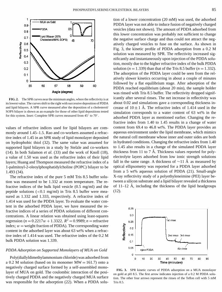
C
i
tee
mft
e
ip
b
lueh-x
a
efr
l
o
bedgedromngeneg-n inMsig-lu-
DA
rel-teshelderif-
wass in-ethere-
teranicsboth.40yer
y-onsybedglee-nessps
erlu-
PHOSPHATIDYLSERINE/
FIG. 2. The SPR curves near the minimum angles, where the reflectivityits lowest value. The curves shift to the right with successive deposition of PDand lipid bilayers. A SPR curve measured after the deposition of a cholesSOPS bilayer is shown as an example for those of other lipid depositions tfor this system. Inset: Complete SPR curves measured from 45◦ to 70◦.
values of refractive indices used for lipid bilayers are comonly around 1.45–1.5. Rao and co-workers assumed a retive index of 1.45 in an SPR study of lipid monolayer deposion hydrophobic thiol (32). The same value was assumedsupported lipid bilayers in a study by Stelzle and co-work(14). In both Salamonet al. (33) and the work of Knoll (18),a value of 1.50 was used as the refractive index of their llayers; Huang and Thompson measured the refractive index1-palmitoyl-2-oleoylphosphatidylcholine (POPC) bilayer to1.493 (34).
The refractive index of the pure 5 mM Tris 8.5 buffer sotion was measured to be 1.332 at room temperature. Thfractive indices of the bulk lipid vesicle (0.5 mg/ml) and tpeptide solutions (<0.1 mg/ml) in Tris 8.5 buffer were measured as 1.332 and 1.333, respectively. A refractive inde1.414 was used for the PDDA layer. To evaluate the water ctent in the adsorbed PDDA layer, we have measured thefractive indices of a series of PDDA solutions of different cocentrations. A linear relation was obtained using least-squregression (n= 0.2217w+ 1.3322, R2= 0.9989;n= refractiveindex;w=weight fraction of PDDA). The corresponding watcontent in the adsorbed layer was about 63 wt% when a retive index of 1.414 was used. The refractive index of the 0.2bulk PDDA solution was 1.339.
PDDA Adsorption on Supported Monolayers of MUA on Go
Poly(diallyldimethylammonium chloride) was adsorbed froa 0.2 M solution (based on its monomer MW= 161.7) onto anegatively charged surface formed by a self-assembled mlayer of MUA on gold. The coulombic interaction of the pos
tively charged PDDA and the negatively charged MUA surfawas responsible for the adsorption (22). When a PDDA soHOLESTEROL BILAYERS 85
s atDArol/sted
-rac-edforrs
idof ae
-re-
e
ofon-re-
n-res
rac-M
d
m
no-i-
tion of a lower concentration (20 mM) was used, the adsorPDDA layer was not able to induce fusion of negatively charvesicles (data not shown). The amount of PDDA adsorbed fthis lower concentration was probably not sufficient to chathe negative surface charge and thus could not attract theatively charged vesicles to fuse on the surface. As showFig. 3, the kinetic profile of PDDA adsorption from a 0.2solution was measured by SPR. The reflectivity increasednificantly and instantaneously upon injection of the PDDA sotion, mostly due to the higher refractive index of the bulk PDsolution (n= 1.339) than that of the Tris 8.5 buffer (n= 1.332).The adsorption of the PDDA layer could be seen from theatively slower kinetics occurring in about a couple of minufollowed by a flat equilibrium stage. After adsorption of tPDDA reached equilibrium (about 20 min), the sample howas rinsed with Tris 8.5 buffer. The reflectivity dropped signicantly after several rinses. The net increase in reflectivityabout 0.02 and simulations gave a corresponding thicknescrease of 10± 1 A. The refractive index of 1.414 used in thsimulation corresponds to a water content of 63 wt% inadsorbed PDDA layer as mentioned earlier. Changing thefractive index from 1.40 to 1.45 results in a change of wacontent from 69.4 to 46.8 wt%. The PDDA layer providesaqueous environment under the lipid membrane, which mimthe natural cell membrane whose inner and outer sides arein hydrated conditions. Changing the refractive index from 1to 1.45 also results in a change of the simulated PDDA lathickness from 11 to 7A. Thickness values reported for polelectrolyte layers adsorbed from low ionic strength solutifall in the same range. A thickness of∼11 A as measured boptical ellipsometry was reported for a PDDA layer adsorfrom a 5 wt% aqueous solution of PDDA (21). Small-anX-ray reflectivity study of a polyethyleneimine (PEI) layer btween a silicon substrate and a lipid bilayer revealed a thickof 11–12A, including the thickness of the lipid headgrou(12).
FIG. 3. SPR kinetic curves of PDDA adsorption on a MUA monolayon gold at pH 8.5. The first arrow indicates injection of a 0.2 M PDDA so
celu-tion. The other four arrows represent the rinses of the Teflon cell with 5 mMTris 8.5.

,
es
d
ee
a
n
/l)
OPS/t pHresentrs in-sing
aticandthead-ofo-
pureeous
ay-
in-
in-dexbi-s.adeessofIn-
ard-endon-inedandainstions incon-with
86 ZHANG, BOOTH
Deposition of Lipids on PDDA
Lipid layers of pure SOPS and SOPS/cholesterol mixtuwere deposited on the positively charged PDDA layers by vcle fusion. The kinetic profiles of the adsorption procesare shown in Figs. 4a and 4b. The final membrane thicknvalues are plotted in Fig. 5. The thickness values weretained by fitting the SPR curves before and after vesiclesion (after rinsing) using the WASPLAS 2.1 software. Lipiwith different compositions of SOPS and cholesterol showdifferent results in both kinetic profiles and thickness valuPure SOPS followed a sharp and fast deposition before thflectivity reached an equilibrium state within an hour’s timThe thickness of the adsorbed SOPS layer was 32± 2 A assimulated by the software WASPLAS 2.1 assuming a refrtive index of 1.49 for the bilayer. With an increasing amouof cholesterol composition up to 20%, the thicknesses of
FIG. 4. SPR kinetic profiles of lipid adsorption with different compositioof SOPS and cholesterol (a: 100% SOPS; 95% SOPS/5% cholesterol;SOPS/10% cholesterol; 80% SOPS/20% cholesterol. b: 75% SOPScholesterol; 70% SOPS/30% cholesterol; 60% SOPS/40% cholestero
PDDA/MUA-covered gold surfaces at pH 8.5. The kinetic profiles of the rinsiprocesses are not shown since the reflectivity changes after rinsing are negligAND STROEVE
ressi-esessob-fu-seds.re-
e.
c-ntthe
s90%25%
on
FIG. 5. Thickness changes versus percentage of cholesterol in Scholesterol mixtures deposited on PDDA/MUA-covered gold surfaces a8.5. Each datum point represents several experiments. The error bars repthe standard deviations of each datum point. The thickness of the lipid layecreases from a single bilayer thickness to that of multilayers, with an increapercentage of cholesterol in the mixture.
lipid layers slightly increased to about 45A, which still fallsin the range of a single bilayer thickness. The electrostattraction between the positively charged PDDA surfacethe negatively charged lipid vesicles was most probablydriving force of adsorption. The negative charges of thesorbed lipid layer electrostatically repel further depositionlipid molecules, resulting in a single bilayer structure. Flurescence images of the lipid membranes formed by bothSOPS and 80% SOPS/20% cholesterol showed homogendistribution of the fluorescent probe molecules in the bilers. The thickness of the SOPS bilayer, 32A, simulated us-ing a refractive index of 1.49, is lower than the usual sgle bilayer thickness, 40–50A, for long-chain phospholipids(35). As mentioned earlier, different values of refractivedices can be found in the literature. Using a refractive inof 1.45 for the bilayers in this work, the simulated SOPSlayer thickness is 43A, a reasonable single bilayer thicknesHowever, using 1.45 as the refractive index for the bilayer mup of 90% SOPS and 10% cholesterol results in a thicknof 51 A, which is thicker than a single bilayer. A thickness39 A is obtained when a refractive index of 1.49 is used.deed, the thickness of the bilayer varies by about 10A whenthe refractive index values of 1.45 and 1.50 are used. Regless of the value of the refractive index, Fig. 5 shows the trof increasing bilayer thickness with increasing cholesterol ctent. It is assumed that cholesterol molecules have constrafreedom due to the fused steroid ring system in its structuremay decrease the motional freedom of the hydrocarbon chof neighboring SOPS molecules. In other words, incorporaof cholesterol molecules might reduce the incidence of kinkthe SOPS molecules (9) and thus make the bilayer moredensed. Since the same refractive index is used for bilayers
ngible.
different compositions of cholesterol, the increase in reflectivitycan also to some extent be an indication of an increase in the
/C
e
ic
llilcdsa,g
e
te
eeao)ns
n
n
t
uin
d
le
flilt
ach-ture.ture;/20%
asles-os-tssup-tedtedlidaseirectng
0%s at
PHOSPHATIDYLSERINE
packing density of lipid molecules rather than an increaslayer thickness.
When the cholesterol composition is over 25%, the thnesses of the adsorbed lipid layers exceed the typical vaof single bilayers. For 70% SOPS/30% cholesterol and 6SOPS/40% cholesterol lipid layers the average thickness is a66 A, as shown in Fig. 5. Multilayers or undisrupted vesicmay have formed on the PDDA-covered substrate. Thelayer thickness did not increase significantly when the choterol composition is increased from 30% to 40%. This resultbe explained by the limited solubility of cholesterol in PS lipiIt has been reported that phase separation into a PS–cholephase and an almost pure cholesterol phase takes placebelow molar ratios of approximately 2 : 1 PS : cholesterol (2Thus, the composition of cholesterol in the negatively charPS–cholesterol phase, which mainly contributes to the lipidsorption to the positively charged polymer surface, saturatabout 30% and does not increase further when more cholesis used. For 75% SOPS/25% cholesterol, the standard deviaof the thickness measurement are rather large. The experimdata cover both the single bilayer range and beyond, showinintermediate adsorption behavior. As shown in Figs. 4a andkinks are present in the kinetic profiles for cholesterol contof more than 20%, but not when less cholesterol is involvThe effect is obvious at cholesterol compositions of 30%40%. A possible reason for these kinks is the SPR line brening and sharpening due to an inhomogeneous layer (36consequence of the formation of multilayers or the adsorptioundisrupted vesicles. The uncharged cholesterol moleculelute the charge density of the lipid mixture, and thus there mnot be sufficient electrostatic repulsion for further adsorptiolipid molecules to the formed bilayer.
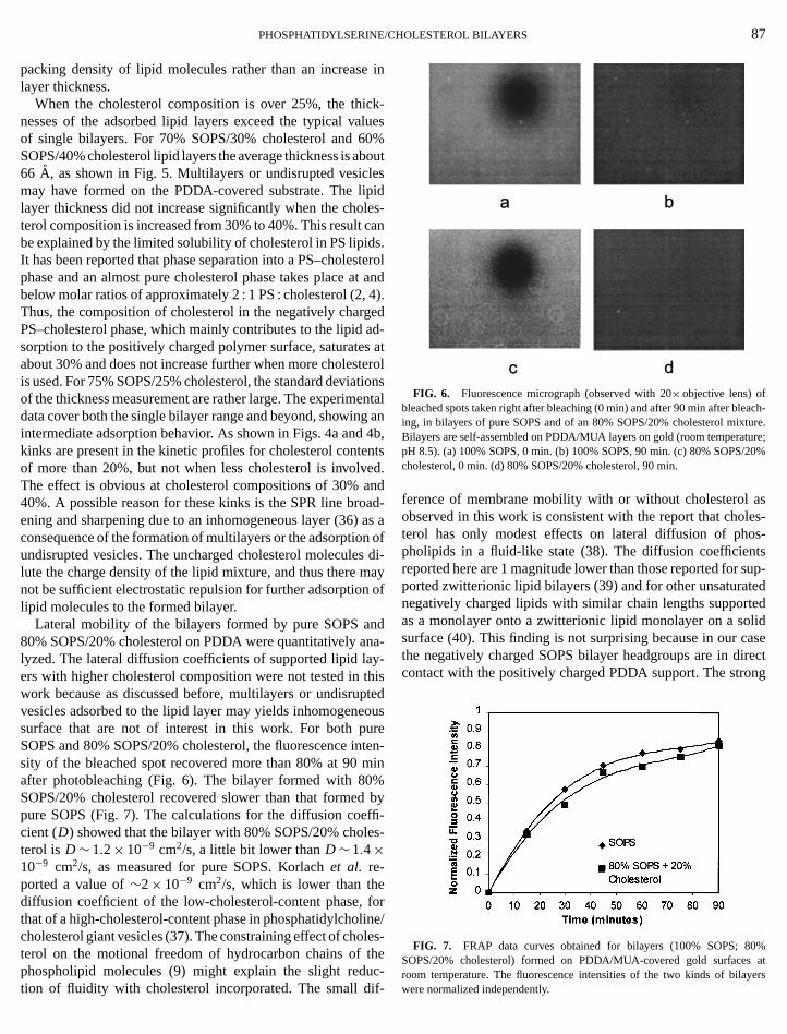
Lateral mobility of the bilayers formed by pure SOPS a80% SOPS/20% cholesterol on PDDA were quantitatively alyzed. The lateral diffusion coefficients of supported lipid laers with higher cholesterol composition were not tested inwork because as discussed before, multilayers or undisruvesicles adsorbed to the lipid layer may yields inhomogenesurface that are not of interest in this work. For both pSOPS and 80% SOPS/20% cholesterol, the fluorescencesity of the bleached spot recovered more than 80% at 90after photobleaching (Fig. 6). The bilayer formed with 80SOPS/20% cholesterol recovered slower than that formepure SOPS (Fig. 7). The calculations for the diffusion coecient (D) showed that the bilayer with 80% SOPS/20% choterol is D∼ 1.2× 10⁻⁹ cm2/s, a little bit lower thanD∼ 1.4×10⁻⁹ cm2/s, as measured for pure SOPS. Korlachet al. re-ported a value of∼2× 10⁻⁹ cm2/s, which is lower than thediffusion coefficient of the low-cholesterol-content phase,that of a high-cholesterol-content phase in phosphatidylchocholesterol giant vesicles (37). The constraining effect of choterol on the motional freedom of hydrocarbon chains of
phospholipid molecules (9) might explain the slight redution of fluidity with cholesterol incorporated. The small difHOLESTEROL BILAYERS 87
in
k-lues0%boutespides-ans.terolt and4).ed
ad-s atterolionsntal
g an4b,ntsd.ndad-as aofdi-
ayof
dna-y-hisptedousreten-
min%
byffi-s-
orne/es-he
FIG. 6. Fluorescence micrograph (observed with 20× objective lens) ofbleached spots taken right after bleaching (0 min) and after 90 min after bleing, in bilayers of pure SOPS and of an 80% SOPS/20% cholesterol mixBilayers are self-assembled on PDDA/MUA layers on gold (room temperapH 8.5). (a) 100% SOPS, 0 min. (b) 100% SOPS, 90 min. (c) 80% SOPScholesterol, 0 min. (d) 80% SOPS/20% cholesterol, 90 min.
ference of membrane mobility with or without cholesterolobserved in this work is consistent with the report that choterol has only modest effects on lateral diffusion of phpholipids in a fluid-like state (38). The diffusion coefficienreported here are 1 magnitude lower than those reported forported zwitterionic lipid bilayers (39) and for other unsaturanegatively charged lipids with similar chain lengths supporas a monolayer onto a zwitterionic lipid monolayer on a sosurface (40). This finding is not surprising because in our cthe negatively charged SOPS bilayer headgroups are in dcontact with the positively charged PDDA support. The stro
FIG. 7. FRAP data curves obtained for bilayers (100% SOPS; 8SOPS/20% cholesterol) formed on PDDA/MUA-covered gold surface
c--room temperature. The fluorescence intensities of the two kinds of bilayerswere normalized independently.
th
i
aiovr
t
te
b
l
iny
therrt
t itsho-a-nd))betedtheon-ay
hanex-on-theers.f an
uesrsue
bi-s. Ita sig-y-nnel
tionow-on-oned
t bytiontedit is
rtedThenedtectep-the
tedin.0 of44).e-ys-cantem
88 ZHANG, BOOTH
electrostatic interaction between the two opposite chargesreduce the lateral diffusion of the lipid molecules. Despiterelatively low diffusion coefficients observed in this work, tvalues measured here fall in the range of diffusion coefficiein fluid bilayers and are well above the characteristic diffusties for lipid membranes in the rigid Lβ phase (partly ordereduntilted,D< 10−11 cm2/s) (35).
Peptide Insertion
Gramicidin has been used as a model for understandingmolecular basis for the folding and dynamics of membrproteins, due to its well-defined ion-channel-forming funct(23, 24). This hydrophobic peptide achieves its antibiotic actiby forming cation-selective channels in lipid bilayer structu(23). The addition of the channel-forming peptide gramicidinthe supported bilayers was studied with single bilayers formby 90% SOPS/10% cholesterol and 80% SOPS/20% cholesTwo milliliters of saturated gramicidin solution (<0.1 mg/ml)in Tris 8.5 buffer was injected into the Teflon cell. The refletivity increased and reached equilibrium within 30 min. Afrinsing with Tris 8.5 buffer, the reflectivity did not show a mesurable decrease (Fig. 8). The net reflectivity increases onbilayers suggest that a certain amount of gramicidin remainethe bilayer. For the bilayer formed by 80% SOPS/20% choterol, the thickness increased from 39 to 42A after the peptideinsertion. In the case of 90% SOPS/10% cholesterol, the thness increased from 33 to 36A. It should be noted here thathe same value of refractive index has been used for lipidlayers before and after peptide insertion. Therefore, the thness increases should be regarded as increases of materthe lipid membranes. There have been reports on the thickmismatch between gramicidin and bilayers. The lipid bila
FIG. 8. SPR kinetic profiles of gramicidin (in Tris 8.5 buffer) insertion inbilayers composed of 90% SOPS/10% cholesterol and 80% SOPS/20% cterol formed on PDDA/MUA-covered gold surfaces. The kinetics measuremwere actually stopped before rinsing to make thickness measurements. Foity purposes, the profiles shown in this figure connect the peptide insekinetics and the rinsing kinetics together. Data of thickness measuremen
listed beside the curves for bilayer thicknesses before and after peptide inseThickness data are in units of Angstroms., AND STROEVE
mayheentsvi-,
thenen
ityestoed
erol.
c-r
a-oth
d ines-
ick-tbi-
ick-als inesser
ooles-ntsclar-
tions are
surrounding an embedded peptide or protein might adjushydrocarbon thickness to match the length of the hydropbic surface of the biomolecule, which results in local deformtion of the lipid membranes (41). Gramicidin has been fouto stretch 1,2-dilauroyl-sn-glycero-3-phosphocholine (DLPCand thin 1,2-dimyristoyl-sn-glycero-3-phosphocholine (DMPCwhen embedded in those free lipid membranes (42). It willchallenging to investigate the deformation of solid-supporlipid membranes caused by the hydrophobic matching ofpeptide and the lipid bilayers. The solid support may exert cstraints to the deformation of the lipid membranes, which mresult in a larger energy penalty of membrane deformation tthat in free membranes. The deformation energy cost mayceed that of the hydrophobic mismatch. In other words, the cstraint of the solid support may result in less deformation ofsupported lipid membrane than those observed in free bilaySince the thickness detected by SPR can only be a result oaddition of materials, refractive index independent techniqsuch as cyclic voltammetry may be a better technique to puthis in a future study.
The net increases in thickness were the same for bothlayer systems with different SOPS/cholesterol compositionappears that the concentration of cholesterol does not havenificant effect on the amount of gramicidin addition to the bilaers. This appears to be consistent with the report that the chastructure is remarkably insensitive to the detailed composiof the membrane environment (23). The channel function, hever, may be different with different amounts of cholesterol ctent in the bilayer. A reduction of gramicidin channel duratiin bilayers with cholesterol was reported (26). It was believthat an increase in stiffness of the lipid membrane broughthe incorporation of cholesterol molecules altered the funcof the ion-channel-forming peptide (26). It needs to be poinout here that, with optical techniques, such as SPR alone,not easy to determine whether gramicidin molecules inseinto the bilayer structure or attached to the bilayer surface.position of the gramicidin molecules may be better determiby electrochemical methods, e.g., cyclic voltammetry, to dethe increased conductivity with ion channels formed by the ptide (43). Nonetheless, the bilayer system constructed andkinetic information obtained by SPR in our work may contributo better understandings of the insertion behavior of gramiciRecent results show that a membrane fusion peptide wt-2the influenza virus also can be bound to supported bilayers (With the potential of gramicidin as a pore-forming material bing incorporated into this polyelectrolyte-supported bilayer stem, the bilayer may become more permeable to ions thatselectively pass through the ion channel. The bilayer systested here may have applications as a biosensor for ions.
SUMMARY
rtion.A SOPS/cholesterol bilayer on a polycation/alkylthiol layerpair was investigated by SPR and FRAP. A thin, hydrated PDDA

C
mrt
ee%p
Fh
t
nu
th
C
.
n
and
–96.
age,
t.
ev,
O. S.,
.,
.
. W.,
.
ton,
W.,
PHOSPHATIDYLSERINE/
layer was deposited on a self-assembled monolayer of Mon gold to lift lipid bilayers away from solid substrate. By thmethod of vesicle fusion, pure SOPS and SOPS/cholesteroltures adsorbed on the PDDA/MUA layer pair through electstatic interactions. With increasing cholesterol content inlipid mixture, the supported bilayer increased from a singlelayer thickness to those of multilayers, likely with inhomogneous surfaces. FRAP experiments showed a slight decrof the lateral diffusion coefficient for the bilayers with 20cholesterol than for those formed by pure SOPS. SPR eximents on the gramicidin addition to the bilayers showeddifference in kinetics or thickness changes with two differeSOPS/cholesterol compositions. In both cases, the ion-chanforming peptide remained bound to the lipid membranes.ture work on this system will focus on the investigation of tactivity of the peptide molecules of forming ion-selective chanels through the supported bilayers, for example, by the meof cyclic voltammetry, and possible controlling factors suchcholesterol content will be tested.
ACKNOWLEDGMENTS
This work was supported by the MRSEC Program of the National ScieFoundation under Award DMR-9808677. C. Booth was a CPIMA SUREdergraduate student for the summer of 1999. Prof. M. Longo is thankeduseful discussions. We are grateful to Dr. A. McKiernan for help withfluorescence microscopy and to B. Argo for assistance with the SPR.would also like to thank Dr. Z. Hou for the help with depositing gold on glaslides.
REFERENCES
1. Bach, D.,Chem. Phys. Lipids35,385–392 (1984).2. Bach, D., and Wachtel, E.,Biochim. Biophys. Acta979,11–19 (1989).3. Barnes, J. P., and Freed, J. H.,Biophys. J.75,2532–2546 (1998).4. Wachtel, E. J., Borochov, N., and Bach, D.,Biochim. Biophys. Acta1066,
63–69 (1991).5. Stuart, M. C. A., Reutelingsperger, C. P. M., and Frederik, P. M.,Cytometry
33,414–419 (1998).6. Yeagle, P. L.,in “Biology of Cholesterol” (P. L. Yeagle, Ed.), Chap. 6. CR
Press, Boca Raton, FL, 1988.7. McMullen, T. P. W., Lewis, R. N. A. H., and McElhaney, R. N.,Biochemistry
32,516–522 (1993).8. Dhal, C., and Dhal, J.,in “Biology of Cholesterol” (P. L. Yeagle, Ed.), Chap
7. CRC Press, Boca Raton, FL, 1988.9. Yeagle, P. L.,in “Advances in Cholesterol Research” (M. Esfahani a
J. B. Swaney, Eds.), pp. 114–116. Telford Press, Caldwell, NJ, 1990.
10. Wachtel, E. J., and Bach, D.,Biochim. Biophys. Acta922, 234–238(1987).
HOLESTEROL BILAYERS 89
UAeix-
o-hebi--ase
er-nontnel-u-en-hodas
cen-foreWess
d
11. Majewski, J., Wong, J. Y., Park, C. K., Sietz, M., Israelachvili, J. N.,Smith, G. S.,Biophys. J.75,2363–2367 (1998).
12. Sohling, U., and Schouten, A. J.,Langmuir75,3912–3919 (1996).13. Sackmann, E.,Science271,43–48 (1996).14. Stelzle, M., Weissmuller, G., and Sackmann, E.,J. Phys. Chem.97,
2974–2981 (1993).15. Sen, A., Ghosh, P. K., and Mukherjee, M.,Mol. Cell. Biochem.187,
183–190 (1998).16. Gyorvary, E., Wetzer, B., Sleytr, U. B., Sinner, A., Offenh¨ausser, A., and
Knoll, W., Langmuir15,1337–1347 (1999).17. Knoll, W.,Annu. Rev. Phys. Chem.49,569–638 (1998).18. Knoll, W.,MRS Bull.16,29–39 (1991).19. Aust, E. F., Ito, S., Sawodny, M., and Knoll, W.,Trends Polym. Sci.2,
313–323 (1994).20. Ulman, A.,in “An Introduction to Ultrathin Organic Films, from Langmuir
Blodgett to Self-Assembly,” Chap. 3. Academic Press, San Diego, 1921. Kleifeld, E. R., and Ferguson, G. S.,Science265,370–373 (1994).22. Decher, G.,in “Comprehensive Supramolecular Chemistry” (J. P. Sauv
Ed.), Vol. 9, Chap. 14. Pegamon Press, New York, 1996.23. Koeppe, R. E., II, and Anderson, O. S.,Annu. Rev. Biophys. Biomol. Struc
25,231–258 (1996).24. Mou, J., Czajkowsky, D. M., and Shao, Z.,Biochemistry35, 3222–3226
(1996).25. Schagina, L. V., Blasko, K., Grinfeldt, A. E., Korchev, Y. E., and L
A. A., Biochim. Biophys. Acta.978,145–150 (1989).26. Lundbæk, J. A., Birn, P., Girshman, J., Hansen, A. J., and Andersen,
Biochemistry35,3825–3830 (1996).27. Aust, E. F., Sawodny, M., Ito, S., and Knoll, W.,Scanning16, 353–361
(1994).28. Spinke, J., Yang, J., Wolf, H., Liley, M., Ringsdorf, H., and Knoll, W
Biophys. J.63,1667–1671 (1992).29. Mercel, R., Sackmann, E., and Evans, E.,J. Phys. Fr.50,1535–1555 (1989)30. Axelrod, D., Koppel, D. E., Schlessinger, J., Elson, E., and Webb, W
Biophys. J.16,1055–1069 (1976).31. Schouten, S., Stroeve, P., and Longo, M.,Langmuir15,8133–8139 (1999)32. Rao, N. M., Plant, A. L., Silin, V., Wight, S., and Hui, S. W.,Biophys. J.
73,3066–3077 (1997).33. Salamon, Z., Huang, D., Cramer, W. A., and Tollin, G.,Biophys. J.75,
1874–1855 (1998).34. Huang, C., and Thompson, T. E.,J. Mol. Biol.13,183–193 (1965).35. Marsh, D., “CRC Handbook of Lipid Bilayers.” CRC Press, Boca Ra
FL, 1990.36. Silin, V., and Plant, A.,Trends Biotechnol.15,353–359 (1997).37. Korlach, J., Schwille, P., Webb, W. W., and Feigenson, G. W.,Proc. Natl.
Acad. Sci. USA96,8461–8466 (1999).38. Yeagle, P. L.,Biochim. Biophys. Acta822,267–287 (1985).39. Tamm, L. K., and McConnell, H. M.,Biophys. J.47,105–113 (1985).40. Gilmanshin, R., Creutz, C. E., and Tamm, L. K.,Biochemistry33,
8225–8232 (1994).41. Huang, H. W.,Biophys. J.50,1061–1070 (1986).42. Harroun, T. A., Heller, W. T., Weiss, T. M., Yang, L., and Huang, H.
Biophys. J.76,937–945 (1999).
43. Plant, A. L.,Langmuir15,5128–5135 (1999).44. Zhang, L., Longo, M. L., and Stroeve, P.,Langmuir16,5093 (2000).