(phomosis) sp, (xanthium italicum - home:...
TRANSCRIPT
HELU,13, Nr. 13, p.p. 93-106 (1990)
Diaporthe (Phomosis) SP, A NEW PATHOGEN OFCOCKLBBUR (Xanthium italicum Moretti) AND OF
SUNFLOWER (Helianthus annuus L. )'
Jean-Bernard Carriere', Marija Pet
' Setnences C argill, Pqtrehoradc, France' htstinrc of Field and Vegaable Crops, 21000 Novi Sae yugoslavia
SUMMARY
InJuly 1988,we have observed symptoms onXal, thiurnitalicun Moreal, similarto thoseprovoked by Phomopsis helianthi Munt.-CveL on sunflowers. The causative agent of thesesymptoms on X italicum isDiaponhe (Phomopsis) rp thât we have arbitrarily naÀe d phornop-sis (Diaponhe ) "xanthium".
' This publication describes the anamorphic and telomorphic forms of this parasite, andgives the first results concerning its biological cycle on X italictun.
- The peculiarity of this fungus is to be equally pathogenic on sunflowers (results provedb-y ascospores tests), and to provoke on the stems of this plant identical symptoms to thosedeveloped by P. helianthi. Then, what is the relation between p "xanthium" and p hetianthi?
The comparison of their anamorphic forms brings them togerher. But, on the otherhând, their telomorphic forms present quite important differences. Flowwer, one of theP helianthi biotypes isolated by Yang (1983) is characterised by typical traits ofD. helianthiand of P'\anthiarn". Consiclcring lhese various observations, and the fact thàt the Xanthiwnand Helianthw genera arc hoth narive to the American continent, we are presently working onthe following hypothesis: cou,ltl t'hornosis (Diaporthe).helianthi be J ro.rn ôr phornop-sis (Diaponhe)."xanthitun" adaptcri ro a new host (the sunflowc:r)?
INTRODUCTION
The pathogic fungus of Helianthus annuus L. (Asteracea, tribe of Heliantheae),Phomosis (Diaporthe) helianthi Munt.-Cvet., has been studiecl since 1980. So, we have inhand numerous results:^o^n!".tlilg its morphological an<t biological properties (Mun-tanola-cverkovié et al., 1981, 1985; Aéimovié and Srrasser, 19g2;Mariêet àt., tgsz; uerret al., 1983; Yang et al., 1984; Fayret and Assemat, 19g7).
During observations on the Danubian flora, we have noticed on an otherAsteraceaeof Heliantheae trlbe - Xanthium italicum Moretti - symptoms similars to those provokeclby P helianthi on sunflowers. The causative agent for these symptoms on Xanthiumitalicum belongs to the Phomopsri genus.
For all these different reasons (similarity of symptoms, belonging of these twoparasites to the Phomopsis genus, taxonomic position of the Helianthi and Xanthium1 The authors are most gratefull to Prof. O. SKORIé and to Semences Cargill for financial support.
They also wish to thank Prof. J. FAyRET (univerry p. Sabarier, Toul-ousE, FRANCE) for hisadvice and Mm. B. KoLIAZINSKI for the determination of x italicum Moretti.
HELIA,13, Nr. 13, p.p. 93-106 (1990)
genera) we have decided to study this new parasite, its possible pathogenicity on thesunflower and its taxonomical relation to P helianthi.
MATERIAL AND METHODS
1- Stains isolarion (July-August 1988): Xanthium italicum infected leaves comingfrom 34 places in Vojvodina (Yugoslavia) have allowed the constitution of a 25-isolatecollection (P1 to P25). The isolations have been made according to the method described
by Altman (1966) on potatoe-dextrose-agar (PDA) and have been cultivated at
Z3oC +zoc in the dark. In November 1988, infected stems have been harvested in the
same location. From this material kept in natural conclitions we have constituted a 53
ascospore isolate collection.
2- Nutrient media: For the composition and the preparation of the media we have
refered to the work by Smith and Onions (1971). Concerning Fayret's medium we have
introduced a cold sterilization of amino acids and of vitamins.
3- Biometrical study:* Stem fragments bearing mature perithecia have been fixed in the Bouin's solution
and dehydrated in graded series of alcohol, then embeded in paraffin. Sections
5pm thick have been cut out transversally to the perithecium level (microtomMC2).After being mounred in Canada Balsam 50, identical preparations tothese presented in Figure 11. have been mcasured (magnification 16 x 10,
F:10).*ffcnidiu: a similar study is being carricd out.*Asci:1ft) mcasurements havebeen carric<l out (magnification 1,6x4O,F=L,515).*Ascorpores and conclia: we have used the immersion technique (magnifica-tion:
16 x 1fi), F=0,628, impresion oil nD(20):1,515). Only the ascopores freed
from their asci have been measured.
4- Pathogenicity tests: they have been carried out according to the followingmethod:
* Spraying of ascopore solution: the preparation of suspension has been realized- afrer Mihaljèèvié (19Saa). The plants wrapped in a plastic foil after spraying
have been putT1iiours in growth chamber (20oC-/+ zoc, L6 hours day / 8hours night,light intensity 2500lux, relative humidity lNVo),
* mycelin mats on the apical part of the leaves (Bertrand and Ttrurvielle, 1987),* mycelin mats on severed petioles (Bertrand and Tbuwielle, 1987)'* mycelin mats directly inserted in wounded stems (Mihaljëevié et al., 1984a).
5- Vegetable material:Xanthium italicum has been produced from seeds harvested
in October. Ttr suppress the dormancy of the seeds gathered from October to February,
it has been necessâry to stock them at 5oC during a minimum of 21 days. The plants have
been grown on a mixture of compost and river sand (5OVo-50Vo), in plastic pots(30 x 20 x 10 cm) with 6 or 9 plants in each pot. In the greenhousse, the average tempera-ture was 25oc-l+ 5oC, 16 hours day, 8 hours night, light intensity: 35000lux.
HELU,13, Nr. 13, p.p.93-106 (1990)
Concerning the tests on the sunflower we have used three lines sensitive to Phelianthi L-1, L-3, CMS-20. Cultivated on compost, they have undergone growingconditions similar to these described above.
Statistical processing has been carrided out on the SYSTAT program (SYST1[INC., 1987) kindly provided by Dr. Mihaljëevié. Confidence intervals have been measuredwith a 5Vo risk.
RESULTS
1- Symptoms observed onXanthium italicum Moretti:The first symptoms appeared on the edges of foliar limbs. There is a tissue
discoloration which presents a light dehydration. Then this zone darkens, necroses takea roughtly triangular shape, very typical. One of the corners of this triangle is centred onthe main foliar vein (Fig. 1). They expand toward the petiole, perceeded by a light green,sometimes golden yellow margin (Fig. 1). The first phase leads to the petiole coloniza-tion, a transversal section of which shows a darkening of the vascular system. At this stage,the foliar limb is completly parched (Fig. 2).
On the stem there appear dry cankers, reddish brown, circular to elliptical, centrerlon the initially infected petiole (Fig a). A longitudinal section of this zone reveals that adarknening of the vascular system preceeds the necroses visible on the epidermis. Thedifferent necroses can merge to form a large ring around the stem. From the petioleinsertion point, the parasite also destroys medular parenchyma by making cavities. Thesetwo phenomena provoke a sharp plant withering, which frequently brcaks under theaction of wind or various impacts. In spring, the infected plants can bc rccognized by thesilver-grcy to reddish-brown colour of the epidermis.
2- First observations of the anamorphic stage:The isolations carried out from infccted leaves and stcms have allowed to observe.
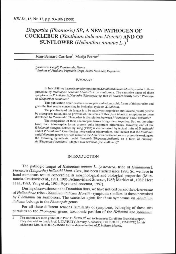
under pure culture, white colonies, roughly circular, oily looking (Fig. 6) ancl composedof septate mycelium. Aerial mycelium and substrated ones are to be found in variableproportions. On the fourth day of culture, outlines of pycnidia differentiate under theshape of globulous or subglobulous structures, colourless. After the wall melanizationand the occurrence of one or several ostioles, liquid exudates, colourless to greenish-beige appear (sixth/seventh day - Figs. 7, 8). These exuclates like the pycnidia bocly, arerich in beta conidia, hyaline cells, filiform, wirh one of rheir.rips bent (Fig. 9). The dailyobservation of the pycnidia content from the fifth to the fifteenth day of culture on PDAhas not allowed the observation of alpha conidia on the isolates of our collection. At thestage of developing, the mycelium allows to differentiate two isolate types, the first onekeeping this whitish colouring on a light beige substrat, the second one producing golclendrops and a brown-dark brown substrate (Fig. 6). These two types have been observedeither pure or mixed. We have been able to link these morphological particularities ofmycelium to morphological variability observed at the level of pycniclia (work inprogress).
Thcsc morphological elements, according to the classification criteria proposed byBarnctt and Hunter (1972), indicate the Phomosis genus.
95

HELU,13, Nr. 13, p.p.93-106 (1990)
HELU,13, Nr. 13, p.p.93-106 (1990) 97

98 HELU,13, Nr. 13, p.p.93-106 (1990)
Fig. 1: Phomopsis,,xantium", natural infection: leaf symptom (arrow: limb tissues discoloration).
Fig 2: idem: petiole colonisation.Fig. 3: idzm artificial infection with ascospores: symptome on leaves.
Fig. 4: Photnopsis"xantiurn", natural infection: stem cankers.
Fig 5: Diaponhe (Phornopsis) helianthi Mrurt.-Cva.: symptoms on leaf du to a natural infection.
Fig. 6: P "mnthiutn", two mycelial froms found in PDA.Fig. 7,8: idem,pycnidia on PDA (arrow: exudat, x32).Fig.9: idnn, beta conidia (from natural host, x1600).Fig 10 D. "mntium", mature perilhecia from natural infected stemof Xantium italicumMoretti (r32).Fig 11l.idetn,Perithecium munted in Canada balsam (x51,2)
Fig. 12: idnn, asci (arrow: location of the apical chitinoide ring, x1600).
Fig 13: idem,ascospores (x1600).Fig. 14: symptomsprovoked byPhomopsis (Diaporthe)"mntium" onsunflowerleave (arrow: colonisedvein).
Fig 15: idem, calonisation of the petiole.Fig 16: idem, necros€s on the sunflower stem.Fig. 17: Phomopsis helianthi: stem cânker (natural infection).

HELU,13, Nr. 13, p.p.93-106 (1990)
In order to be clearer, we have decided to arbitrarily name the anamorphic stage ofthis parasite as Phomopsis'xanthiumn.
On natural host, the pycnidia develop on the necrotic tissues of the stem. Thepycnidia are brown-dark brown, globulose, variable (work in progress). However, on fiveinfected stem samples (October 1988 to February 1989) coming from 17 differentlocations, we have noticed:
* an abundant production of beta conidia morphologically iclentical to thoseproduced on PDA,
* thc absence ofalpha conidia.Thble I presents the measurements of beta conidia produced on natural host.3- First observations made on the telomorphic stage:
On naturally infected stems harvested at the end of August and put in humidchamber at 23"C, we have observed, after 30 days of incubation, perithecia caracteristicfor the Diaporthe genus.
The globose bodies set in the corticâl tissues are topped by a beak carrying an ostiole(Fig. 10). The outside tissues are black and are not supportecl by any real stromaricformation.
The perithecial cavity contains elongated asci presenting an apical chitinoid ring(Fig. 12). Each ascus produced eight two-celled ascospores, hyaline, the cytoplasm ofwhich contains two fatty and very refrin_gent drops (Fig. 13). The ascorspores germinationcan be observedafter2or3 hours at23oC. The infected stems coming fiomvaiious placeshave identically produced perithecia, confirming that the perithecial incluction takesplace before the winter period. Afterwar<ls, thc lime necessary for perithecial develop-ment decreases progressively. In March-April, it is only .l or 3 days at 23oc. 'rheperithecia, asci and ascorporcs' biometical characteristic arc given in Tàblc 1. Themorphological characteristic of the ascospore cultures (53 isolats) leads us to the sameconclusions drawn above.
Table I. Dimensions of beta conidia form Pâ ornopsis "xmtthiurn ", perithecia, asci and ascopores fromDiaporthe"mnthium" (fuom natural host). L: Iength in7rm, | : width inpm
For all the 88- isolate collection the telomorphic form has been systematically andabundantly observed on the medium (PDA). The perithecia appear either directly on thebottom of Petri dishes, or in the me<lium under the pycnidia. In both c:lses, they aremorphologically identical to the perithecia produced on the natural host. They contain
99
Beta conidia Perithecia Asci AscosporaIL IL IL L
n 1000 50 100 100011.39.3
t3.60.6
rt.211.3
2.9')<3.6o.22.92.9
0.8 307 253 52.O 8.30.3 2N 180 36.0 4.81.8 400 320 64.8 t4.4o.2 46 34 5.6 1.50.8 ?a9 239 5.0 8.00.9 324 266 53.0 8.6
Mean 16.6M min 7.9M maxs
24.52.9
M-2o/,/1 16.5M+2o/11 16.8

Date of inoculation Sunflower linesStage
plant/inoculation 7o necrosed plants Time of incubation'
9.12.19885.01.198916.01.198924.03.198913.3.1989
t-ll-3
CMS-20t-Lt-l
star budbud 3.5 cmbegin. flow.
8 leaves
bud 1 cm
8379908598
392911
46JJ
100 HELU,13, Nr. 13, p.p.93-106 (1990)
ascospores. A study is in progress to determine the ascospore size, its germination andpathogenic power onX. italicum.
The Fiayret's medium is favorable for the development of the tclomorphic fromwhich will arbitrarly be named Diaporthe "xlnthium".
Table 2 Detail of experimentation concerning the Helianthus annuus L.'s inoculation with Phomopsisi
"xanthium" (method of ascospores)
i in days
4- Pathogenicity tests:* Pathogenicity of Phomosis (Diaporthe) nxantium" onXanthium italicum Moretti:The inoculation by ascospores suspension has allowed us to reproduce the whole
of the infection process obsened under natural conditions. After 20 days of incubation,we have obtained typical foliar symptoms (Fig 3) which have progressed into necroses
on the stem with a l\OVo success rate. A pyncidia and perithecia formation has occurredon the necrosed tissue. The used reisolations have reproduced colonies characteristic forP. "xanthiumn in pure culture.
Beta coniclia suspension on unwounded plants has not given any results. Repeti-tions of these manipulations are in progress.
Whith the mycelium tests on leaves, wc have not be able to induce the infectionprocess. After 45 clays of incubation, 2 to 3 cm of the leaf were necrosed but the mainvein has not been colonized. The necrosed zone was outlinecl by a conspicuous brightyellow margin.
The mycelium mats on petioles and wounded stems have provoked the formationof cankers and fructifications of pyncidia rich with beta conidia, then perithecia contain-ing ascospores. As indicated in Thble 3 the stem method is the more drastic of these twomethods (cf. detail in the next paragraph).
Pathogenicity of Phomopsis (Diaporthe) nxanthittm" ôn sunflower:In the field (August 1988), we have used the tree mycelial tests at the buddingstage,
3-5 cm (line l-1).The inoculation on leaves has ended by the same phenomenon observed with P
nxanthiumn on X. italicun (necroces limited to a few centimeters, main foliar veinuncolonized, bright yellow margin). On the other hand, the petiole and the stem methodshave allowed the formation of necroses wholly identical to these provoked by Phomosis(Diaporthe) helianthi onsunflowers. Morphologically it has not been possible to differen-ciate the pyncidia on the infected tissues from those produced by P. 'xanthium' on X.italicum or from thoseofP âelianthionsunflowers. They exclusivelycontain beta condia.The pure cultures issued from re-isolation of P "xanthium" on sunflowers have kept thcirspecificity, i.e., golden mycelial exudate for the isolates possessing this feature.

HELU,13, Nr. 13, p.p. 93-106 (1990)
In the greenhouse, during winters 1988 and 1989, we have carriecl out inoculationsby sprayingDiaporthe "xanthiurn ' ascospores. Details of the experimentation are summedup in'Iàble 2.T\e lenght of incubation has been calculated in relation to the occurenceof stem necroses and not in relation to the occurence of the first foliar symptoms. As itis, because of their atypical aspect, the symptoms have been difficult to observe,moredifficult than the symptoms.developecl by P helianthi on sunflowers.
On green leaves we have not observed any necrotic phenomena whatsoever. Someatypical necroses have formed on leaves, presenting symptoms of senescence (yellowingof limbs). The colonization of the vein has taken place (Figs. 14,15) but rhe necrobes onthe limb were diffused, not preceded by a tissues tliscoloration or by a yellow margin. Atthe same time on plants inoculated with D. helianthi ascospores we have witnessecl thetypigal development of foliar necroses. Similar tests are presently being carried out toconfinn I hese observations.
In spite of atypical foliar symptoms, P "xenthium" has developed stem cankersindistinguishable from the ones produced byP â elianthi onsunflowers(Figs 16, 17). Thisinfection process has lead to the formation of pycnidia - containing beta conidia - andperithecia. The ascospores of the first Phomosis (Diaporthe) 'xantiium" generation onsunflowers are being the object of a very precise biometric study (in progress).
Defined this way, the incubation periocl varies according to the host plantsphysiological stage at the time of inoculation. The shorrest delay corresponds io thebegining of the flowering stage.
* Tcst of pathogenicity of Phomosis (Diaporthe) hetianthi on Xanthiunt italicumMoreni (Tàble 3):
Taàle 3. Results of the inoculati on of Xanthium ituticum (x.i) with Phelianthi (PH), using the wounded stemmethod and swered petiole method (l = enght in mm)
l0t
Inoculation on stem f1) lrroculnl;.t on Detiole ( IPHD(i PXr(i Con. PIIÀ.i PX.D(i Con.
Mean 14.7 226.3 0 2.6 )ô.) 0o 23.5 46.3 0 7.2 10.7 0n 24 u 6 30 60 6Confidence interval 523 207?44 o/5.2 30/84Coefficient ofvar. (7a) r58 z0 279 l87a plants withsvmptoms ?5 100 0 t6 75 07o destroved olants 0 r00 0 0 18 o
Inoculation by mycelial mats on the stem: the percentage of plants infected by phelianthi is very low (25vo) as compare<l wirh p nxanthiumn (100%). Besides, the tengntof the necroses is not a point in favour of typical infeciion process (14.7 mm înaverage-confidence interval 5-23 mm) which is confirmed by the absence of completlydestroyed plants. P nxanthiumn,the pathogenicity of which has been proved by ascosporeitest, has destroyed lffiVo of plants with necroces of 226.3 mm on average (confTâenceinterval of 2O8.6, 6-244.6).
When we examine the results obtained with the mycelial tests on petioles it is clearthat the stem tissues have not been significantly colonized byP heliinthi (necroses of
toz HELU,13, Nr. 13, p.p.93-106 (1990)
2.6 mm on average, conficlence interval 0-5 mm). P nxanthium' has developped cankers
of 56.5 mm on average (confidence interval29.5-83.5 mm).
Mycelial tests on leaves: as wc have demonstrated for P "xanthiunrn onX. italicumand on iunflowers, P. helianthi has not been able to reproduce typical symptoms on X.
italicum.
Ascospores tests: these tests which can alone confirm the pathogenicity of Phelianthi onX. italicum are now bcing made.
In conclusion: the ascospores tests realized allow us to confirm:
* Phomosis (Diaporthe) "xanthiumn is pathogenic of X. italicum,* this parasite is also pathogenic of H. annuus.
DISCUSSION
Barnet and Hunter (1972) define the Phomopsls genus according to the structureof pyncidia containing alpha ancl beta conidia (sometimes c conidia) on one hand and
acoording to the imperfect form of the Diaporthe genus on the other.
The pyncidia of the parasite isolated onX. italicum correspond to the morphologi-cal criteria describecl by Barnett and Hunter but until now have not produced alpha
conidia on natural host or on PDA. Only beta coniclia have been observetl. Before we
give a definite verdicr on the capacity of this parasitc to produce or not alpha conidia, we
have to test mcdia preparecl from host planl (stoms, leaves, seeds). These media have
exceprionally provokctl alpha conidia production withP helianthi (Muntanola-Cvetkoviéet al., 1985).
We can affirm, however, thc beloning of our parasite to the Phomopsis genus
because in purc culture on PDAwe have observed the systematic pasage of the anamor-phic form to the telomorphologic stage (Diaporthe).T\is perfect form has been charac-
ierized by the presence of pseudostromata (in pure culture) and by the morphology ofits perithecia (according to Nitsch in Muntanola-Cvetkovié et al.' 1981a).
About the taxonomy, we also have to precise the significance of the two mycelialforms observed on PDA The notecl difference has been kept during successive subcul-turing. A comparative study will allow to define their morpho-physiological charac-
teristics and their biological cycle (existence of distinctive telomorphic forms).
According to the progress of our bibliographical reseach concerning the mycofloraof Xanthium spp. , only three representatives of the P/r omopsis genus are known on these
weeds. Voros (in Ubrizsy and Voros, 1968) discribes Phomopsis xanthii Voros on Xan-th,ium strumarium L., a parasitewhich is characterized by its alpha conidia (7.2-9.0x2.2-2.7 pm),beta conidia (13.0-23.5 x0.7-0.9 pm) and the absence of perfect form on thenarural host. Mihaljëeviéand Muntanola-Cvetkovié (1984b) have isolated onX. italicumand on X. strumarium, two Phomopsis species with the following characteristic (inMihaljëevié and Muntanola-CVetkovié, 1984b) :

HELU,13, Nr. 13, p.p.93-106 (1990) 103
alpha conidiabeta conidia
perithecia:on host
lrom X.italicutn(l)
fomXsmutnriutn1
on mediaa = present; (*) : ocçasionaly present; - : absent
These three parasites do not have any known perfect form on natural host andmedium. They produce alpha condia in variable proportion. Those two observationsallowed to differentiate them lvithout hesitation - so it seems to us - from our parasite.Howcvcr we are presently checking the morpho-physiological properties of these iso-lates.
The specifity of P "xanthiurn" will be proved after testing its pathogenicity ondifferents hosts, in particular on the two spccies of Xanthiunt present in Yugoslavia (X.strumarium andX. cafforusL. according to Koljazinski, personal communication, 1989).
The second aspect of this publication concerns the pathogenicity of P "xanthiunrnonH. annuus.
According to Koch's postulate, we havc provcd the pathogenicity of P "xanthiuntnon cultivated sunflowers. Then. what is the rclation between P "xenthiunf' and P helian-thi?
There are some common points between these two fungi:
- the symptoms provoked by these two parasitcs on their respective natural hostsare similar on leaves, petioles and stems. Thc confrontation of the descriptionconcerningP helianthi (Muntanola-Cvetkovié et al., 1981, 1985; Herr ct al., 1983;Yang et al., 1984; Fayret and Assemat, 1987) with ourown tlocumcnts is sufficientto provc it (Figs. 1,3,5). In both cases, it seems that thcse fungi synthesize atoxin which provokes a limb tliscoloration and the lbrmation of a marginbetween necrosed and hcalthy fissucs.
- The infection process lcaf-petiole-stcm admitted ftrr P helianthi(Petrov et al.,1981; Bertrand and Ttrurvielle, 1987) is iclentical to the one developed by P'xanthùtm" on X. italicum. T\e similarity is underlined by Mihaljëevié (1984b)who remarks on numerous Phomopsis spp. which feature the absence of visiblesymptoms during the vegetative phase of their host. This remark also applies tothe two previously quoted isolates found onX. stntmaium andX. italicum.
- One of the two mycelial forms ofP "xanthiLtnr" scems morphologically very closeto the one of P helianthi. A comparative study is necessary, pariculary withrespect to this form on malt-agar which, according to Muntanola-Cvetkovié(1981, L985) allows to characterize P. helianthi.
- The two parasites would produce exclusively beta conidia. Concerning P helian-thi, the isolates of Herr (1983) and Fayret (1987) contradict Muntanola-Cvet-kovié's conclusion (1988) on this point. As underlined in Yang (1983) and Herr(1983), it is important to determine if this character is controlled genetically orby ecological factors.
- The similarity of pyncidia is logical in as much as these two parasites belong tothe Phomopsrs genus. Besides, the dimension of the fructification on P. helianthidiffers in function of their developing stage and of other factors (in Muntanola-
104 HELU,13, Nr. 13, p.p.93-106 (1990)
CVetkovié et al., 1988). It is probable that P "xanthiltntn's pycnidia are subject tothe same phenomcnon which makes all biomctrical comparisons uncertain.
- Themaindifferences noticed bctwccnPâe lianthiandP."xanthium" concern theirtelomorphic stage.
- The perithecial induction in D. "xanthiunt" on X. italicum takes place ealier thanthe one of D. helianthi on sunflowers. In the humid chamber, D.'xanthiumndevelops mature perithecia as soon as the end of August, beginning of Septem-ber. If the perithecial induction in D. helianthi occurs during autumn (Mun-tanola-Cvetkovié er al., 1981, 1985; Herr er al., L983; Mihaljëevié et al., 1984)the maturation in humid chamber takes place only in February-March (sameauthors). On the suuflower, Phomopsis (Diaporthe) "xanthium'keeps this fea-ture. Indeed, on the sunflower stems inoculated at the same time byP "xanthiumnandbyPhelianthiandharvested inOctober,theperitheciaofD."xanthiumnhaveappeared in November and those of D. helianthi at the end of Niarch. In naturalconditions, the D. "xanthiunt' ftucliÎications come to maturity on X. italicum inMarch-April (according to climatic conditions) when a new generation of planthas germinated. The same synchronism exists for the D. Helianthi- H. annuuscouple (Muntanola-Cvetkovié et al., 1988).
- The ascospore size: there is a hightly significant difference (student's test:t.obs:49,23; ddl=998; n=500) between theD."xanthurn" ascospores (11,7 x2,9pm; 9,3-16,6 x 2,5-3,6 4m) and those of D. helianthi (13,4 x 3,3 pm; lO,7-1.6,5x1,7-3,9,r2m). Our measures are in concordance with the ones of Mun-tanola-Cvetkovié (1981) and Fayret (1987).
The induction of the D. "xathiumn form on PDA is a striking feature whichdifferentiates it from the majority of P helianrh i isolatr:s (Muntanola-Cvetkovié et al.,1981, 1985; Herr et al., 1984; Fayret and Assemar, 1987;. On the other hand, Yang (1984),in the Unitetl States has found on the sunflowcr onc isolate o[ P helianthi which like Pxanthium easily produces perithecia on PDA. According to Muntanola-Cvetkovié(1985), this isolate can be considcrcd as a special P helianthi biotype. Then among thespeciesPhomopsis (Diaporthe) helianthiMunt.-Cvet., isolates appear, native to the USA,which have one ofthe characteristicofP.'xanthiurl". These observations, considering theAmerican origin of Xanthium and Helianth?J genera, have lead us to consider thefollowing working hypothesis: coulcl P helianthi be a form of P 'xanthiun " adapted to anew host (sunflower)? In this perspective we have already produced and studied twogenerations of P. 'xanthiun " on sunflowers.
CONCLUSION
The pathogenicagent responsible for the symptoms observedonXanthiumitalicumMoreui belongs to the Phomopsrs genus. It has perfect Diaporthe stage. We have tem-porarily named it Phomos is (Diaporthe )'xanthiumn .
Althought incomplete, these studies undertaken to characterize it allow us toconclude the following.

HELU,13, Nr. 13, p.p. 93-106 (1990) 105
- The conidial stage presents two types o[ mycelia, one of them is whitish on beigesubstrate, the other produces golden mycelial cxudate with a brown substrate.On the host and media their pycnidia woukl only produce bera conidia.
- The ascospores (11.7'r 2.9 pm) constitute the primary inoculum in the absenceof pathogenicity of beta conidia. The perithecial inducrion takes place on thenatural host very early (August, scptembcr) but the maturing of peritheciacorresponds to the development of a new host generation. The infection pathwayoccurs as follows: necrosis on the leaves, colonization of the petioles, and cankerson the stems. The cankers bear in the course of vegetation pycniclia rich in betaconidia.
First comparisons with other pathogens belonging to the Phomosis genus andp-resent onXanthium qpp. indicate that we have probably cletermined a new pathogen onXanthium italicum.
The particularity of Phomosis (Diaporthe) "xanthium" is its equal pathogenicity onHelianthus annuus L.. The symptoms it provokcs on sunflower stemàre in all pointsidentical to those due t o Phomosis (Diaporthe) helianthi Munt.-Cvet..These two paiasitesdo not produce alpha conidia, and one of the mycelial type ofp "xenthium,,is wery similarto P helianthi. Their telomorphicl forms oppose them: the perithecial incluction is veryearly with D. "xanthium" in relation to D.helianthi (onX-italicuLn as we ll as on sunflowersj,th,ey significantly differ in the size of ascospores andD."xanthium" hasan easy productionof perithecia on PDA. This last feature nevertheless brings our parasite near ine biotypeof P.helianthi isolated by Yang in the United States. And cônsidering the fact iùtXanthium and Helianth?J are bol.h native to the North American continent, we arepresently working on the following hypothesis: could Phomosis helianthi be an adapteclform of Pkanthium" on a new host. Hclionthus onnuus?
BIBLIOCRAPIIY
Aéimovié, M. & Snaser5l{ (1982). Zairira bilja 33. 117-158.Alonan, J. (1966). Puett Press INC. Boulder Colorado.Assemat, P. & Fayret, L (7987). C. R. Acad. Sci. paris, t. 305. Serie lll, p.221-224. l9B7 .Beman$ E & Tbunielle, D. (1987). Informations techniques du CETiOM. 96.1Fayet, J. &Assemat, P (1987). Informations techniques CETIOM. 93.1.1997.Her4 L. J., Lipps, P E. & Watters, B. L. (1983). plant Disease 67.gl|-gt3.Marié,A. , Malirevié, S. & SULI (tgSZ).7_ar1'jâ bitja 33. 403-419.Mihaljëevié, M., Muntanola-cvetkovié, M. & Petroy M. (1980). Savremena poljoprivreda 2E. 531-539.Mihaljëevié, M' & Muntanola'Cvetkovié, M. (7984a). XI Internationnal Sunflower Conference. 10-13 March
1984, Mar del Plara, Argenrin a., 413-418.Mihaljéevié, M. & Muntanola-Cvetkovié, M. (7984b). XI Internationnal Sunflower Conference. 10-13 March
1984, Mar del Plara, Argentin a., 419-423.Muntanola-cvetkovié, M., Mihaljéevié, M. & Petro4 M. (l99r). Savremena poljoprivreda 29:293-306.Muntanola-cve;kovié, M., Mihaljëevié, M. & Petro4 M. (7981). Nova Hedwigia ,^34: 417-435.Muntanola-Cvetkovié, M., Mihaljëevié, M., Vukojevic,II. & Petrou, M. (1985). lians. Br. Mycol. Soc. E5: 477-483.Muntanola-cvetkovié, M., vukojevic, J. & M., Mihaljéevié, M. (1988). can. j. uot. 67:1119-1125.Smith" D., Otriorc, A.H. S. (1971). Commonwealth Mycological Insiitute.Petrou, M., Muntanola-Cverkovié, M. & Mihaljëevit, U. çS:try.Arh. biol. nauka, 33: l3-19.Ubrizsy, G. Berry, R.W, Luttrel, E.S. & Vongkaysone T, (1984). plant diseases, 6g:254_255.

106 HELU,13, Nr. 13, p.p.93-106 (1990)
Diaporthe (Phornopsis) ^SP, NOUVEAU PARÀSITE DE Xanthiurn italicum Moretti,PATHOGENE D' Heliantus arntuus L.
Caniere, J.8., Petrou, M.
En juillet 1988, nous avons observé des symptomes sur xanthium italicum Morettiidentiques à ceux provoqués par Phottopsis helianthi Munt.-Cvet sur tournesol. I-lagent
pathogéne re*pon.àble de ces symptomes sur X.italicurn est un Photnopsis (Diaporthe) sp.
nommé arbitrai rement Phomopsis (Diaponhe) "xanthiurn".cette publication décrit les formes anamorphes et telemorphes de ce parasite et
présente les piemiers résultâts concernant son rycle biologique str Xinlicurn.La pàrticularité de ce champignon est d'être également pathogéne sur tournesol
(résultat prôuve pâr tests âscospores), et de provoquer sur cette plante des symptomes sur tige
identique" à ceux developpes par P.helianthi. Quelle est alors la relation enlre"Phelianthi et
P"mnthiutn"?La comparalson ties formes anamorphes de ces deux parasites les rapproche. Par
contre, la comparaison de leun formes télémorphes les oppose. Cependant, un d-es biotypes
dePhelianthi iiolé par Yang (1983), posséde des caracteristiques typiques deD. helianthi etdeD.,'mnthiun,.Ces diverses observations, compte tenu del'origineNord Americaine desgÊmes
Xanthiwn er llelianthus nous ont conduit a considerer I'hypothése suivante: Phornopsis
(Diaporthe) helianthi serait il une lorme de Phornopsis (Diaponhe) "xanthiutn" a(ilptée à un
nouvel hote (le tournesolf
Diaporthe (Phonopsis) sP, NUEVO PATOGENO DE Xanthiurn italicum Moreti Y DE
GIRASOL (Ilelianns annuw L)
Carriere, J.8., Petro4 M.
!,1 .lutrr de 1988 hemos observad o en Xanthium italicurn Moretti, slntomas similares
a los producitltrs por Phornopsis helianthi Munt-CVet. en girasoles. El agente câusal de estos
sintomasenX itulicwnesuiDiaporthe(Phonopsis)s.p.qtrchemosdenominadoarbitraria-mcnte Phomopsis (Diaporthe) "xa nthiuttr,".
Esta pùUticaciOn describe ananrorfo y telemorfo de este parâsito y ofrcce los primeros
resultados dc su ciclo biologico en X. italicwn.La particularidad de este hongo reside en ser igualmente patogénico en girasoles (segûn
los ensayoi de inoculacidn con ascoporas), y en gue produce en los tallos de esta planta sitoma
idénticos a los que desarrolla P helianthi.La comparaci6n de sus anamorfos los sitûa en un mismo grupo. Pero, por otro lâdo'
los teleomorfos presentan diferenciâs bastantes importantes. Sin embargo, uno de los biotipos
de P. helianthi aiilado por Yang (1983) se caracteriza por caracteristicas tipicâs de D. heliathiy de P,,xanthium". Considerando estâs observacionesy el hecho de ser tanto Xanthium como
Helianthw gêneros nativos del continente americano, trabajamos actuelmente en la siguiente
hipdtesis: {odrta ser Photnopsis (Diaporthe) helianthi una forma de Phomopsis (Diaporthe)
"xanthiurn" adaptada a un nuevo huésped (el girasol)?