phenotypic and functional comparison between...
TRANSCRIPT
ISSN 1735-1383
Iran. J. Immunol. June 2012, 9 (2), 98-108
Nowruz Delirezh, Ehsan Shojaeefar
Phenotypic and Functional Comparison
between Flask Adherent and Magnetic
Activated Cell Sorted Monocytes Derived
Dendritic Cells
Article Type: RESEARCH ARTICLE
The Iranian Journal of Immunology is a quarterly Peer-Reviewed Journal Published by Shiraz Institute for Cancer Research and the
Iranian Society of Immunology & Allergy, Indexed by Several World Indexing Systems Including:
ISI, Index Medicus, Medline and Pubmed
For information on author guidelines and submission visit: www.iji.ir
For assistance or queries, email:

Delirezh N,et al
Iran.J.Immunol. VOL.9 NO.2 June 2012 98
Phenotypic and Functional Comparison
between Flask Adherent and Magnetic
Activated Cell Sorted Monocytes Derived
Dendritic Cells Nowruz Delirezh*, Ehsan Shojaeefar Department of Microbiology, Faculty of Veterinary Medicine, Urmia University, Urmia, Iran
ABSTRACT Background: Generation of an effective dendritic cell (DC) based cancer vaccine depends on appropriate differentiation of monocytes in vitro. Objective: To compare the effects of monocyte separation methods, flask adherence (Flask-DC) and magnetic activated cell sorting (MACS-DC), on phenotypic and functional characteristics of resultant DCs. Methods: DCs from healthy volunteers were generated from plastic adherence and MACS isolated monocytes in the presence of GM-CSF and IL-4 as well as TNF-α and monocyte conditioned medium (MCM) in 7 day cultures. Mature DCs were then subjected to phenotypic analysis using anti-CD14, anti-CD83 and HLA-DR monoclonal antibodies. Functional and cytokine release assays were carried out using [3H] thymidine uptake test and commercially available ELISA kits for the determination of IL-12, IL-10, IFN-γ and IL-4, respectively. Results: We found that MACS-DCs were more homogenous and the yield and viability were fairly higher than Flask-DCs. MACS-DCs expressed higher levels of CD83 and HLA-DR as well as CD14 compared to the Flask-DCs. Induction of T cell proliferative responses were higher in Flask-DCs and also they elicited higher levels of IL-12: IL-10 and IFN-γ: IL-4 ratios in cytokine generation assays. Conclusion: MACS method was superior for mass production of viable homogenous and fully mature DCs but their cytokine profile had the potential to polarize the immune system toward Th2 type immune response. Keyword: Adherence, Cell Separation, Dendritic Cell, Phenotype, Th2
-------------------------------------------------------------------------------------------------------------- *Corresponding author:
Dr. Nowruz Delirezh, Department of Microbiology, Faculty of Veterinary Medicine, Urmia
University, Urmia, Iran, Tel: (+) 98 441 2972648, Fax: (+) 98 441 3440199, e-mail: [email protected]

Monocyte isolation method affects DC polarization
Iran.J.Immunol. VOL.9 NO.2 June 2012 99
INTRODUCTION Successful usage of dendritic cell (DC) based immunotherapy for numerous types of tumors (1,2,3) as well as specific cytotoxic T cell priming for passive immunotherapy or adoptive transfer (4), generation of fully mature and functional DC and their successful in vitro loading by tumor antigens are the goals of many reported studies. Cancer patient’s own immature immune cells are suppressed by tumor cells, and there are needs for mature and functional DCs as cell based tumor vaccines to trigger full immunologic responses against tumor cells (3). Mature DCs express a full array of costimulatory molecules including CD40, CD80/86 as well as a maturation marker CD83, upregulated HLA-DR and secrete polarizing cytokines including IL-12, and IL-10. These maturation properties of DCs enable them to exert stimulating effect on T cells through multiple signals. Therefore, stimulated T cells proliferate and secrete IFN-γ or IL-4 in response to an effective action of mature and functional DCs. Higher IL-10 and IL-4 profile represents polarizing toward Th2 and the subsequent humoral immune response whereas the dominance of IL-12 and IFN-γ in the reaction of DCs and T cells, reveals a Th1 followed by cell mediated immune response (5). Among the many reported methods on bulk DC preparation, such as isolation of DC from peripheral blood (6) and differentiation of DC from CD34+ cells as well as peripheral blood monocytes in the presence of appropriate cytokines such as IL-4, GM-CSF and TNF-α (7). Recently, methods have established the clinical phase of DC based immunotherapy and are accepted as standard ways to produce DC in vitro (8). There are multiple methods for obtaining monocytes as an abundant and simply available cells in the peripheral blood. Some of these methods include plastic/glass adherence (9), density gradient centrifugation (10), and specific marker based separations such as magnetic activated cell sorting (MACS) (Miltenyi Biotec, Germany), fluorescent activated cell sorting (FACS) and bipolar tetrameric antibody (Ab) based separation (11). In this study we compared the effects of two monocyte separation methods including MACS and cell culture flask adherence methods on the production of DCs by several functional and phenotypic analyses. The MACS method is based upon a monocyte specific marker i.e. CD14 and the cell culture flask adherence method is based upon the fact that β2 integrin expressing cells can adhere to plastic or glass. The population of human monocytes are divided into two different subsets including CD14low CD16+ (5- 10%) and CD14+ CD16¯ (90-95%) (12,13). Therefore, the difference in CD14 expression in these subsets, which plays a central role in MACS method, and their reported difference in CD11c expression (12) involved in plastic adherence, may produce monocytes with variable composition. MATERIALS AND METHODS Media and Reagents. Complete medium (CM) including RPMI-1640 (Gibco, Germany) supplemented with 10% human AB serum (Blood Transfusion Organization, Tehran, Iran), 2.5×10⁻⁵ M 2ME, 2mM L- glutamine (Sigma Chemical Co, Munich, Germany), 100 U/ml penicillin, and 100 µg/ml streptomycin (Sigma Chemical Co, Munich, Germany) were used
Delirezh N,et al
Iran.J.Immunol. VOL.9 NO.2 June 2012 100
to culture cells from peripheral blood mononuclear cells (PBMCs). Recombinant human GM-CSF (Novartis-Basel, Switzerland), IL-4 (Peprotech, USA), TNF-α and monocyte conditioned medium (MCM) which is the supernatant solution from an overnight culture of adherent PBMCs (25% V/V) were used to obtain mature DCs (mDCs) from peripheral blood monocytes. Preparation of Apoptotic Tumor Cell. 6×10⁶ T47-D breast cancer cell line were cultured in t T25 flasks (2×10⁶ cells in each flask) in DMEM medium supplemented with 10% human AB serum (Iranian Blood Transfusion Organization, Tehran, Iran). The optimum dose and post irradiation incubation period to induce maximum number of apoptotic tumor cells were as mentioned before (4). The cells were irradiated up to 8 Gy (optimum dose) by a gamma emitting 60Co radioisotope source (Omid Hospital, Urmia, Iran).The irradiated cells were incubated for 48 hours (optimum incubation period) at 37C and 5% CO2. The resultant apoptotic tumor cells were frozen in liquid nitrogen until use. Monocyte Isolation. Fresh peripheral blood was taken with informed consent from five healthy volunteers into sterile falcon tubes containing heparin (200 IU/ml) (Sigma Chemical Co, Munich, Germany). Peripheral blood mononuclear cells (PBMC) were isolated by using 1.077 g/ml Ficoll/Hypaque (Sigma Chemical Co, Munich, Germany), as previously described (14). Monocytes were isolated from PBMC either by positive selection of CD14+ cells using a MACS system (Miltenyi Biotech, Bergisch Gladbach, Germany), according to the manufacturer’s protocol, or by cell culture flask adherence as plastic adherence method (Flask). For monocyte isolation by Flask, 10-15×106 PBMC per flask were distributed into T25 cell culture flasks, and allowed to adhere in a 5% CO2 incubator at 37°C for 2 hrs in 5 ml of CM. Non-adherent cells were removed and the adherent cells were carefully washed, twice with CM. DC Generation. For the generation of immature DCs (immDCs), monocytes isolated by either MACS or flask adherence methods were cultured in cell culture flasks containing 5 ml of CM per flask supplemented with 800 U/ml human granulocyte-macrophage colony-stimulating factor (GM-CSF) (Sandoz Basel, Switzerland) and 400 U/ml human IL-4 (Peprotech, USA). Additions of these amounts of cytokines were repeated on day 3, and again on day 4. Apoptotic Ag of tumor were added to immature DC's at a ratio of 1:1 and incubated overnight. On day 5, maturation factors including MCM (25% V/V) and TNF-α (10 ng/ml) were added to all flasks and tumor antigen-pulsed mature DCs were harvested on day 7. At the same time, the supernatants of mature DCs were removed and kept frozen at -80°C for cytokine assay. Such, DC's generated from monocytes obtained with either MACS or Flask methods will be referred to as MACS-DCs and Flask-DCs. Estimation of DC Yield and Viability from Plated PBMC. Separated DCs from cell culture flasks in day 7 were submitted to counting and assessment of their viability by trypan blue exclusion test. The percentage of yield was estimated by the following formula:
%����� =��
����× 100
Microscopic Analysis. The bottom of the culturing flasks were observed by inverted microscope daily. Shape and size of cells as well as their composition were compared in both groups. Extended cells without projections were considered as macrophages (15) and

Monocyte isolation method affects DC polarization
Iran.J.Immunol. VOL.9 NO.2 June 2012 101
the round and unchanged cells were accounted as lymphocytes. Platelets were considered as the smallest cells with irregular surfaces. Phenotypic Analysis. Immunophenotyping of monocyte-derived DCs was performed by direct immunofluorescence staining of cell surface antigens using FITC-conjugated mouse antibodies against CD14, CD83, HLA-DR and the appropriate isotype matched controls (DAKO, Denmark). Samples were analyzed on FACS DAKO (Partec, Germany) using FlowMax software. Allogenic Mixed Leukocyte Reaction (MLR). Allogenic MLR was performed using mature DCs which irradiated with 3000 rad as a stimulator and the allogenic T cells as responder cells in the ratios of 1:5, 1:10 and 1:20. A 2.5% phytohemagglutenin stimulated T cells (Sigma Chemical Co Munich Germany) and the DC or T cells alone served as positive and negative controls respectively. Cultures were made in V bottom 96 well plates at a final volume of 200µl of CM supplemented with 10% AB serum for 5 days and [3H] thymidine was added at a concentration of 1µCi/well 18 hours before the end of the culture. Proliferative responses were measured by a liquid scintillation counter (Wallac Inc Turku Finland) and expressed as mean count per minute (CPM) obtained for triplicate wells. Cytokine Release Assay. Concentration of IL-10 and IL-12 in the supernatants of 7-days cultured DC and IL-4 and IFN-γ in MLR supernatant were analyzed using commercially available ELISA kits according to the manufacturer’s instructions (Peprotech, USA). Cytokine release was reported as ng/ml. The IL-12: IL-10 and IFN-γ: IL-4 ratios were also calculated as a polarizing parameter for the generated DCs. The sensitivity of ELISA kit was under 1ng/ml. Statistical Analysis. The data shown in each figure corresponds to a representative experiment of at least three independently performed experiments. Data were expressed as mean ± standard deviation (S.D.). Statistical analyses were done by two-tailed Paired t-test and Bonferroni One-way ANOVA test. RESULTS Yield and Viability. Our results revealed that upon using MACS or Flask methods, either 6.56 ± 2.49 or 5.69 ± 1.75 percent of plated PBMCs were differentiated into DCs, respectively. The viability of MACS-DC and Flask-DC were 89.66 ± 10.4 and 88.66 ± 8.08 percent, respectively. The differences in either yield or the viability of resultant DCs by the two methods were not significant (p<0.05). Microscopic Analysis. Daily observation of cells in both groups showed that in the Flask method, the cell culture contained more non DC cells including lymphocytes, macrophages and platelets, whereas, in the MACS method more homogenous cell culture dominated with dendritic cells was observed (Figure 1). Immunophenotyping of Generated DCs. Flow cytometric analysis of DCs showed significantly increased CD83 expressing DCs among MACS-DCs compared with Flask-DCs (19.4 ± 3.9 vs. 52.3 ± 5.5) (p<0.05). Also a higher percentage of MACS-DCs expressed HLA-DR (70.6 ± 7 vs. 58.5 ± 1.01) and CD14 (54.4 ± 3.1 vs. 36.4 ± 2.3) compared with Flask-DCs, but their differences were not significant (Figure 2).
Delirezh N,et al
Iran.J.Immunol. VOL.9 NO.2 June 2012 102
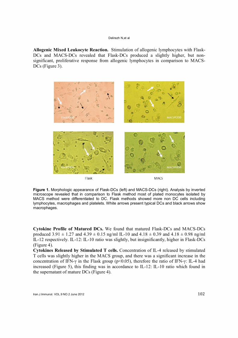
Allogenic Mixed Leukocyte Reaction. Stimulation of allogenic lymphocytes with Flask-DCs and MACS-DCs revealed that Flask-DCs produced a slightly higher, but non-significant, proliferative response from allogenic lymphocytes in comparison to MACS-DCs (Figure 3). Figure 1. Morphologic appearance of Flask-DCs (left) and MACS-DCs (right). Analysis by inverted microscope revealed that in comparison to Flask method most of plated monocytes isolated by MACS method were differentiated to DC. Flask methods showed more non DC cells including lymphocytes, macrophages and platelets. White arrows present typical DCs and black arrows show macrophages.
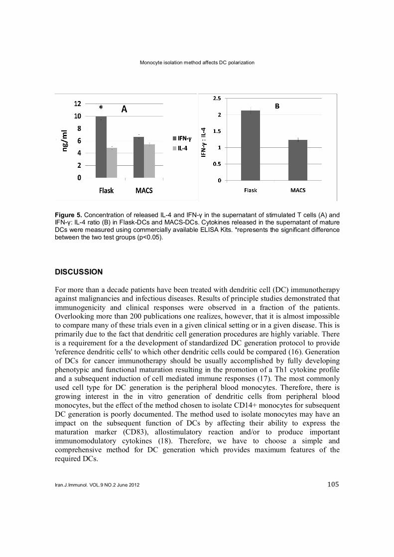
Cytokine Profile of Matured DCs. We found that matured Flask-DCs and MACS-DCs produced 3.91 ± 1.27 and 4.39 ± 0.15 ng/ml IL-10 and 4.18 ± 0.39 and 4.18 ± 0.98 ng/ml IL-12 respectively. IL-12: IL-10 ratio was slightly, but insignificantly, higher in Flask-DCs (Figure 4). Cytokines Released by Stimulated T cells. Concentration of IL-4 released by stimulated T cells was slightly higher in the MACS group, and there was a significant increase in the concentration of IFN-γ in the Flask group (p<0.05), therefore the ratio of IFN-γ: IL-4 had increased (Figure 5), this finding was in accordance to IL-12: IL-10 ratio which found in the supernatant of mature DCs (Figure 4).

Monocyte isolation method affects DC polarization
Iran.J.Immunol. VOL.9 NO.2 June 2012 103
Figure 2. Immunophenotyping of DCs. A: Representative flow cytomeric histograms, obtained from MACS-DCs and Flask-DCs, stained with FITC conjugated mAb against CD14, CD83, and HLA-DR. As shown in histograms the MACS-DCs (Right) produced a higher fluorescent intensity relative to Flask-DCs (Left) using the three stained markers. B: Flow cytometric analysis showing increased CD83, CD14 and HLA-DR expression among MACS-DCs compared with Flask-DCs. *represents a significant difference between two test groups (p<0.05).

Delirezh N,et al
Iran.J.Immunol. VOL.9 NO.2 June 2012 104
Figure 3. Allogenic MLR response induced by MACS-DCs and Flask-DCs. Allogenic T cells were incubated with MACS-DCs and Flask-DCs at a ratio of 5:1, 10:1 and 20:1 for 5 days. Uptake of [
3H]
thymidine during the last 18 hrs of incubation was then measured.
Figure 4. Concentration of IL-10 and IL-12 in the supernatant of 7-day cultured DCs. Cytokines released in the supernatant of mature DCs were measured using commercially available ELISA Kits. Concentration of cytokines (A), and IL-12: IL-10 ratio (B) in Flask-DCs and MACS-DCs.
Monocyte isolation method affects DC polarization
Iran.J.Immunol. VOL.9 NO.2 June 2012 105
Figure 5. Concentration of released IL-4 and IFN-γ in the supernatant of stimulated T cells (A) and IFN-γ: IL-4 ratio (B) in Flask-DCs and MACS-DCs. Cytokines released in the supernatant of mature DCs were measured using commercially available ELISA Kits. *represents the significant difference between the two test groups (p<0.05).
DISCUSSION For more than a decade patients have been treated with dendritic cell (DC) immunotherapy against malignancies and infectious diseases. Results of principle studies demonstrated that immunogenicity and clinical responses were observed in a fraction of the patients. Overlooking more than 200 publications one realizes, however, that it is almost impossible to compare many of these trials even in a given clinical setting or in a given disease. This is primarily due to the fact that dendritic cell generation procedures are highly variable. There is a requirement for a the development of standardized DC generation protocol to provide 'reference dendritic cells' to which other dendritic cells could be compared (16). Generation of DCs for cancer immunotherapy should be usually accomplished by fully developing phenotypic and functional maturation resulting in the promotion of a Th1 cytokine profile and a subsequent induction of cell mediated immune responses (17). The most commonly used cell type for DC generation is the peripheral blood monocytes. Therefore, there is growing interest in the in vitro generation of dendritic cells from peripheral blood monocytes, but the effect of the method chosen to isolate CD14+ monocytes for subsequent DC generation is poorly documented. The method used to isolate monocytes may have an impact on the subsequent function of DCs by affecting their ability to express the maturation marker (CD83), allostimulatory reaction and/or to produce important immunomodulatory cytokines (18). Therefore, we have to choose a simple and comprehensive method for DC generation which provides maximum features of the required DCs.

Delirezh N,et al
Iran.J.Immunol. VOL.9 NO.2 June 2012 106
As mentioned above, when choosing a DC generation protocol for vaccination purposes, several critical parameters must be considered. The first parameter which is important for the generation of DC vaccines is the number of DCs that needs to be generated. Relying on the results obtained in the present study, higher but not significant yield and viability were obtained using MACS method. The second factor that affects resultant product is contamination of the DC preparation, which affects the efficacy of the DC vaccination. Since the type of contaminating cells present in the DC preparation can vary among protocols and in order to minimize the effect of the contaminants, it is necessary to aim for the highest possible DC purity. Based on the data from the literature, we propose to aim for DC purities of >75% (10,19). In the present study we obtained 65% and 56% DCs from the seeded monocytes by MACS and Flask methods, respectively. However, very homogenous DCs were obtained by the MACS method in comparison to the Flask method (Figure 1). Indeed more monocytes were driven to form DCs in the MACS method indicating that the differentiation capacity of this method is higher (20). Expression of β2 integrins on the surface of granulocytes and natural killer cells, and that of CD11b on lymphocytes and CD18 on all leukocytes (21) make these cells to remain close to monocytes in the flask adherence method and make the resultant culture heterogenous (22,23,24). An important feature of mature DCs is the upregulation of CD83 as a specific marker (25); however, the role of this surface molecule is not fully understood. It is believed that these markers may be involved in the regulation of cell mediated immune response (26). We found that in contrast to the Flask-DCs, the MACS-DCs significantly increased the expression of CD83, an indication of a more maturated status (Figure 2). On the other hand, CD14 expression in mature DCs is down regulated upon adding IL-4 which suppresses the transcription of the CD14 gene (27). The flow cytometric immunophenotype of generated DCs, however, showed a decreased CD14 expression in both groups in contrast to the estimated quantities of this marker in the original monocytes, but its expression was slightly higher in MACS-DCs. This difference is most probably the result of a higher positive selection of CD14+ CD16¯ subset of monocytes in the MACS method (12,13,28) and should not be interpreted as more maturation of the Flask-DCs, because the expression of other maturation markers such as HLA-DR and CD83 were higher in MACS-DCs. MHC II (HLA-DR) expression is another maturation marker in DCs which is upregulated during maturation process from intra cellular sources. Our results showed that expression of HLA-DR was slightly higher in MACS-DCs. Higher expression of maturation markers, in particular HLA-DR, is well correlated to allostimulatory capacity of generated DCs. We have subjected Flask-DCs and MACS-DC to allogenic MLR. Our results revealed that Flask-DCs caused a more but insignificant proliferation response in allogenic lymphocyte reaction. This response probably arose from a more co-stimulation by flask adherence derived cells which may contain other HLA-DR expressing PBMCs such as B cells in addition to DCs. It is reported that the positive selection of monocytes by anti-CD14-coated microbeads inhibits the lipopolysaccharide (LPS)-induced production of interleukin (IL)-12, IL-10 and tumour necrosis factor-α (TNF-α) from human DCs. However, when DC was grown from monocytes isolated by plastic adherence, LPS induced the production of much higher levels of these cytokines. DCs derived from adherence-isolated monocytes induced the
Monocyte isolation method affects DC polarization
Iran.J.Immunol. VOL.9 NO.2 June 2012 107
development of potent cytotoxic T lymphocytes and a Th1 cytokine profile as confirmed by interferon-γ production (10,18). In accordance to these findings, our results showed a significantly higher IFN-γ production which resulted in a higher IFN-γ: IL-4 ratio and also in a slightly higher IL-12 and a subsequent IL-12: IL-10 ratio in the Flask group. It seems that Flask-DCs polarized the immune response towards a Th1 cytokine profile and a cell mediated immune response which is a favorite feature in cancer immunotherapy. The easiest and most cost-effective way for DC generation is through adherence of monocytes to plastic which has also been developed for use in closed systems. However, the variability in DC purity in this method remains as an important shortcoming. However, highly purified monocytes can also be obtained by positive immunomagnetic selection of CD14+ cells, but the reagents required are expensive, and limits their clinical use. Furthermore, positive selection of monocytes raises concerns about the use of xenogeneic antibodies and possible activation/alteration of the monocytes (29,30). However, it has never been investigated if monocyte activation has a negative effect on DC generation (23,24). In conclusion, MACS-DCs are preferred in terms of mass production, purity and maturity in particular, but their lower IFN-γ: IL-4 and IL-12: IL-10 ratios may decrease their chance of selection for cancer immunotherapy. However, they may be suitable for the treatment of autoimmune diseases due to Th2 the polarization of the immune response. ACKNOWLEDGMENTS This work was supported by the Institute of Biotechnology, Urmia University. We are grateful to Dr. A. Rezaie and Mr. M. Ojaqzadeh for blood donation. We also thank Mr. Mehmannavaz at the Transfusion Organization of Iran, Dr. Ranjkeshzadeh and his colleagues at the radiotherapy center of Omid hospital, Urmia, Iran. REFERENCES
1 Nestle FO, Farkas A, Conrad C. Dendritic-cell-based therapeutic vaccination against cancer. Curr Opin Immunol. 2005;
17:163-9. 2 Li YL, Wu YG, Wang YQ, Li Z, Wang RC, Wang L, et al. Bone marrow-derived dendritic cells pulsed with tumor lysates
induce anti-tumor immunity against gastric cancer ex vivo. World J Gastroenterol. 2008; 14:7127-32. 3 Kalinski P, Urban J, Narang R, Berk E, Wieckowski E. Muthuswamy R. Dendritic cell-based therapeutic cancer vaccines:
what we have and what we need. Future Oncol. 2009; 5:379-90. 4 Delirezh N, Moazzeni SM, Shokri F, Shokrgozar MA, Atri M, Kokhaei P. Autologous dendritic cells loaded with apoptotic
tumor cells induceT cell-mediated immune responses against breast cancer in vitro. Cell Immunol. 2009; 257:23-31. 5 Colino J, Shen Y, Snapper CM. Dendritic cells pulsed with Intact Streptococcus pneumoniae elicit both protein and
polysaccharide specific immunoglobulin isotype resposes in vivo through distinct mechanisms. J Exp Med. 2002; 195:1-14. 6 Freudenthal PS, Steinman RM. The distinct surface of human blood dendritic cells, as observed after an improved isolation
method. Proc Natl Acad Sci U S A. 1990; 87:7698-702. 7 Sallusto F, Lanzavecchia A. Efficient presentation of soluble antigen by cultured human dendritic cells is maintained by
granulocyte/macrophage colony stimulating factor plus interleukin-4 and downregulated by tumor necrosis factor alpha. J Exp Med. 1994; 179:1109-18.
8 Berges C, Naujokat C, Tinapp S, Wieczorek H, Hoh A, Sadeghi M, et al. A cell line model for the differentiation of human dendritic cells. Biochem Biophys Res Commun. 2005; 333:896-907.
9 Davis GE. The Mac-1 and p150/95 beta 2 integrins bind denatured proteins to mediate leukocyte cell-substrate adhesion. Exp Cell Res. 1992; 200:242-52.
10 Lehner M, Holter W. Endotoxin-Free Purification of Monocytes for Dendritic Cell Generation via Discontinuous Density Gradient Centrifugation Based on Diluted Ficoll-Paque Plus. Int Arch Allergy Immunol. 2002; 128:73-6.
Delirezh N,et al
Iran.J.Immunol. VOL.9 NO.2 June 2012 108
11 Mucci I, Legitimo A, Compagnino M, Consolini R, Migliaccio P, Metelli MR, et al The methodological approach for the generation of human dendritic cells from monocytes affects the maturation state of the resultant dendritic cells. Biologicals. 2009; 37:288-96.
12 Geissmann F, Jung S, Littman DR. Blood monocytes consist of two principal subsets with distinct migratory properties. Immunity. 2003; 19:71-82.
13 Strauss-Ayali D, Conrad SM, Mosser DM. Monocyte subpopulations and their differentiation patterns during infection. J Leukoc Biol. 2007; 82: 244-52.
14 Boyum A. Separation of leukocytes from blood and bone marrow. Introduction. Scand J Clin Lab Invest Suppl. 1968; 97:7-11. 15 Mazzucchelli R, Amadio M, Curreli S, Denaro F, Bemis K, Reid W, et al. Establishment of an ex vivo model of monoctytes-
derived macrophages differentiated from peripheral blood mononuclear cells (PBMC) from HIV-1 transgenic rats. Mol Imm. 2004; 41:979-84.
16 Erdmann M, Schuler-Thurner B. Towards a standardized protocolfor the generation of monocyte-derived dendritic cell vaccines. Methods Mol Biol. 2010; 595:149-63.
17 Frankenberger B, Schendel DJ. Third generation dendritic cell vaccines for tumor immunotherapy. Eur J Cell Biol. 2012; 91:53-8.
18 Elkord E, Williams PE, Kynaston H, Rowbottom AW. Human monocyte isolation methods influence cytokine production from in vitro generated dendritic cells Immunology. 2005; 114:204-12.
19 Figdor CG, de Vries IJ, Lesterhuis WJ, Melief CJ. Dendritic cell immunotherapy: mapping the way. Nat Med. 2004; 10:475-80.
20 Yamahira A, Narita M, Nakamura T, Watanabe N, Kaji M, Taniguchi T, et al. Generation of antigen – specific cytotoxic T lymphocytes using a leukemic plasmacytoid dendritic cell line as antigen presenting cells. Leuk Res. 2011; 35:793-9.
21 Roitt IM, Delves PJ. Roitt’s Essential Immunology, 10th ed. UK: Blackwell Science. Ltd; 2001. p. 451-62. 22 Tuyaerts S, Aerts JL, Corthals J, Neyns B, Heirman C, Breckpot K, et al. Current approaches in dendritic cell generation and
future implications for cancer immunotherapy. Cancer Immunol Immunother. 2007; 56:1513-37. 23 Suen Y, Lee SM, Aono F, Hou S, Loudovaris M, Ofstein G, et al. Comparison of monocyte enrichment by immuno-magnetic
depletion or adherence for the clinical-scale generation of DC. Cytotherapy. 2001; 3:365-75. 24 Meyer-Wentrup F, Burdach S. Effcacy of dendritic cell generation for clinical use: recovery and purity of monocytes and
mature dendritic cells after immunomagnetic sorting or adherence selection of CD14+ starting populations. J Hematother Stem Cell Res. 2003; 12:289-99.
25 Zhou LJ, Tedder TF. CD14+ blood monocytes can differentiate into functionally mature CD83+ dendritic cells. Proc Natl Acad Sci USA. 1996; 93:2588-92.
26 Scholle N, Hayden-Ledbetter M, Dahlin A, Hellstrom I, Hellstrom KE, Ledbetter JA. Cutting edge: CD83 regulates the development of cellular immunity. J Immunol. 2002; 168:2599-02.
27 Lauener RP, Goyert SM, Geha RS, Vercelli D. Interleukin 4 downregulates the expression of CD14 in normal human monocytes. Eur J Immunol. 2009; 20:2375-8.
28 Sanchez-Torres C, Garcia-Romo GS, Cornejo-Cortes MA, Rivas-Carvalho A, Sanchez-Schmitz G. CD16+ and CD16- human blood monocyte subsets differentiate in vitro to dendritic cells with different abilities to stimulate CD4+ T cells. Int Immunol. 2001; 13:1571-81.
29 Breckpot K, Corthals J, Heirman C, Bonehill A, Michiels A, Tuyaerts S, et al. Activation of monocytes via the CD14 receptor leads to the enhanced lentiviral transduction of immature dendritic cells. Hum Gene Ther. 2004; 15:562-73.
30 Elkord E, Williams PE, Kynaston H, Rowbottom AW. Human monocyte isolation methods inXuence cytokine production from in vitro generated dendritic cells. Immunology. 2005; 114:204-12.