ph.d. synopsis - gtu.ac.ingtu.ac.in/uploads/synopsis_129990931004_rutvishah_997648.pdf ·...
TRANSCRIPT

Decision Support System for Automatic Identification of ROI (Region of Interest)
for Medical Images
Ph.D. Synopsis Submitted to
Gujarat Technological University
For the Award of Doctor of Philosophy
In Computer Science
By:
Rutvi Shah Enrollment No: 129990931004,
Registration No: 4043, Batch: 2012
Under the Guidance of: Dr. Priyanka Sharma
Professor (IT), Raksha Shakti University, Ahmedabad.
Doctoral Progress Committee Experts:
Dr. Bankim Patel Dr. Sanjay Kumar Vij Director, Dean (Academics), Shrimad Rajchandra Institute of ITM-Universe, Managementand Computer Applications, Vadodara, Gujarat. Maliba Campus, Bardoli, Surat.

INDEX Sr. No. Topic Page No.
1. Title of Thesis 1 2. Motivation 1 3. Abstract 2 4. Brief description of the state of the art of the research
4.1 Medical Imaging X-ray images
4.2 Segmentation Segmentation Types Segmentation of bone from X-ray
4.3 Automated Identification of ROI (Bone breakage) in bone X-ray images
4.4 Decision support system for prescription of a course of treatment
4
5. Problem Definition 7 6. Objective of work 7 7. Original contribution by the thesis 8 8. The methodology of research, results/comparisons
Proposed algorithm 8.1 Methodology of research 8.2 Proposed Algorithm 8.2.1 Preprocessing 8.2.2Background removal 8.2.3 Mask generation 8.2.4 Bone detection 8.2.5 Bone Extraction 8.2.6 Fracture Detection 8.2.7 Decision support system 8.3 Results
9
9. Conclusion 17 10. Copies of papers published and a list of all publications
arising from the thesis 19
11. Patent 12. Achievements 20 13. References 21
1
1. Title of the Thesis
Decision Support System for Automatic Identification of ROI (Region of Interest) for Medical Images
2. Motivation: One fine night at around 11:00 P.M, one boy of 2 years was playing joyfully
with his father. The boy was climbing on the back of his father and jumping on
the bed and laughing loudly. The father was very happy watching his son play.
Once again the boy jumped on the bed, but this time he did not laugh. While
jumping the boy could not balance and got hurt. There was a severe swelling in
his hand which indicated that there must be a bone breakage. The father
rushed him to the near most orthopedic hospital. As it was late in the night,
the doctor was not available. The attendant at the hospital took the X-Ray, but
could not understand what treatment should be given to the child. So he called
up the doctor. It took more than an hour for the doctor to reach the hospital
and start the treatment. The boy had suffered a major fracture. As the boy was
young only fixation and casting would have cured his fracture but the delay in
the treatment resulted in surgery.
Such delay in treatment for adults and elderly people can prove to be fatal.
Especially such surgical delay is associated with a significant increase in the
risk of death. Certain fractures can cause severe hemorrhage or predispose to
other life-threatening complications. Femur fractures that disrupt the femoral
artery or its branches are potentially fatal. Pelvic fractures can damage pelvic
arteries or veins causing life-threatening hemorrhage; the more displaced the
pelvic fracture, the greater the potential blood loss. Hip fractures, particularly
in the elderly, may prevent ambulation, resulting in potentially life-threatening
complications, such as pneumonia, thromboembolic disease, if there is a
prolonged period of immobility. Patients with multiple rib fractures are at
substantial risk for pulmonary contusion and related complications.

2
All these facts have motivated us to focus on the proposed fracture
identification and detection system that can automatically determine the
presence and absence of fractures and if present, suggest the preliminary
decision support system by suggesting the immediate course of treatment for
the fracture.
3. Abstract: Today bone fractures are very common in our country because of road
accidents or through other injuries. Patients with bone fractures who go into
shock state have a mortality of 30-50%. When combined with other injuries in
the body, for example, an abdominal injury, the chance of mortality rises even
higher, approaching 100% in some cases. Traumatic injuries in this region can
result in severe hemorrhage, multiple organ dysfunction syndromes (MODS),
injury to the nerves, internal organ damage, thus resulting in a higher
mortality rate. Even if injuries at this magnitude don’t occur, severe pain and
impaired mobility are normally associated with pelvic fractures.
According to the market report of India Today, the fracture features more in
Indian hospital records and the incidences have increased 3-folds over the past
3 decades with more than 4.4 lakh people and is expected to increase to more
than 6 lakh in 2020.
The X-Ray images are the most common accessibility of peoples during the
accidents. X-rays are the oldest and most frequent form of medical imaging.But
the minute fracture detection in the X-Ray image is not possible due to the
complexity of bone structure and the difference in visual characteristics of
fracture by their location. So it is difficult to accurately detect and locate the
fractures also determine the severity of the injury.
3
The automatic detection of fractures in X-Ray images is a significant
contribution to assisting the physicians in making faster and more accurate
patient diagnostic decisions and treatment planning.
The major challenges in medical imaging are 1) the segmentation process, 2)
automatic identification of the region of interest 3) evaluation 4) suggestive
course of action. Segmentation allows the partitioning of an image into regions
with cohesive properties. The Region of Interested (ROI) consists of the most
important part of the medical image that is diagnostically important. In today's
revolution oriented medical environment, computer-aided disease detection
plays a vital role in a wide range of applications and services in day-to-day
activities. Among fractures, automatic detection is considered more challenging
because they are different and variable in presentation and their outcomes are
unpredictable. The numerous incidences necessitate the health care
professionals to analyze a huge number of x-ray images.
The main aim of this research work is to minimize this delay by providing a
preliminary decision support system. The system will automatically identify the
region of interest (bone breakage). The system will evaluate the type of
breakage from the region of interest identified and also suggest the immediate
course of treatment required.
The research proposes an automatic algorithm (ROIDMI) for detecting bone
fracture in the X-Ray image. It uses the 7 step algorithm for detecting the
fracture. The results of this novel system are promising, demonstrating that the
proposed method is capable of automatically detecting hairline, minor and
major type’s fractures accurately, and shows potential for clinical application.
The system also gives the prescription for the course of treatment to be
followed based on the type of fracture identified. Results obtained demonstrate
the performance of the system for automatic identification of bone fracture in
the X-ray image.

4
4. Brief description of the state of the art of the research
4.1 Medical Imaging During the recent years, much attention has been focused on medical imaging
due to the appearance of less invasive and more accurate medical devices.
Modern medical imaging offers the potential and promise for major advances in
science and medicine, as higher fidelity images are produced. Digital images
are more and more used by medical practitioners to help them during the
disease diagnosis and decision-making process because they display various
body organs. There are several imaging technologies used in radiology to
diagnose or treat diseases: X-ray radiography, ultrasound, computed
tomography (CT), nuclear medicine and magnetic resonance imaging (MRI). X-
ray images (radiographs) represent one of the oldest and more frequently used
noninvasive medical tests that help physicians during various stages of
treatment, including fracture diagnosis, skeletal maturation evaluation, hip
replacement surgery, or other bone diseases. Medical imaging has led to
improvements in the diagnosis and treatment of numerous medical conditions
in children and adults. X-ray images
The discovery of X-rays and the invention of CT represented major advances in
medicine. X-ray imaging exams are recognized as a valuable medical tool for a
wide variety of examinations and procedures. They are used to:
noninvasively and painlessly help to diagnose disease and monitor
therapy;
support medical and surgical treatment planning; and
guide medical personnel as they insert catheters, stents, or other devices
inside the body, treat tumors or remove blood clots or other blockages.

5
4.2 Segmentation Segmentation is an important as well as the crucial process in medical image
processing. Image segmentation is a process of isolating an object or region of
interest from an entire image. Segmentation is done to represent an image in
more meaning manner and easier to analyze. The result of segmentation is a
set of segments or pixels that collectively cover the entire image and can be
characterized by image properties like color, intensity, texture etc.
Segmentation Types:
Image segmentation is broadly categorized into two types: Local segmentation In image segmentation, local segmentation is at the
lowest level and operates only on a small number of pixels. Local segmentation
is widely used to achieve a number of image processing tasks such as image
de-noising, pixel classification, edge detection and pixel interpolation. Local
segmentation is concerned with segmenting sub-image or the portions in the
image. Local segmentation can be called an image enhancement process.
Global Segmentation Global segmentation is opposite of local segmentation. It
is at the highest level and deals with segmenting a whole image. In global
segmentation, an image is partitioned into groups of pixels which are
homogeneous with respect to some characteristics and each group is called a
segment. Global segmentation is further categorized into three types: Region
approach, Boundary approach, and Edge approach. Clustering is one of the
widely used techniques in which clustering of pixels is done according to some
homogeneity criteria. We have used edge approach i.e. Level Set Active Contour
method for image segmentation.
General medical image segmentation methods can be categorized into the
following classes: classical image segmentation methods (thresholding, region-
based, and edges-based), pattern recognition-based, deformable models,
wavelet-based methods, and atlas-based techniques [1]-[5].

6
Segmentation of Bone from X-ray
X-Ray image analysis techniques to access bone content and bone structure is
an area of research that has attracted many researchers. [6-8]. Almost all these
techniques, extract features from x-ray images for bone analysis. One
important a sub-field of image analysis is the segmentation of bone structure
from the x-ray image. Image segmentation is used to classify or assign each
pixel into a group (label), where each group represents membership to a set of
pixels that define an object or region in the image. Thus, the main aim of
segmentation is to subdivide the various portions, so that it can help medical
practitioners during the study of bone structure, identification of bone fracture,
measurement of fracture treatment and treatment planning prior to surgery. It
is considered as a challenging task because the bone x-ray images are complex
in nature and the output of segmentation algorithm is affected due to various
factors like partial volume effect, the intensity in the homogeneity, the presence
of noise and artifacts and Closeness in gray level of different soft tissues.
4.3 Automated Identification of ROI (Bone breakage) in bone X-ray images
The most difficult part of medical image analysis is the automated localization
and delineation of structures of interest. Automated data evaluation is one way
of enhancing the clinical utility of measurements. A crucial role for automated
information extraction in medical imaging usually involves the segmentation of
regions of the image in order to quantify volumes and areas of interest of
biological tissues for further diagnosis and localization of pathologies.
A bone x-ray makes images of any bone in the body, including the hand, wrist,
arm, elbow, shoulder, foot, ankle, leg (shin), knee, thigh, hip, pelvis or spine.
Detection and correct treatment of fractures are considered important, as a
wrong diagnosis often leads to ineffective patient management, increased
dissatisfaction, and expensive treatment. During literature survey, we did not
get any such research paper addressing automated identification of the bone
fractures from the X-ray images. However, only a few inventions have claimed
automated segmentation of the bone part from the X-ray image. Patent No:

7
US9275469 US claims compositions and methods that allow for the analysis of
bone mineral density and/or bone structure from x-ray images, wherein the
bone structure includes trabecular bone structure. Patent No: US 8715187 B2
claims at automatically identifying and segmenting different tissue
types in ultrasound images. Patent No: US 8135200 B2 relates to an imaging
system using auto-shutter, a method for finding a region of interest in a digital
image.
4.4 Decision support system for prescription of course of treatment
The importance of automated fracture detection comes from the fact that in
clinical practice when an orthopedic doctor is not available, it becomes difficult
in providing timely treatment. This can lead to fatal consequences.
Computer detection of fractures can assist in identifying the type of fracture
and the decision support system can give the primary course of treatment and
thus improve the timeliness and fasten the healthcare for the patient.
5. Problem Definition
To design and develop a decision support system for automatic identification of ROI (Region of Interest) in medical images.
6. Objective of work
The main focus of this work is to automatically detect a region of interest (bone
fracture) from the bone X-ray images and in particular suggest the course of
treatment based on the type of crack identified in the region of interest using a
series of sequential steps. The objectives can be stated as follows:
1. To extract the bone part from the X-ray image.
2. To develop an algorithm that automatically identifies the region of
interest (the breakage in bone) in the image.
3. To develop an algorithm to analyze and evaluate the type of breakage.
4. To develop decision support system to suggest the course of action
based on the type of breakage.

8
7. Original contribution by the thesis The entire work in this synopsis, as well as thesis, is the original work, with
the journal papers and research papers as the backbone. The proposed system
and algorithms have been visualized as a collection of various modules, each of
which with relevant publications. The details of the book chapters, journal
papers, and conference papers are as follows:
Book Chapters: 1. R. Shah et al. “Bone Segmentation from X-ray images: Challenges and
Techniques” Information Systems Design and Intelligent Applications:
Proceedings of Fourth International Conference India 2017. Vol. 672.
Bhateja, Vikrant and S. Das, Eds. Cham: Springer International
Publishing, 2018, pp. 878-887.
2. R. Shah et al., “Comparative Performance Study of Various Content-
Based Image Retrieval Methods,” in Proceedings of First International
Conference on Information and Communication Technology for
Intelligent Systems: Volume 1, vol. 50, S. C. Satapathy and S. Das,
Eds. Cham: Springer International Publishing, 2016, pp. 397–407.
International Journal
1. R. Shah, Prof. R Shah, Dr. Priyanka Sharma, “Survey of Region of
Interest and its Applicative Utility in various areas of the medical field,”
Published at International Journal of Advanced Technology in
Engineering and Science www.ijates.com, Volume No 3, Special Issue
No 01, ISSN 2348-7550, March 2015.
2. Dr. Priyanka Sharma, R Shah, Prof. R Shah, “Implementation of E-
learning Using Mobile Technology”, Published at International Journal
of Information and Computing Technology, Volume 3, Issue No 1, ISSN
0976-5999, December 2013.
9
International Conference
1. Shah, Rutvi, and Priyanka Sharma. "Bone Segmentation from X-Ray
Images: Challenges and Techniques." Information Systems Design and
Intelligent Applications. Springer, Singapore, 2018. 853-862.
2. Shah et al., “Comparative Performance Study of Various Content-
Based Image Retrieval Methods,” in Proceedings of First International
Conference on Information and Communication Technology for Intelligent
Systems: Volume 1, vol. 50, S. C. Satapathy and S. Das, Eds. Cham:
Springer International Publishing, 2016, pp. 397–407.
3. R. Shah, Prof. R Shah, Dr. Priyanka Sharma, “Survey of Region of
Interest and its Applicative Utility in various areas of the medical field,”
presented at the International Conference On Recent Trends In
Engineering Science And Management, 2015.
4. R. Shah, P. Sharma, and R. Shah, “Performance Analysis of Region of
Interest Based Compression Method for Medical Images,” 2014, pp.
53–58.
National Conference
1. R. Shah, Prof. RR Shah, Dr. Priyanka Sharma, “Clinical Decision
Support System: An Efficient Structure for providing High Quality
Patient Care,” presented at the National Conference On Emerging
Trends in Information and Communication Technology 2013.
8. The methodology of research, results/comparisons, Proposed algorithm 8.1 Methodology of research
The methodology consists of the following steps for automatic identification
of fractures in bones from X-ray images:
(i) Preprocessing – Procedure that contains steps that enhance the x-ray
input image in a way that its result improves the fracture detection process.
10
(ii) Background removal – Procedure to remove the unwanted background
data by using Otsu's multi-thresholding method.
(iii) Mask generation- Procedure to convert the image into black and white
masked image.
(iv) Bone detection– Innovative algorithm to segment the bone from the
source image. This procedure takes source and masks Image as input and
produces segmented. Bone is segmented using the Active Contour method.
(v) Bone Extraction-procedure to extract the segmented bone part from the
image.
(vi) Fracture Detection – Innovative algorithm to analyze the segmented bone
part to identify the type of breakage in the bone. The system scans the
segmented bone part for the presence of the breakage. If the breakage exists
then the system identifies the breakage as hairline, minor and major.
(vii) Decision support system-
8.2 Proposed Algorithm 1. Load image
The image is loaded from the database.
2. Grayscale conversion
Gray Scale Conversion: In computing, a grayscale or greyscale digital
image is an image in which the value of each pixel is a single sample,
that is, it carries only intensity information. The gray scale histogram
can be used for the further processing of the image like segmentation,
edge detection etc.
3. Background removal
Thresholding: The simplest method of image segmentation is called
the thresholding method. This method is based on a clip-level (or a
threshold value) to turn a gray-scale image into a binary image. This
11
allows for the removal of other surrounding artifacts which exist in
the original image, e.g. X-Ray table, cables, hands, and lower
extremities.
We perform automatic thresholding to find the threshold for
segmenting background. We calculate Multi threshold (7 thresholds)
from the image using Otsu’s method and selects the first threshold for
background segmentation.
We perform COLUMN wise segmentation with a selected threshold
and separate background and foreground pixels of the image.
4. Mask generation
Next, the image is enhanced to emphasize bone structures. For this
technique, image contrast is increased by mapping grayscale values to
a wider range; the bone is enhanced to a higher intensity.
We use active contour method for bone detection, this method
requires two images as input first one is source image and the second
one is mask image. The mask image is a Black and white image
5. Bone detection
We propose a novel algorithm to segment the bone part from the
image. The algorithm uses Active Contour method for segmentation.
Active Contour Segmentation
The Active contour extraction process identifies the bone regions in
the images based on the differences in the gray level pixels in the
images.
In contour extraction process the initial seed points, internal energy,
and external energy were taken as input. The initial seed points were
grown in order to match the internal energy to the external energy.
The initial energy term helps in the exact identification of the shape of

12
the segmented region. Deformation of the shape and control on
iteration of the process can be done with the help of the external
energy term. Active contours will identify the object regions [8]. The
curve will move around the boundaries of the objects and finally the
exact shape around the object. The process is repetitive in iterations
resulting in the identification of the exact region of input images. The
number of iterations of the process depends on the image type and
the grayscale concentration of the input images [9].
Medical image segmentation is a fundamental problem in image
processing and computer vision. Segmentation algorithms play a vital
role in many biomedical imaging applications, such as diagnosis and
treatment planning, localization of pathology, the study of anatomical
structure, and computer-integrated surgery [1]. However, most of the
existing articles on medical image segmentation are focused on CT
and MRI and less on the segmentation of X-ray images. X-Ray image
segmentation techniques are treated in [3], [4], [5], [10]. The goal in
these papers is the segmentation of bone structures from the X-ray
images. This task is considered challenging because these types of
images are complex in nature since the regions delineated by bone
contours are highly nonuniform in intensity and texture. Deformable
models (snakes, level set based models, or active shape models) can
be used for X-ray image segmentation. An interesting solution is to
combine two or more segmentation techniques.
6. Bone extraction (column-wise segmentation)
The fractured bone tissue is more complex to recognize because it has
some additional features to be considered. Due to the fact that the
bone fragments may have an arbitrary shape and can belong to any
bone in a nearby area, it is essential to label all the fragments during
the segmentation process. This labeling requires expert knowledge. A
prior knowledge cannot be easily used because it is rare to find two
identical fractures and therefore it is difficult to guess the shape of the
13
bone fragments, especially in comminuted fractures. In our case, a
fractured region in the input images was identified by finding the
threshold for the particular region [8]. The intensity of gray level is
found lower than 150 pixels in the fracture region. The morphological
opening operation is employed in order to remove the useless portion
of the segmented image. It also removes the image region boundary
pixels thus helps in the removal of the excess regions segmented
along with the bone regions. The smaller blobs in the images which
will thereby be formed will be the fracture regions [11].
7. Fracture (ROI) detection
ROI is detected by column-wise operation and marking the entire
row for the discontinuity in white and black pixels values.
8. Image restoration
The image restoration process is the phase where the final image is
generated by superimposing the identified ROI on the original
image. The final image now shows the exact place of fracture in red
color.
9. Prescription
When the image restoration phase completes and the type of
fracture is identified, the algorithm fetches the prescription related
to the fracture and display it on the screen along with the final
image. It clearly states the type of fracture (hairline, minor or
major) and the immediate course of treatment as per the fracture
type.
8.3 Results
The research proposes a decision support system and – step algorithm for
automatic detection of bone fracture in the X-Ray image. The results of this
novel system are promising, demonstrating that the proposed method is

14
capable of automatically detecting hairline, minor and major type’s fractures
accurately, and shows potential for clinical application.
The proposed algorithm has been applied on a dataset which consists of
fractured & Non-fractured bones. The dataset is of total 90 original X-ray
images. It comprises of 10 images of 8 different types of fractures and 10
images without any fracture.
The system is tested for the time required to produce the resultant image
and the percentage of recall. The mean of time for the total number of
images of each type of fracture is obtained. The recall for each set of images
is calculated. The proposed algorithm is able to achieve 100% recall for 4
different types of fracture types (Comminuted, Impacted, Supracondylar and
Avulsion). The mean of the recall the whole system gives the overall system
performance which is 83%.
Input Image Fracture Type Time Elapsed (in Seconds)
Output Image Prescription
Compound Fracture (of1.jpg)
2.414892
Major Crack/Supracondylar fracture (External Fixation (Emergency))
CompoundFractures (of2.png)
8.335972
Consult Doctor Immediately
15
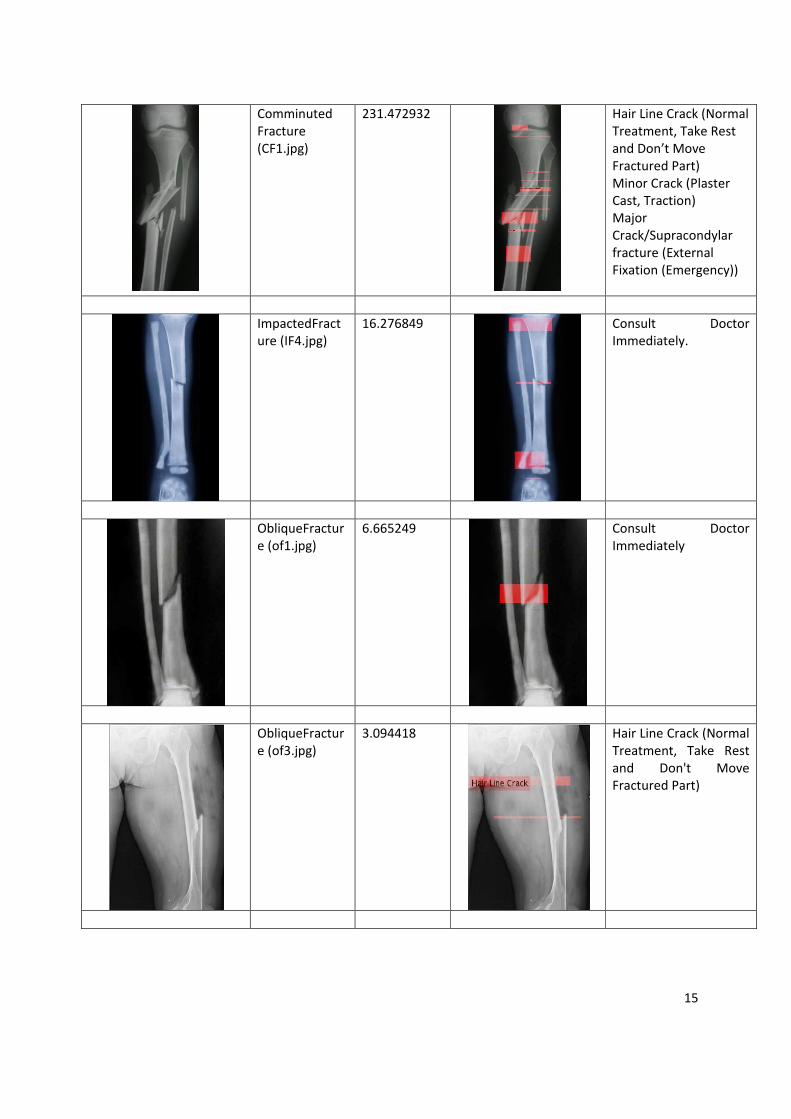
Comminuted Fracture (CF1.jpg)
231.472932
Hair Line Crack (Normal Treatment, Take Rest and Don’t Move Fractured Part) Minor Crack (Plaster Cast, Traction) Major Crack/Supracondylar fracture (External Fixation (Emergency))
ImpactedFracture (IF4.jpg)
16.276849
Consult Doctor Immediately.
ObliqueFracture (of1.jpg)
6.665249
Consult Doctor Immediately
ObliqueFracture (of3.jpg)
3.094418
Hair Line Crack (Normal Treatment, Take Rest and Don't Move Fractured Part)
16
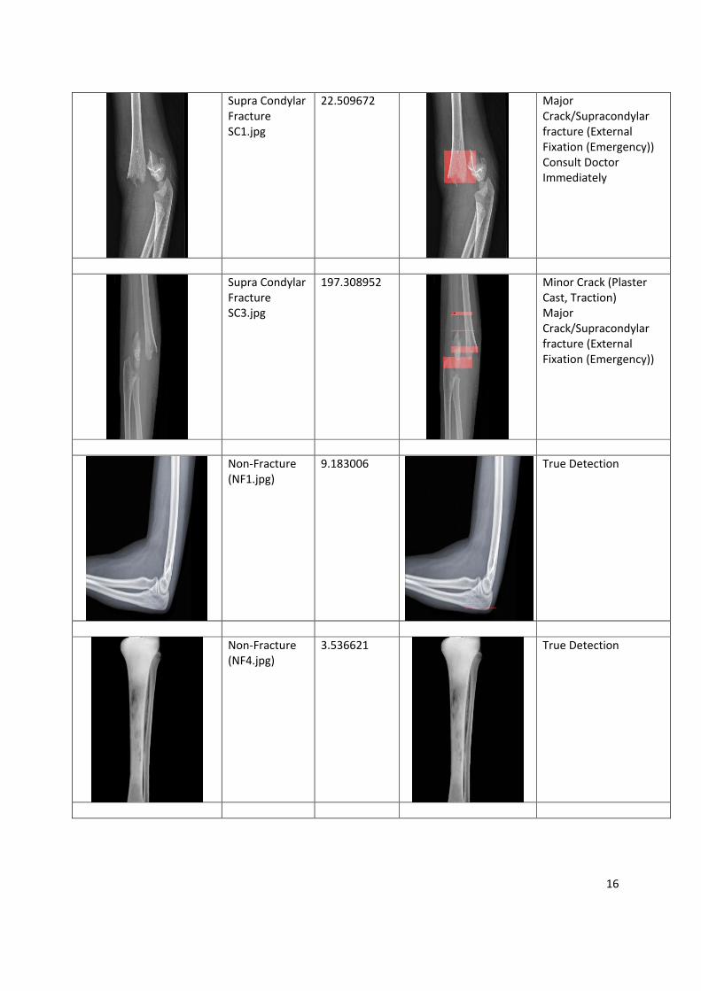
Supra Condylar Fracture SC1.jpg
22.509672
Major Crack/Supracondylar fracture (External Fixation (Emergency)) Consult Doctor Immediately
Supra Condylar Fracture SC3.jpg
197.308952
Minor Crack (Plaster Cast, Traction) Major Crack/Supracondylar fracture (External Fixation (Emergency))
Non-Fracture (NF1.jpg)
9.183006
True Detection
Non-Fracture (NF4.jpg)
3.536621
True Detection
17
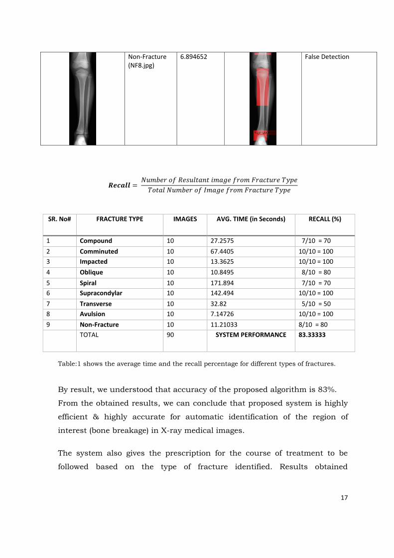
Non-Fracture (NF8.jpg)
6.894652
False Detection
=
SR. No# FRACTURE TYPE IMAGES AVG. TIME (in Seconds) RECALL (%)
1 Compound 10 27.2575 7/10 = 70 2 Comminuted 10 67.4405 10/10 = 100 3 Impacted 10 13.3625 10/10 = 100 4 Oblique 10 10.8495 8/10 = 80 5 Spiral 10 171.894 7/10 = 70 6 Supracondylar 10 142.494 10/10 = 100 7 Transverse 10 32.82 5/10 = 50 8 Avulsion 10 7.14726 10/10 = 100 9 Non-Fracture 10 11.21033 8/10 = 80 TOTAL 90 SYSTEM PERFORMANCE 83.33333
Table:1 shows the average time and the recall percentage for different types of fractures.
By result, we understood that accuracy of the proposed algorithm is 83%.
From the obtained results, we can conclude that proposed system is highly
efficient & highly accurate for automatic identification of the region of
interest (bone breakage) in X-ray medical images.
The system also gives the prescription for the course of treatment to be
followed based on the type of fracture identified. Results obtained
18
demonstrate the performance of the system for automatic identification of
bone fracture in the X-ray image.
9. Conclusion Medical image processing is a field of science that is gaining wide acceptance in
healthcare industry due to its technological advances and software
breakthroughs. A bone x-ray makes images of any bone in the body, including
the hand, wrist, arm, elbow, shoulder, foot, ankle, leg (shin), knee, thigh, hip,
pelvis or spine. The importance of fracture detection comes from the fact that
clinical practice, a tired radiologist has been found to miss fracture cases after
looking through many images containing healthy bones. In a time of
unavailability of an orthopedic doctor, automatic fracture detection system can
be of great importance in providing the patient the required preliminary course
of treatment. The Computer detection of fractures can also assist the doctors
by flagging suspicious cases for closer examinations and thus improve the
timeliness and accuracy of their diagnosis. In our literature review we found
that despite newer innovations, automatic detection of bone fractures
essentially remains unresolved as these injuries are different and variable in
presentation and their outcomes are unpredictable.
The research proposes an automatic hierarchical algorithm for detecting bone
fracture in the X-Ray image. It uses the 5 step algorithm for detecting the
fracture. The results of this novel system are promising, demonstrating that the
proposed method is capable of automatically detecting hairline, minor and
major type’s fractures accurately, and shows potential for clinical application.
The system also gives the prescription for the course of treatment to be
followed based on the type of fracture identified. Results obtained are
promising and shows the accuracy of 100% for 4 types of fracture out of 8
types.
The system helps to minimize the delay in diagnosis by providing a preliminary
decision support system as the system not only automatically identifies the

19
region of interest (bone breakage), it also evaluates the type of breakage and
suggests the prescription for an immediate course of treatment required.
10. Copies of papers published and a list of all publications arising from the thesis
Book Chapters:
1. R. Shah et al. “Bone Segmentation from X-ray images: Challenges and
Techniques” Information Systems Design and Intelligent Applications:
Proceedings of Fourth International Conference India 2017. Vol. 672.
Bhateja, Vikrant and S. Das, Eds. Cham: Springer International
Publishing, 2018, pp. 878-887.
2. R. Shah et al., “Comparative Performance Study of Various Content-
Based Image Retrieval Methods,” in Proceedings of First International
Conference on Information and Communication Technology for
Intelligent Systems: Volume 1, vol. 50, S. C. Satapathy and S. Das,
Eds. Cham: Springer International Publishing, 2016, pp. 397–407.
International Journal
1. R. Shah, Prof. R Shah, Dr. Priyanka Sharma, “Survey of Region of
Interest and its Applicative Utility in various areas of the medical
field,” Published at International Journal of Advanced Technology in
Engineering and Science www.ijates.com, Volume No 3, Special Issue
No 01, ISSN 2348-7550, March 2015.
2. Dr. Priyanka Sharma, R Shah, Prof. R Shah, “Implementation of E-
learning Using Mobile Technology”, Published at International Journal
of Information and Computing Technology, Volume 3, Issue No 1,
ISSN 0976-5999, December 2013.

20
International Conference
1. Shah, Rutvi, and Priyanka Sharma. "Bone Segmentation from X-Ray
Images: Challenges and Techniques." Information Systems Design and
Intelligent Applications. Springer, Singapore, 2018. 853-862.
2. Shah Rutvi, Shah Rushabh, Lavingiya Kruti, “GDLC: A Software
Approach in Game Development,” presented at the International
Journal of Advance Research in Science and Engineering, Vol. No. 6
Issue No 06, June 2017.
3. Shah et al., “Comparative Performance Study of Various Content-
Based Image Retrieval Methods,” in Proceedings of First International
Conference on Information and Communication Technology for
Intelligent Systems: Volume 1, vol. 50, S. C. Satapathy and S. Das,
Eds. Cham: Springer International Publishing, 2016, pp. 397–407.
4. R. Shah, Prof. R Shah, Dr. Priyanka Sharma, “Survey of Region of
Interest and its Applicative Utility in various areas of the medical
field,” presented at the International Conference On Recent Trends In
Engineering Science And Management, 2015.
5. Shah, Rushabh, Priyanka Sharma, and Rutvi Shah. "Performance
analysis of region of interest based compression method for medical
images." Advanced Computing & Communication Technologies
(ACCT), 2014 Fourth International Conference on. IEEE, 2014.
Received “BEST PAPER AWARD” for the paper.
National Conference
1. R. Shah, Prof. RR Shah, Dr. Priyanka Sharma, “Clinical Decision
Support System: An Efficient Structure for providing High Quality
Patient Care,” presented at the National Conference On Emerging
Trends in Information and Communication Technology 2013.
MOOC Courses Done
1. Coursera Course on “Fundamentals of Digital Image and Video Processing” in 2016.
21
2. Coursera Course on “Introduction to Programming with Matlab”
by Vanderbilt University in 2015.
3. Coursera Course on “Image and Video Processing: From Mars to Hollywood with a stop at the Hospital” by Duke University in 2014.
11. Patents “Decision Support System for Automatic Identification of Region of
Interest for Medical Images”, Application Number 201721037688; Date of
filing October 21, 2017; Date of publication November 10, 2017.
12. Achievements
Patent filled for the research work done.
Presented and Published 5 papers at International conferences and 1
at the National conference.
Published 2 papers at International journals.
Received “Best Paper Award” for the paper presented at IEEE
international conference and published in IEEE Digital Explore.
Two of the presented papers has been published as a book chapters in
Springer books.
The papers published in IEEE and Springer are listed in Google
Scholar and Scopus.
The presented papers have been cited by 13 authors on Google Scholar
and by 3 authors on Scopus. The H index of citations is1.
Successfully completed the course of “Image and Video Processing”
from Coursera.org in collaboration with Duke University, North
Carolina with distinction.
Successfully completed the course of “Fundamentals of Digital Image
and Video Processing” from Coursera.org with distinction.
Successfully completed the course of “Image and Video Processing”
from Coursera.org in collaboration with Duke University, North
Carolina with distinction.
22
13. A partial list of References 1. Pham, D. L., Xu, C., Prince, J. L.: Current methods in medical image
segmentation. Rev. Biomed. Eng. 2, 315{337 (2000) 2. Dougherty, G.: Medical Image Processing: Techniques and Applications
(Biological and Medical Physics, Biomedical Engineering). In: Dougherty, G. (eds.). Springer, New York (2011)
3. Mahendran, S. K., Baboo, S. S.: Enhanced Automatic X-Ray Bone Image Segmentation using Wavelets and Morphological Operators. In: International Conference on Information and Electronics Engineering, vol. 6, pp. 125{129. Singapore (2011)
4. Feng, D.: Segmentation of Bone Structures in X-ray Images. Thesis proposal to the School of Computing National University of Singapore, supervisor Dr. Leow Wee Kheng, (2006)
5. Seise, M., McKenna, S. J., Ricketts, I. W., Wigderowitz, C. A.: Segmenting Tibia and Femur from Knee X-ray Images. Med. Image Underst. Anal., 103-106 (2005)
6. Lai, C.C. (2006) A novel image segmentation approach based on particle swarm optimization, IEICE Trans Fundamentals, Vol.E89A, Pp.324-327.
7. Pham, D.L., Xu, C. and Prince, J.L. (2000) Current methods in medical image segmentation. Ann Rev Biomed Engg., Vol. 2, Pp. 315-37.
8. V. Zharkova, S. Ipson, J. Aboudarham and B. Bentley, “Survey of image processing techniques”, EGSO internal deliverable, Report
9. number EGSO-5-D1_F03-20021029, October 2002, 35p 10. J. C. Russ. Image Processing Handbook, the Sixth Edition. CRC Press Taylor &
Francis Group, 2011. 11. Sharma N, Ray AK, Sharma S, Shukla KK, Pradhan S, Aggarwal LM.
Segmentation and classification of medical images using texture-primitive features: Application of BAM-type artificial neural network, J Med Physics 2008, Vol 33, Pp.119-26.
12. Chen, Y., Ee, X., Leow, W. K., Howe, T. S.: Automatic Extraction of Femur Contours from Hip X-ray Images. In: Liu, Y., Jiang, T., Zhang, C. (eds.) Computer Vision for Biomedical Image Applications. LNCS, vol.3765, pp. 200{209. Springer, Heidelberg (2005)
13. Sarita, Vikas Sandhu. "Survey of Various X-Ray Bone Image Segmentation Approaches." (2016).
14. Stolojescu-CriŞan, Cristina, and Ştefan Holban. "A comparison of X-Ray image segmentation techniques." Advances in Electrical and Computer Engineering Engineering 13.3 (2013).
15. Kaushik, Akanksha, Parkash C. Mathpal, and Vandini Sharma. "Edge Detection and Level Set Active Contour Model for the Segmentation of Cavity Present in Dental X-Ray Images." International Journal of Computer Applications 96.9 (2014).
16. Joshi, M. U., and S. T. Gandhe. "Bone fracture detection using active contour segmentation." International Journal of Applied Engineering Research 11.6 (2016): 4230-4234.
17. C. Stolojescu-Crisan, S. Holban, “A Comparison of X-ray Image Segmentation Techniques,” Advances in electrical and computer engineering, vol. 13, no. 3, pp. 85-92, 2013.
18. N. Otsu, “A Threshold Selection Method from Gray-Level Histograms,” IEEE Transactions on Systems, Man, and Cybernetics, vol. 9, no. 1, pp. 62-66, 1979.
23
19. H. P. Narkhede, “Image Segmentation Methods: A Comparative Study”, International Journal of Science and Modern Engineering (IJISME), ISSN: 2319-6386, Volume-1, Issue-8, July-2015, pp. 54-61.
20. A. M. Khan and Ravi. S, “Review of Image Segmentation Techniques”, International Journal of Soft Computing and Engineering (IJSCE), ISSN: 2231-2307, Volume-3, Issue-4, September-2015, pp. 84-92.
21. Tallapragada, VV Satyanarayana, et al. "A Novel Medical Image Segmentation and Classification using Combined Feature Set and Decision Tree Classifier." International Journal of Research in Engineering and Technology 4.9 (2016): 83-86.
22. Iassonov, P., T. Gebrenegus, and M. Tuller (2009), Segmentation of X-ray computed tomography images of porous materials: A crucial step for characterization and quantitative analysis of pore structures, Water Resour. Res., 45, W09415, doi:10.1029/2009WR008087.