pharmacometrics modelling in type 2 diabetes...
TRANSCRIPT

ACTAUNIVERSITATIS
UPSALIENSISUPPSALA
2017
Digital Comprehensive Summaries of Uppsala Dissertationsfrom the Faculty of Pharmacy 226
Pharmacometrics Modelling inType 2 Diabetes Mellitus
Implications on Study Design and Diabetes DiseaseProgression
SITI MAISHARAH SHEIKH GHADZI
ISSN 1651-6192ISBN 978-91-554-9844-3urn:nbn:se:uu:diva-317040
Dissertation presented at Uppsala University to be publicly examined in B42, BiomedicalCenter, Uppsala University, Uppsala, Friday, 28 April 2017 at 09:15 for the degree of Doctorof Philosophy (Faculty of Pharmacy). The examination will be conducted in English. Facultyexaminer: PhD Sandra Visser (Early Stage Quantitative Pharmacology & Pharmacometrics,Merck, USA).
AbstractGhadzi, S. M. S. 2017. Pharmacometrics Modelling in Type 2 Diabetes Mellitus. Implicationson Study Design and Diabetes Disease Progression. Digital Comprehensive Summaries ofUppsala Dissertations from the Faculty of Pharmacy 226. 93 pp. Uppsala: Acta UniversitatisUpsaliensis. ISBN 978-91-554-9844-3.
Pharmacometric modelling is widely used in many aspects related to type 2 diabetes mellitus(T2DM), for instance in the anti-diabetes drug development, and in quantifying the diseaseprogression of T2DM.
The aim of this thesis were to improve the design of early phase anti-diabetes drugdevelopment studies with the focus on the power to identify mechanism of drug action(MoA), and to characterize and quantify the progression from prediabetes to overt diabetes,both the natural progression and the progression with diet and exercise interventions, usingpharmacometrics modelling.
The appropriateness of a study design depends on the MoAs of the anti-hyperglycaemic drug.Depending on if the focus is power to identify drug effect or accuracy and precision of drugeffect, the best design will be different. Using insulin measurements on top of glucose hasincrease the power to identify a correct drug effect, distinguish a correct MoA from the incorrect,and to identify a secondary MoA in most cases. The accuracy and precision of drug parameterestimates, however, was not affected by insulin. A natural diabetes disease progression modelwas successfully added in a previously developed model to describe parameter changes ofglucose and insulin regulation among impaired glucose tolerance (IGT) subjects, with thequantification of the lifestyle intervention. In this model, the assessment of multiple short-termprovocations was combined to predict the long-term disease progression, and offers apart fromthe assessment of the onset of T2DM also the framework for how to perform similar analysis.Another previously published model was further developed to characterize the weight changein driving the changes in glucose homeostasis in subjects with IGT. This model includes thecomplex relationship between dropout from study and weight and glucose changes.
This thesis has provided a first written guidance in designing a study for pharmacometricsanalysis when characterizing drug effects, for early phase anti-diabetes drug development. Thecharacterisation of the progression from prediabetes to overt diabetes using pharmacometricsmodelling was successfully performed. Both the natural progression and the progression withdiet and exercise interventions were quantified in this thesis.
Keywords: Pharmacometric, type 2 diabetes mellitus, impaired glucose tolerance, prediabetes,anti-diabetes drug development, insulin, glucose, natural diabetes disease progression,lifestyle intervention, short-term provocation, long-term effect, glucose homeostasis, glucoseand insulin regulation, weight, dropout
Siti Maisharah Sheikh Ghadzi, Department of Pharmaceutical Biosciences, Box 591, UppsalaUniversity, SE-75124 Uppsala, Sweden.
© Siti Maisharah Sheikh Ghadzi 2017
ISSN 1651-6192ISBN 978-91-554-9844-3urn:nbn:se:uu:diva-317040 (http://urn.kb.se/resolve?urn=urn:nbn:se:uu:diva-317040)

To my family :past, present and future


List of Papers
This thesis is based on the following papers, which are referred to in the text by their Roman numerals.
I Ghadzi SMS, Karlsson MO, Kjellsson MC. The impact of in-
sulin measurements in oral glucose tolerance test: a simulation study in type 2 diabetes to assess power to characterize drug effects. (Submitted)
II Ibrahim MMA, Ghadzi SMS, Kjellsson MC, Karlsson MO. Study design selection in early clinical anti-diabetic drug development: a simulation study of glucose tolerance tests. (Manuscript)
III Ghadzi SMS, Karlsson MO, de Mello VD, Uusitupa M,
Kjellsson MC, Finnish Diabetes Prevention Study Group. Mathematical diabetes disease progression modelling in the integrated glucose-insulin (IGI) model among impaired glucose tolerance subjects from the Finnish Diabetes Prevention Study. (Manuscript)
IV Ghadzi SMS, Karlsson MO, de Mello VD, Uusitupa M,
Kjellsson MC, Finnish Diabetes Prevention Study Group. Mod-el-based quantification of the natural diabetes disease progression and lifestyle intervention effects on weight, beta cell function and insulin sensitivity for subjects with impaired glucose tolerance. (Manuscript)
Reprints were made with permission from the respective publishers.


Contents
Introduction ................................................................................................... 13 Diabetes mellitus ...................................................................................... 13
Prediabetes states ................................................................................. 15 Disease progression of T2DM ............................................................. 16 Non-pharmacological intervention in T2DM ...................................... 18
Antidiabetic drug development in T2DM ................................................ 19 Pharmacological treatment of T2DM .................................................. 19 Glucose tolerance tests in the antidiabetic drug development ............. 21
Pharmacometrics ...................................................................................... 23 Non-linear mixed effect (NLME) models ........................................... 24 Maximum likelihood estimation method ............................................. 25 Pharmacometrics modelling in drug development .............................. 25 Pharmacometrics modelling in T2DM ................................................ 26
Aims .............................................................................................................. 28
Methods ........................................................................................................ 29 Data .......................................................................................................... 29
Simulation from the prior developed model (Paper I & II) ................. 29 Finnish Diabetes Prevention Study (FDPS) (Paper III & IV) .............. 31
Study power calculation (Paper I & II) .................................................... 31 ............... 31
Monte Carlo Mapped Power (MCMP) Method (Paper I) .................... 32 Accuracy and precision of parameter estimates (Paper I & II) ................ 33
Drug parameters (EmaxD & EC50D) (Paper I) and x) (Paper II) ........ 33 Glucose area under the curve ratio (AUGCD/AUGCPL) ...................... 33
Diabetes disease progression modelling ................................................... 34 IGI model (Paper III) ........................................................................... 34 WHIG model (Paper IV) ..................................................................... 36
Sensitivity and specificity analyses (Paper III) ........................................ 38 Logistic regression dropout model (Paper IV) ......................................... 39 Data analyses and model evaluation ........................................................ 40

Results ........................................................................................................... 42 Study power.............................................................................................. 42
Inclusion vs. exclusion of insulin measurements (Paper I).................. 42 Most appropriate study design based on the drug MoAs (Paper II) ... 45
Accuracy and precision of parameter estimates (Paper I & II) ............... 46 Drug parameter EmaxD & EC50D x .............................................. 46 Glucose area under the curve ratio (AUGCD/PL) .................................. 49
Diabetes disease progression modelling ................................................... 52 IGI model (Paper III) ........................................................................... 52 WHIG model (Paper (IV) .................................................................... 63
Discussion ..................................................................................................... 71 Study design selection for the early phase clinical trial of anti-diabetes drug development ..................................................................................... 71
Inclusion vs. exclusion of insulin measurements ................................. 71 Most appropriate study design based on the drug MoAs ..................... 72
.................. 72 Recommendation on study design selection based on drug MoAs and purpose of analysis ........................................................................ 73
IVGTT-OGTT-IGI model in IGT population .......................................... 74 Diabetes disease progression and lifestyle intervention effects in the IGT population ......................................................................................... 75
Insulin sensitivity ................................................................................. 75 Beta cell function ................................................................................. 76 Weight ................................................................................................. 77 FPG ...................................................................................................... 77 FSI ....................................................................................................... 77 HbA1c .................................................................................................. 78
Specificity and sensitivity analyses .......................................................... 78 Logistic regression dropout model ........................................................... 79
Perspectives .................................................................................................. 81
Conclusion .................................................................................................... 83
Acknowledgements ....................................................................................... 84
References ..................................................................................................... 87

Abbreviations
ABSG50 AD AGI AUGC AUGCD AUGCPL AUGCD/PL AUGCD/PL,Cx B B0 BC BINS BIOG BLWT BMI CGI CLG CLGI CLI df DP DPP-4 DPP-4i DPw DWGT EC50D EFDI EFs EFw EGP EmaxD FDA FDPS FO FOCE FOCE INTER
Absorbed glucose at 50% maximum incretin effect Anno domini Alpha-glucosidase inhibitor Area under the glucose curve Area under the glucose curve for drug Area under the glucose curve for placebo Ratio of AUGCD/AUGCPL Ratio of AUGCD/AUGCPL for various glucose tests Beta cell function Baseline beta cell function Before Christ Basal insulin secretion Glucose bioavailability Baseline body weight Body Mass Index Combined glucose intolerance Insulin-independent glucose clearance Insulin-independent glucose clearance Insulin clearance Degree of freedom Disease progression Dipeptidyl peptidase-4 Dipeptidyl peptidase-4 inhibitors Effect of disease progression on weight Change of weight from baseline Drug concentration at 50% of drug effect Total effect of disease progression and intervention Change in weight on insulin sensitivity Effect of disease progression and intervention on weight Endogenous glucose production Maximum drug effect (United States) Food and Drug Administration Finnish Diabetes Prevention Study First-order First-order conditional estimation First-order conditional estimation with interaction

FPG FPS FSI g GABS GDM GGI gi GLP-1 GPR40 GPRG GSEN Gss GTT HbA1c HDL HOMA IFG iIFG IGI IGT iIGT IIV INCR INCRmax INT INTRB INTw iOFV IOV IS IS0 ISS ISS0 IVGTT kGE1 kGE2 kIE kin kIS KM kout LRT MBDD
Fasting plasma glucose First-phase secretion of insulin Fasting serum insulin Glucose only data Glucose absorption Gestational diabetes mellitus Graded glucose insulin infusion Glucose and insulin data Glucagon-like peptide-1 G-protein-coupled receptor 40 Glucose effect on its own production Glucose sensitivity Baseline glucose Glucose tolerance test Glycated haemoglobin High density lipoprotein Homeostatic model assessment Impaired fasting glucose Isolated impaired fasting glucose Integrated glucose-insulin Impaired glucose tolerance Isolated impaired glucose tolerance Inter-individual variability Incretin effect Maximum incretin effect Intervention Intervention effect on the rate of beta cell function Intervention effect on weight Individual objective function value Inter-occasion variability Insulin sensitivity Baseline insulin sensitivity, logit scale Baseline insulin concentration Baseline insulin sensitivity Intravenous glucose tolerance test Rate constant of glucose effect on its own production Rate constant of glucose effect on insulin secretion Rate constant of insulin effect on glucose clearance Rate constant for production Rate constant of first phase insulin secretion Kaplan-Meier Rate constant for elimination Likelihood ratio test Model-based drug development

MCMP MoA MODY MPG MTT MTT-24 NLME NGT NO NONMEM NWPRI OFV OGTT OHA PD PND PPAR- PPG PROB PsN PTime Q RB REE RESE RESG RESGPO RESI RSE SGLT-2 SGLT-2i sMTT SSE STime1 STime2 SU t1/2 T1DM T2DM TRT TZD VG VI VP
Monte Carlo Mapped Power Mechanism of action Maturity-onset of diabetes in youth Mean plasma glucose Meal tolerance test or mean transit time 24 hours meal tolerance test Nonlinear mixed effects Normal glucose tolerance No glucose provocation NON-linear Mixed Effect Modelling® software Normal-Inverse Wishart Prior Objective function value Oral glucose tolerance test Oral hypoglycaemic agent Probability of dropping out Probability of not dropping out Peroxisome proliferator-activated receptor- Post-prandial glucose Probability of dropping out (logistic function) Pearl-speak NONMEM Power function for time effect Inter-compartmental clearance Rate of beta cell function deterioration Relative estimation error Residual error of early observation Residual error of intravenous glucose Residual error of oral glucose Residual error of insulin Relative standard error Sodium glucose co-transporter 2 Sodium glucose co-transporter 2 inhibitor Single meal tolerance test Stochastic simulation and estimation Parameter related to the time effect (first slope) Parameter related to the time effect (second slope) Sulphonylurea Half-life, weight component Type 1 diabetes mellitus Type 2 diabetes mellitus Indicator variable of treatment Thiazolidinedione Volume of distribution for glucose Volume of distribution for insulin Volume of distribution for peripheral glucose
VPC WGT WHIG WHO
x
Visual predictive check Weight Weight-HbA1c-Insulin-Glucose World Health Organisation Significance level Difference of objective function value Parameter of a linear drug effect

13
Introduction
Diabetes mellitus Diabetes mellitus is a group of metabolic disease characterized by the hyperglycaemic condition. The hyperglycaemia may be resulting from physiological defects of either insulin secretion, insulin action or both. The chronic hyperglycaemia in diabetes is associated with long-term damages, dysfunction, and failure of various organs, especially the eyes (diabetic retinopathy), kidneys (diabetic nephropathy), nerves (diabetic neuropathy), heart, and blood vessels.1
The origin of diabetes can be traced back to 1500 BC, through an Egyp-tian manuscript, which described the disease by the ‘too great emptying of urine’. In around 230 BC, Apollonius of Memphis used the word ‘diabetes’, which means ‘to pass through’, in which he and his colleagues considered diabetes as the disease of kidneys. In around the 30-50 AD, the first com-plete clinical description of diabetes was made appeared by Aulus Cornelius Celsus, in his important work, De medicina. The first person to distinguish between conditions of what known now as the diabetes mellitus and diabetes insipidus was Aretaeus of Cappadocia, a Greek physician, in the second century.2,3 In his work On the Causes and Indications of Acute and Chronic Disease, there were detailed descriptions and observations on diabetes melli-tus, and among his writings were2:
‘Diabetes is a dreadful affliction, not very frequent among men, being a melting down of the flesh and limbs into urine. The patients never stop mak-ing water and the flow is incessant, like the opening of the aqueducts. Life is short, unpleasant and painful, thirst unquenchable, drinking excessive and disproportionate to the large quantity of urine, for yet more urine is passed…. If for a while they abstain from drinking, their mouth become parched and their body dry; the viscera seem scorched up, the patients are affected by nausea, restlessness and burning thirst, and within a short time they expire.’2
The ancient physician and surgeon in India (400-500 AD), Sushruta and Charaka had observed that the urine from the people with diabetes attracted ants and flies, and they named the condition as ‘madhumeha’ or ‘honey urine’. They were also able to distinguish the two types of diabetes, that later to be known as type 1 (T1DM) and type 2 diabetes mellitus (T2DM). A Per-
14
sian physician, Avicenna (980-1037 AD), referred the diabetes condition as abnormal appetite and observed gangrene. He created a mixture of seeds (lupin, fenugreek, zedoary) as a remedy, recorded in his work, The Canon of Medicine. The term ‘mellitus’ was invented by a British Surgeon General, John Rollo in 1798, to differentiate the diabetes from the other form, diabe-tes insipidus, in which the urine was tasteless.2,3
In the current time, globally, the prevalence of diabetes is exponentially increasing as obesity is reaching pandemic levels. In fact, the number of adults with diabetes was estimated to be 422 million in 2014 as compared to 108 million in 1980. The global prevalence in adults has almost doubled since 1980, from 4.7 to 8.5%, with the association of being overweight and obese as the strongest risk factors. In 2012, 1.5 million deaths caused by diabetes were reported, with additional deaths of 2.2 million people from cardiovascular disease, chronic kidney disease and tuberculosis, related to the higher-than-optimal blood glucose. By the latest definition, diabetes is diagnosed in three conditions: 1. fasting plasma glucose (FPG) (126mg/dL) or 2. 2-hour plasma glucose after 75g oral glucose tolerance test
haemoglobin A1C (HbA1c) 4
Based on the American Diabetes Association, there are other classifica-tions of diabetes mellitus and other categories of glucose regulation, which are the T1DM, T2DM, and other specific types of diabetes, such as genetic defects of the beta-cells and insulin actions, endocrinopathies, drug or chem-ical-induce diabetes, infections, uncommon forms of immune-mediated dia-betes, other genetics syndrome sometimes associated with diabetes and ges-tational diabetes mellitus. For T1DM, it is also known as insulin-dependent and juvenile-onset diabetes, which occurs in 5-10% of those who are diag-nosed with diabetes. This type of diabetes occurs as a result of a cellular-mediated autoimmune destruction of the beta cell of the pancreas, and the destruction rate is quite variables among individuals. There are also some idiopathic cases of T1DM, in which some of patients have permanent insu-linopenia and prone to ketoacidosis, without evidence of autoimmunity.
In T2DM, the pathophysiology ranging from predominantly insulin re-sistance with relatively insulin deficient to a predominant insulin secretory defect with insulin resistance. T2DM is also known as non-insulin-dependent diabetes and accounts for 90-95% of diabetes cases. Most of the patients are obese or have a high fat distribution at the abdominal area, and it is known that obesity and central obesity could cause some degree of insulin resistance. This type of diabetes is in most cases, goes undiagnosed for many years as the hyperglycaemia develops gradually and seldom becoming se-vere enough for the diabetes symptoms to appear. Whereas patients with T2DM may have insulin level that seems normal or elevated, a higher blood glucose levels in these patients would be expected to result in even higher insulin levels had their beta cell function been normal. This showed the de-

15
fect of insulin secretion in these patients, in which it is insufficient to com-pensate for insulin resistance. Insulin resistance may improve with weight reduction and/or anti-hyperglycaemic agents, but is seldom restored to nor-mal. The risk of developing T2DM increases with age, obesity, and lack of physical activity.1
Besides than T1DM and T2DM, there are a few other specific types of diabetes. Genetic defects of beta-cells, for example, frequently characterized by the onset of hyperglycaemia at an early age, usually before 25 years old, which is also referred to as maturity-onset diabetes of the young (MODY). It is characterized by impaired insulin secretion with minimal or no defects in insulin action, in which abnormalities at six genetic loci on different chro-mosomes have been identified. Another type of diabetes, disease of the exo-crine pancreas (injury to the pancreas), can be acquired through pancreatitis, trauma, infection, pancreatectomy, and pancreatic carcinoma. Endocrinopa-thies can also cause diabetes, such as in the acromegaly, Cushing’s syndrome, glucagonoma and pheochromocytoma. The excessive of some hormones, for example growth hormone, cortisol, glucagon and epinephrine in those cases, can antagonize insulin secretion, which then leads to the development of diabetes. Besides, drug- or chemical-induced diabetes can also occur. Drugs such as nicotinic acid and glucocorticoids can impair insulin action, and certain toxin such as intravenous pentamidine can permanently destroy pancreatic beta cells. Diabetes caused by infections can be related to certain viruses that has been associated with the beta cell destruction, for example coxsackievirus B, cytomegalovirus, adenovirus and mumps. The uncommon form of immune-mediated diabetes exists in the form of “stiff-man” syndrome, an autoimmune disorder of the central nerv-ous system, and anti-insulin receptor antibodies binding to the insulin recep-tor, and blocking the insulin binding to its receptor in target tissues. There are also other genetic syndromes that sometimes associated with diabetes, such as Down’s syndrome. In addition, there is also another type of diabetes, known as gestational diabetes mellitus (GDM), with the prevalence of 1 to 14% of pregnancies, depending in the studied population. The deterioration of glucose tolerance usually occurs in the 3rd trimester of pregnancy.1
Prediabetes states For the prediabetes or intermediate hyperglycaemia such as impaired glucose tolerance (IGT), the criteria are FPG <7.0 mmol/L (126 mg/dL) and 2–hour
mg/dL and 200 mg/dL). The other type of prediabetes state, known as the impaired fasting glucose (IFG) is when the FPG is in between 6.1 to 6.9 mmol/L (110 mg/dL to 125 mg/dL), and if measured, the 2-hour plasma glucose <7.8 mmol/L (140 mg/dL).4 The IGT and IFG can exist as isolated,
16
e.g. isolated IGT (i-IGT) and IFG (i-IFG), or in combination, e.g. combined glucose intolerance (CGI).5
Both IGT and IFG (isolated or combined) are in the insulin-resistance states, but people with IGT have a moderate to severe muscle insulin re-sistance and slightly reduced hepatic insulin sensitivity, whereas people with IFG have a normal muscle insulin sensitivity but a high hepatic insulin re-sistant.6 Results of studies comparing the two prediabetes states are incon-clusive regarding the differences between the changes in insulin resistance, first- and second-phase insulin secretions related to the beta cell function, and incretin function between IGT and IFG.5,7–15 For example, the insulin resistance was reported to be higher in IGT than IFG subjects in a study,10 but lower in some other studies.5,11,13,15 For the first-phase insulin secretion, it was reported to decrease in both IGT and IFG,11,13 but another study con-tradicted the result, reporting an absolute first-phase insulin secretion de-crease in IFG, but not in IGT subjects.8 Similar contradictory results can be obtained for the second- or late-phase insulin secretion, in which it was re-ported to decrease in IGT and IFG,11 but reported to be normal in the IFG subjects in another study.13 The glucagon-like peptide-1 (GLP-1) hormone (incretin) was reported to reduce in response to oral glucose in IGT and IFG,12,14 but an increase in the GLP-1 in IFG populations was also reported.8
The prediabetes is the earliest stage of T2DM, in which the glucose level is normal or higher than normal, but not in the range of diabetes. If left un-treated, it tends to progress to T2DM and later leads to the complications in T2DM, as a results of persistent hyperglycaemia.16
Disease progression of T2DM As mentioned earlier, about 90-95% of diabetes cases diagnosed in the world is the T2DM. T2DM develops as part of a wider health problem namely metabolic syndrome, which is characterized by central obesity, dyslipidae-mia, hypertension and IGT.17,18 IGT is associated with hyperinsulinemia; a compensatory response for increased insulin resistance in the cells. In gen-eral, underlying insulin resistance precedes the onset of T2DM and it is also frequently found with low high density lipoprotein (HDL) as well as high triglyceride level and both conditions are the risk factors for coronary heart diseases.17,18
Progression from healthy to overt diabetes has been explained in five stages: 1. compensation, 2. adaptation, 3. unstable early decompensation, 4. stable decompensation 5. severe decompensation.19 In stage 1 (compensa-tion), the insulin secretion increases to compensate the increase in insulin resistance to maintain normoglycaemia, in which the beta cell mass was reported to be normal or increased. The most common examples are the in-sulin resistance due to obesity that accompanied by higher rate of insulin secretion and increased acute glucose-stimulated insulin secretion following

17
an intravenous glucose load. In stage 2, beta cells can no longer compensat-ing as the normoglycemia can no longer be maintained. However, the glu-cose level continues to rise, and the changes in beta cell function and differ-entiation occur, especially in the form of loss of acute glucose-stimulated insulin secretion. Usually, the individuals in stage 2 usually escape the pro-gression to diabetes for years. For the stage 3 (unstable early decompensa-tion), the beta cells are no longer able to keep the glucose level in the predi-abetes range due to a marked decline of beta cell mass and/or increase in insulin resistance. In stage 4, the beta cell mass was reported to decrease by about 50%, but the insulin secretion is still enough to prevent ketoacidosis. For the patients with T2DM, this stage lasts for a relatively long time. The last stage, which is stage 5 (severe decompensation), marked by a severe beta cells loss, and the people tend to become ketotic and truly dependent on insulin. This condition is usually occurs in patients with T1DM, but rarely occurs in T2DM.19
In addition, there are a few clinical characterizations of diabetes disease progression, which are progression from prediabetes to overt diabetes, the lack of acute insulin response, progression to medication, loss of glycaemic control on medication, declining of beta cell function measured by homeo-stasis model assessment (HOMA), and progressive weight gain.20 Based on a study, progression from prediabetes to overt diabetes mostly happens in the subjects with a high baseline body mass index (BMI) and high FPG with a high increase in FPG and 2-hours glucose levels.21
The acute insulin response has been known as the major determinant of the glucose tolerance status over time. The impairment of acute insulin response occurs when the compensatory mechanism fails to increase insulin level, leading to the failure in maintaining the normal glucose tolerance (NGT). Failure in increasing insulin secretion leads to the IGT and decreased insulin secretion leads to the development of overt diabetes.20 Additionally, the progression of diabetes could also be measured by the need for medica-tions; and clinical trials revealed that loss of glycaemic control has happened in medication therapy. Matthews et al. showed that patients with diabetes needed additional therapy by six years of sulphonylurea monotherapy.22 It was also revealed that individuals with higher FPG, younger, and those with lower beta cell reserve, developed higher rates of glycaemic control loss by sulphonylurea monotherapy.20,22 The declining of beta cell function is the major sign of diabetes disease progression, which begins around 12 years before diagnosis. Beta cell function decline is associated with increased hy-perglycaemia despite appropriate treatment. Genetic predisposing factors, beta cell mass deficit and inflammation are the theoretical explanations of the decrease in beta cell function.20 Other than characteristics above, disease progression of diabetes is also predicted by progression of weight gain. It is known that weight loss is associated with the improvement of beta cell func-tion and decreased the need for treatment.20,23

18
Non-pharmacological intervention in T2DM The non-pharmacological intervention has been investigated as a prevention as well as treatment for T2DM. As a prevention of T2DM, the benefit effects of lifestyle intervention such as changes of dietary intake, weight reduction management, as well as physical activities have been investigated among pre-diabetic subjects. 24–32 The dietary intake examples were total fat intake <30% of energy, saturated fat intake <10% of energy, fibre intake
24 intake <30-35 energy (%), and protein intake 10-15 energy (%), and fibre
26. The weight was aimed to be reduced in the range of 5-10% from baseline,24,25,27 or to be reduced if the subjects had a high BMI ( or 25kg/m2). 30,32 The examples of physical activity interventions were having
24 25, brisk walking of 30 minutes/day31, and physical activity 30-40 minutes/day32. Improvements in glucose tolerance, beta cell function and insulin sensitivity were docu-mented with the lifestyle intervention due to weight loss and increased in physical fitness.26–28 A significant reduction of the progression from IGT to T2DM was also reported in the lifestyle intervention group throughout the follow-up duration of 2 to 6 years. In a study, the cumulative 6-year inci-dence of T2DM was 41-46% in the intervention as compared to 68% in the control group.30 In other studies, the incidence of T2DM was reduced by 58% in the intervention group, after a mean of 2.8 and 3.2 years.24,25
The treatment of T2DM today focuses on reducing the glucose concentra-tions measureable in blood or plasma. The reduction is done through the non-pharmacological intervention such as the diet and exercises, and com-monly also drug treatments. It is known that despite interventions, independ-ent of if it is dietary behaviour or drug therapy, the disease, once manifested progress. However, new studies have shown some remission of T2DM in some individuals after the lifestyle intervention, but the physiological mech-anism is still unclear, and more works need to be done for a strong conclu-sion to be made.33–37 For example, in a study, the patients with T2DM in the intensive lifestyle intervention group were more likely to achieve a partial or complete remission of T2DM, with the prevalence of 11% and 7.3% at the first and the fourth year. However, it was concluded that the absolute remis-sion rates were modest.33 In a separate study, 80% of the patients (8 out of 10 subjects) went into partial remission with the decrease in HbA1c by a mean of 0.5% in a 6-months program of weight loss and exercise. The au-thors suggested for further studies to be done on these interventions, with randomized controls and longer-term follow-up period.35
19
Antidiabetic drug development in T2DM Pharmacological treatment of T2DM Most patients with T2DM will eventually require the pharmaco-logical treatment with oral hypoglycaemic agents (OHA) and/or insulin therapy, after the non-pharmacological approach. Currently, there are a few major antidiabetic drugs classifications, which are: biguanides, sulfonylureas (SUs), thiazolidinediones (TZDs), -glucosidase inhibitors (AGIs), megliti-nides, GLP-1 receptor agonists, dipeptidyl peptidase-4 inhibitors (DPP-4i), and sodium glucose co-transporter 2 (SGLT-2) inhibitors.38,39
Biguanide Biguanide such as metformin, decreases hepatic glucose production as the main mechanism of action, and also mildly increases insulin-stimulated glu-cose uptake.40 Metformin was introduced in 1959 as an antihyperglycaemic agent, and the only clinically significant biguanide. It is also the most widely used antihyperglycaemic agent in the world, generally well-tolerated and typically associated with a significant reduction in HbA1C level (about 1.5%).38,39
Sulphonylureas (SUs) The main mechanism of action (MoA) of SUs is to stimulate insulin release
-cells.40,41 Tolbutamide, the first SU was marketed in Germany in 1950. The second generation of SUs, such as glipizide and glyburide were released in 1984 in the United States, although these drugs has been in the Europe market more than 14 years earlier. In 1995, glimepiride, which is sometimes known as the third generation SU, was released in the United States. SUs are widely used, generally safe, pre-dictable and inexpensive, although the side effect of hypoglycaemia may limit their use. A reduction of HbA1c by 1-2% can be expected with the SUs therapy.38,39
Thiazolidinediones (TZDs) The TZDs has been shown to improve insulin sensitivity through the activation of the peroxisome proliferator-activated receptor- - activators and increase insulin-stimulated glucose disposal in muscle.42 The first TZD agent approved by the Food Drug and Administration (FDA) was troglitazone. However, it was withdrawn in 2000 due to the side effect of liver damage. Another TZDs, which were pioglitazone and rosiglitazone were approved in 1999. In 2010, the FDA restricted the use of rosiglitazone, due to its potential to cause cardiovascular ischemia and increase the risk of bladder cancer.38,39 Later in 2013, the FDA started to ease the restriction based the result of a study, concluded that people treated with rosiglitazone

20
did not have an elevated risk of myocardial infarction as compared to pa-tients taking any other anti-hyperglycaemic agents. The use of TZDs are typically associated with an increase in HbA1c level by 0.5-1.0% and is not associated with hypoglycaemia.38
-glucosidase inhibitors -glucosidase inhibitors acts as competitive, reversible inhibitors of
-glucosidase, and produce the net effect of reducing absorption and production of monosaccharides in the small intestines.43 The first drug in this class was acarbose, approved by the FDA in 1995, followed by miglitol, approved in 1996. These drugs have a modest impact on HbA1c, need for multiple daily doses, and have profound gastrointestinal side effects.38,39
Meglitinides (glinides) The meglitinides (glinides) have MoA similar to SUs, but with a different structure than the SUs. It lowers the blood glucose by stimulating the insulin release from the functioning pancreas beta cells. The effect of this drug is glucose-dependent, which diminishes at a low glucose concentration. The first agent was repaglinide, and was approved by the FDA in 1997, followed by the second one, nateglinide, approved in 2000.Glinides can cause hypo-glycaemia and need for multiple daily dosing, but it is associated with the reduction of HbA1c by 1.0-1.5%.38,39
Glucagon-like peptide-1 (GLP-1) receptor agonists The GLP-1 receptor agonist binds to GLP-1 receptor, causing increased glu-cose-dependent insulin secretion and glucagon suppression.40,44–46 The first GLP-1 receptor agonist that became available for clinical use is exenatide, in 2005. In 2010, the second agent, liraglutide was approved, followed by the long-acting (once weekly) exenatide, approved in 2012.38 According to the FDA press releases, a few others of GLP-1 receptor agonist were approved, which were dulaglutide (2014), albiglutide (2014) and lixisenatide (2016). The GLP-1 receptor agonists are administered subcutaneously and associat-ed with weight loss and a 0.5-1.0% reduction in HbA1c.38
Dipeptidyl Peptidase-4 inhibitors (DPP-4i) The DPP-4i blocks the destruction of incretin hormone, resulting in pro-longed incretin activity, which in turn enhanced the glucose-dependent insu-lin release. Incretin regulates the glucose-dependent insulin secretion, where it increases insulin secretion when glucose level increased, particularly post-prandial.46–48 Sitagliptin (2006) was the first DPP-4i to be approved in the United States, followed by saxagliptin and linagliptin. In 2013, alogliptin was approved by the FDA. Another DPP-4i, vildagliptin was approved in
21
Europe, but not in the United States. These agents are taken orally, and asso-ciated with about 0.8% reduction in HbA1c.38
Sodium glucose co-transporter 2 (SGLT-2) inhibitors SGLT-2 inhibitors reduce glucose reabsorption in the kidney thus increase urinary glucose excretion.49,50 Canagliflozin was the first SGLT-2 inhibitors approved by the FDA in 2013, followed by dapagliflozin (2014)38 and em-pagliflozin (2016). These agents are associated with a 0.5-0.6% reduction in HbA1c, as well as a slight reduction in weight and BMI.38 Recently, based on the FDA press release in 2016, the FDA has strengthened the existing warning about the risk of acute kidney injury for canagliflozin and dapagli-flozin.
Others Besides than the existing major drug classification of antidiabetic agents, there are also other classes of OHAs which are currently available, but less used particularly in T2DM, including amylin agonists (pramlintide), bromo-criptine and colesevelam.38,39 Another antidiabetic agent is the G-protein-coupled receptor 40 (GPR40) agonist, with the MoA of enhancing in vitro and in vivo insulin secretion as a response to glucose level (glucose-dependent effect).51,52 The agent, for example TAK-875 have reached the clinical trials, however the Phase III clinical trial of TAK-875 was recently terminated due to the side-effect of liver toxicity in patients.53,54 Despite this challenge, GPR40-based therapy provides an interesting alternative in the antidiabetics drug development. Currently, another GPR40 agents, which are the JTT-851 and P11187 are currently in the Phase II and I trials, respective-ly.53
Glucose tolerance tests in the antidiabetic drug development In the diabetes drug development studies with the aim to detect drug effects, various glucose provocations have been used, such as the intravenous and oral glucose tolerance test (IVGTT and OGTT, respectively), meal test, which are the single (sMTT) and 24-hour meal tolerance test (MTT-24), as well as the graded glucose insulin infusion (GGI) or the clamp test. The standard protocol involving the subjects to perform fasting for about eight to twelve hours before a baseline blood sample taken from the subjects. This is followed by the glucose provocation study of interest, with a specific glu-cose dose and administration route, and later, the subsequent blood sam-pling.55–60 Commonly, only glucose levels are measured in the pre-clinical and animal studies, and both glucose and insulin are measured in the clinical trials.

22
Intravenous glucose tolerance test (IVGTT) In the IVGTT, commonly, a bolus glucose dose (0.3 g/kg) is administered over 1-2 minutes, followed by 5-minute insulin infusion 20 minutes later (for insulin-modified IVGTT). The insulin dose is added because the endogenous insulin secretion may be too low to appropriately counteract with a sudden increase of blood sugar level. Blood samples are taken pre-dose, at baseline, every 2 minutes from 2 to 55 minutes, and later at 60, 70, 80, 100, 120, 140, 160, 180, 210 and 240 minutes.56 In this glucose provocation test, the first 10 minutes represents the initial distribution phase of the glucose in the blood circulation. This is followed by the stimulation of insulin secretion by the pancreatic beta cells, and a peak of glucose-stimulated endogenous insulin secretion is observed, particularly in healthy subjects. The peak might be missing in patients with some degree of beta cell impairment. At this point, the endogenous glucose production by the liver ceases. An additional insulin peak can be observed, if an exogenous dose of insulin is added during the IVGTT. After that, a marked decrease of glucose concentration and increase in the glucose clearance can be observed. The IVGTT is highly reliable and reproducible, however, it is more invasive than the OGTT or the sMTT.61
Oral glucose tolerance test (OGTT) For the OGTT, there are variations in oral glucose doses and sampling times. Commonly, the standard oral glucose doses are 50, 75 and 100 g, taken oral-ly by the subjects (usually within 5 minutes). For an individualized ap-proach, 1.75 g/kg glucose dose is used, to a maximum of 75 g glucose.61 The blood samples are usually taken at baseline, and every 30 minutes later until 180, up to 240 minutes.52 In the clinical setting, however, only the baseline (fasting) and the 2-hour blood samples are collected, especially for diabetes screening. In the OGTT, the glucose levels increase after a lag period, reach a peak, and then declining during the elimination phase. The OGTT is wide-ly used in the clinical setting as well as the antidiabetic drug development, as it is non-invasive, easily performed, low in cost and mimics the glucose ab-sorption profile after a meal, with the activation of incretin effect. However, it was shown to have variability in the rate of gastric emptying and glucose absorption, and these may affect the reproducibility of the results.61–63
Meal tolerance tests (MTTs) The meal tolerance test includes the sMTT and MTT-24. In the sMTT, the subjects were usually given a meal equivalent to 62.5g glucose, with the blood samplings at baseline, and every 30 minutes later until 180, up to 240 minutes.54 In MTT-24, the glucose provocations involve three main stand-ardized meals equivalent to 62.5g glucose per meal, and three snacks equiva-lent to 12.5g glucose per snack. Blood samples are drawn at baseline, every 30 minutes for 480 minutes (16 hours) and then every 120 minutes (2 hours)

23
until 1440 minutes (24 hours).64 These non-invasive MTTs are the closest to resemble the normal physiological behaviour, thus enable the researchers to study the effects of antidiabetic drugs, as close as the real-life situation. However, as the OGTT, MTTs may have a high variability in the glucose absorption and gastric emptying time.61,65
Graded glucose insulin infusion (GGI) The graded glucose insulin infusion (GGI) is also known as the euglycemic hyperinsulinemic clamp study. In this glucose provocation test, glucose solution is continuously infused at five stages each of 20 to 40 minutes, start-ing with 2mg/kg up to 32mg/kg with blood samples drawn at baseline, 7 minutes, and every 20 minutes from 10 to 230 minutes.59 The insulin level is aimed to raise until the usual postprandial level, so that the endogenous glu-cose production from the liver is suppressed. Therefore, it is important to add glucose infusion in various amount, to keep the glucose levels within a physiological range. This glucose provocation test is relatively invasive, labour intensive, difficult to perform and expensive. Besides, it is far from the physiological glucose and insulin regulations especially after a meal in-take, as it is done in the steady-state condition.61,63 Other types of clamp studies are the hyperglycaemic and hyperinsulinemic hypoglycaemic clamp.61 The hyperglycaemic clamp is less commonly used than the euglycaemic clamp. The subjects are stimulated with the same level of glu-cose concentration throughout the study to maintain hyperglycaemia, there-fore, the beta cell and peripheral insulin sensitivity, as well as the non-insulin-mediated glucose uptake can be assessed.61,66 For the hyperinsuline-mic hypoglycaemic clamp, the technique is very similar to the euglycaemic hypoglycaemic clamp, except for the hypoglycaemia was maintained in this study. It is useful to address research questions related to hypoglycaemia and counter regulatory responses.61,67
Pharmacometrics Pharmacometrics has been defined as ‘the science of developing and apply-ing mathematical and statistical methods to: (a) characterize, understand, and predict a drug’s pharmacokinetics and pharmacodynamics behaviour, (b) quantify uncertainty of information about that behaviour, and (c) ration-alize data-driven decision making in the drug development process and pharmacotherapy.’68,69 Later, a broader definition of pharmacometrics has been proposed by Barret et al. in 2008, as ‘the branch of science concerned with mathematical models of biology, pharmacology, disease, and physiolo-gy used to described and quantify interactions between xenobiotics and pa-tients, including beneficial effects and side effects resultant from such inter-faces.’69,70

24
Non-linear mixed effect (NLME) models Pharmacometrics analysis often used the non-linear mixed effects (NLME) models. It involves simultaneous estimation of the parameter’s mean and variance based on the data from all individuals.71,72 These models containing a mathematical description of a system, with a structural component describ-ing the typical behaviour called fixed effects and a stochastic component describing the variability of the behaviour, called random effects. This com-bination of fixed and random effects contributes to the “mixed effects” term. For the random effects, at least three levels of variability are identified, and differentiated. One level explains the different between the parameter values for different individuals, often referred to as inter-individual variability (IIV). The second one represents the unexplained residual variability that accounts for the differences between individual prediction and observation that maybe related to measurements error, assay imprecision or model mis-specification. The third level of residual error may describes the differences between occasions in the same subjects (intra-individual), often known as inter-occasion variability (IOV).73 The structural component is commonly described as compartmental models with parameters defining the compo-nents, known as population parameters.
An example of NLME model with an IIV for continuous data can be de-scribed by equation 1. In equation 2, the addition of the unexplained residual variability on a parameter for an individual is described.
= xp( ) Eq. 1
In the equation 1, Pi is the parameter value for a P represents the population mean parameter value, with its log-normally dis-tributed random effect or IIV, iP. The iP represents the difference of the individual’s parameter from the population mean, and it is assumed to be normally distributed 2.
= , + Eq. 2
Equation 2 describes the jth observation in an individual, i. The f(xij,Pij) is the individual prediction describes by a function determined by the parameter vector Pi (all parameters of an individual) and the independent variables xij (study design characteristics, such as time and dose). ij represents the ran-dom effect (residual error), describing the differences between observations
2. Besides than an additive residual error model, the proportional or both
additive and proportional residual model can also be implemented. In the additive, the magnitude of residual error is the same irrespective of the pre-
25
dicted value (homoscedastic error). For the proportional, the magnitude of the residual error varies proportionately with the predicted values (hetero-scedastic error). And for the combination of additive and residual, the error is proportional at high predictions and additive at low predictions, often re-ferred to as the slope-intercept model.
Maximum likelihood estimation method The computer software NON-linear Mixed Effect Modelling® NONMEM was used in all projects of this thesis.74 NONMEM uses a parametric maxi-mum likelihood estimation method for parameter estimation, in which the parameters of a model are estimated by maximization of the extended least squared objective function. The objective function value (OFV) is approxi-mately proportional to -2 the logarithm of the likelihood of the data. In the case of two nested models or hierarchical models, the difference in OFV is
2-distributed, with the assumption that the model is correct and the errors are normally distributed.75
The likelihood ratio test (LRT) can be used to discriminate between hier-archical models. A significant improvement in model fit (e.g. with the addi-tion of a drug effect), can be concluded when the difference of OFV
2-
distribution, with the degree of freedom (df) corresponding to the difference
example, a decrease of 3.84 in OFV between hierarchical models with df =1 and is considered statistically significant at = 5%.
Pharmacometrics modelling in drug development Pharmacometrics model-based analysis is increasingly used in drug devel-opment process as a complement to traditional analysis due to its important role in accelerating the costly and time-consuming process.76–79 Several ex-amples has shown that model-based drug development (MBDD) has reduced the sample size and the length of study with maintained study power, in clin-ical trials.76,77 The high study power is achieved by the fact that the model based analysis involves the simultaneous analysis of every subject’s meas-urements over time, with variability between individuals taken into account. On the other hand, the traditional analysis involves the comparison between a certain value (for example mean or the maximum value) of each study arm at a specific time point, thus lead to lower power.80

26
Pharmacometrics modelling in T2DM Integrated models There are a few integrated models in diabetes that have been developed, describing the glucose-insulin regulations, as well as glucose-HbA1c rela-tionship.
The first model describing the glucose-insulin regulation was introduced by Bergman et al. in 1979, called the minimal model, developed in the mon-grel dogs, using the IVGTT.81 In the minimal model, two main parameters are described, which are the insulin sensitivity and glucose effectiveness. Insulin sensitivity was measured by the sensitivity of the glucose clearance to insulin concentration, and glucose sensitivity is the insulin-independent glucose elimination. The complexity of the glucose-insulin interaction was addressed by having a fixed input insulin concentration, while modelling the parameters related to glucose concentration. The minimal model was adapted to the nonlinear mixed-effect model by Denti et al. in 2010,82 and further developed by Largajolli et al. in 201283 among T2DM patients. How-ever, this model had a limited ability for prediction and simulation purposes, as it is lacking in the simultaneous analysis of the glucose and insulin dy-namics. In 2000, de Gaetano et al. had proposed a one-compartment model for glucose and insulin. In this model, the glucose and insulin were modelled simultaneously, with the control mechanisms between the two components.84 Later, in 2007, Silber et al. had published the integrated glucose-insulin model (IGI), developed in the healthy and T2DM patients. This model was developed with the integration of glucose and insulin as proposed by de Gaetano et al. with the use of tracer glucose and a higher complexity.56 It has also been developed in OGTT of healthy55 and T2DM patients57, MTT (24-hours64 and single60). The IGI model has shown to have a good mechanistic basis, with excellent simulation and estimation abilities, thus becoming a precious tool be applied in the diabetes drug development.
The models describing the glucose and HbA1c relationship was also in-troduced in the form of FPG-HBA1c and mean plasma glucose (MPG)-HbA1c. Hamren et al. had introduced a mechanism-based pharmacodynam-ics model for the FPG-HbA1c, to describe the glycosylation of haemoglobin to HbA1c among patients with T2DM.85 Besides than FPG, the MPG rela-tionship with HbA1c was also investigated, as in the publications by Lledo-Garcia et al.86 and Moller et al. in 2013.87 The MPG was shown to be better than FPG in predicting the HbA1c changes, as the MPG is derived from multiple glucose measurements, thus it is less sensitive to measurements errors. Besides, the changes in HbA1c was more influenced by the changes in post-prandial glucose level than the FPG.87 The model by Lledo-Garcia et al. was developed among healthy, as well as patients with T1DM and T2DM. It describes the relationship between MPG and HbA1c by using the glycosylation rate constant, with the red blood cell’s lifespan and its precur-

27
sors.86 In the model developed by Moller et al., a good HbA1c prediction at 24-28 weeks of trial was obtained using 12-week data from the T2DM pa-tients. This model provides a useful tool for the late-stage antidiabetic drug development, such as in improving phase III dose selection based on the phase II data.87
Diabetes disease progression models A few models have been developed to describe the progression of T2DM, which is known to progress over a long time-span. A model developed by Topp et al. has described the dynamics of glucose, insulin and beta cell mass in determining the glycaemic control among healthy subjects.88 The model was further developed by Ribbing et al. in T2DM patients, with the addition of treatment effects and impacts of disease state on insulin sensitivity and beta cell mass.89 Another model developed by Frey et al. in T2DM patients describes a linear disease progression as the changes in FPG with the drug effect.90 A more mechanistic approach was demonstrated by de Winter et al., in describing the disease progression by the integration of FPG, fasting se-rum insulin (FSI), HbA1c, insulin sensitivity and beta cell function in the patients with T2DM.91 This model was further developed by Choy et al., with the addition of weight component, diet and exercise effects, as well as the transit compartment for HbA1c formation with the postprandial glucose factor.92
There is a lack of diabetes progression modelling among the prediabetes subjects, such as in the IGT population. This population is known to have different physiological characteristics than the patients with T2DM, such as in term of severity of hyperglycaemia, insulin sensitivity and resistance level as well as beta cell function. This may influence the diabetes disease pro-gression and responses to the pharmacological and nonpharmacological in-terventions. In this thesis, the disease progression and nonpharmacological intervention effects were investigated in the glucose and insulin regulations among IGT subjects, using the pharmacometrics analysis.

28
Aims
General aim The general aims of this thesis were to improve the design of early phase anti-diabetes drug development studies with the focus on the power to identi-fy drug MoAs, and to characterize and quantify the progression from predia-betes to overt diabetes, both the natural progression and the progression with lifestyle intervention. These were done using pharmacometrics modelling.
Specific aims The specific aims were:
to compare the study power between pharmacometrics analysis using
longitudinal measurements of both glucose and insulin as opposed to on-ly glucose in: identifying a drug effect, distinguishing a correct drug MoA from the competing incorrect MoA, and identifying a secondary drug MoA in addition to a primary drug MoA.
to investigate the most appropriate study design in phase I for the anti-
diabetes drug development for several hypothetical MoAs of a study drug, using pharmacometrics model-based simulation and estimation.
to develop the IGI model to include disease progression model for glu-
cose and insulin in subjects with IGT and to quantify the effect of life-style intervention.
to investigate natural disease progression and lifestyle intervention ef-
fects on the body weight, insulin sensitivity, and beta cell function among IGT subjects using the WHIG Model.

29
Methods
Data Simulation from the prior developed model (Paper I & II) Study design In Paper I, data were simulated based on standardized protocol of oral glu-cose tolerance test (OGTT) among patients with T2DM, according to a cross-over design with placebo and study compound, using the OGTT-IGI Model. In the first occasion, a placebo was administered at time 0, followed by 75 g oral glucose 30 minutes later. A washout period was simulated after the first occasion and the second occasion started with the administration of hypothetical drug compound (50 mg) at time 0, followed by 75g oral glucose 30 minutes later. Blood samplings were simulated from baseline to every 30 minutes, until 240 minutes. Two set of datasets were produced, one with glucose and insulin, and the other with only glucose measurements.
For Paper II, same study design as in Paper I used, which was the crosso-ver design, involving placebo and drug compound administered at time 0 for different occasion, followed by glucose provocations. The data was simulat-ed for patients with T2DM based on six glucose provocations, which were the insulin-modified IVGTT, OGTT, sMTT, MTT-24, GGI and repeated sampling of fasting values or no oral glucose tolerance test (NO). IVGTT-IGI model56 structure was used for the simulation of IVGTT and GGI, OGTT-IGI model57 for OGTT, and MTT-IGI model64 for sMTT and MTT-24 data. In the IVGTT, a bolus glucose dose (0.3 g/kg) was administered, followed by 5-minutes insulin infusion, 20 minutes later. Blood samples were drawn at baseline, every 2 minutes from 30 to 50 minutes, every 5 minutes until 85 minutes, every 20 minutes starting from 100 to 180, 210 and 240 minutes.56 For the OGTT and sMTT, simulated patient received oral glucose dose of 75 g and 62.5 g, with the blood sampling at baseline, 30, 60, 120, 180, and 210 minutes.55,57 In the study design of MTT-24, simulated patients received three main meals containing 62.5g glucose and three snacks of 12.5g glucose. Blood sampling of both glucose and insulin was simulated at baseline, every 30 minutes for 480 minutes (16 hours) and then every 120 minutes (2hrs) until 1440 minutes (24hrs).64 In GGI, glucose solu-tion was continuously infused at five stages each of 40 minutes, starting with 2mg/kg up to 32mg/kg with blood sampling at baseline, 7 minutes, and eve-
30
ry 20 minutes from 10 to 230 minutes.59 For NO, no glucose test simulated, but the blood samples were taken at the same time intervals as the OGTT.
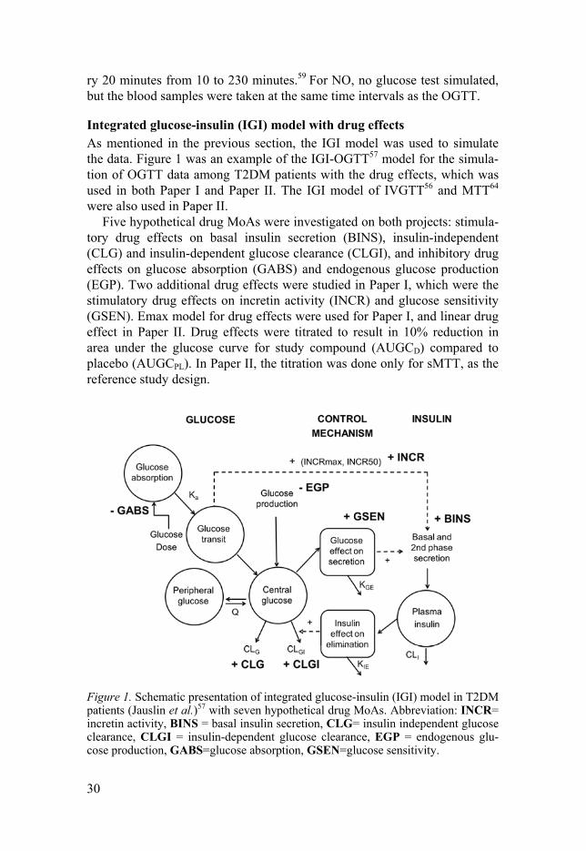
Integrated glucose-insulin (IGI) model with drug effects As mentioned in the previous section, the IGI model was used to simulate the data. Figure 1 was an example of the IGI-OGTT57 model for the simula-tion of OGTT data among T2DM patients with the drug effects, which was used in both Paper I and Paper II. The IGI model of IVGTT56 and MTT64 were also used in Paper II.
Five hypothetical drug MoAs were investigated on both projects: stimula-tory drug effects on basal insulin secretion (BINS), insulin-independent (CLG) and insulin-dependent glucose clearance (CLGI), and inhibitory drug effects on glucose absorption (GABS) and endogenous glucose production (EGP). Two additional drug effects were studied in Paper I, which were the stimulatory drug effects on incretin activity (INCR) and glucose sensitivity (GSEN). Emax model for drug effects were used for Paper I, and linear drug effect in Paper II. Drug effects were titrated to result in 10% reduction in area under the glucose curve for study compound (AUGCD) compared to placebo (AUGCPL). In Paper II, the titration was done only for sMTT, as the reference study design.
Figure 1. Schematic presentation of integrated glucose-insulin (IGI) model in T2DM patients (Jauslin et al.)57 with seven hypothetical drug MoAs. Abbreviation: INCR= incretin activity, BINS = basal insulin secretion, CLG= insulin independent glucose clearance, CLGI = insulin-dependent glucose clearance, EGP = endogenous glu-cose production, GABS=glucose absorption, GSEN=glucose sensitivity.

31
Finnish Diabetes Prevention Study (FDPS) (Paper III & IV) For Paper III and IV, the data came from the Finnish Diabetes Prevention Study (FDPS)93,94 consisting 522 overweight (average BMI=31kg/m2), mid-dle-aged (average age=55 years old) subjects with IGT; randomly assigned to control and lifestyle intervention groups. The subjects in the intervention group were given an intensive individual lifestyle intervention with counsel-ling on diet, weight reduction and exercise with associated goals of interven-tion. The goals of interventions were 5% reduction of weight, total intake of fat and saturated fat less than 30% and 10% of energy consumed, increased in fibre intake to at least 15g per 1000kcal, and moderate exercise for at least 30 minutes per day. On the other hand, the subjects in the control group were given a general oral and written information (a two-page leaflet) about diet and exercise, at baseline and during annual visits, without specific individual program. The subjects were recruited from five different centres in Finland, namely Helsinki, Kuopio, Turku, Tampere, and Oulu.
The glucose and insulin concentrations after the OGTT was collected yearly, with sampling performed at 0, 30, 60 (for some subjects) and 120 minutes post glucose-dose. In addition, at one of the study centres, Kuopio, frequently sampled intravenous glucose tests (IVGTT) were performed for 87 of the subjects at the start (year 0) and end of the study (year 4). Glucose and insulin were sampled at 2, 4, 6, 8, 10, 12, 14, 16, 19, 22, 24, 27, 30, 40, 50, 60, 70, 90, 100, 120, 140, 160, and 180 minutes post-glucose dose. Sub-jects who developed diabetes were excluded from the study at the time of diagnosis based on the 1985 World Health Organization (WHO) technical report series,95 after a second OGTT was performed for the diagnosis con-firmation.93,94
In Paper III, all the glucose and insulin observations from the glucose provocations (OGTT and IVGTT) performed at the Kuopio centre (101 and 82 subjects) were used, while in Paper IV, the components of baseline weight, FPG, FSI and baseline HbA1c from all centres (522 subjects) were used for the analysis.
Study power calculation (Paper I & II)
Analysis of data, both with and without insulin (Paper I) as well as for vari-ous glucose provocation study designs (Paper II) was made using the same IGI models used for the creation of the data.56,57,64 The analysis was conduct-ed on both models with drug effect (full model) and without drug effect (re-duced model). However, in the analysis, many parameters in the models were fixed and the IIVs were greatly reduced, especially for the system-specific parameters, as to reduce the run times. In specific, the estimated
32
parameters were glucose clearance (both insulin-independent, CLG and insu-lin-dependent, CLGI), insulin clearance (CLI), baseline glucose (GSS), base-line insulin (ISS), and the drug effect parameter, which were the maximum drug effect (EmaxD), drug potency (EC50D) or linear drug effect ( x) with the IIVs on all estimated parameters, except for the EmaxD. All remaining pa-rameters of the IGI model were fixed to the published values with removed IIVs. In the absence of insulin measurements (only in Paper I), ISS and CLI with their associated IIVs were fixed to the published values.
Specifically, in Paper I, there are 3 main parts of investigation. For Part 1 (identify a drug effect) and Part 2 (distinguishing the correct MoA), data were simulated using one at a time of the seven hypothetical MoAs. In Part 3 (identifying a secondary MoA on top of a primary MoA), two MoAs were combined in the simulations. The drug effects in Part 3 were simulated as independent of each other and only a selection of the most likely combina-tions of MoAs were simulated.
full-reduce in which full represents the model with drug effect and the reduced model is without drug effect. The Likelihood Ratio Test (LRT) was used to assess the signifi-cance of adding the drug effect in the model, with the chosen of 5%, and the df set to the number of differing parameters between the competing models. By using the LRT principle to discriminate between full and re-duced models, the between full models (OFVfull) and reduced models (OFVred) need to be at least 3.84, at = 5% and df =1, for the drug effect to be significant. In addition, for Part 2 of Paper I, the competing models are non-hierarchical and all have the same number of parameters, so the LRT could not be used. To determine superiority between the competing models, an arbitrary critical value of 10 was used, i.e if was larger than 10 in favour for the correct model, it was deemed superior, else models were deemed to be of similar quality.
In Paper 1, the difference in study power between analysis with and without insulin was calculated by the relative ratio of the for analysis with insulin to the for analysis without insulin, denoted /g. In Paper II, study power was assessed by calculating by the relative ratio be-tween the sMTT for analysis of data from the sMTT design and the
y for all other designs, where y denotes the designs: MTT-24, OGTT, NO, IVGTT and GGI. This ratio, denoted sMTT/y, can be interpreted as how many times more individuals are needed for an sMTT to achieve equiv-alent power to the compared design.
Monte Carlo Mapped Power (MCMP) Method (Paper I) Specifically in Paper I, in addition to the gi/g, the power was also assessed using the MCMP method.80 This was done to compare the two dif-ferent methods of calculating study power. In the MCMP76,80, the individual

33
sums of n n number of subjects. The procedure is repeated 10,000 times and the study power is assessed as the p of 10,000 greater than 7.81 ( =0.05, df=3).
Accuracy and precision of parameter estimates (Paper I & II) Drug parameters (EmaxD & EC50D) (Paper I) and ( x) (Paper II) In Paper 1, the accuracy and precision of parameter estimation were ana-lysed for the EmaxD and EC50D for all drug MoAs in Part 1 of the study, and for x in Paper II. The analyses were performed by using the estimated pa-rameters values of EmaxD, EC50D x , obtained from stochastic simulation and estimation (SSE) of 500 studies with 15 subjects each.96,97 The sample size of 15 subjects was chosen as it is an acceptable number of subjects in the early clinical phase of anti-hyperglycaemic trials.98
In Paper I, the parameter accuracy and precision were investigated by cal-culating the relative estimation error (REE) of the estimated value of EmaxD and EC50D from the full models of analysis using glucose and insulin data (gi) and the full models of analysis using only glucose data (g), as in equa-tion 2a and 2b. It was expected that the values of EmaxD and EC50D should be the same, with and without insulin measurements. ( ) = ( , , )/ , Eq. 2a ( 50 ) = ( 50 , 50 , )/ 50 , Eq. 2b
x x ob-tained from the SSE with the ‘true’ value used for the simulation, as in equa-tion 3. ( ) = ( )/ Eq. 3
Glucose area under the curve ratio (AUGCD/AUGCPL) Estimation of accuracy and precision of the glucose AUC were also per-formed, by assessing the ratio between the area under the glucose curve of drug (AUGCD) and area under the glucose curve with placebo (AUGCPL), and this ratio is denoted as AUGCD/PL in Paper I and AUGCD/PL,Cx in Paper II. The CX in AUGCD/PL,Cx represents different drug MoAs.

34
Firstly, a simulation was performed to compute the AUGCD and AUGCPL for 15 subjects, for 500 simulations of estimated placebo and full models. The simulation was done using the parameter estimates resulted from SSE, with the inclusion and exclusion of insulin data. For each simulation using SSE estimates, the mean (AUGCD/PL or AUGCD/PL,Cx) from 15 subjects was calculated, producing 500 mean values of AUGCD/PL for gi (AUGCgi) and g (AUGCg) for Paper I, and 500 mean values of AUGCD/PL,Cx for Paper II.
A second simulation of placebo and full models with 15 subjects and 500 simulations was performed, to compute the AUGCD/PL or AUGCD/PL,Cx as the ‘true’ ratio, using the parameter values set for the original simulations. The mean AUGCD/PL or AUGCD/PL,Cx was calculated as in the first simulation step. REE calculated for the AUGCD/PL or AUGCD/PL,Cx obtained from the SSE and AUGCD/PL or AUGCD/PL,Cx from the true simulation, as in the equa-tion 4a, 4b and 4c. In Paper I, the AUGCgi and AUGCg were expected to be the same values. ( ) = ( , , , , )/ , ,
Eq. 4a ( ) = ( , , , , )/ , , Eq. 4b
( , ) = ( , , , )/ , ,
Eq. 4c
Diabetes disease progression modelling IGI model (Paper III) The combination of IVGTT- and OGTT-IGI model among IGT subjects The combination of intravenous and oral IGI model55–57 (Figure 2) was used to fit the data for baseline in describing IGI model among the IGT subjects, incorporating prior information on the parameters from the published IVGTT- and OGTT-IGI models55–57 using prior99 functionality ($PRIOR) in NONMEM 7.374. The parameters with the same values for oral, intravenous, healthy volunteers and patients with T2DM were estimated with prior infor-mation.
In addition, the parameters that differed in previous populations of the IGI model between provocations or populations55–57 were estimated without prior information. These parameters were the CLG, CLGI, first-phase insulin secre-

35
tion (FPS), shape effect of glucose on its own production (GPRG), oral glu-cose bioavailability (BIOG), rate of first-phase insulin secretion (kIS), maxi-mum incretin effect (INCRmax), absorbed glucose at 50% maximum incretin effect (ABSG50), as well as GSS and ISS, with their related variability. The estimation was done with a lower and upper boundary of diabetics’ and healthy subjects’ values, to mimic the physiological state of the IGT subjects that is not as optimum as healthy state, but also not as deteriorated as the patients with T2DM.16
In addition to the parameter estimates and their variability, parameter cor-relations were also investigated in the model, which were the off-diagonal matrix between the central glucose volume of distribution (VG), inter-compartmental glucose clearance (Q) and insulin volume of distribution (VI), as the published IGI model.55–57 Specific to IGT population in this study, an additional off-diagonal matrix was investigated for the correlation between CLGI, FPS, ISS and GSS.
All the parameter and prior values can be found in Table 5 of the results section, with the parameter uncertainty or the df related to every prior value. The $PRIOR functionality is a restricted maximum-likelihood function for constraining parameter estimation based on the prior information. The Nor-mal-Inverse Wishart Prior (NWPRI) was used, described as a normal distri-bution for fixed effect parameters (typical parameters) and an Inverse-Wishart for the IIVs.99 For the priors on the fixed effect parameters, the pa-rameter uncertainty (standard error2 (SE2)) was calculated as in the equation 5a, incorporating each typical parameter value and its relative standard error (RSE). The df was calculated using equation 5b for the priors on the IIV,
2) of the random effect value (deviation between the individual’s and population’s value) and its RSE.
2 = ( % 100 )2 Eq. 5a = 2 2/( % 100 )2 + 1 Eq. 5b
The typical parameter values with the associated RSE were obtained from the published IGI model.55–57 The prior values, their associated variability, uncertainty and df were fixed during estimation.
Natural disease progression and intervention effects model For the modelling of the natural disease progression and intervention effects, the data from baseline until the fourth year were used. The disease progres-sion model was set to initiate 24 hours after the end of year 0 study in the dataset, to allowing the glucose and insulin concentrations to return to the baseline values before the next glucose provocations. The disease progres-sion and intervention effects were investigated on the most reasonable pa-rameters from the pathophysiological perspective. These parameters were

36
the FPS as an early insulin responsiveness in intravenous provocation, CLGI as a reflection of insulin sensitivity, ISS, reflecting changes in basal insulin secretion and INCRmax, as the changes of incretin hormones in the diabetic state. The effect parameters were added one at a time (stepwise manner), to assess the significance of adding additional parameters in the nested models, using LRT with = 5%. Figure 2 is the illustration of the disease progres-sion (DP) and lifestyle intervention (INT) effects in the combination of IVGTT- and OGTT-IGI.
Figure 2. The combination of oral and intravenous integrated glucose-insulin (IGI) model55–57 with the disease progression (DP) and intervention (INT) effects modelon the first-phase insulin secretion (FPS), insulin-dependent glucose clearance (CLGI), baseline insulin concentration (Iss), and maximum incretin effect (INCRmax). The final model was chosen with a linear DP and INT for FPS and CLGI, and a step function from the baseline to the first year for Iss, in which the Iss level was un-changed after the first year. No significant effects of DP and INT were found for the INCRmax.
WHIG model (Paper IV) In Paper IV, the IGT characteristics were described using the integrated WHIG model developed by Choy et al.92 Some of the system parameters were fixed to the published values during the estimation process, as they were assumed to be the same in the healthy and patients’ populations. The

37
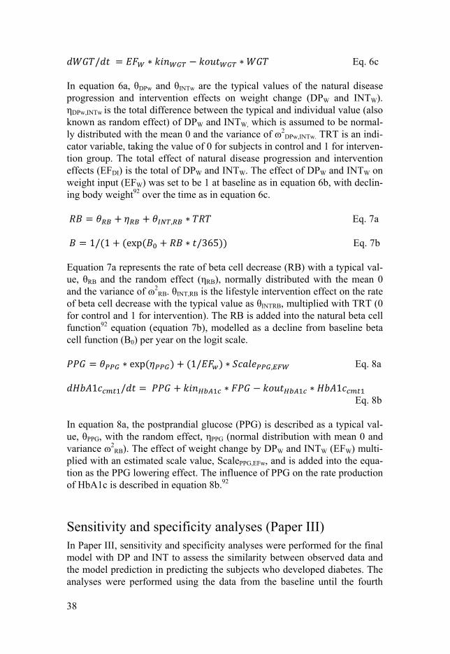
fixed parameters were the turn-over time of weight changes, represented in the model by the half-life of the weight compartment (t1/2, WGT), the effect of weight change (per kg) on insulin sensitivity, represented by the scaling factor of change in weight on insulin sensitivity (Scale EFs), the time life-span of HbA1c, represented by the mean transit time of HbA1c (MTT) and the rate of glycation in relation to FPG, i.e. the rate constant of HbA1c com-partments production (Kin, HbA1c).
Figure 3. The investigated weight-HbA1c-insulin-glucose (WHIG) model92 with the effects of natural disease progression and lifestyle intervention (EFW, (DPW&INTW) over time on the weight component. The intervention effect on the beta cell function (EFB, (INTRB)) is investigated together with the natural loss of beta cell function (B), that later influenced the rate of insulin production. The effect of EFW on post-prandial glucose (PPG) input (EFPPG, (EFW)) is added in the model. The fasting plasma glucose (FPG) and PPG drive the production of HbA1c that is described using 3 transit compartments.
The natural disease progression (DPW) and lifestyle intervention (INTW) ef-fects over time were investigated on the weight (WGT) (equation 6a and 6b), beta cell function (B) (equation 7a and 7b) and postprandial glucose (PPG) input components (equation 8a and 8b), with the expectation of decreasing weight (equation 6c), increasing beta cell function and decreasing PPG input with intervention due to lower carbohydrate intake. A decrease in PPG input is later expected to decrease the rate production of HbA1c.
= + + , Eq. 6a = (100 )/100 Eq. 6b
38
/ = Eq. 6c
In equation 6 DPw INTw are the typical values of the natural disease progression and intervention effects on weight change (DPW and INTW).
DPw,INTw is the total difference between the typical and individual value (also known as random effect) of DPW and INTW, which is assumed to be normal-ly distributed with the mean 0 and the variance of 2
DPw,INTw. TRT is an indi-cator variable, taking the value of 0 for subjects in control and 1 for interven-tion group. The total effect of natural disease progression and intervention effects (EFDI) is the total of DPW and INTW. The effect of DPW and INTW on weight input (EFW) was set to be 1 at baseline as in equation 6b, with declin-ing body weight92 over the time as in equation 6c.
= + + , Eq. 7a
= 1/(1 + (exp ( + /365)) Eq. 7b
Equation 7a represents the rate of beta cell decrease (RB) with a typical val-RB and RB), normally distributed with the mean 0
and the variance of 2RB INT,RB is the lifestyle intervention effect on the rate
INTRB, multiplied with TRT (0 for control and 1 for intervention). The RB is added into the natural beta cell function92 equation (equation 7b), modelled as a decline from baseline beta cell function (B0) per year on the logit scale.
= exp ( ) + (1/ ) , Eq. 8a 1 / = + 1
Eq. 8b
In equation 8a, the postprandial glucose (PPG) is described as a typical val-PPG PPG (normal distribution with mean 0 and
variance 2RB). The effect of weight change by DPW and INTW (EFW) multi-
plied with an estimated scale value, ScalePPG,EFw, and is added into the equa-tion as the PPG lowering effect. The influence of PPG on the rate production of HbA1c is described in equation 8b.92
Sensitivity and specificity analyses (Paper III) In Paper III, sensitivity and specificity analyses were performed for the final model with DP and INT to assess the similarity between observed data and the model prediction in predicting the subjects who developed diabetes. The analyses were performed using the data from the baseline until the fourth

39
year. The model-based diagnosis of diabetes was made based on the diag-nostic criteria of FPG or the 2-hour postprandial glucose values after a 75g OGTT. There were 3 main scenarios investigated for the model prediction, which were: A. using all the observed data of glucose and insulin concentrations at 0, 30,
60 and 120 minutes for the OGTT and IVGTT from baseline to the fourth year
B. using the data as in the scenario A, but excluding the glucose and insulin concentrations of the IVGTT at the fourth year
C. using the data as in the scenario A, but excluding the glucose and insulin concentrations at 0, and 120-minutes for OGTT.
For the calculation of the specificity and sensitivity analyses, the subjects who developed diabetes from the model prediction were compared to the ones who developed diabetes based on the records of doctors’ diagnosis, as well as on the observed fasting and the 2-hour glucose concentrations from the data.
The sensitivity analysis represents the model ability to correctly identify the subjects who developed diabetes at the fourth year (equation 9a). For example, a value of 0.9 was translated to model’s ability to detect 90% sub-jects who developed diabetes (true positive) with 10% of subjects went un-detected (false negative). Specificity analysis represents the model’s ability to correctly identify the subjects who did not develop diabetes until the fourth year (equation 9b). The value of 0.8, for instance, represents the abil-ity of the model to identify 80% of the subjects who had not developed dia-betes (true negative), with 20% falsely identified to develop diabetes (false positive), at the fourth year.100
= /( + ) Eq. 9a = /( + ) Eq. 9b
Logistic regression dropout model (Paper IV) A logistic regression dropout model was used to describe the dropout in the data. For the subjects who dropped out, the exact date of dropout was not documented, and only the information that the dropout occurred between two subsequent visits was available. Thus, the logistic regression was chosen to develop a model for the probability of the dropout event over a time-interval, in addition to its simplicity and reliability to be implemented as a dropout model. Each potential predictor of dropout with its corresponding
inciple was used to assess the significance of adding the predictors into the model.
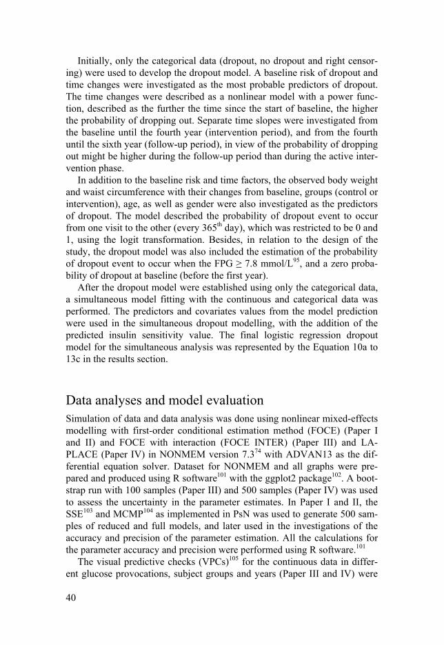
40
Initially, only the categorical data (dropout, no dropout and right censor-ing) were used to develop the dropout model. A baseline risk of dropout and time changes were investigated as the most probable predictors of dropout. The time changes were described as a nonlinear model with a power func-tion, described as the further the time since the start of baseline, the higher the probability of dropping out. Separate time slopes were investigated from the baseline until the fourth year (intervention period), and from the fourth until the sixth year (follow-up period), in view of the probability of dropping out might be higher during the follow-up period than during the active inter-vention phase.
In addition to the baseline risk and time factors, the observed body weight and waist circumference with their changes from baseline, groups (control or intervention), age, as well as gender were also investigated as the predictors of dropout. The model described the probability of dropout event to occur from one visit to the other (every 365th day), which was restricted to be 0 and 1, using the logit transformation. Besides, in relation to the design of the study, the dropout model was also included the estimation of the probability of dropout event to occur when the FPG 7.8 mmol/L95, and a zero proba-bility of dropout at baseline (before the first year).
After the dropout model were established using only the categorical data, a simultaneous model fitting with the continuous and categorical data was performed. The predictors and covariates values from the model prediction were used in the simultaneous dropout modelling, with the addition of the predicted insulin sensitivity value. The final logistic regression dropout model for the simultaneous analysis was represented by the Equation 10a to 13c in the results section.
Data analyses and model evaluation Simulation of data and data analysis was done using nonlinear mixed-effects modelling with first-order conditional estimation method (FOCE) (Paper I and II) and FOCE with interaction (FOCE INTER) (Paper III) and LA-PLACE (Paper IV) in NONMEM version 7.374 with ADVAN13 as the dif-ferential equation solver. Dataset for NONMEM and all graphs were pre-pared and produced using R software101 with the ggplot2 package102. A boot-strap run with 100 samples (Paper III) and 500 samples (Paper IV) was used to assess the uncertainty in the parameter estimates. In Paper I and II, the SSE103 and MCMP104 as implemented in PsN was used to generate 500 sam-ples of reduced and full models, and later used in the investigations of the accuracy and precision of the parameter estimation. All the calculations for the parameter accuracy and precision were performed using R software.101
The visual predictive checks (VPCs)105 for the continuous data in differ-ent glucose provocations, subject groups and years (Paper III and IV) were

41
performed as implemented in PsN (version 4.6.12) with 500 simulations. In Paper III, the censoring option105 for the subjects who develop diabetes based on the FPG or the 2-hour plasma glucose concentrations from the model prediction was added in the VPC. This was done in order to mimic the original study design, in which the subjects who developed diabetes were excluded at the time of diagnosis24. Additionally, in Paper IV, a VPC with 100 simulations was performed for analysis of the categorical data only. The visualization of the data and results was done using Xpose4 (version 4.5.0).106–108 The LRT was used to discriminate between nested models, with the chosen =5%. Based on LRT, the OFV is assumed to be chi-square dis-tributed, and a decrease of 3.84 in OFV between hierarchical models with df =1 is considered statistically significant at =5%. The final model was cho-sen based on the lowest OFV, the best VPCs, specificity and sensitivity analyses (Paper III) as well as the scientific plausibility.
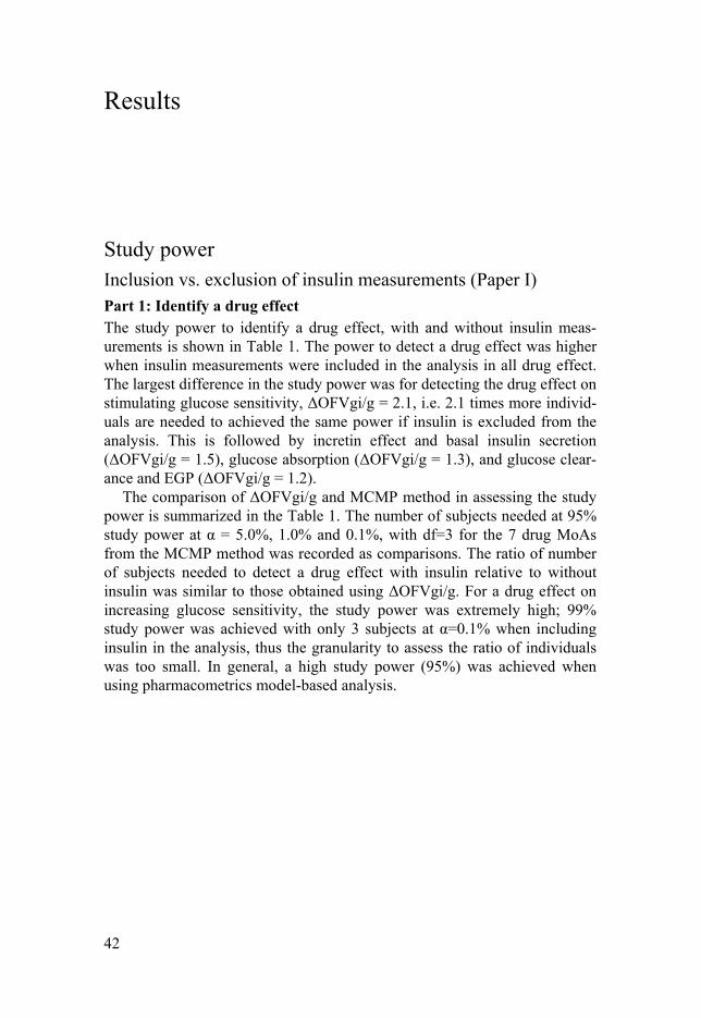
42
Results
Study power Inclusion vs. exclusion of insulin measurements (Paper I) Part 1: Identify a drug effect The study power to identify a drug effect, with and without insulin meas-urements is shown in Table 1. The power to detect a drug effect was higher when insulin measurements were included in the analysis in all drug effect. The largest difference in the study power was for detecting the drug effect on
d-uals are needed to achieved the same power if insulin is excluded from the analysis. This is followed by incretin effect and basal insulin secretion
r-ance
The comparipower is summarized in the Table 1. The number of subjects needed at 95% study power at = 5.0%, 1.0% and 0.1%, with df=3 for the 7 drug MoAs from the MCMP method was recorded as comparisons. The ratio of number of subjects needed to detect a drug effect with insulin relative to without insulin was similar increasing glucose sensitivity, the study power was extremely high; 99% study power was achieved with only 3 subjects at =0.1% when including insulin in the analysis, thus the granularity to assess the ratio of individuals was too small. In general, a high study power (95%) was achieved when using pharmacometrics model-based analysis.
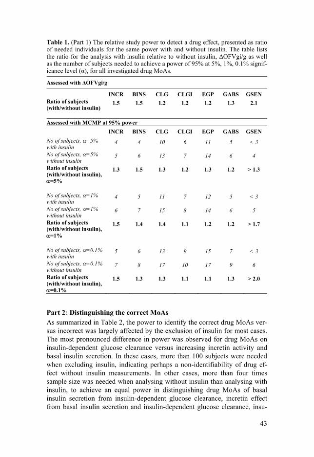
43
Table 1. (Part 1) The relative study power to detect a drug effect, presented as ratio of needed individuals for the same power with and without insulin. The table lists the ratio for the analysis with insulin relative to without insulinas the number of subjects needed to achieve a power of 95% at 5%, 1%, 0.1% signif-icance level ( ), for all investigated drug MoAs.
INCR BINS CLG CLGI EGP GABS GSEN Ratio of subjects
1.5 1.5 1.2 1.2 1.2 1.3 2.1
Assessed with MCMP at 95% power
INCR BINS CLG CLGI EGP GABS GSEN No of subjects, =5% with insulin
4 4 10 6 11 5 < 3
No of subjects, =5% without insulin
5 6 13 7 14 6 4
Ratio of subjects
=5%
1.3 1.5 1.3 1.2 1.3 1.2 > 1.3
No of subjects, =1% with insulin
4 5 11 7 12 5 < 3
No of subjects, =1% without insulin
6 7 15 8 14 6 5
Ratio of subjects
=1%
1.5 1.4 1.4 1.1 1.2 1.2 > 1.7
No of subjects, =0.1% with insulin
5 6 13 9 15 7 < 3
No of subjects, =0.1% without insulin
7 8 17 10 17 9 6
Ratio of subjects
=0.1%
1.5 1.3 1.3 1.1 1.1 1.3 > 2.0
Part 2: Distinguishing the correct MoAs As summarized in Table 2, the power to identify the correct drug MoAs ver-sus incorrect was largely affected by the exclusion of insulin for most cases. The most pronounced difference in power was observed for drug MoAs on insulin-dependent glucose clearance versus increasing incretin activity and basal insulin secretion. In these cases, more than 100 subjects were needed when excluding insulin, indicating perhaps a non-identifiability of drug ef-fect without insulin measurements. In other cases, more than four times sample size was needed when analysing without insulin than analysing with insulin, to achieve an equal power in distinguishing drug MoAs of basal insulin secretion from insulin-dependent glucose clearance, incretin effect from basal insulin secretion and insulin-dependent glucose clearance, insu-

44
lin-independent glucose clearance from basal insulin secretion, as well as glucose sensitivity from incretin effect and basal insulin secretion. In addi-tion, at least 1.5 times fewer subjects were needed in other cases, to distin-guish true from false MoAs with the inclusion of insulin measurements.
The study power however, may not be affected by insulin measurements to distinguish drug MoAs of insulin-dependent from insulin-independent glucose clearance, EGP and glucose absorption, and EGP from glucose ab-sorption. Besides, the correct drug MoA of increasing insulin-independent glucose clearance could not be discriminate from the incorrect of decreasing EGP, even when insulin measurements were included in the study. This con-clusion was based on more than 100 subjects were needed, both when in-cluding and excluding insulin measurements.
Table 2. (Part 2) The relative study power to distinguish correct drug mechanism of action (MoA) from incorrect, represented as ratio of subjects needed to achieve a same power with and without l-
Correct MoA Competing Incorrect MoA
INCR BINS CLG CLGI EGP GABS GSEN INCR - 4.7 1.7 4.1 2.5 2.5 1.6 BINS * - 1.8 6.0 2.5 3.1 2.0 CLG 3.8 4.7 - 1.5 ** 1.6 2.4 CLGI * * 1.0 - 1.0 1.0 2.5 EGP 2.9 3.1 1.0 1.4 - 1.1 1.6 GABS 3.7 3.8 1.2 1.4 1.2 - 2.3 GSEN 4.4 4.5 2.5 3.5 2.6 3.1 - Abbreviation: MoA = Mechanism of action, INCR = incretin activity, BINS = basal insulin secretion, CLG = insulin-independent glucose clearance, CLGI = insulin-dependent glucose clearance, EGP = endogenous glucose production, GABS = glucose absorption, GSEN = glucose sensitivity.
were needed to distinguish the drug effect without insulin were needed to distinguish the drug effect with and without insulin
Part 3: Identifying a secondary drug MoA on top of a primary MoA As shown in Table 3, the overall power to detect a secondary true drug MoA was higher with the use of glucose and insulin as opposed to only glucose measurements in the analysis. This is most pronounced for detecting a sec-ondary drug MoA of basal insulin secretion on top of the primary drug MoA of incretin effect, and insulin-dependent glucose clearance on top of basal insulin secretion. More than 100 subjects were needed to detect the second-ary drug MoAs from the primary ones when excluding insulin in these cases, indicating perhaps a non-identifiability of the secondary effect without insu-lin. Furthermore, a major difference, at least twice as many subjects were needed when insulin measurements were excluded from the study, to detect

45
insulin-dependent glucose clearance on top of glucose sensitivity
sensitivity on top of all tested primary drug MoA6.0). In other cases, a slight increase of power to detect secondary drug MoA was observed, which were for insulin-independent on top of insulin-
f-
as glucose absorption on top of insulin-independent glucose clearance
There was no difference of study power to detect insulin-independent glu-cose clearance on top of EGP, and EGP on top of glucose clearance. In addi-tion, in identifying a secondary drug MoA on insulin-independent glucose clearance on top of primary MoA on EGP, more than 100 subjects were re-quired, independent of sampling insulin.
Table 3. (Part 3) The relative study power to detect correct secondary correct drug effect with the primary already included, represented as ratio of subjects needed to achieve a same power without and with insulin. The table lists
Secondary MoA Competing Incorrect MoA INCR BINS CLG CLGI EGP GSEN BINS * - - - - - CLG - 1.1 - 1.4 ** - CLGI - * - - 1.1 3.7 EGP 1.6 2.1 1.0 1.0 - 1.8 GABS 8.1 - 1.5 - - - GSEN 4.3 6.0 - 4.9 4.9 - Abbreviation: MoA = Mechanism of action, INCR = incretin activity, BINS = basal insulin secretion, CLG = insulin-independent glucose clearance, CLGI = insulin-dependent glucose clearance, EGP = endogenous glucose production, GABS = glucose absorption, GSEN = glucose sensitivity.
were needed to detect the secondary drug MoA without insulin were needed to detect the secondary drug MoA with and without insulin
Most appropriate study design based on the drug MoAs (Paper II) Table 4 shows the study power of each study design as a relative ratio to the study power of sMTT model. As mentioned earlier, this ratio is the represen-tation of how many times more individuals are needed for a sMTT to achieve equivalent power to the compared designs. Intravenous provocations were always more powerful than oral provocations except for MTT-24 in certain MoAs. The power of MTT-24 was higher than sMTT for all MoAs except for increasing insulin-independent glucose clearance. Repeated fast-

46
ing samples or NO had for many MoAs higher power than sMTT. Notable though is that safety was not assessed in this study, and no glucose provoca-tion may be associated with more hypoglycaemia than other designs. The MTT-24 and OGTT were the least powerful study designs for drug effect on increasing insulin-independent glucose clearance, potentially due to the stimulation of insulin release at a higher rate by incretin activity made the insulin–independent glucose clearance less important.
Table 4. The study power of each model for each drug effect is described as a ratio relative to the study power of single meal tolerance test (sMTT).
Drug effects Study designs sMTT MTT-24
OGTT NO IVGTT GGI
BINS 1 8.66 1.49 2.02 5.82 5.43 CLG 1 0.60 0.29 0.78 2.81 3.89 CLGI 1 17.28 2.82 2.74 17.98 7.93 EGP 1 1.98 0.58 1.73 4.16 4.77 GABS 1 3.87 0.40 - - - Abbreviation: IVGTT = intravenous glucose tolerance test; GGI = graded glucose infusion; NO = no provocations test; OGTT = oral glucose tolerance test; sMTT = single meal toler-ance test; MTT-24 = 24-hours meal tolerance test, INCR = incretin activity, BINS = basal insulin secretion, CLG = insulin-independent glucose clearance, CLGI = insulin-dependent glucose clearance, EGP = endogenous glucose production, GABS = glucose absorption, GSEN = glucose sensitivity. Yellow represents the most powerful, and the green represents the second most powerful designs.
Accuracy and precision of parameter estimates (Paper I & II) Drug parameter EmaxD & EC50D and x In Paper I, an acceptable accuracy and precision of the parameter EmaxD without insulin measurements was observed for the drug effects with MoA related to glucose: glucose clearance, EGP and glucose absorption. For the EC50D, the only drug MoA where accuracy was acceptable without insulin measurements was for glucose absorption. The precision of EC50D for this drug MoA was however poor. Moreover, in all other drug MoAs for both parameters, the estimates were both biased and poor in precision. The results are illustrated in Figure 4.
x parameter are visualized in Figure 5. The parameter estimation derived from OGTT, IVGTT and GGI were overall good for all MoAs. Thus, the most powerful study designs, albeit including the two most invasive, were also producing accurate and precise estimates. The precision and accuracy of MTT-24 and sMTT were dependent on the MoA of the drug. The precision of MTT-24 was the best

47
for all MoAs. The worst precision and accuracy in all MoAs were the sMTT for drug effect on insulin-dependent glucose clearance, and NO for drug effect on insulin-independent glucose clearance, despite these designs being rather powerful.
Figure 4. (Part 1) Relative estimation error (REE) of the drug parameters of maxi-mum drug effect (EMAXD) and drug concentration at 50% EMAX (EC50D). Ab-breviation: SSE = stochastic simulation and estimation, INCR = incretin activity, BINS = basal insulin secretion, CLG = insulin-independent glucose clearance, CLGI = insulin-dependent glucose clearance, EGP = endogenous glucose produc-tion, GABS = glucose absorption, GSEN = glucose sensitivity.

48
Figure 5. A descriptive representation of the distribution of relative estimation error of each study design model for five investigated drug MoAs. The different study design models are intravenous glucose tolerance test (IVGTT), oral glucose tolerance test (OGTT), single meal tolerance test (sMTT), 24-hours meal tolerance test (MTT-24), graded glucose infusion (GGI) and no-provocations test (NO). Abbreviation: BINS = basal insulin secretion, CLG = insu-lin-independent glucose clearance, CLGI = insulin-dependent glucose clearance, EGP = glucose production, GABS = glucose absorption. Yellow represents the most powerful, and green represents the second most powerful designs.

49
Glucose area under the curve ratio (AUGCD/PL) The AUGCD/PL for Paper I was investigated and reported in Figure 6. The AUGCD/PL was found to be estimated with an adequate accuracy to the true simulation value and with a good precision for most drug effects, both in-cluding and excluding insulin data in the analysis, despite the inaccuracy and imprecision of the EmaxD and EC50D.
In addition, the same observation found in Paper II, in which despite the differences between the estimation and simulation x values, both models can precisely simulate the same AUGCD/PL,Cx as shown in Figure 7. The worst accuracy was NO for drug effects on insulin-dependent glucose clear-
x estimated in sMTT were the least precise and accurate for drug MoAs on insulin-dependent glucose clearance, sMTT can still simulate AUGCD/PL,Cx precisely as simulation models. The same observation was seen for NO when the drug effect is on insulin-independent glucose clearance. NO can simulate plasma glucose AUGCD/PL,Cx with good precision and accu-racy, but it was still the worst among all other models for this drug MoA. In conclusion, the study design with the highest power for a drug MoA was not always the most precise neither was the most accurate one. The most power-ful, accurate and precise study designs are the intravenous ones which are also the most invasive. From power perspective, MTT-24 is a good alterna-tive, unless we are expecting effect on insulin-independent glucose clearance like SGLT-2-inhibitors, but it seems to be resulting in a biased parameter estimate.

50
Figure 6. (Part 1) Relative estimation error (REE) for AUGC ratio, in seven hypo-thetical drug MoAs. Abbreviation: Glu + Ins = glucose and insulin data, Glu = glucose only data, INCR = incretin activity, BINS = basal insulin secretion, CLG = insulin-independent glucose clearance, CLGI = insulin-dependent glucose clear-ance, EGP = endogenous glucose production, GABS = glucose absorption, GSEN = glucose sensitivity.
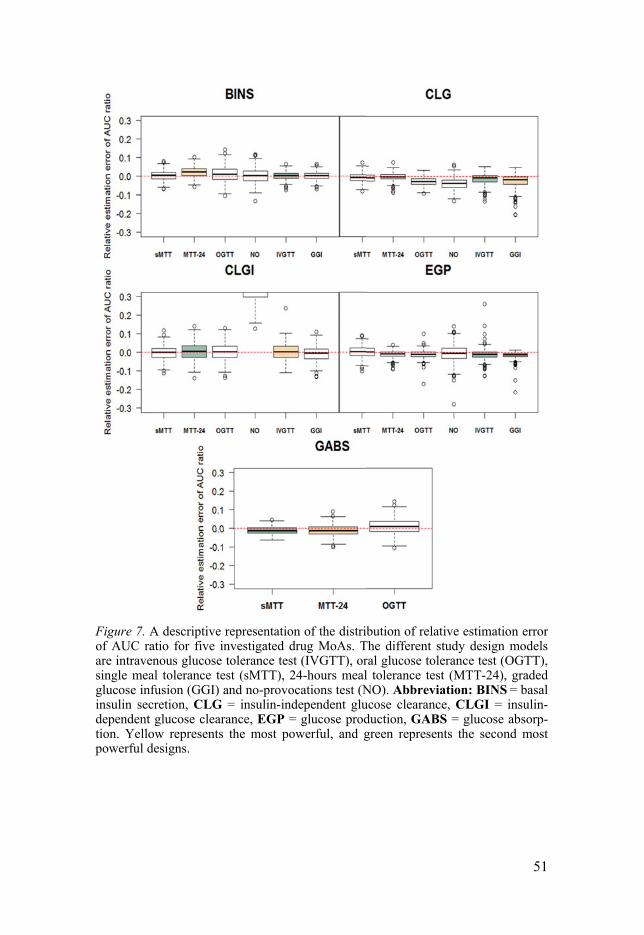
51
Figure 7. A descriptive representation of the distribution of relative estimation error of AUC ratio for five investigated drug MoAs. The different study design models are intravenous glucose tolerance test (IVGTT), oral glucose tolerance test (OGTT), single meal tolerance test (sMTT), 24-hours meal tolerance test (MTT-24), graded glucose infusion (GGI) and no-provocations test (NO). Abbreviation: BINS = basal insulin secretion, CLG = insulin-independent glucose clearance, CLGI = insulin-dependent glucose clearance, EGP = glucose production, GABS = glucose absorp-tion. Yellow represents the most powerful, and green represents the second most powerful designs.
52
Diabetes disease progression modelling IGI model (Paper III) The combination of IVGTT- and OGTT-IGI model among IGT subjects Final parameter estimates for the subjects with IGT were summarized in Table 5, under the baseline data final estimates. In general, the fixed effect parameters with prior information were similar with or only slightly deviated from the initial values. For the parameters without prior information, the values were estimated to be in between the values of healthy subjects and patients with T2DM, except for the rate constant of glucose effect on its own production (kGE1) and CLG. The kGE1 was fixed to the previously published value of 0.0573 min-1 as it was estimated to be unreasonably higher than the published one. CLG was fixed to the published diabetic value due its tenden-cy to reach the lower boundary during estimation. The CLGI, FPS, and GPRG were closer to diabetic values, whereas BIOG was closer to healthy subjects’. The values for ISS and GSS were in agreement with the definition of IGT population. An adequate model description of the data on the parame-ters level was achieved, indicated by the reasonable parameter estimations in the IGI model.
For the inter-individual variability, similar parameter values with the ones published were estimated in most cases. For some parameters as VG, IN-CRmax, CLI, VP, effect delay rate constant of glucose on insulin secretion (kGE2), shape effect of glucose on insulin secretion (IPRG) and effect delay rate constant of insulin effect on glucose clearance (kIE) were decreased from the published values. Parameter correlation between the Q with VG and VI was increased in the IGT subjects, whereas the correlation between VG and VI was lower.
Natural disease progression and intervention effects model Natural disease progression effect The effects of natural disease progression and intervention on chosen param-eters are summarized in Table 5, as all final parameter estimates. The FPS and CLGI were decreased by 3.0% and 8.1% per year in all subjects as a re-sult of the natural disease progression. The ISS was observed to increase by 68% from baseline to Year 1, and remain unchanged after the first year.
Lifestyle intervention effect On top of the natural disease progression, the lifestyle intervention increased the FPS value by 2.9% per year, producing a net effect of 0.1% decrease per year. This can be interpreted as a much slower deterioration of the acute response of beta cell function to produce insulin following an IVGTT. The CLGI had an increment of 6.0% per year with the lifestyle intervention, pro-ducing a 2.1% decrease per year, representing a slower deterioration of insu-

53
lin sensitivity with intervention. The ISS was affected by the intervention with a net effect of 153% increase from baseline to the first year and re-mained constant after that, reflecting the improvement of beta cell function to secrete insulin during the specific period.
No significant effects of natural disease progression and lifestyle inter-vention were found for the INCRmax. The model fit was no better than the chosen final model, in which an insignificant OFV improvement and worse VPCs were observed. Besides, the specificity and sensitivity results were also worse than the chosen final model, indicating a poor model prediction.
Table 5. Final parameter estimates for only baseline and all data (baseline until the fourth year) in the combination of integrated glucose-insulin for intravenous and oral glucose tolerance test (IVGTT-OGTT-IGI) among the subjects with impaired glu-cose tolerance (IGT) with the use of prior information from the published IGI mod-el. 55–57
Baseline data All data
Parameter
Initial esti-
*Prior
Uncer-
df
estimates (RSE )||
i-mates (RSE )||
Glucose VG , L Central volume of
distribution 9.33* 0.313376 8.92 (2.3) 9.13 (1.4)
Vp, L Peripheral volume of distribution
8.56* 0.183184 8.01 (3.0) 8.78 (0.6)
CLG, L/min Insulin-independent clearance
0.0287† - 0.0287† 0.0287†
CLGI, L/min/ (mu/L)
Insulin-dependent clearance
(0.00297, 0.00829) ‡
- 0.00405 (4.4)
0.00389 (9.5)
Q, L/min Inter-compartmental clearance
0.442* 0.005001 0.35 (10.1) 0.25 (9.6)
kGE1, min-1 Effect delay rate constant, glu. on its own prod.
0.0573† 0.000040 0.0573† 0.0573†
kGE2, min-1 Effect delay rate constant, glu. on ins. Sec.
0.0289* 0.000016 0.0335 (3.6) 0.0307 (3.2)
GPRG (-) Shape effect, glu on its own production
(-2.79,0) ‡ - -0.587 (38.2) -1.37 (11.7)
IPRG (-) Shape of effect, glu. on ins. secretion
1.42* 0.029036 1.59 (2.4) 1.86 (1.5)
BIOG, % Bioavailability (0.722, 0.811) ‡
- 0.754 (6.2) 0.723 (3.0)
MTT, min Mean transit time of glucose absorption
34.9* 14.738 44.2 (4.1) 37.9 (2.9)
n Number of glucose transit compartment
1.27* 0.0929 1.8 (8.9) 0.9 (9.0)
INCRmax Maximal incretin effect
1.47§
- 1.93 (19.7) 1.26 (15.7)

54
ABSG50 Absorbed glucose at 50% INCRmax
14.8§ - 15.1 (49.8) 18.4 (49.0)
Gss, mg/dL Baseline glucose concentration
105.6§ - 108 (1.1) 107 (1.1)
Insulin VI, L Volume of distribu-
tion 6.09* 0.300414 4.0 (2.2) 4.02 (2.0)
CLI, L/min Insulin clearance 1.22* 0.003721 0.805 (1.7) 0.852 (2.1) kIE, min-1 Effect delay rate
constant, insulin on glucose clearance
0.0213* 0.000008 0.0218 (5.6) 0.0155 (9.0)
kIs, min-1 Rate constant, delay compartment of first-phase secretion and plasma compartment
0.384§ - 0.824 (12.8) 0.678 (8.1)
FPS, mU First-phase insulin secretion
(0,704) ‡ - 119 (12.9) 155 (6.1)
Iss, mU/L Baseline insulin concentration
13.5§ - 13.5 (4.7) 6.35 (2.8)
IssI,NT, mU/L Baseline insulin concentration, inter-vention group
- - - 4.11 (3.5)
Disease progression and intervention effects DP_FPS Disease progression
on FPS - - - -0.030 (35.3)
DP_CLGI Disease progression on CLGI
- - - -0.081 (40.3)
DP_ INCRmax
Disease progression on INCRmax
- - - -
DP_ISS Disease progression on Iss
- - - 0.683 (14.9)
INT_FPS Intervention effect on FPS
- - - 0.029 (79.0)
INT_CLGI Intervention effect on CLGI
- - - 0.060 (68.4)
INT_ INCRmax
Intervention effect on INCRmax
- - - -
INT_ISS Intervention effect on Iss
- - - 0.849 (24.4)
Residual error RESG, % Residual error, iv
glucose 7.32* 0.000003 3.81 (4.1) 6.5 (3.4)
RESGPO, % Residual error, oral glucose
8.58* 0.000027 9.15 (2.0) 14.8 (1.4)
RESI, % Residual error, insu-lin
25.2* 0.000229 14.9 (3.8) 25.2 (3.0)
RESE (-) Residual error, early 3.31* 0.483164 1.64 (5.9) 1.25 (3.7) Inter- Glucose VG Central volume of
distribution 30* 63 24.4 (3.8) 23.8 (5.4)
Vp Peripheral volume of distribution
30* 27 28.2 (16.4) 97.0 (37.3)

55
IOV, Vp Peripheral volume of distribution, IOV
15* 1 50.6 (21.6) 93.7 (18.8)
CLG Insulin-independent clearance
59† 59 59† 59†
CLGI Insulin-dependent clearance
46 - 46.5 (10.1) 65.0 (26.1)
Q Inter-compartmental clearance
85* - 83.9 (8.8) 77.6 (8.1)
kGE2 Effect delay rate constant, glu. on ins. sec
85* 12 58.1 (9.6) 59.9 (13.2)
IPRG Shape of effect, glu. on ins. secretion
35* 11 28.1 (14.0) 20.6 (9.5)
MTT Mean transit time of glucose absorption
11* 12 19.3 (26.3) 28.1 (32.5)
INCRmax Maximal incretin effect
55§ - 19.7 (88.5) 49.4 (19.3)
ABSG50 Absorbed glucose at 50% INCRmax
114§ - 131.9 (121.4)
137.5 (401.2)
Gss Baseline glucose concentration
14§ - 9.8 (11.6) 10.3 (11.5)
Insulin
VI Volume of distribu-tion
41* - 31.8 (4.8) 31.8 (2.7)
CLI Insulin clearance 29* 29 26.9 (10.0) 26.9 (10.6)
kIE Effect delay rate constant, insulin on glucose clearance
58* - 57.4 (7.0) 75.2 (39.9)
FPS, (mU) First-phase insulin secretion
67§ - 127.7 (14.8) 110.9 (13.5)
Iss Baseline insulin concentration
49§ - 50.5 (13.1) 46.5 (11.1)
Parameter correlations CorrVG-Q Correlation between
VG and Q -0.19* - -0.732 (6.7) -0.743 (8.8)
CorrVG-VI Correlation between VG and VI
0.7225* - 0.624 (5.6) 0.653 (4.2)
CorrQ-VI Correlation between Q and VI
-0.122* - -0.225 (19.3) -0.313 (11.9)
CorrCLGI-FPS Correlation between CLGI and FPS
-0.3364§ - -0.389 (24.4) -0.492 (20.5)
CorrCLGI-ISS Correlation between CLGI and ISS
-0.7181§ - -0.737 (15.3) -0.566 (17.3)
CorrCLGI-GSS Correlation between CLGI and GSS
-0.0948§ - -0.103 (60.0) 0.160 (83.4)
CorrFPS-ISS Correlation between FPS and ISS
0.2386§ - 0.214 (47.0) 0.338 (19.9)
CorrFPS-GSS Correlation between FPS and GSS
-0.6058§ - -0.622 (19.3) -0.645 (14.0)
CorrISS-GSS Correlation between ISS and GSS
0.3957§ - 0.447 (22.8) 0.355 (25.0)
* Prior values † Fixed to published value, CLG (diabetic), KGE1 (healthy)55–57 ‡ Estimated with lower and upper boundary using healthy and diabetic values § Free estimation || RSE values (variance scale) were obtained from the bootstrap (n=100)

56
Visual predictive checks IVGTT-OGTT-IGI model among IGT subjects The VPCs for the IVGTT-OGTT-IGI model in this IGT subjects were shown in Figure 8. For both glucose and insulin components, the model fitted the median data of IVGTT and OGTT fairly well. An adequate model fit can be observed at the 2.5th and 97.5th percentile of the data for every component, especially for the insulin. A small model misspecification, however, was observed for the variability of the glucose component (at the 2.5th and 97.5th percentile), especially at the end of time profile. This might be explained by a large heterogeneity in term of glucose control in this populations, in which some of the subjects may be at the beginning of the IGT state close to the healthy state, whereas the other may be at the end, close to T2DM. Apart from the small misfit, this combination of IVGTT-OGTT-IGI model could adequately describe the data of IGT subjects.
Figure 8. VPCs of the IVGTT and OGTT for the glucose and insulin for the subject with IGT. Solid lines represent the median of observed data; dashed lines represent the 2.5th percentile (lower part) and 97.5th percentile (upper part) for observed data. Dark shaded areas are the 95% confidence interval for median of simulated data, light shaded areas are the 95% confidence interval for 2.5th percentile (lower part) and 97.5th percentile (upper part) from 500 simulated datasets.

57
IVGTT-OGTT-IGI model with disease progression and intervention The VPCs for glucose and insulin components in control and intervention groups are shown in Figure 9 for IVGTT. The IGI model with the inclusion of natural disease progression and intervention effects has fairly well de-scribed the data for baseline and the fourth year for IVGTT. An adequate overall model fit can be seen for both glucose and insulin components in the baseline and the fourth year, among subjects in the control as well as life-style intervention groups. The median and the 5th percentile model prediction were very good for all cases. Only a small over-prediction can be found in the 95th percentile of the model prediction for the baseline glucose compo-nent. Possible explanation might be due to the heterogeneity factor. On the fourth year, the confidence interval for the model prediction was wider than at baseline, as the subjects who developed diabetes were excluded from the VPC analysis. The VPCs for OGTT are illustrated in Figure 10. A very good model fit can be observed on the median, 5th and 95th percentile of simulated data in all cases, except for the over-prediction of the 5th percentile of base-line glucose concentrations. The same possible explanation as for the misfit of baseline glucose for IVGTT may be applied in this case.

58
Figure 9. VPCs for IVGTT from baseline and the fourth year for final model of IVGTT-OGTT-IGI among IGT subject, with the diabetes disease progression and intervention effects. Solid lines represent the median of observed data; dashed lines represent the 5th percentile (lower part) and 95th percentile (upper part) for observed data. Dark shaded areas are the 95% confidence interval for median of simulated data, light shaded areas are the 95% confidence interval for 5th percentile (lower part) and 95th percentile (upper part) from 500 simulated datasets.

59

60
Figure 10. VPCs for OGTT from baseline until the fourth year for final model IVGTT-OGTT-IGI among IGT subject, with the diabetes disease progression and intervention effects. Solid lines represent the median of observed data; dashed lines represent the 5th percentile (lower part) and 95th percentile (upper part) for observed data. Dark shaded areas are the 95% confidence interval for median of simulated data, light shaded areas are the 95% confidence interval for 5th percentile (lower part) and 95th percentile (upper part) from 500 simulated datasets.
Glucose and insulin concentrations of fasting and 2-hours profiles The overall model fit for the baseline and the 2-hour glucose and insulin was adequate, as illustrated in Figure 11. In specific, the model fit for the 2.5th percentile of the baseline glucose was a bit under-predicted in the control group. For the 2-hour glucose concentration, a misfit can be found at base-line (year 0), in which an under-prediction can be found. This misfit may be explained by a condition known as the regression toward the mean109. The subjects were selected based on their 2–hour postprandial glucose in be-tween 160-220 mg/dL, based on the IGT criteria form the WHO technical report series 198595, therefore, a tight variability for the baseline was ob-served with a specific mean value. However, for the subsequent observations (for example at the first year), the variability may increase by chance, in

61
which the subjects who had a value close to the mean at the first occasion, had a value that was further from the mean at the second one. For the insulin component, a small misfit can be found at the 97.5th percentile and the later time point for the median data.
Figure 11. VPCs for baseline and 2-hour of plasma glucose and serum insulin con-centration for final model of IVGTT-OGTT-IGI among IGT subject, with the diabe-tes disease progression and intervention effects. Solid lines represent the median of observed data; dashed lines represent the 2.5th percentile (lower part) and 97.5th percentile (upper part) for observed data. Dark shaded areas are the 95% confidence interval for median of simulated data, light shaded areas are the 95% confidence interval for 2.5th percentile (lower part) and 97.5th percentile (upper part) from 500 simulated datasets.
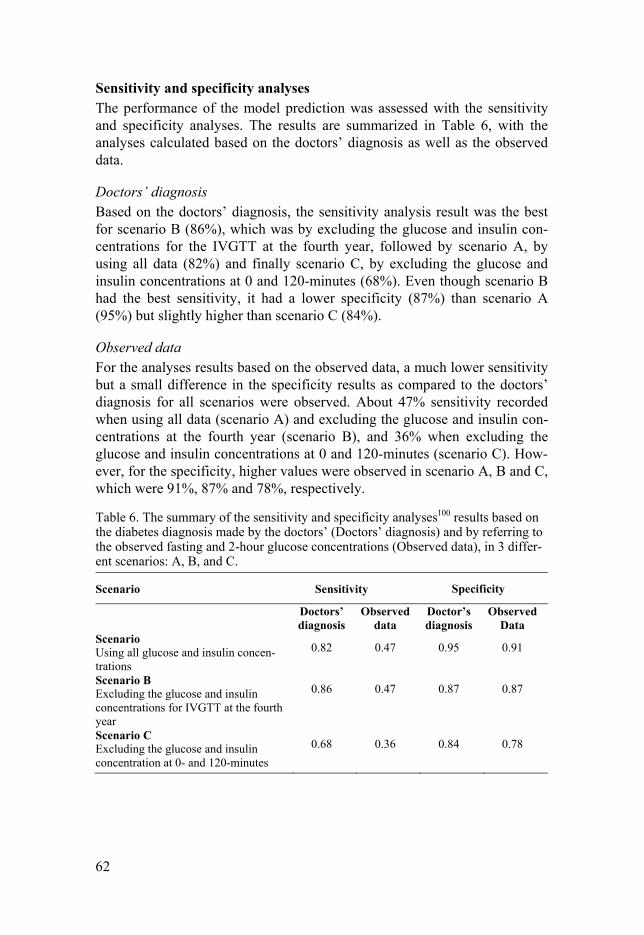
62
Sensitivity and specificity analyses The performance of the model prediction was assessed with the sensitivity and specificity analyses. The results are summarized in Table 6, with the analyses calculated based on the doctors’ diagnosis as well as the observed data.
Doctors’ diagnosis Based on the doctors’ diagnosis, the sensitivity analysis result was the best for scenario B (86%), which was by excluding the glucose and insulin con-centrations for the IVGTT at the fourth year, followed by scenario A, by using all data (82%) and finally scenario C, by excluding the glucose and insulin concentrations at 0 and 120-minutes (68%). Even though scenario B had the best sensitivity, it had a lower specificity (87%) than scenario A (95%) but slightly higher than scenario C (84%).
Observed data For the analyses results based on the observed data, a much lower sensitivity but a small difference in the specificity results as compared to the doctors’ diagnosis for all scenarios were observed. About 47% sensitivity recorded when using all data (scenario A) and excluding the glucose and insulin con-centrations at the fourth year (scenario B), and 36% when excluding the glucose and insulin concentrations at 0 and 120-minutes (scenario C). How-ever, for the specificity, higher values were observed in scenario A, B and C, which were 91%, 87% and 78%, respectively.
Table 6. The summary of the sensitivity and specificity analyses100 results based on the diabetes diagnosis made by the doctors’ (Doctors’ diagnosis) and by referring to the observed fasting and 2-hour glucose concentrations (Observed data), in 3 differ-ent scenarios: A, B, and C.
Scenario Sensitivity Specificity
Doctors’ diagnosis
Observed data
Doctor’s diagnosis
Observed Data
Scenario Using all glucose and insulin concen-trations
0.82 0.47 0.95 0.91
Scenario B Excluding the glucose and insulin concentrations for IVGTT at the fourth year
0.86 0.47 0.87 0.87
Scenario C Excluding the glucose and insulin concentration at 0- and 120-minutes
0.68 0.36 0.84 0.78

63
WHIG model (Paper (IV) Natural disease progression and intervention effects model The estimated parameters of the WHIG model in subjects with IGT are summarized in Table 7.
Body weight For a typical individual in this IGT population, the estimated baseline body weight (BLWT) was 85.4 kg. The weight was estimated to decrease by 0.9%, and further decreased with the effect of intensive lifestyle intervention with individualized counselling by 3.2%, producing a net decreasing effect of 4.1%. The lifestyle intervention was more effective in lowering the weight than the standard counselling for the controls, as expected.
Insulin sensitivity The mean estimated baseline insulin sensitivity in this population was 63.2% and 63.7% of normal for subjects in the control and intervention groups. A mean increase of 69.0% of normal in the control and 77.9% of normal in the intervention group was observed at the end of the study. A higher improve-ment of insulin sensitivity achieved with the intervention than without, as a result of weight changes.
Beta cell function The baseline beta cell function in this population was estimated as a mean of 50.1% and 51.7% of normal for subjects in the control and intervention groups. At the end of the study, the mean beta cell function was decreased to 41.1% in the control and 40.1% of normal in the intervention group. A de-crease beta cell function values for both groups in this model may be ex-plained by the improvement of insulin sensitivity achieved from the weight changes. This may result in less need of insulin secretion, thus resulting in an apparent reduction in beta cell function from baseline.
FSI Baseline FSI was estimated to be similar for the subjects on both control and intervention groups, as a mean value of 9.9 mU/L. A mean decrease of 0.9 mU/L (control) and 1.6 mU/L (intervention) was estimated at the end of study.
FPG Baseline FPG was estimated to be as a mean value of 6.1 mmol/L for control and 6.0 mmol/L for intervention groups. A mean increase of 0.3 mmol/L (control) and 0.2 mmol/L (intervention) for FPG was estimated at the end of the study.

64
HbA1c Baseline HbA1c was estimated to be similar for the subjects in the control and intervention groups, which was a mean of 5.8%. At the end of the study, a mean of 0.1% decrease in HbA1c was estimated for the subjects in the intervention group, whereas the mean of HbA1c for the subjects in the con-trol group remained unchanged. In a typical individual, the total effect of both natural disease progression and lifestyle intervention has resulted in a 0.4% decrease in PPG input that influenced the rate production of HbA1c.
Table 7. Final parameter estimates with relative standard errors (RSE, %) and their respective inter-individual variability (IIV, %) of the WHIG model among subjects with impaired glucose tolerance with the logistic regression dropout model.
Typical value (RSE †
IIV (%) †
Parameter Weight t1/2 , WGT, d Half-life of weight component 96.9* - BLWT, kg Baseline weight 85.4 (0.04) 15.6 (2.7) Insulin sensitivity IS0 Baseline insulin sensitivity, logistic
function -0.418 (8.1) 94.8‡ (7.3)
Scale EFS Scaling factor for change in weight on insulin sensitivity
0.0514* 19.5 (4.2)
Beta cell function B0 Baseline beta cell function, logistic
function -0.117 (6.8) 64.7‡ (0.2)
RB, y Rate of baseline beta cell function decrease per year, logits
0.0693 (0.7) 2.4‡ (17.9)
HbA1c MTT, d Mean transit time of HbA1c 38.9* - kin, HbA1c, %/d L/mmol
Rate constant, HbA1c compart-ments production
0.0129* -
PPG, %/d Residual HbA1c production rate independent of FPG
0.0653 (0.02) 13.4 (0.8)
Scale PPG,EFw Scaling factor on PPG when time
-0.00379 (6.9) -
Disease progression and treatment effect DPW, % Effect of natural disease progres-
sion (DP) on weight input 0.908 (13.0) 398.7‡ (3.9)
INTW, % Effect of intervention (INT) on weight input
3.18 (13.1) -
INTRB Effect of intervention (INT) on beta cell function
0.0191 (27.9) -
Residual errors Weight Proportional residual error on
weight 0.0344 (3.0) -
FSI Proportional residual error on fasting serum insulin
0.251 (0.3) 22.3 (2.7)

65
FPG Proportional residual error on fasting plasma glucose
0.067 (0.9) -
HbA1c Proportional residual error on glycated haemoglobin
0.049 (0.9) 37.8 (1.0)
M_FSI Magnitude of FSI eps-eta 0.969 (0.1) - Logistic regression dropout model Baseline Parameter related to the baseline
probability of dropout -14.5 (0.2) -
STime1, /year Parameter related to the time effect 5.4 (0.6) - STime2, /year Parameter related to the time effect
after 4 years 0.842 (6.7) -
PTime Power function for time effect (Hill factor)
0.197 (1.7) -
DWGT, /kg Parameter related to body weight change from baseline
0.0321 (13.1) -
IS Parameter related to for insulin sensitivity
-0.907 (2.6) -
T2DM Probability of dropping out when FPG 7.8mmol/L
0.42 (7.0) -
* Value fixed to the published model92 † RSE values (variance scale) were obtained from bootstrap (n=500) ‡ The parameters with an additive IIV (B0, RB, IS0, DPW) The logistic regression dropout model The final logistic regression dropout model is described in the equation 10a until 13c, describing the implementation of the dropout predictors, and the calculation of the probability of the dropout event in the logistic function (PROB) to occur and to not occur. = + + ( ) +
Eq. 10a > 1460 = + ++ ( ) + Eq. 10b = ( ) / ( ( + 1)) Eq. 11
P in the equation 10a represents the probability of dropping out with linear Base is the estimated parameter of the baseline pre-
dictor. Time is a predictor of dropout and it was investigated as a power function, with the probability of dropout increased with increasing time. The time effect on dropout was observed to be nonlinear, with estimated PTime

66
value of 0.3. The estimated parameter related to the time is described as STime1. The changes of weight from baseline (DWGT) and insulin sensitivity
value were the most significant covariates in the dropout model and their DWGT and IS. The insulin sensitivity was calculat-
ed as the baseline insulin sensitivity (ISS0) multiplied by the effect of weight change on the insulin sensitivity (EFFS). In equation 10b, an additional slope
STime2 was added to differ-entiate the time effect in the active study period with intervention (0-4 year) and the follow-up (4-6 year).
An improvement of the OFV by 12.9 was observed by adding the second slope of time (STime2), and further improvements by 5.1, 10.7 and 21.2 were obtained with the addition of the power function (PTime), change of body weight from baseline (DWGT) and insulin sensitivity (IS), respective-ly. Equation 11 represents the estimated probability of dropout in the logistic function (PROB), and it was constrained to lie between 0 and 1. The esti-mated parameters with their relative standard error are summarized in Table 7.
= 1 (1 ) Eq. 12a 7.8 / = Eq. 12b < 365 = 0 Eq. 12c
Equation 12a to 12c describe the probability of dropout (PROB), scaled to the time interval (DTime), which was 365 days. In addition, a separate prob-ability of dropout was estimated when the FPG probability of dropout because of diabetes, as described in equation 12b. In equation 12c, a zero probability to dropout was also implemented when time < 365 days, indicating no dropout was allowed to occur at time 0. = (1 ) Eq. 13a
7.8 / = 1 Eq. 13b < 365 = 1 Eq. 13c

67
The probability of dropout event to not occur was implemented as in the equation 13a to 13c, as a counterbalance from the probability of dropout event to occur.
All the parameter estimates for the logistic regression dropout model are summarized in Table 7. As expected, the baseline dropout was estimated to be -14.5, indicating a very low probability of dropout in the first interval (occasion = 0 to 1). A slightly lower probability of dropout in the active in-tervention than the follow-up period was observed. An increase in DWGT increased the probability of the dropout event to occur. In addition, the prob-ability of dropping out 7.8 mmol/L, indicating less than 50% probability of subjects were diagnosed with diabetes based on the FPG.95
Visual predictive checks The VPCs for the final model can be found in Figure 12 for each biomarker, and Figure 13 for their changes from baseline. The plots in the A section represent the final model with the inclusion of dropout model, whereas for the plots in B are without. An overall model fit was good for each bi-omarker, especially with the inclusion of dropout model. In specific, the inclusion of dropout model (A) was able to overcome the model over-prediction in the 95th percentile (B) of the FPG component. For the change from baseline, the same good model fit was illustrated in the VPCs, with or without the dropout model, except for the under-prediction at the 95th per-centile of the HbA1c component, as well as a small over-prediction of the weight component for the first year, also at the 95th percentile.

68
Figure 12. VPCs of biomarkers measured in the WHIG model among IGT subjects, based on control and intervention group. The biomarkers are fasting plasma glucose (FPG), glycated haemoglobin (HbA1c), weight and fasting serum insulin (FSI). A) VPCs for the final model with dropout model, and B) VPCs for the model without dropout model. Black circles indicate observed data. Solid lines represent the medi-an of observed data; dotted lines represent the 5th percentile (lower part) and 95th percentile (upper part) of the observed data. Shaded areas are the 95% confidence interval for 5th percentile (upper part), median (middle part) and 95th percentile (up-per part) from 500 simulated datasets.

69
Figure 13. VPCs of the change from baseline (cfb) of the biomarkers measured in the WHIG model among subjects with impaired glucose tolerance (IGT) subjects, based on control and intervention group. The biomarkers are fasting plasma glucose (FPG), glycated haemoglobin (HbA1c), weight and fasting serum insulin (FSI). C) cfb VPCs for the final model with dropout model, and D) cfb VPCs for the model without dropout model. Black circles indicate observed data. Solid lines represent the median of observed data; dotted lines represent the 5th percentile (lower part) and 95th percentile (upper part) of the observed data. Shaded areas are the 95% confi-dence interval for 5th percentile (upper part), median (middle part) and 95th percen-tile (upper part) from 500 simulated datasets.

70
Figure 14 is the illustration of the Kaplan-Meier-VPCs (KM-VPCs) for the final dropout model (overall- and groups- stratified survival curves) by using only the categorical data. These KM-VPCs show a good model prediction on both overall survival and groups-stratified survival curves. The rate of drop-out for the subjects in the intervention group was slower than the control for both the active part of the study and for the follow-up period. An adequate dropout model was successfully developed to describe the dropout event in the study, by using only categorical data, as well as with simultaneous analy-sis.
Figure 14. Kaplan-Meier-VPCs for the logistic regression dropout model as an overall survival curve (upper panel) and as stratified on groups (lower panels), using only the categorical data. The solid blue line represents the observed data, and the shaded area is the 95% prediction intervals of the Kaplan Meier plot from 100 simu-lated datasets. The estimated parameters for baseline, STime1, STime2, PTime, and DWGT were -12.8, 3.36/year, 0.86/year, 0.27, and 0.031/kg.

71
Discussion
Study design selection for the early phase clinical trial of anti-diabetes drug development Inclusion vs. exclusion of insulin measurements In this study, a smaller needed samples size has been translated to a higher study power with unchanged sample size. In most cases, the inclusion of insulin measurements added power to detect drug effects and to distinguish the correct drug MoA from incorrect ones. As expected, the increase in power with inclu-sion of insulin was strongest when MoAs were related to insulin (BINS and INCR), and weaker when the MoAs were related to the glucose (CLG, EGP, GABS). The insulin-dependent glucose clearance (CLGI) had little or no gain in power of including insulin when tested against the glucose related parameters. When discriminating CLGI from basal insulin secretion, either as competing MoAs or as a secondary MoA, insulin measurements have consistently contrib-uted to a large improvement in power. A similar trend was also seen with CLGI and incretin. This behaviour is possibly related to this MoA being related both to glucose and insulin. Glucose sensitivity (GSEN), which also is related to both glucose and insulin, unlike CLGI, behaved much more as a pure insulin related parameters, with a large power increase with the inclusion of insulin measure-ments in the analysis.
The setting of this project was not feasible to distinguish a correct MoA on CLG from an incorrect MoA on EGP. There were more than 100 subjects were needed in a study, both with and without insulin measurements. This is not sur-prising as the insulin-independent glucose clearance and the endogenous glucose production (also insulin-independent) are two sides of the same coin. A decrease of insulin-independent input of glucose is indistinguishable from an increase of insulin-independent output of glucose, without the tracer glucose in the design.
An adequate accuracy and a good precision for AUGCD/PL were obtained in all MoAs, with and without insulin measurements. This reflects a good model per-formance for the estimation of AUGCD/PL. The insulin measurements did not affect the precision of AUGCD/PL, reflected by the similar trends of accuracy and precision distribution when including and excluding insulin measurements.

72
Most appropriate study design based on the drug MoAs The difference in number of observations between the designs could ex-plained the results of the study power analysis. The designs differed greatly in number of observations: n=37, 26, 14 and 6 for MTT-24, IVGTT, GGI and OGTT/sMTT/NO, respectively. Thus, the IVGTT, GGI and MTT-24 have the highest study power due to the number of observations.
Besides than the number of observation, the study power is also depend-ent on the way glucose and insulin responses are provoked in the design. All drug effects, except on glucose absorption, affects the fasting concentrations of glucose and insulin. Thus, repeated fasting sampling at steady state or NO is expected to be powerful, with higher power the larger the change in fast-ing concentrations. Hypothetically, as insulin secretion increases, the relative importance of basal EGP, BINS and CLG will decrease. Thus, the power to detect drug effect on these MoAs will decrease the stronger insulin response the design provokes and the opposite will apply for CLGI.
The results showed that MTT-24 is the most powerful study design for drug MoA on basal insulin, due to the high number of observations. Fur-thermore, MTT-24 was not powerful for CLG while extremely powerful for CLGI because of the stimulation of high amount of insulin that makes CLGI more dominant. Moreover, the MTT-24 was more powerful to detect the drug MoA of decreasing GABS than sMTT because of the rich sampling, and more powerful than OGTT because of the slower glucose absorption rate that allows the detection of changes in absorption process.
GGI was the better study design for detecting CLG and EGP than IVGTT, but less powerful for CLGI. This may be explained by the fact that GGI stimulates insulin secretion in a step-up manner with longer period to reach high insulin levels compared to IVGTT. Additionally, IVGTT is the most powerful study design for CLGI, as it produces a rapid increase in glu-cose plasma level that stimulate high insulin secretion in an instant. The rich sampling of the intravenous glucose provocations may contribute to the high power to detect all drug MoAs, except for GABS.
In term of precision of parameter estimation, it has been shown that de-spite the biased parameter estimates in some of the models, a precise estima-tion of AUGC was observed reflecting a good model performance in term of simulation and estimation of the AUGCD/PL,Cx.
MCMP method vs. OFVgi/g in calculating study power There are some methods used in pharmacometrics analysis to calculate the study power, such as the SSE103 and MCMP method80. For SSE, it involves an intensive simulation and estimation methods, which is time-consuming and computer-intensive, thus making it less extensively used in the current time.

73
A newer method known as MCMP developed by Vong et al., is a faster and lesser computer-invasive alternative of calculating study power than the gold standard SSE76,80. The MCMP method was initially used in Paper I for the study power calculation, however, the study power obtained was very high, with 3 included subjects giving 90% power at 5% in most MoAs. Further-more, in some cases, less than 3 subjects were needed to achieve 95% study power at , especially when insulin measurements were included in the analysis.
The high power is related to the method of analysis (model-based), the size of the drug effect, and the design (cross-over, no. of observations and no. of subjects). Changing the method of analysis was not an option as the purpose was to investigate the performance of model-based analysis. The size of the drug effect had been selected based on what was a reasonable response for a new compound in development and a smaller drug effect would not be taken forward in the development.
The design in anti-hyperglycaemic drug development is fairly standard, with cross-over in early phase studies with observations every 30 minutes. This left the number of subjects as the comparable entity and an alternative approach of calculating full-
reduce full-reduce for analysis without insu-lin, which reflects the fraction of subjects needed to achieve equal study power with and without insulin.
To evaluate the reliability of suggested approach, the example of results method for
Part 1 of Paper I (refer to Table 1), in which similar results obtained for the ratio of number of subject needed, for both methods. This reflected the abil-
ratio power calculation to produce the results with an adequate similarity to the MCMP, even without a specific predetermined study power.
Recommendation on study design selection based on drug MoAs and purpose of analysis Based on the results from Paper I and II, an overall written summary was developed to aid designing the studies of anti-diabetes drug development using model-based analysis. This guidance can be used in a scenario when the IGI model is used, involving a certain drug MoAs. The best study design with the inclusion or exclusion of insulin measurements for different analy-sis purposes and its less invasive alternatives for each hypothetical drug MoA was summarized in Table 8.

74
Table 8. Recommendation on design in relation to purpose of the analysis and hy-pothesized site of drug actions.
Purpose of analysis MoA Study power
and AUGC PL
1
Drug parame-ter estimates1
Identifica-tion of drug action
Identification of correct site of drug action
Identification of secondary true site of drug action
CLGI IVGTT (OGTT is the less invasive alternative)
IVGTT (OGTT is the less invasive alternative)
Insulin reduced sample size for all MoAs
Insulin reduced sample size for all but competi-tion with CLG, EGP, GABS
Insulin reduced sample size for all
EGP GGI (NO is the less invasive alter-native)
GGI (NO is the less invasive alter-native)
Insulin reduced sample size for all but competi-tion with CLG
Insulin reduced sample size for all, but competi-tion with prima-ry CLG and CLGI
CLG GGI (sMTT is the less invasive alternative)
MTT-24 (sMTT is the less invasive alternative)
Indiscriminable from EGP
Indiscriminable with primary EGP
GABS MTT-24 (sMTT is the less invasive alternative)
MTT-24 (sMTT is the less invasive alternative)
Insulin reduced sample size for all
Insulin reduced sample size for all
BINS MTT-24 (NO is the less invasive alter-native)
IVGTT (NO is the less invasive alter-native)
Insulin reduced sample size for all
Insulin reduced sample size for all
INCR 2 Insulin reduced sample size for all
3
GSEN 2 Insulin reduced sample size for all
Insulin reduced sample size for all
1Insulin did not improve accuracy or precision 2Not tested in Paper II 3Not tested in Paper I (Part3) Abbreviation: SSE = stochastic simulation and estimation, gi = glucose and insulin, g = glucose only, INCR = incretin activity, BINS = basal insulin secretion, CLG = insulin-independent glucose clearance, CLGI = insulin-dependent glucose clearance, EGP = endog-enous glucose production, GABS = glucose absorption, GSEN = glucose sensitivity. IVGTT-OGTT-IGI model in IGT population The IGI model was published with the final parameter estimates for OGTT and IVGTT among diabetes subjects and healthy volunteers.55–57 The use of NONMEM’s prior functionality to develop the IGI model among IGT popu-lation was considered as the best, as the functionality incorporates the pub-

75
lished parameter values with their uncertainty taken into consideration dur-ing estimation. Therefore, the final parameter estimates in the new popula-tion (IGT) are comparable to the published (healthy and T2DM). Besides, a more stable population data analysis has been produced by this approach, especially in the case of a complex pharmacokinetics/pharmacodynamics model as the IGI.99
The CLG was observed to reach the lower boundary during estimation, most probably due to its non-identifiability resulting from the absence of hot glucose data in this IGT population. The hot glucose was used with the glu-cose and insulin data during the first development of IGI model,55–57 to dif-ferentiate between the exo- and endogenous glucose sources and to help discriminate the CLG from the CLGI. A similar observation seen for the kGE1, as it was estimated to be unreasonably high, probably as a compensation of the nearly absence of GPRG value in this IGT population.
On the population level, the CLGI, FPS and GPRG that translate to insulin sensitivity, insulin secretion function as a result of high glucose load, and feedback mechanism function of glucose on its own production were close to the state of diabetes mellitus. The BIOG however, was close to the healthy state.
Diabetes disease progression and lifestyle intervention effects in the IGT population Insulin sensitivity In this thesis, the change of insulin sensitivity was assessed as the change of the parameter CLGI in Paper III, and directly as insulin sensitivity estimation in Paper IV. In Paper III, a decrease in CLGI over time due to the natural disease progression indicating the depletion of the insulin sensitivity over the years. Similar result found in Paper IV, as the insulins sensitivity in this IGT population was about 63% of normal for the baseline, reflected as the deteri-oration of insulin sensitivity compared to healthy people. This was in agree-ment with other studies’ results that reported a reduction in the insulin sensi-tivity translated from 20-30% reduction of the insulin-dependent glucose clearance among IGT, from the NGT subjects,10,15,110–112 even after BMI adjustment.110 The combination of the insulin secretion depletion and muscle insulin resistance increase were determined to be the most prominent causes of decreasing insulin-dependent glucose clearance among the IGT subjects.113
With lifestyle intervention, a slower deterioration of insulin sensitivity was observed in Paper III, translated from the final net effect of a smaller reduction of the CLGI over time. In Paper IV, as a result of weight reduction, the insulin sensitivity was significantly increased at the end of the study,

76
with a higher increment in the intervention group than the control. These results complement the results from previous studies among the IGT sub-jects, regarding the benefit of lifestyle interventions (counselling on dietary intake, weight reduction, and moderate intensity physical activities for at least 150 minutes per week) on insulin sensitivity and insulin secretion as a result of improving beta cell function.26–28 A study reported the improve-ments of glucose tolerance and insulin sensitivity in the lifestyle intervention group subjects, determined by weight loss and increased physical fitness.26 Another study concluded that the lifestyle intervention significantly reduced the diabetes incidence among IGT subjects, due to the improvement of beta cell function and insulin sensitivity.27 In addition, two studies reported a 58% reduction of the progression from IGT to T2DM throughout the mean follow-up duration of 2.6 to 3.2 years with lifestyle intervention.24,25
Beta cell function In Paper III, the beta cell function was assessed from the change of FPS pa-rameter. It was found to decrease every year, representing the deterioration of acute beta cell function response to secrete insulin following a high glu-cose concentration after the IVGTT. In Paper IV, the baseline beta cell func-tion was about half of normal and reduced to a significant extent as a result of natural disease progression. The current results were supported by a num-ber of studies reported the suboptimal beta cell function among IGT subjects in the form of lesser first- and second-phase insulin secretions, as compared to the NGT subjects.10,15,26,27,110,111 Other study concluded a higher reduction of the first-phase insulin secretion among IGT subject with the first degree relative with T2DM than those without.114
The acute insulin secretion from pancreatic beta cell had a much slower deterioration with lifestyle intervention, marked by a very low decrease of the estimation on the FPS following the IVGTT over the years. Previous publications have recorded the improvement of beta cell function among IGT subjects, with the effect of lifestyle intervention.26–28 However, in Paper IV, the beta cell function was estimated to decrease more in the intervention group than the control. These contradictory results can possibly be explained by the fact that the beta cell reduced its insulin production due to increased insulin sensitivity in the glucose-insulin regulation system, in which not equally much insulin secretion is needed to maintain the blood glucose, and this may be seen as a reduction of beta cell function. A paper by Bergman et.al has illustrated this condition in which the improvement of insulin action (decreased insulin resistance) has resulted in a downregulation of beta cell sensitivity to secrete insulin.115 In addition, in the WHIG model (Paper IV), the old implementation of the HOMA equation is used to calculate the beta cell function, which has been shown to produce a slightly lower beta cell function than expected.

77
Weight The weight change was investigated in Paper IV, and it was shown to de-crease more effectively for the subjects in the lifestyle intervention group than control. The weight had slightly reduced without intervention, possibly due to the placebo effect, as the subjects in the control group were also pro-vided with brief oral and written information about diet and exercise recom-mendation at baseline and subsequent visits. This might encouraged the sub-jects to lead a healthier lifestyle, for example changing the dietary and phys-ical activities, which might reduce the overall weight.24 The subjects in the lifestyle intervention group, however, were provided with the individualized detailed advice to achieve the goals of intervention regarding diet and exer-cise recommendations. They were observed to benefit directly from the in-tervention, as reflected by a greater extent of weight reduction as compared to the subjects in the control group. Previous studies have shown the benefit of lifestyle intervention, for example with counselling on dietary intake and moderate intensity physical activities for at least 150 minutes per week, that effectively reduced weight among IGT subjects.24,26,27 A similar results on the weight reduction obtained from a study involving the NGT, IGT and T2DM subjects.25
FPG The FPG was estimated to be around a mean of 6.0 mmol/L at baseline, which can be categorized into the prediabetes state.4 As explained earlier, the FPG value was not as high as in T2DM patients, probably due to increasing baseline insulin secretion to prevent the increment of glucose level among IGT subjects.28,116,117 The FPG was almost unchanged through the end of the study, which is expected as subjects who developed diabetes were excluded and only those with unchanged or even decreased FPG remained in the study. Thus, only those subjects who had improved insulin sensitivity or insulin secretion and managed to maintain normoglycaemic levels stayed in the study.
FSI The change of FSI was investigated as the change in Iss in Paper III, and a direct FSI estimation in Paper IV. The Iss was found to increase from the baseline to the first year, and the estimated FSI value (mean = 9.9 mU/L) suggesting an increase in the beta cell function burden to secrete higher insu-lin secretion than normal. This may be explained by the feedback mecha-nism of beta cell to secrete more insulin as a result of the decreasing CLGI (insulin sensitivity) and increasing insulin resistance, especially in the early progression of T2DM. In this condition known as compensatory hyperinsu-

78
linemia, a higher insulin secretion is needed to suppress the increase of base-line glucose concentration due to increased insulin resistance. This feedback mechanism phenomenon has been well documented.118 Besides, it was doc-umented in previous studies that fasting plasma insulin concentration was higher in the IGT than in NGT subjects.10,110,113,119,120 Another study reported a higher baseline insulin secretion rate among the IGT than the normal glu-cose tolerance counterparts.121
Lifestyle intervention affected the Iss value by an increment from baseline until the first year, and later maintained throughout the remaining years. The estimated FSI in Paper IV, however, was decreased at the end of six years in this study. For both of these conditions, the possible explanation might be related to the physiological improvement of beta cell to secrete insulin at the baseline level, with the effect of lifestyle intervention.26–28 Furthermore, it may also be related to the improvement of insulin sensitivity, as the indirect effects of the lifestyle intervention and weight reduction. Previous studies reporting the reduction of FSI with the effect of lifestyle intervention, which related to the improvement of insulin sensitivity.26,27
HbA1c In Paper IV, the baseline HbA1c was estimated to be a mean of 5.8%, denot-ed a prediabetes state4, and slightly improved by a 0.1% decrease at the end of the study. This slight improvement may be related to the unchanged glu-cose value throughout the six years of study that influence the production of a stable HbA1c value. Previous studies have documented a decreased HbA1c value among prediabetes subjects and T2DM patients who received lifestyle intervention, due to the improvement of glucose control.24–27 Besides than the FPG, the PPG input was also contributed to the HbA1c production. In this study, the effect of lifestyle intervention resulted in a decrease in PPG that drove the rate production of HbA1c. A few studies have documented the improvement of meal intake, reduced energy intake, decreased consumption of and quantity of fat, increased vegetables intake, as well as decreased sug-ar, salt and alcohol among subjects who underwent lifestyle intervention programme.24–26
Specificity and sensitivity analyses In the Paper III, the sensitivity and specificity analyses were performed to assess the model prediction performance, on 3 difference scenarios: A. using all data from 0- until 120-minutes for the OGTT and IVGTT from
baseline to the fourth year, as a reflection of best-case scenario. B. excluding the glucose and insulin concentrations of the IVGTT at the
fourth year, to eliminate the influence of the rich IVGTT data on the

79
natural disease progression and intervention effects, and on the model predictability of subjects who will developed diabetes.
C. excluding glucose and insulin concentrations at the 0- and 120-minutes, to let the model predict the missing observations based on the estimation of the natural disease progression and intervention effects, as the base-line and 2-hour glucose are used for the diagnosis of T2DM. Thus, the predictions of the values for diagnosis were purely model-driven.
Based on the doctors’ diagnosis, an acceptable sensitivity and specificity analyses results were obtained. Specifically, the final model could identify 68% to 86% subjects who developed T2DM and only 14% to 32% went undetected. The model was also able to identify 84% to 95% subjects who did not develop T2DM, with only 16% to 5% falsely identified to develop T2DM at the fourth year. These results were also suggesting that the glucose and insulin concentrations at the fourth year may have an influence on the specificity, but not sensitivity analysis, by referring to the 86% sensitivity and 87% specificity for the scenario B, as compared to 82% sensitivity and 95% specificity for the scenario A. Besides, the model could predict an ac-ceptable 0- and 120-minutes glucose and insulin concentrations from the baseline until the fourth year, reflecting from a good sensitivity (68%) and specificity (84%) of the model.
Based on the data, however, a poor sensitivity with less than 50% was ob-tained for all scenarios. The specificity, on the other hand, was good, with 78% to 91%, indicating a high model performance to correctly identify the person who were never progress into T2DM, but quite poor to identify the subjects who developed T2DM at the fourth year. From the diabetes diagno-sis record, it was observed that more subjects were supposed to developed diabetes if the diagnosis was made directly on the observed data (FPG and 2-hour glucose), than from the diabetes diagnosis done by the doctors. This may be related to the fact that a second OGTT was performed to confirm the T2DM diagnosis if any subjects had a high FPG or the 2-hour glucose level on the first reading. Unfortunately, the data of the second OGTT were not available. Most probably in most cases, the second OGTT had ruled-out the diabetes diagnosis, therefore less subjects were diagnosed as T2DM, as compared to when the diagnosis were made directly from the observed data.
Logistic regression dropout model In the dropout model, the probability of dropout event to occur was higher as the time progress, but lower in the active study than the follow-up period. This could be explained by the possibility that the subjects were more moti-vated to stay in the study during the intervention period than after the end of the intervention. In addition, an increase in body weight from baseline has

80
shown to increase the probability of dropping out. This may be related to a higher probability to progress into T2DM for the subjects who had increased in weight, thus leads to diabetes dropout. A decrease in motivation to stay in the study when failing to reduce weight could also be one of the factors. Besides, in the simultaneous analysis, the higher the insulin sensitivity, the lower probability of dropping out. The improvement of insulin sensitivity may be reflected by a better glucose control, especially for the 2-hour glu-cose level. A better glucose control leads to a lower progression to T2DM, thus reduced the probability of dropping out due to T2DM. For the dropout event related to the value of FPG, it was independent on the time component (constant), and 42% of the subjects who had FPG had dropped out because of T2DM. This low probability of being diagnosed with T2DM based only on FPG is also seen in the observed data, where there are a fair number of subjects with FPG 7.8 mmol/L who have remained in the study.

81
Perspectives
Currently, the pharmacometrics analysis in the anti-diabetes drug develop-ment has becoming more common, and may be considered as the standard approach. The studies related to the study design in the early clinical trial in this thesis have provided an insight on the most appropriate study design depending on the MoAs of the drug and the purpose of analysis, specifically when using the model-based analysis and the IGI model. A written guidance was produced for the future references, with the hope to maximise the bene-fit of using pharmacometrics analysis in the process of developing anti-diabetes drugs. An alternative way of calculating study power has also been used in this thesis, which has shown to be comparable to the standard ap-proach of calculating study power. The future investigations may include mixed study designs, for example a study design of repeated fasting samples followed by intravenous glucose provocations such as the GGI or IVGTT. The mixed study design is expected to be informative on both dynamic and steady state effects, however, if the drug is expected to produce one of the studied MoAs as in the thesis work, intravenous provocations may be suffi-cient. Furthermore, the work in the thesis has also describes a methodology to investigate more combinations of drug MoAs in the future. The combina-tion of two drugs with different MoAs could also be investigated. In this case, as the secondary drug effects has been well detected and differentiated from the primary ones in this thesis, we could expect that the separate effects of two combined agents could also be well, or might be even better, charac-terised. This is because the differences in pharmacokinetics between two drugs would improve the power to separate drug effects.
The quantification of the natural disease progression and lifestyle inter-vention effects in this thesis has helped us understand the physiological changes of the glucose and insulin regulation in the IGT population. In this thesis, the assessment of multiple short-term glucose provocations (IVGTT and OGTT) was combined to predict the long-term disease progression, and offers apart from the assessment of the onset of T2DM, also the framework for how to perform similar analysis, combining short-term data to predict long-term outcome. The developed IGI model in the IGT population may be useful for clinical trial simulations related to the IGT population. With the additional disease progression model and the quantification of the effect of lifestyle intervention, the model had also been used to quantify the long-term effects of non-medication lifestyle intervention on the changes on the glu-

82
cose and insulin regulations among the IGT subjects in this thesis. Further investigations on the effects of medication interventions can be conducted in the future. A longer longitudinal data, for example a 10 years’ study period and a more frequent sampling of OGTT, for example every 30 minutes from 0 until 240 minutes could benefit the analysis in better quantifying the dis-ease progression and intervention effects. Moreover, the thesis work has also helped us to understand the effect of weight changes on the glucose and in-sulin homeostasis, specifically on the insulin sensitivity, beta cell function and HbA1c among IGT population, from the pharmacometrics point of view. Different approaches on analysing insulin sensitivity and beta cell function had also been performed in this thesis using different pharmacometrics mod-els.
The implementation of the logistic dropout model in this thesis provides the understanding of the complex relationship between dropouts from study and weight as well as glucose changes. In the investigations, the most signif-icant predictors of overall dropout have been identified. Future investiga-tions may include the time-to-event modelling with the use of parametric models to describe the absolute risk of developing T2DM and the relation-ship to weight change, and changes in glucose covariates. This model could then be coupled with the model developed in the thesis and then used to simulate the risk of diabetes for various scenarios in terms of weight reduc-tion.
83
Conclusion
This thesis has provided a first written guidance on how to design a study for pharmacometrics analysis when characterising drug effects for further im-proving the early phase of anti-diabetes drug development, and has charac-terised and quantify the progression from prediabetes to overt diabetes, both the natural progression and the progression with diet and exercise interven-tions, using pharmacometrics modelling.
In specific:
The need of insulin measurements in designing a clinical trial of anti-diabetes drug using the pharmacometrics analysis and the IGI model was assessed using the study power analysis. Excluding the insulin meas-urements from analysis using the IGI model will affect the power to de-tect a drug effect, distinguish a correct drug MoA from an incorrect and detect a secondary MoA from a primary. However, insulin measure-ments will not impact the estimates of drug effect parameters.
The most appropriate study design in phase I for the anti-diabetes drug
development for several hypothetical MoAs was different for each MoA, depending on if the focus is the power the identify drug effect or accura-cy and precision of drug effect parameter.
A natural diabetes disease progression model was successfully added in
the combination of IVGTT-OGTT-IGI model to describe parameter changes of FPS, CLGI and Iss among the IGT subjects, with the quantifi-cation of the effects of lifestyle intervention.
The effects of natural disease progression and lifestyle intervention on
the body weight, insulin sensitivity, and beta cell function among IGT subjects were quantified in the WHIG model, with the addition of those effects on the PPG input. The model also includes the complex relation-ship between dropout from study as well as weight and glucose changes.
84
Acknowledgements
The work presented in this thesis was carried out at the Department of Pharmaceutical Biosciences, Faculty of Pharmacy, Uppsala Universitet, Sweden. I am grateful to the Ministry of Higher Education and Universiti Sains Malaysia for the study leave and scholarship for the financial support during my studies, and the DDMore Consortium for the research funding. I would also like to thank Apotekarsocieteten and the Anna Maria Lundin Foundation at Smaland Nation to provide me with travel grants to attend international conferences and courses. During these 4 years, there are many people who have contributed to this thesis, directly and indirectly. I wish to express my deepest gratitude to: My main supervisor, Assoc. Prof. Dr Maria Kjellsson for your utmost guid-ance and support to make this PhD thesis possible. Thank you so much for all the help and discussions in our weekly Tuesday and diabetes meetings. You have made every manuscript becoming wayyy better than the first draft. I am so grateful to have such an excellent, dedicated and reliable supervisor like you. And also, thanks for many exciting stories about your study life back then.
My co-supervisor, Prof. Mats Karlsson, for accepting me to be a part of your excellent pharmacometrics group. Thank you very much for every fruitful discussion during weekly diabetes meeting, and for every recommendation and manuscript reviews.
My co-authors: Moustafa, Prof. Matti Uusitupa and Dr. Vanessa D. de Mello for being such pleasant persons to work with.
My half-time opponents: Assoc. Prof. Stilna Siv, Dr. Ola Caster and Dr. Martin Bergstrand, for a great session and many valuable feedbacks.
Prof. Margareta Hammarlund Udenas (I’ll never forget your life advices), Prof. Ulrica Simonsson, Assoc. Prof. Andy Hooker, and Prof. Lena Friberg, for your leadership. Siv, Kristin, Elisabeth, Martin, Joachim, Elodie, Sebas-tian, and Irena for contributing to a great working atmosphere, science and non-science related. Kajsa and Rikard, for an efficient works on PsN. Mag-

85
nus, Tobias and Jerker for IT support. Marina, Ulrica and Karin for admin support. Agneta Ekholm, for formatting the manuscripts in this thesis.
Diabetes group member (past and present): Steve, Oskar A, Rikke, Gustaf, Moustafa, Margreke, Tore, Fadli, Sara, and Erik, for our scientific discus-sion during weekly meeting, and for the nice time travelling to the diabetes seminars and conferences.
My first officemates in the PhD student’s office: Ari, Benjamin, Ida, Nebojsa, Xiao Mei, followed by: Maryam P, Gustaf, Robin, Marina, Erik, Irene, Henrik, Moustafa, Yang and Shijun. I have enjoyed every single mo-ment in that office. To Ida, thanks for being so welcoming on my first week in the department, and to share your adventure experience in Malaysia. To Xiao Mei, for being a generous soul. To Nebojsa, Ari and Benjamin, I know this is sooo unbelievable, but I enjoyed listening to all your discussions.
My long-term office-mate, Robin, thanks for being a great office-mate. I hope you’re happy to be in our office, because I am! Benjamin, for your abundance help and course on R and for the lesson on how to eat Tim Tam as a cookie straw, Emilie for helping me a lot with the logistic regression dropout, and Brendan for teaching me skiing for the first time.
Irene and Chunli, for our unforgettable experiences when travelling together to Switzerland and Alicante. Irene, thanks for inviting us to Greece for your department’s seminar, with excellent hospitality. Chunli, for always being my good friend in the department. Maryam P and Irene, for our cruise trip to Helsinki, Anders T, Marina, Moustafa, Joao, Ida, Elisabeth, Elin, Thomas, David, Henrik and Eva (thanks for the after-thesis-submission chocolate!) for the fun time in Dublin and Malahide. Svetlana, Lenaig, Chenyan and Chunli for the fascinating roommates chatting in Salen. My Campus 1477 buddies: Khadijah and Eva. Thanks for your company!
The one I considered sisters: Maryam T, Gulbeyaz, and Khadijah. Thank you for all the lunch time, gathering time with our families, the foods, chats and for being you.
All my colleagues in the department (past and present), thank you so much for contributing towards a lovely and positive working environment, for all the presentations and discussion during meetings and Friday seminars, for the parties and activities, as well as the coffee weeks and go-fikas.

86
James and Kerstin, for welcoming and accommodating us in your lovely house during our first few months in Sweden. We will never forget your kindness and the warmth of your heart for accepting us.
My Malaysian friends in Sweden (past and present): Ami (thanks for the long phone chat!), Jesmin & Shan, Mona & Duen (I will never forget our roadtrip!), Yasmin & Ahmad, Salman, Kamal & Alyn, Dalina, Muzafar, Na-sir & An, Shahar & Syikin, Adila & Izwan, Syaiful, Khadijah, Ida, fellow Malaysians in Stockholm, Malaysian Ambassador to Sweden, and Embassy staffs. I am grateful for the celebrations, foods, chats and the feeling to be at home no matter where we are.
My Mubasyirin: Hafzan, Sabariah, Hadzliana, Fatima, Aizati, Sarah, Amirah and Atiqah. Let’s continue supporting and motivating each other!
My colleagues in the Discipline of Clinical Pharmacy, School of Pharma-ceutical Science Malaysia, I’ll be back soon! And lastly, I would like to dedicate my appreciations to my family, without whom I probably could not be where I am today. A thousand thanks to: My mother, Jamilah for your support, trust and pray for me, and your pa-tience to travel almost 19 hours by flight just to visit me here (thanks Ikin for accompanying Ma). My late father, Sheikh Ghadzi, you’re never far. My mother-in-law, Adariah, for allowing me to kidnap your youngest son and your beloved first grandchild/granddaughter far, far away from home. My siblings (Along, Angah, Ira, Ikin, Ada), to always keep me updated on mat-ters back home. My daughter Alya, my BFFL, for always give me the reasons to love, laugh and smile, for your drawings in my office, snowball-fights and snowman-building, for our exciting moment travelling to so many places, and for being you, the best daughter a mother could ask for. And, to my unborn baby, for always being a loving part of me, and being the best company during this thesis writing. Hope to meet you in August! And, last but definitely not least, the love of my life, my husband, Fadli, for always being there for me. You’re never having doubts of moving to Sweden with me, leaving behind our comfort life, home, families and friends for 4 years. I am thankful for your patience and supports, for our life experiences, our travelling time and the lessons that we’ve learned, for sharing with you my scientific world during your project time in the department, and simply for our time together. I love you, and I’m forever grateful that I’ve shared this journey with you. Let’s go home, and continue our journey together!

87
References
1. American Diabetes Association. Diagnosis and classification of diabetes mellitus. Diabetes Care S5–S10 (2004).
2. Zajac, J., Shrestha, A., Patel, P. & Poretsky, L. in Principle of diabetes, second edition 3–16 (2010). doi:10.1017/CBO9781107415324.004
3. Lakhtakia, R. The history of diabetes mellitus. Sultan Qaboos Univ. Med J 368–370 (2013).
4. World Health Organization. Global Report on Diabetes. (2016). at
5. Tripathy, D. et al. Insulin secretion and insulin sensitivity in lessons from the Botnia study. Diabetes 975–980 (2000).
6. Nathan, D. M. et al. Impaired fasting glucose and impaired glucose tolerance: Implications for care. Diabetes Care 753–759 (2007).
7. Davies, M. J., Raymond, N. T., Day, J. L., Hales, C. N. & Burden, A. C. Impaired glucose tolerance and fasting hyperglycaemia have different characteristics. Diabet. Med. 433–40 (2000).
8. Færch, K. et al. Impaired fasting glycaemia vs impaired glucose tolerance: similar impairment of pancreatic alpha and beta cell function but differential roles of incretin hormones and insulin action. Diabetologia 853–861 (2008).
9. Faerch, K. et al. GLP-1 response to oral glucose is reduced in prediabetes, screen-detected type 2 diabetes, and obesity and influenced by sex: the ADDITION-PRO Study. Diabetes 2513–2525 (2015).
10. Festa, A. et al. Differences in insulin resistance in nondiabetic subjects with isolated impaired glucose tolerance or isolated impaired fasting glucose. Diabetes 1549–1555 (2004).
11. Hanefeld, M. et al. Insulin secretion and insulin sensitivity pattern is different in isolated impaired glucose tolerance and impaired fasting glucose: the risk factor in impaired glucose tolerance for atherosclerosis and diabetes study. Diabetes Care 868–874 (2003).
12. Laakso, M. et al. Insulin sensitivity, insulin release and glucagon-like peptide-1 levels in persons with impaired fasting glucose and/or impaired glucose tolerance in the EUGENE2 study. Diabetologia 502–511 (2008).
13. Meyer, C. et al. Different mechanisms for impaired fasting glucose and impaired postprandial glucose tolerance in humans. Diabetes Care 1909–1914 (2006).
14. Rask, E. et al. Insulin secretion and incretin hormones after oral glucose in non-obese subjects with impaired glucose tolerance. Metabolism. 624–631 (2004).
15. Abdul-Ghani, M. A., Jenkinson, C. P., Richardson, D. K., Tripathy, D. & DeFronzo, R. A. Insulin secretion and action in subjects with impaired fasting glucose and impaired glucose tolerance: Results from the veterans administration genetic epidemiology study. Diabetes 1430–1435 (2006).

88
16. Phillips, L. S., Ratner, R. E., Buse, J. B. & Kahn, S. E. We can change the natural history of type 2 diabetes. Diabetes Care 2668–2676 (2014).
17. Alberti, S. G., Zimmet, P., Shaw, J. & Grundy, S. M. The IDF consensus worldwide definition of the metabolic syndrom. (2006). at
18. Ncep. Executive summary of the third report of the national cholesterol
education program expert panel on detection, evaluation and treatment of high blood cholesterol in adults (Adult Treatment Panel III). Jama 2486–2497 (2001).
19. Weir, G. C. & Bonner- -cell dysfunction during progression to diabetes. Diabetes S16–S21 (2004).
20. Fonseca, V. A. Defining and characterizing the progression of type 2 diabetes. Diabetes Care S151–S156 (2009).
21. Ferrannini, E. et al. Mode of onset of type 2 diabetes from normal or impaired glucose tolerance. Diabetes 160–165 (2004).
22. Matthews, D. R., Cull, C. a, Stratton, I. M., Holman, R. R. & Turner, R. C. UKPDS 26: Sulphonylurea failure in non-insulin-dependent diabetic patients over six years. UK Prospective Diabetes Study (UKPDS) Group. Diabet. Med. 15 297–303 (1998).
23. Kahn, S. E. et al. Glycemic durability of rosiglitazone, metformin, or glyburide monotherapy. N. Engl. J. Med. 2427–2444 (2006).
24. Tuomilehto, J. et al. Prevention of type 2 diabetes mellitus by changes in lifestyle among subjects with impaired glucose tolerance. N. Engl. J. Med. 1343–1350 (2001).
25. Knowler, W. C. et al. Reduction in the incidence of type 2 diabetes with lifestyle intervention or metformin. N. Engl. J. Med. 393–403 (2002).
26. Mensink, M., Feskens, E. J. M., Saris, W. H. M., De Bruin, T. W. a & Blaak, E. E. Study on Lifestyle Intervention and Impaired Glucose Tolerance Maastricht (SLIM): preliminary results after one year. Int. J. Obes. Relat. Metab. Disord. 377–384 (2003).
27. Snehalatha, C. et al. Changes in insulin secretion and insulin sensitivity in relation to the glycemic outcomes in subjects with impaired glucose tolerance in the Indian Diabetes Prevention Programme-1 (IDPP_1). Diabetes Care 1796–1801 (2009).
28. Wilcox, G. Insulin and insulin resistance. Clin. Biochem. Rev. 19–39 (2005).
29. Tuomilehto, J. Nonpharmacologic therapy and exercise in the prevention of type 2 diabetes. Diabetes Care 189–193 (2009).
30. Pan, X.-R. et al. Effects of diet and exercise in preventing NDDM in people with impaired glucose tolerance.The Da Qing IGT and Diabetes Study. Diabetes Care 537–544 (1997).
31. Ramachandran, A. et al. The Indian Diabetes Prevention Programme shows that lifestyle modification and metformin prevent type 2 diabetes in Asian Indian subjects with impaired glucose tolerance (IDPP-1). Diabetologia 289–297 (2006).
32. Kosaka, K., Noda, M. & Kuzuya, T. Prevention of type 2 diabetes by lifestyle intervention: A Japanese trial in IGT males. Diabetes Res. Clin. Pract. 152–162 (2005).
33. Gregg, E. W. et al. Association of an intensive lifestyle intervention with remission of type 2 diabetes. JAMA 2489–96 (2012).

89
34. Mottalib, A., Sakr, M., Shehabeldin, M. & Hamdy, O. Diabetes remission after nonsurgical intensive lifestyle intervention in obese patients with type 2 diabetes. J. Diabetes Res. 468704 (2015).
35. Ades, P. A., Savage, P. D., Marney, A. M., Harvey, J. & Evans, K. A. Remission of recently diagnosed type 2 diabetes mellitus with weight loss and exercise. J Cardiopulm Rehabil Prev 193–197 (2015).
36. Courcoulas, A. P. et al. Three-year outcomes of bariatric surgery vs lifestyle intervention for type 2 diabetes mellitus treatment. JAMA Surg. 1–9 (2015).
37. Karter, A. J., Nundy, S., Parker, M. M., Moffet, H. H. & Huang, E. S. Incidence of remission in adults with type 2 diabetes: the diabetes & aging study. Diabetes Care 3188–3195 (2014).
38. White J.R. A brief history of the development of diabetes medications. Diabetes Spectr. 82 – 86 (2014).
39. Quianzon, C. C. L. & Cheikh, I. E. History of current non-insulin medications for diabetes mellitus. J. Community Hosp. Intern. Med. Perspect. 2–5 (2012).
40. Tahrani, A. A., Bailey, C. J., Del Prato, S. & Barnett, A. H. Management of type 2 diabetes: new and future developments in treatment. Lancet 182–197 (2011).
41. Proks, P., Reimann, F., Green, N., Gribble, F. & Ashcroft, F. Sulfonylurea stimulation of insulin secretion. Diabetes S368–S376 (2002).
42. Kahn, C. R., Chen, L. & Cohen, S. E. Unraveling the mechanism of action of thiazolidinediones. J. Clin. Invest. 1305–1307 (2000).
43. Arungarinathan, G., Mckay, G. A. & Acarbose. Br. J. Cardiol. 78–81 (2011).
44. -like peptide-1 receptor agonists and dipeptidyl peptidase-4 inhibitors in type 2 diabetes. Lancet (2006).
45. MacDonald, P. E. et al. The multiple actions of GLP-1 on the process of glucose-stimulated insulin secretion. Diabetes S434–S442 (2002).
46. Prins, J. B. Experimental and clinical pharmacology Incretin mimetics and s of action. Aust. Prescr. 102–104 (2008).
47. Dicker, D. DPP-4 Inhibitors: Impact on glycemic control and cardiovascular risk factors. Diabetes Care S276–S278 (2011).
48. White, J. R. Dipeptidyl peptidase-IV inhibitors: pharmacological profile and clinical use. Clin. Diabetes 53–57 (2008).
49. Pfister, M., Whaley, J. M., Zhang, L. & List, J. F. Inhibition of SGLT2: a novel strategy for treatment of type 2 diabetes mellitus. Clin. Pharmacol. Ther. 621–5 (2011).
50. Bays, H. Sodium glucose co-transporter type 2 (SGLT2) inhibitors: targeting the kidney to improve glycemic control in diabetes mellitus. Diabetes Ther. 195–220 (2013).
51. Alquier, T. & Poitout, V. GPR40: good cop, bad cop? Diabetes 1035–6 (2009).
52. Burant, C. Activation of GPR40 as a Therapeutic Target for the Treatment of Type 2 Diabetes. Diabetes Care S175–9 (2013).
53. Oh, D. Y. & Olefsky, J. M. G protein-coupled receptors as targets for anti-diabetic therapeutics. Nat. Rev. Drug Discov. 161–172 (2016).
54. Mancini, A. D. & Poitout, V. GPR40 agonists for the treatment of type 2 diabetes: Life after ‘TAKing’ a hit. Diabetes, Obes. Metab. 622–629 (2015).

90
55. Silber, H. E., Frey, N. & Karlsson, M. O. An integrated glucose-insulin model to describe oral glucose tolerance test data in healthy volunteers. J. Clin. Pharmacol. 246–56 (2010).
56. Silber, H. E. et al. An integrated model for glucose and insulin regulation in healthy volunteers and type 2 diabetic patients following intravenous glucose provocations. J. Clin. Pharmacol. 1159–1171 (2007).
57. Jauslin, P. M. et al. An integrated glucose-insulin model to describe oral glucose tolerance test data in type 2 diabetics. J. Clin. Pharmacol. 1244–55 (2007).
58. Piche, M.-E. et al. What is a normal glucose value? Differences in indexes of plasma glucose homeostasis in subjects with normal fasting glucose. Diabetes Care 2470–2477 (2004).
59. Kjems, L. L., Holst, J. J., Vølund, A. & Madsbad, S. The influence of GLP-1 on glucose-stimulated insulin secretion. Effects on B-cell sensitivity in type 2 and nondiabetic subjects. Diabetes 380–386 (2003).
60. Choy, S., Henin, E., Van Der Walt, J. S., Kjellsson, M. C. & Karlsson, M. O. Identification of the primary mechanism of action of an insulin secretagogue from meal test data in healthy volunteers based on an integrated glucose-insulin model. J. Pharmacokinet. Pharmacodyn. 1–10 (2013).
61. Jauslin-stetina, P. Mechanism-based modeling of the glucose-insulin regulation during clinical provocation experiments. (2008).
62. Myers, G. B. & Mckean, R. M. The oral glucose tolearance test: a review of the literature. Am. J. Clin. Pathol. 299–312 (2015).
63. Trout, K. K., Homko, C. & Tkacs, N. C. Methods of measuring insulin sensitivity. Biol. Res. Nurs. 305–318 (2007).
64. Jauslin, P. M., Frey, N. & Karlsson, M. O. Modeling of 24-hour glucose and insulin profiles of patients with type 2 diabetes. J. Clin. Pharmacol. 153–164 (2011).
65. Brodovicz, K. G. et al. Postprandial metabolic responses to mixed versus liquid meal tests in healthy men and men with type 2 diabetes. Diabetes Res. Clin. Pract. 449–455 (2011).
66. DeFronzo, R. a, Tobin, J. D. & Andres, R. Glucose clamp technique: a method for quantifying insulin secretion and resistance. Am. J. Physiol. E214–E223 (1979).
67. Kinsley, B. T., Widom, B. & Simonson, D. C. Secretion and Symptoms Effect of glycemic control. Diabetes Care 17–26 (1995).
68. Ette, E. I. & William, P. J. Pharmacometrics: The Science of Quantitative Pharmacology. (John Wiley & Sons Inc, 2007).
69. van der Graaf, P. H. CPT: Pharmacometrics and Systems Pharmacology. CPT Pharmacometrics Syst. Pharmacol. e8 (2012).
70. Barrett, J. S., Fossler, M. J., Cadieu, K. D. & Gastonguay, M. R. Pharmacometrics: A multidisciplinary field to facilitate critical thinking in drug development and translational research settings. J. Clin. Pharmacol. 632–649 (2008).
71. Sheiner, L. B. & Ludden, T. M. Population pharmacokinetics/dynamics*. Annu Rev Pharmacol Toxicol. 185–209 (1992).
72. Davidian, M. & Giltinan, D. M. Nonlinear models for repeated measurement data: an overview and update. J. Agr. Biol. Envir. St. 387–419 (2003).
73. Karlsson, M. O., Beal, S. L. & Sheiner, L. B. Three new residual error models for population PK/PD analyses. J. Pharmacokinet. Biopharm. 651–672 (1995).

91
74. Beal, S. L., Sheiner, L. B. & Boeckman, A. J. NONMEM Users Guides. (1989–2006) (Icon Development Solutions, Ellicott City, MD, 2008). (2008).
75. Beal, S. L. Population pharmacokinetic data and parameter estimation based on their first two statistical moments. Drug Metab. Rev. 173–93 (1984).
76. Karlsson, K. E., Vong, C., Bergstrand, M., Jonsson, E. N. & Karlsson, M. O. Comparisons of analysis methods for proof-of-concept trials. CPT pharmacometrics Syst. Pharmacol. e23 (2013).
77. Milligan, P. et al. Model-based drug development: a rational approach to efficiently accelerate drug development. Clin. Pharmacol. Ther. 502–14 (2013).
78. Reeve, R. et al. Benefits of model-based drug development: a rigorous, planned case study. Commun. Stat. - Simul. Comput. 141217112719001 (2014).
79. Stone, J. a et al. Model-based drug development survey finds pharmacometrics impacting decision making in the pharmaceutical industry. J. Clin. Pharmacol.
20S–30S (2010). 80. Vong, C., Bergstrand, M., Nyberg, J. & Karlsson, M. O. Rapid sample size
calculations for a defined likelihood ratio test-based power in mixed-effects models. AAPS J. 176–86 (2012).
81. Bergman, R. N., Yder, Y. Z., Bowden, C. R. & Cobelli, C. Quantitative estimation of insulin sensitivity. J. Clin. Invest. E667 – E677 (1979).
82. Denti, P., Bertoldo, A., Vicini, P. & Cobelli, C. IVGTT glucose minimal model covariate selection by nonlinear mixed-effects approach. Am. J. Physiol. Endocrinol. Metab. E950–60 (2010).
83. Largajolli, A., Bertoldo, A. & Cobelli, C. Identification of the glucose minimal model by stochastic nonlinear-mixed effects methods. in Conference proceedings: Annual International Conference of the IEEE Engineering in Medicine and Biology Society 5482–5485 (2012).
84. De Gaetano, A. & Arino, O. Mathematical modelling of the intravenous glucose tolerance test. J. Math. Biol. 136–168 (2000).
85. Hamrén, B., Björk, E., Sunzel, M. & Karlsson, M. O. Models for plasma glucose, HbA1c, and hemoglobin interrelationships in patients with type 2 diabetes following tesaglitazar treatment. Clin. Pharmacol. Ther. 228–235 (2008).
86. Ledo-Garcia, R., Mazer, N. A. & Karlsson, M. O. A semi-mechanistic model of the relationship between average glucose and HbA1c in healthy and diabetic subjects. J. Pharmacokinet. Pharmacodyn. 129–142 (2013).
87. Møller, J. B. et al. Longitudinal modeling of the relationship between mean plasma glucose and HbA1c following antidiabetic treatments. CPT Pharmacometrics Syst. Pharmacol. e82–88 (2013).
88. Topp, B., Promislow, K., Devries, G., Miura, R. M. & Finegood, D. T. A -cell mass, insulin, and glucose kinetics: pathways to diabetes. J.
Theor. Biol. 605–619 (2000). 89. Ribbing, J., Hamrén, B., Svensson, M. K. & Karlsson, M. O. A Model for
glucose, insulin, and beta-bell dynamics in subjects with insulin resistance and patients with type 2 diabetes. J. Clin. Pharmacol. 861–872 (2010).
90. Frey, N. et al. Population PKPD modelling of the long-term hypoglycaemic effect of gliclazide given as a once-a-day modified release (MR) formulation. Br. J. Clin. Pharmacol. 147–157 (2003).

92
91. de Winter, W. et al. A mechanism-based disease progression model for comparison of long-term effects of pioglitazone, metformin and gliclazide on disease processes underlying type 2 diabetes mellitus. J. Pharmacokinet. Pharmacodyn. 313–343 (2006).
92. Choy, S., Kjellsson, M. C., Karlsson, M. O. & de Winter, W. Weight-HbA1c-insulin-glucose model for describing disease progression of type 2 diabetes. CPT Pharmacometrics Syst. Pharmacol. 11–19 (2016).
93. Uusitupa, M., Lindi, V., Louheranta, A., Salopuro, T. & Lindstro, J. Long-term improvement in insulin sensitivity by changing lifestyle of people with impaired glucose tolerance. 4-year results from the Finnish Diabetes Prevention Study. Diabetes (2003).
94. Lindstrom, J. et al. The Finnish Diabetes Prevention Study (DPS). Lifestyle intervention and 3-year results on diet and physical activity. Diabetes Care 3230–3236 (2003).
95. Diabetes mellitus: report of a WHO study group. WHO techical report series 1985. 727:7–113 (1985).
96. SSE user guide PsN 4.6.0. 1–17 (2016). 97. Ueckert, S., Karlsson, M. O. & Hooker, A. C. Accelerating Monte Carlo power
studies through parametric power estimation. J. Pharmacokinet. Pharmacodyn. 223–234 (2016).
98. Jauslin, P. M., Karlsson, M. O. & Frey, N. Identification of the mechanism of action of a glucokinase activator from oral glucose tolerance test data in type 2 diabetic patients based on an integrated glucose-insulin model. J. Clin. Pharmacol. 1861–71 (2012).
99. Gisleskog, P. O., Karlsson, M. O. & Beal, S. L. Use of prior information to stabilize a population data analysis. J. Pharmacokinet. Pharmacodyn. 473–505 (2002).
100. Lalkhen, A. G. & McCluskey, A. Clinical tests: sensitivity and specificity. Contin. Educ. Anaesthesia, Crit. Care Pain 221–223 (2008).
101. R Core Team. R: A language and environment for statistical computing. R Foundation for statistical computing,Vienna, Austria: Available at: http://www.R-project.org/ . (2016). at
102. Wickham, H. & Chang, W. An implementation of the grammar of graphics. (2014).
103. SSE user guide PsN 4.5.12. 1–13 (2013). 104. MCMP user guide PsN 4.5.1. 1–9 105. VPC and NPC user guide. PsN 4.6.12. 1–44 (2016). 106. Keizer, R. J., Karlsson, M. O. & Hooker, A. Modeling and Simulation
Workbench for NONMEM: Tutorial on Pirana, PsN, and Xpose. CPT pharmacometrics Syst. Pharmacol. e50 (2013).
107. Jonsson, E. N. & Karlsson, M. O. Xpose - An S-PLUS based population pharmacokinetic/pharmacodynamic model building aid for NONMEM. Comput. Methods Programs Biomed. 51–64 (1998).
108. Hooker, A., Karlsson, M., JW, J. & Jonsson, E. xpose4: Tools for nonlinear mixed-effect model building and diagnostics. R package version 4.X.X. http://xpose.sourceforge.net.
109. Murakawa, Y., Yamashita, T., Ajiki, K., Sezaki, K. & Omata, M. Ostensible day-night difference of QT prolongation during long-term treatment with antiarrhythmic drugs: Reappraisal of the law of ‘regression to the mean’. J. Cardiovasc. Pharmacol. 62–65 (1998).
93
110. Weyer, C., Bogardus, C. & Pratley, R. E. Metabolic characteristics of individuals with impaired fasting glucose and/or impaired glucose tolerance. Diabetes 2197–2203 (1999).
111. Osei, K., Gaillard, T. & Schuster, D. P. Pathogenetic mechanisms of impaired glcuose tolearance and type 2 diabetes in African-Americans. Diabetes Care
396–404 (1997). 112. Bock, G. et al. Pathogenesis of pre-diabetes: mechanisms of fasting and
postprandial hyperglycemia in people with impaired fasting glucose and/or impaired glucose tolerance. Diabetes 3536–3549 (2006).
113. Abdul-Ghani, M. A., Tripathy, D. & DeFronzo, R. A. Contributions of beta-cell dysfunction and insulin resistance to the pathogenesis of impaired glucose tolerance and impaired fasting glucose. Diabetes Care 1130–1139 (2006).
114. Emerson, P. et al. Different pathophysiology of impaired glucose tolerance in first-degree relatives of individuals with type 2 diabetes mellitus. Metabolism.
602–607 (2009). 115. Bergman, R. N., Ader, M., Huecking, K. & Citters, G. Van. Accurate
assessment of beta-cell function. The hyperbolic correction. Diabetes S212–S220 (2002).
116. DeFronzo, R. A. Impaired glucose tolerance: do pharmacological therapies correct the underlying metabolic disturbance? Br. J. Diabetes Vasc. Dis. S24–S40 (2003).
117. Cavaghan, M. K., Ehrmann, D. A. & Polonsky, K. S. Interactions between insulin resistance and insulin secretion in the development of glucose intolerance. J. Clin. Invest. 329–333 (2000).
118. DeFronzo, R. a. Pathogenesis of type 2 diabetes mellitus. Med. Clin. North Am. 787–835 (2004).
119. Osei, K., Rhinesmith, S., Gaillard, T. & Schuster, D. Impaired insulin sensitivity, insulin secretion, and glucose effectiveness predict future development of impaired glucose tolerance and type 2 diabetes in pre-diabetic African Americans: Implications for primary diabetes prevention. Diabetes Care 1439–1446 (2004).
120. van Haeften, T. W. et al. Disturbances in beta-cell function in impaired fasting glycemia. Diabetes S265–70 (2002).
121. Ferrannini, E. et al. Predominant role of reduced beta-cell sensitivity to glucose over insulin resistance in impaired glucose tolerance. Diabetologia 1211–1219 (2003).

Acta Universitatis UpsaliensisDigital Comprehensive Summaries of Uppsala Dissertationsfrom the Faculty of Pharmacy 226
Editor: The Dean of the Faculty of Pharmacy
A doctoral dissertation from the Faculty of Pharmacy, UppsalaUniversity, is usually a summary of a number of papers. A fewcopies of the complete dissertation are kept at major Swedishresearch libraries, while the summary alone is distributedinternationally through the series Digital ComprehensiveSummaries of Uppsala Dissertations from the Faculty ofPharmacy. (Prior to January, 2005, the series was publishedunder the title “Comprehensive Summaries of UppsalaDissertations from the Faculty of Pharmacy”.)
Distribution: publications.uu.seurn:nbn:se:uu:diva-317040
ACTAUNIVERSITATIS
UPSALIENSISUPPSALA
2017