pharmacology and pharmacokinetics - vanderbilt university medical
TRANSCRIPT

Pharmacology and pharmacokinetics
Bassel Abou-Khalil, M.D. Vanderbilt University
Pharmacology and Pharmacokinetics
nMechanisms of drug actions n Pharmacokinetic principles, illustrate PHT n Pharmacokinetic interactions n Liver, renal disease, hemodialysis n Changes through the ages

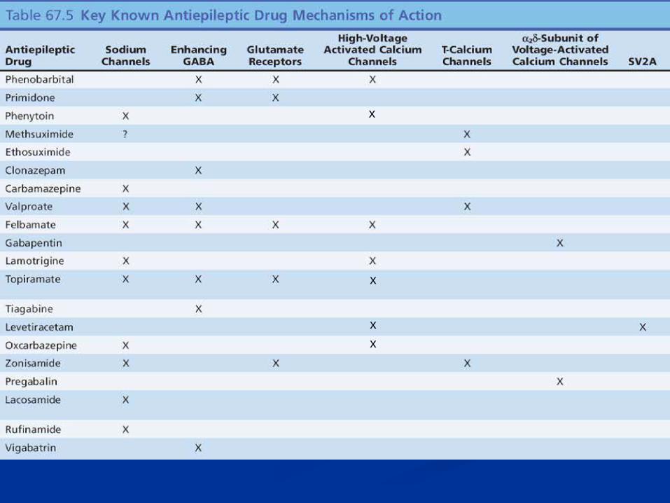
AED main mechanisms of action
n Na channel blocking n Enhancing GABA n Glutamate receptor antagonism n Blocking high voltage activated calcium channels n Blocking T- calcium channels n Binding Alpha-2-delta subunit of voltage-activated
calcium channels n Binding synaptic vesicle protein SV2A n K-channel opening

Na channel blocking
n Enhancement of fast inactivated state- blocking of sustained repetitive firing: n Phenytoin, carbamazepine, oxcarbazepine, lamotrigine,
rufinamide n Selective enhancement of slow inactivation of
voltage-gated sodium channels n Lacosamide
n Multiple mechanisms, including effect on sodium channels n Valproate, felbamate, topiramate, zonisamide

Fast versus slow inactivation of VGSC
n Fast inactivation occurs on a time scale of milliseconds.
n Slow inactivation occurs over the time course of seconds to minutes. n involves modification of the shape of the sodium
channel


All but one of the following AEDs block sodium channels as their main mechanism of action
A- carbamazepine B- lamotrigine C- levetiracetam D- phenytoin E- lacosamide
SAE Question ARS
All but one of the following AEDs block sodium channels as their main mechanism of action
A- carbamazepine B- lamotrigine C- levetiracetam D- phenytoin E- lacosamide
SAE Question ARS

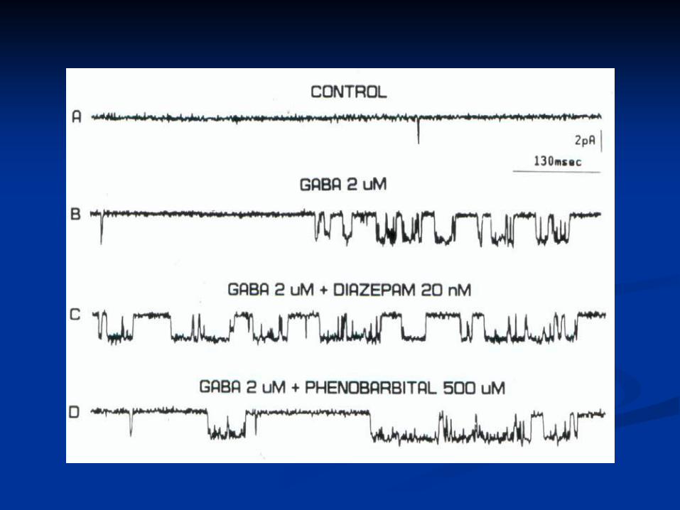
Enhancing GABA n Irreversible inhibition of GABA transaminase: vigabatrin n Inhibition of GABA reuptake at the synapse: tiagabine n Prolongation of GABA-mediated chloride channel
openings: phenobarbital n Increased frequency of GABA-mediated chloride channel
openings: benzodiazepines, topiramate (different binding site- also increases GABA levels in the brain by MRS)
n Enhancing GABA transmission in specific circuits: valproate n Enhancing GABA-elicited Cl − currents: felbamate n Several AEDs are associated with acute elevation of brain
GABA by MRS after single doses: 70% for topiramate, 48% with gabapentin.

Glutamate receptor antagonism
n Kainate and AMPA-type glutamate receptor antagonism: topiramate
n NMDA receptor antagonism: felbamate n Selective AMPA antagonism: perampanel n Phenobarbital may also have glutamate
antagonism- it reduces AMPA/kainate receptor mediated currents

Blocking high voltage activated calcium channels
n Topiramate n Lamotrigine n Levetiracetam n Felbamate n Phenobarbital n Phenytoin n Oxcarbazepine

Binding Alpha-2-delta subunit of voltage-activated calcium channels
n Decrease in neuronal calcium currents by binding of alpha 2 delta subunit of the voltage gated calcium channel n Gabapentin n Pregabalin

Blocking T- calcium channels
n Blocking of low, threshold, “transient” (T-type) calcium channels in thalamic neurons
n Ethosuximide n Valproate n Zonisamide

Binding synaptic vesicle protein SV2A
n Levetiracetam (and related drugs) n Levetiracetam binding to SV2A seems to result
in nonspecific decrease in neurotransmitter release.
n There was a functional correlation between SV2A binding affinity and anticonvulsant potency of levetiracetam analogs
K-channel opening
n Ezogabine (retigabine) n enhances the activity and prolongs the opening of
neuron-specific KCNQ2/3 (Kv7.2/7.3) voltage-gated K+ channels, thereby activating the M-current
n It also potentiates GABA-evoked currents in cortical neurons. Potentiation of GABA-induced currents occurs at much higher concentration than that needed to activate potassium currents

Carbonic anhydrase inhibition
n Topiramate n Zonisamide n Acetazolamide

Multiple mechanisms
n Felbamate n Phenobarbital n Topiramate n Valproate n Zonisamide
x
x x
x

All but one of the following AEDs block T-calcium channels
A- ethosuximide B- valproate C- lamotrigine D- zonisamide
SAE Question ARS

All but one of the following AEDs block T-calcium channels
A- ethosuximide B- valproate C- lamotrigine D- zonisamide
SAE Question ARS

Implications of mechanism of action
n Combining two AEDs with same mechanism may cause adverse experiences even though the levels are in the “therapeutic” range (pharmacodynamic interaction)
n Combining two AEDs with different mechanisms may be more effective than two AEDs with the same mechanism n Synergistic combinations: lamotrigine + valproate;
possibly valproate + ethosuximide; lamotrigine and levetiracetam

Pharmacokinetics
n Refers to changes over time in drug concentration in the body as a function of absorption, distribution and elimination.
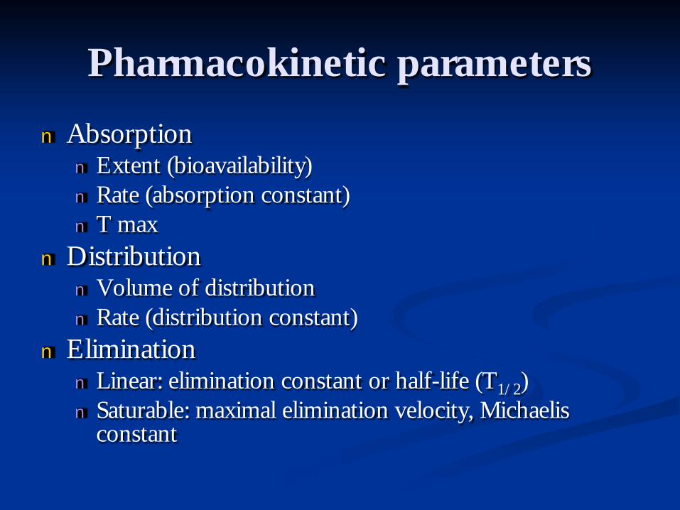
Pharmacokinetic parameters
n Absorption n Extent (bioavailability) n Rate (absorption constant) n T max
n Distribution n Volume of distribution n Rate (distribution constant)
n Elimination n Linear: elimination constant or half-life (T1/2) n Saturable: maximal elimination velocity, Michaelis
constant

Cmax
Tmax

Bioavailability
n The amount of an administered dose that will reach the bloodstream
n It is a function of the route of administration and drug formulation. n 100% after IV administration
n Oral bioavailability is usually determined by calculating the area under the curve (AUC) for the plot of concentration versus time following a single dose, orally (or other routes) versus IV.
n Low for gabapentin, and decreasing with increasing single dose

Absorption rate
n Absorption is generally a first-order process with a half-life (just like elimination) n Rapid absorption desirable for acute treatment n Generally less desirable with long-term therapy (due to
large fluctuations)
n Slow release formulations reduce absorption rates n Delayed release preparations only delay absorption,
which occurs rapidly after the gastric resistant coating is dissolved.

Distribution n After the AED reaches the blood stream, it will diffuse
into other tissues/ body fluids until an equilibrium is reached.
n The extent of distribution is expressed by the volume of distribution (Vd), expressed in liters per kilograms
n Vd is the hypothetical volume whose calculation is based on the assumption that drug concentration equals concentration in blood plasma throughout this volume (often not the case).
n Vd can be calculated by dividing total amount of drug in the body (equal the IV dose) by the blood level associated with this dose.

Volume of distribution
n Body water represents approximately 60% of body weight → an AED with a Vd >0.6 L/kg binds to certain tissues
n Vd is based on total serum level → a decrease in serum protein binding results in a higher value for Vd.
n If the AED binds with adipose tissue (for example diazepam), Vd will be larger in obese individuals. If it does not it will be smaller.

Volume of distribution- clinical value
n Vd allows the calculation of loading dose. n Vd= Dose/concentration → Dose= Vd x
concentration. n Example 1: phenobarbital Vd= 0.55 L/Kg. If
desired serum level is 40 mg/L, loading dose= 40 x 0.55= 22mg/Kg.
n Example 2: phenytoin level is 5 mg/L and the desired level is 15 mg/L. Phenytoin Vd= 0.75 L/Kg. The desired additional load= 0.75 x (15-5)= 7.5 mg/Kg.

Distribution by one vs >2 compartment model
n A one-compartment distribution model exists if the final concentration equilibrium is reached rapidly following IV administration
n >2 compartment distribution model applies if after initial rapid distribution in one compartment the drug diffuses into a second or more compartments.
n The total Vd will correspond to the sum of the compartments.
n An example is diazepam redistributing to adipose tissue. The true T1/2 is 36 hours, but the redistribution half-life is < 1 hour
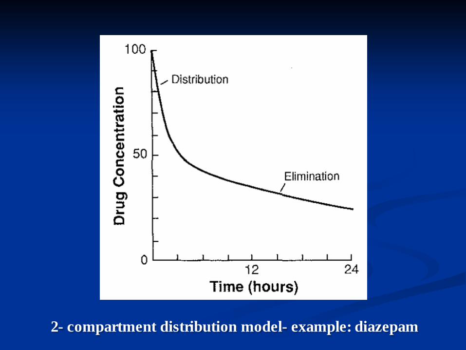
2- compartment distribution model- example: diazepam

Elimination
n Enzymatic biotransformation in the liver n Directly by the kidneys in an unmetabolized
form n Both of the above n Elimination rate determines the rate at which
the drug needs to be replaced to maintain concentration.

Metabolism of AEDs
n Phase I: oxidative reactions, mediated by cytochrome P450 enzymes) or reductive reactions, mediated by aldoketoreductases.
n Phase II: increases water solubility of drug or its phase I metabolite- conjugate a drug with moieties such as glucuronic acid (e.g. microsomal epoxide hydrolase and uridine diphosphate (UDP)-glucuronosyltransferases).

CYP enzymes involved in metabolism of select AEDs
CYP CBZ CZP ESM FBM PB PHT TGB VPA ZNS
1A2 X
2A6 X
2B6 X
2C8 X X
2C9 X X X
2C18 X
2C19 X X X X
2E1 X X
3A4 X X X X X X X
3A5 X X
3A7 X
4B1

Pharmacogenetics, drug elimination and toxicity
n Genetic variability in CYP and other enzymes can alter AED metabolism and predispose to drug toxicity.
n Certain polymorphisms and mutations are associated with slower or faster metabolism.
n Testing for these mutations may play a role in personalized medical treatment of epilepsy.

Linear elimination
n Most drugs (and all but one AED) follow linear elimination kinetics: the elimination rate is linearly proportional to the blood concentration.
n Linear elimination kinetics mean that the half-life is constant.
n Half-life is the time it takes for the concentration to fall to half its value through elimination.

Clinical role of half-life
n The half-life (T1/2) helps determine n how often the AED should be administered for
long-term therapy (at least once every half-life) n how long it takes for steady-state to be reached at a
constant daily dose (97% by 5 half-lives).
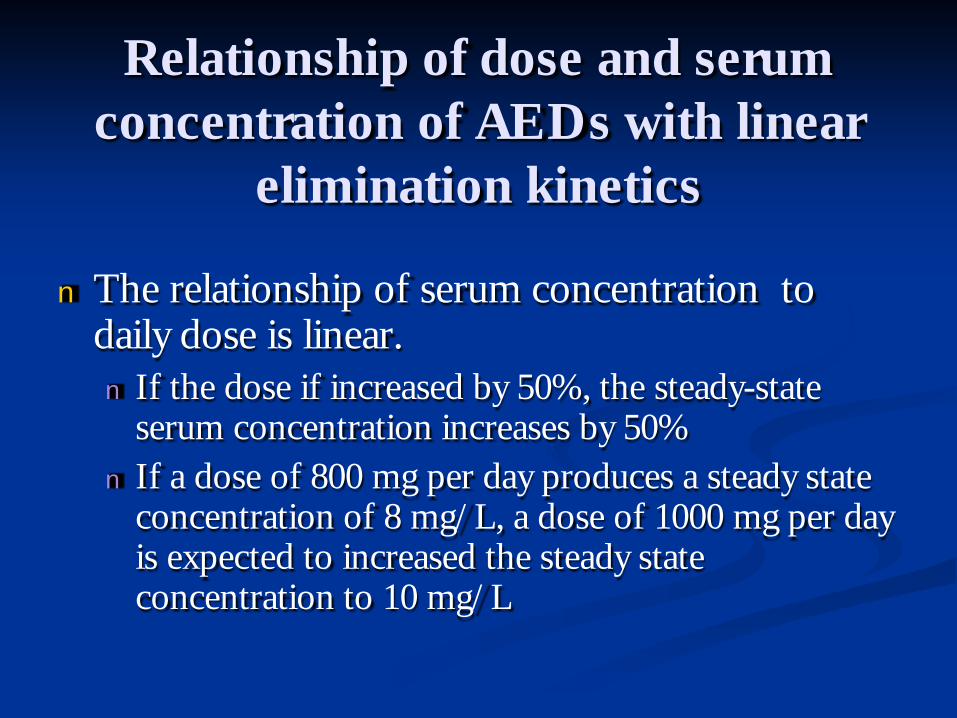
Relationship of dose and serum concentration of AEDs with linear
elimination kinetics
n The relationship of serum concentration to daily dose is linear. n If the dose if increased by 50%, the steady-state
serum concentration increases by 50% n If a dose of 800 mg per day produces a steady state
concentration of 8 mg/L, a dose of 1000 mg per day is expected to increased the steady state concentration to 10 mg/L
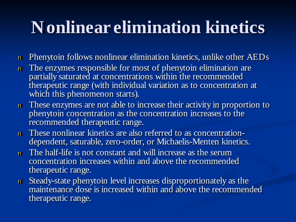
Nonlinear elimination kinetics n Phenytoin follows nonlinear elimination kinetics, unlike other AEDs n The enzymes responsible for most of phenytoin elimination are
partially saturated at concentrations within the recommended therapeutic range (with individual variation as to concentration at which this phenomenon starts).
n These enzymes are not able to increase their activity in proportion to phenytoin concentration as the concentration increases to the recommended therapeutic range.
n These nonlinear kinetics are also referred to as concentration-dependent, saturable, zero-order, or Michaelis-Menten kinetics.
n The half-life is not constant and will increase as the serum concentration increases within and above the recommended therapeutic range.
n Steady-state phenytoin level increases disproportionately as the maintenance dose is increased within and above the recommended therapeutic range.

Phenytoin nonlinear kinetics- Example of consequences
n Example 1: a daily dose of 300 mg per day results in a serum concentration of 9 mg/L. Increasing the dose to 400 mg per day (30% increase) would have increased the steady state concentration by 30% to 12 mg/ml if phenytoin were to follow linear elimination kinetics. With its nonlinear kinetics, the concentration may increase disproportionately by >300% to 31 mg/L with associated toxicity
n Example 2: a patient presents with phenytoin toxicity and a serum concentration of 40 mg/L. The T1/2 was previously estimated at 24 hours. However, after phenytoin was stopped it took 3 days for the serum concentration to go below 20 mg/L

0
10
20
30
100 200 300 400
Non-linear kinetics (ex: phenytoin)
Indi
vidu
al th
erap
eutic
rang
e
Linear kinetics (other AEDs)
0
10
20
30
100 200 300 400
8
16
4
31
9 12
Parameters for nonlinear kinetics
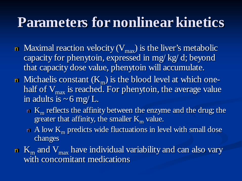
n Maximal reaction velocity (Vmax) is the liver’s metabolic capacity for phenytoin, expressed in mg/kg/d; beyond that capacity dose value, phenytoin will accumulate.
n Michaelis constant (Km) is the blood level at which one-half of Vmax is reached. For phenytoin, the average value in adults is ~6 mg/L. n Km reflects the affinity between the enzyme and the drug; the
greater that affinity, the smaller Km value. n A low Km predicts wide fluctuations in level with small dose
changes n Km and Vmax have individual variability and can also vary
with concomitant medications

0
10
20
30
100 200 300 400
4
31
9
0
10
20
30
100 200 300 400
4
14
8
Lower Km Higher Km
500

Clearance
n The daily AED dose is the amount required to maintain the desired serum concentration, countering elimination
n Elimination rate is measured by “clearance”, (L per day or L/Kg per day) which is defined as dose (mg per day or mg/Kg per day) divided by steady state serum level (mg/L).
n For phenytoin and other drugs with nonlinear kinetics, clearance will vary with serum concentration.

Pharmacokinetic interactions Enzyme-induction
n Interactions most often related to enzyme induction or inhibition.
n Enzyme-inducing AEDs include phenobarbital, primidone, phenytoin, carbamazepine. These AEDs induce multiple p450 enzymes, increasing the metabolism of drugs metabolized by these enzymes.
n Carbamazepine induces its own metabolism (auto-induction)- the process is complete in about 2-3 weeks.
n Some newer AEDs are selective enzyme inducers. For example, oxcarbazepine is a weak inducer of CYP3A4, which metabolizes estrogen.

Enzyme inhibition
n Valproate inhibits the metabolism of phenobarbital, ethosuximide, carbamazepine epoxide (active carbamazepine metabolite), lamotrigine, and rufinamide, resulting in increased serum concentrations of these compounds.
n Felbamate inhibits the metabolism of phenytoin, valproate, and carbamazepine epoxide.
n New AEDs are occasionally selective weak inhibitors. For example, oxcarbazepine and topiramate are weak inhibitor of CYP2C19, which metabolizes phenytoin, and may cause an increase in phenytoin level.

Effect of established AEDs on blood concentration of newer AEDs
AED serum concentration with add-on CBZ PHT VPA
FBM
GPN, PGB
LTG
TPM
TGB
LEV
OXC
ZNS
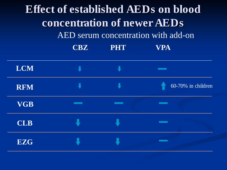
Effect of established AEDs on blood concentration of newer AEDs
AED serum concentration with add-on CBZ PHT VPA
LCM
RFM
VGB
60-70% in children
CLB
EZG

Effect of newer AEDs on blood concentration of established AEDs
AED serum concentration with add-on
PGB FBM GPN LTG TPM TGB LEV OXC ZNS
CBZ
PHT
VPA
*
* an increase in PHT level may occur at high OXC doses

Effect of newer AEDs on blood concentration of established AEDs
AED serum concentration with add-on
LCM addition
CBZ
PHT
VPA
16-20%
8%
RFM addition
VGT addition
CLB addition
EZG addition
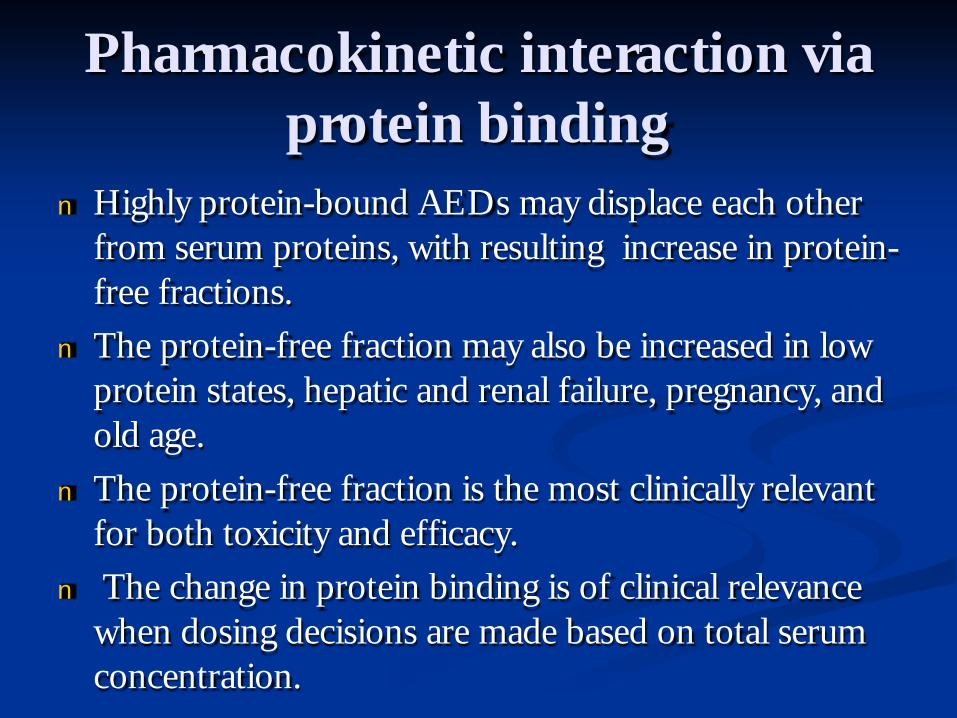
Pharmacokinetic interaction via protein binding
n Highly protein-bound AEDs may displace each other from serum proteins, with resulting increase in protein-free fractions.
n The protein-free fraction may also be increased in low protein states, hepatic and renal failure, pregnancy, and old age.
n The protein-free fraction is the most clinically relevant for both toxicity and efficacy.
n The change in protein binding is of clinical relevance when dosing decisions are made based on total serum concentration.

Example of interaction by displacement from protein binding n In monotherapy, each of phenytoin and valproate are
~90% protein bound; the free phenytoin level is ~10% of the total level.
n With concomitant use, each 1 mg/L of valproate increases the free fraction of phenytoin by approximately 0.1-0.2%.
n At a VPA level of 100 mg/L, the free phenytoin fraction may increase to 20-30%. A phenytoin level of 15 mg/L may actually be toxic, with a free level of 3-4.5 mg/L equivalent to a total level of 30-45 mg/L.
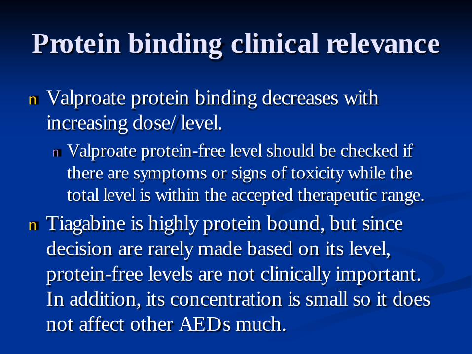
Protein binding clinical relevance
n Valproate protein binding decreases with increasing dose/level. n Valproate protein-free level should be checked if
there are symptoms or signs of toxicity while the total level is within the accepted therapeutic range.
n Tiagabine is highly protein bound, but since decision are rarely made based on its level, protein-free levels are not clinically important. In addition, its concentration is small so it does not affect other AEDs much.

AEDs and liver disease, renal disease, hemodialysis
n Liver disease may have an important effect on disposition of some AEDs, because the liver is a primary site of drug metabolism.
n Changes will vary depending on hepatic blood inflow by the portal vein, hepatocellular mass, and liver functional capacity.
n Most AEDs are capacity-limited drugs, with low extraction ratios. Their rate of metabolism depends on free drug concentration at enzyme receptor sites.

Liver disease
n AEDs metabolized by the liver will be most affected. n Hepatic failure is associated with decreased protein
binding (even if albumin is not reduced). n Larger protein-free fraction and decreased liver enzyme
functional capacity will have competing effects on rate of AED metabolism.
n With progressively lower liver enzyme functional capacity, there will be a longer half-life for AEDs metabolized by the liver, necessitating lower daily doses.

AED hepatic metabolism- dose adjustment to be considered with disease
AED
Metabolism + <50% ++ >50% - <90% >90%
Phenobarbital ++ Liver
Phenytoin +++Liver
Ethosuximide ++Liver
Carbamazepine +++Liver
Valproate +++Liver
Gabapentin None
Lamotrigine +++Liver
Topiramate +Liver
Levetiracetam +Blood
Oxcarbazepine +++Liver
Zonisamide ++Liver
AED
Metabolism + <50% ++ >50% - <90% >90%
Primidone ++Liver
Clonazepam ++Liver
Felbamate ++Liver
Tiagabine +++Liver
Pregabalin None
Lacosamide +Liver
Rufinamide +++Liver
Vigabatrin None
Ezogabine ++Liver
Clobazam +++Liver

Renal disease
n AEDs excreted unchanged by the kidneys will be most affected, with slower elimination rate and longer T1/2.
n Protein binding decreases with renal dysfunction, even without lower albumin level (small molecules may displace AEDs from binding sites and altered binding sites of albumin molecules may play a role). Phenytoin free fraction may be as high as 30%.
n Higher free portion may initially accelerate elimination. n With progressive disease, lower doses and longer inter-
dose intervals may be necessary to prevent accumulation and toxic side effects.

Dialysis
n Hemodialysis may remove some AEDs. n Dialysis may also affect drug activity through
other changes (pH, protein concentration, osmolality, electrolytes, glucose, urea).
n Following hemodialysis, albumin binding of phenytoin and phenobarbital is decreased.

Dosing for renal disease, hemodialysis Dose change with renal impairment
Replacement after dialysis
Phenobarbital Reduce slightly based on level Supplement based on level
Primidone Reduce slightly based on level Supplement based on level
Phenytoin * No change No change
Ethosuximide No change Supplement one dose
Clonazepam No change No change
Carbamazepine No change No change
Valproate * No change No change
Felbamate Reduce dose by 50% ?
Gabapentin Reduce dose by 50-90% Supplement one dose
Lamotrigine Reduce by 0-20% Supplement 20% of dose
Topiramate Reduce by 50% Supplement ?% of dose
* Monitor protein-free level

Dose change with renal impairment
Replacement after dialysis
Tiagabine No change No change
Levetiracetam Reduce dose by 50-70% Supplement one dose
Oxcarbazepine Reduce dose by 0-50% No change ?
Zonisamide Reduce by up to 35% Supplement ?% of dose
Pregabalin Reduce dose by 50-85% Supplement one dose
Lacosamide Reduce dose by 0-30% Supplement 50% of dose
Rufinamide Not affected Supplement 30% of dose
Vigabatrin Reduce dose by 25-75% Supplement half dose
Ezogabine Reduce dose by 0-50% ?
Perampanel Not recommended with severe renal impairment
?
Clobazam No change ?
Dosing for renal disease, hemodialysis

The following AED requires dose change in the presence of renal impairment:
A- Phenytoin B- Valproate C- Carbamazepine D- Gabapentin E- Tiagabine
SAE Question ARS

The following AED requires dose change in the presence of renal impairment:
A- Phenytoin B- Valproate C- Carbamazepine D- Gabapentin E- Tiagabine
SAE Question ARS
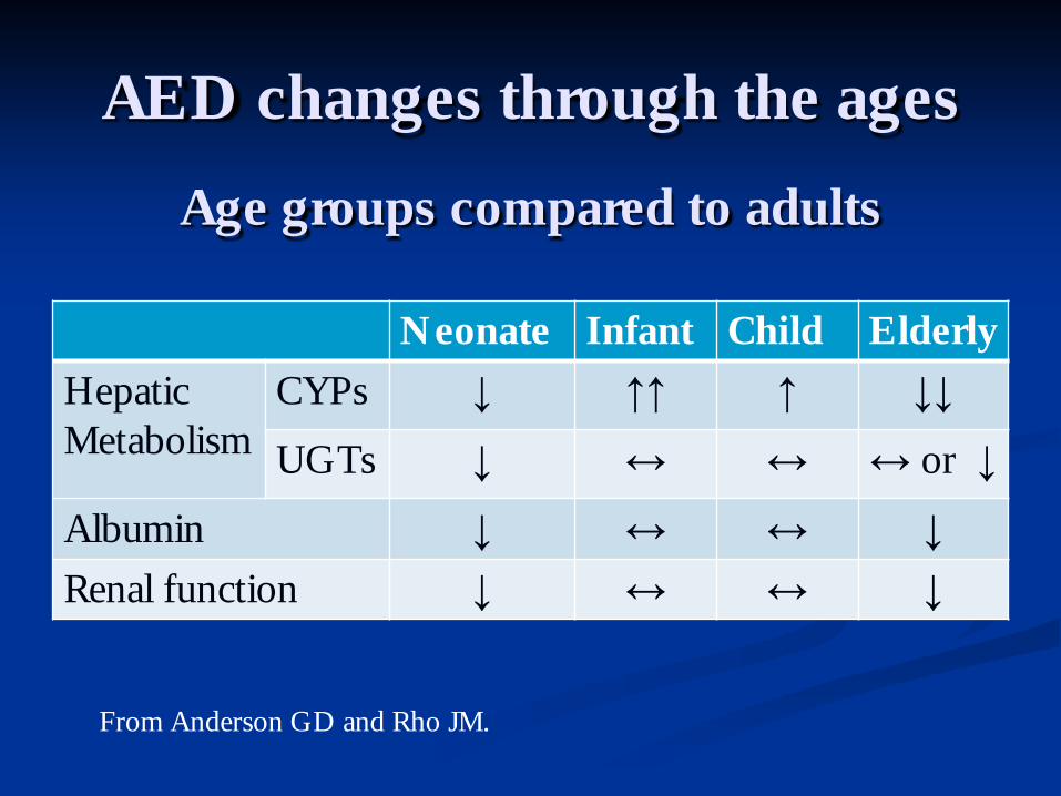
AED changes through the ages
Neonate Infant Child Elderly Hepatic Metabolism
CYPs ↓ ↑↑ ↑ ↓↓
UGTs ↓ ↔ ↔ ↔ or ↓
Albumin ↓ ↔ ↔ ↓ Renal function ↓ ↔ ↔ ↓
Age groups compared to adults
From Anderson GD and Rho JM.

Age-related physiological changes affecting AED therapy
Physiological function
Common changes in the elderly
Drug absorption Reduction in functional absorptive capacity Decreased distal gastric contractions and slowed gastric emptying Increased postprandial gastric pH fluctuations and decreased gastric pH Delayed colonic transit time
Drug distribution Decreased total body water mass Decreased proportion of fat in frail elderly individuals Decrease in serum albumin and reduced protein binding
Metabolism and excretion
Decreased liver size, hepatic blood flow and bile flow Decreased renal blood flow and glomerular filtration rate

Factors affecting AEDs in the elderly
n AED absorption may be reduced or become erratic. n Lean body mass is decreased. n There is progressive reduction in creatinine
clearance and hepatic clearance (outweighs decreased absorption).
n Protein-binding is reduced (increasing protein-free fraction for highly protein-bound AEDs).
n Multiple medications (due to co-morbidities) and herbal supplements present risk of interactions.
The following occur with increasing age except: A- Drug absorption may be reduced and become
erratic B- AED levels decrease C- Lean body mass decreases D- Creatinine clearance and hepatic clearance
decrease E- Protein binding decreases
SAE Question ARS

The following occur with increasing age except: A- Drug absorption may be reduced and become
erratic B- AED levels decrease C- Lean body mass decreases D- Creatinine clearance and hepatic clearance
decrease E- Protein binding decreases
SAE Question ARS