pharmacokinetics of monoclonal antibodies and fc-fusion ... · r eview pharmacokinetics of...
TRANSCRIPT

REVIEW
Pharmacokinetics of monoclonal antibodiesand Fc-fusion proteins
Liming Liu&
Department of Pharmacokinetics, Pharmacodynamics and Drug Metabolism, MRL, West Point, PA 19486, USA& Correspondence: [email protected] (L. Liu)
Received February 20, 2017 Accepted March 23, 2017
ABSTRACT
There are many factors that can influence the pharma-cokinetics (PK) of a mAb or Fc-fusion molecule with theprimary determinant being FcRn-mediated recycling.Through Fab or Fc engineering, IgG-FcRn interactioncan be used to generate a variety of therapeutic anti-bodies with significantly enhanced half-life or ability toremove unwanted antigen from circulation. Glycosyla-tion of a mAb or Fc-fusion protein can have a significantimpact on the PK of these molecules. mAb charge canbe important and variants with pI values of 1–2 unitdifference are likely to impact PK with lower pI valuesbeing favorable for a longer half-life. Most mAbs displaytarget mediated drug disposition (TMDD), which canhave significant consequences on the study designs ofpreclinical and clinical studies. The PK of mAb can alsobe influenced by anti-drug antibody (ADA) response andoff-target binding, which require careful considerationduring the discovery stage. mAbs are primarily absor-bed through the lymphatics via convection and can beconveniently administered by the subcutaneous (sc)route in large doses/volumes with co-formulation ofhyaluronidase. The human PK of a mAb can be rea-sonably estimated using cynomolgus monkey data andallometric scaling methods.
KEYWORDS monoclonal antibody (mAb), Fc-fusionprotein, pharmacokinetics, FcRn, target-mediated drugdisposition (TMDD), glycosylation, anti-drug antibody (ADA),human PK prediction
INTRODUCTION
The ground-breaking discovery of monoclonal antibody(mAb) technology by Kohler and Milstein in 1975 providedthe possibility of creating antibodies as a class of
therapeutics (Kohler & Milstein, 1975). The original obsta-cles of immunogenicity of murine mAbs in humans wasovercome by the revolution in molecular biology in the1980s, which enabled humanization of murine antibodiesand, eventually, the successful development of fullyhumanized antibodies. Humanization greatly reduces atherapeutic antibody’s immunogenicity in humans, makingchronic administration possible. Such advances in antibodytechnologies have resulted in an explosion in the develop-ment of therapeutic mAbs over the last decade. Today, morethan 47 mAbs and derivative drugs have been approved forhuman use with many of them attaining blockbuster status(Ecker et al., 2015). Among the top ten selling drugs in 2014,five were monoclonal antibodies and one was an Fc fusionmolecule, with each having an annual revenue over 6.5 bil-lion US dollars (Nisen, 2015). It is estimated that in the nearfuture, about 30% of the new drugs will be antibodies orantibody derivatives (Elvin et al., 2013). Antibody derivativesinclude Fc-fusion proteins, antibody-drug conjugates(ADCs), immunocytokines (antibody-cytokine fusions), andantibody-enzyme fusions.
The efficacy of a mAb is largely dependent on its phar-macological and pharmacokinetic properties. The pharma-cological effects or pharmacodynamics (PD) of a mAb arebest described as “What a drug does to the body”; a mAbbinds to a target (e.g., receptors, soluble antigens, etc.) andinduces either antagonistic (i.e., blocking or neutralizing) oragonistic effects (i.e., activating), triggering down-streampharmacological effects, leading to efficacy and/or unwantedside effects. The pharmacokinetics (PK) of a mAb is bestdescribed by “What the body does to the drug”. The fate of amAb in vivo will be determined by how the body handles itunder physiological or pathological conditions. The clear-ance or half-life of a mAb will determine the body’s “expo-sure” to the mAb, which in turn will determine the extent ofPD effects. The exposure-response (PK-PD) relationshipdetermines the outcome of a drug’s effects on the body.
© The Author(s) 2017. This article is an open access publication
Protein Cell 2018, 9(1):15–32DOI 10.1007/s13238-017-0408-4 Protein&Cell
Protein
&Cell

Understanding this relationship is an essential and integralpart of drug discovery and development.
Being large proteins of 150 kDa, mAbs possess someunique PK properties, making the discovery and develop-ment pathway of mAbs substantially different from that ofsmall molecule drugs. The unique PK properties are deter-mined by many factors related to an antibody’s structure andfunctions including FcRn mediated recycling, glycosylationpatterns, overall charge and pI, target-mediated clearance,anti-drug antibody response, and off-target binding.
In this review article, general PK properties and thefactors influencing the PK of mAbs and Fc fusion proteinswill be discussed, in addition to PK topics related topreclinical and early clinical development of mAb drugs.The impacts of glycosylation on the PK and PD of mAbsand Fc-fusion proteins have been reviewed extensivelyelsewhere (Liu, 2015) and will be only briefly discussedhere.
FACTORS INFLUENCING THE PK OF MAB ANDFC-FUSION PROTEINS
An antibody is a complex molecule consisting of an antigenbinding region (Fab domain) and a constant region (Fcdomain). Fab binds to antigens and can be responsible fortarget-mediated clearance of a mAb. Glycosylation andcharges in the Fab domain are also important for the PKproperties of a mAb. In the Fc region, a subdomain of theCH2-CH3 domain is responsible for FcRn binding that resultsin recycling of antibodies for a long half-life (Roopenian &Akilesh, 2007). The same CH2-CH3 domain is also respon-sible for Protein A and G binding, which is exploited forantibody purification (Kim et al., 1994b). The CH1-CH2
domain is responsible for FcγR binding, which is critical forantibody effector functions (Daëron, 2014; Jefferis & Lund,2002). A canonical glycosylation site is located in the Fcregion CH2 domain (Asn297) in all IgG subclasses. Glyco-sylation patterns can impact both PK and PD significantly(Arnold et al., 2007; Jefferis, 2009a). The PK propertiesrelated to the structure of Fc are also relevant to Fc-fusion
molecules. In addition, the fusion partner replaces Fab andmay be responsible for target mediated clearance. Figure 1depicts the general structure of IgG1 and the specificdomains that are important for PK properties.
FcRn: key regulator of IgG PK
In the 1960s, Dr. Roger Brambell proposed a mechanism bywhich IgG is salvaged from catabolism by receptors locatedwithin cellular compartments and/or on the surface of cells(Brambell et al., 1964; Brambell, 1966). The “hypothesized”Brambell receptor, later called neonatal Fc receptor (FcRn),was cloned in 1989 (Simister & Mostov, 1989). Experimentsin FcRn knockout mice have definitively confirmed that FcRnis responsible for protecting IgG from catabolism (Junghans,1997; Roopenian & Akilesh, 2007). Over the past two dec-ades, extensive work by many investigators has establishedthe role of FcRn in regulating the levels and transport of IgGin the body (Roopenian & Akilesh, 2007), validating Bram-bell’s hypothesis. It is now established that FcRn binds toIgG at acidic pH (∼6.0) with very low or negligible affinity atpH 7–7.4 (Ghetie & Ward, 1997; Roopenian & Akilesh,
lgG
Fab
FcCH2
VH
V L
CHI
C L
CH2
CH3 CH3
VH
CHI
VL
CL
Binding ligands
Antigen
Glycan receptor
FcRn
Relation to PK
Target mediated disposition; charge/PImediated clearance; off-target binding
Glycan mediated clearance and tissuedistribution
IgG recycling for long half-life
Figure 1. Antibody features that contribute to PK properties.
Figure 2. FcRn mediated IgG recycling pathway and
antibody mediated antigen removal via pH dependent
binding. (A) IgG circulating in the blood is taken up by
endothelial cells or monocytes through either fluid phase
pinocytosis or receptor mediated endocytosis. Once inside
the cells, IgG binds to FcRn in the acidified endosomes.
IgG that binds to FcRn migrates to the cell surface where
the IgG encounters a physiological pH environment (∼pH7.4) and is released back into the blood. IgG that is not
bound to FcRn (due to competition with other IgG) will be
sorted to lysosomes for degradation. (B) mAb (with strong
binding at pH 7.4 and no or weak binding at pH 6) binds to
antigen at neutral pH in the circulation; once endocytosed
into the cell and entered the acidic endosome, the antibody
releases the antigen and binds to FcRn. FcRn bound
antibody recycles back to the blood stream while the
antigen is degraded in the lysosome.
c
REVIEW Liming Liu
16 © The Author(s) 2017. This article is an open access publication
Protein
&Cell

Blood(physiological pH) lgG dissociates
(physiological pH)
IgG or serum proteinsPinocytosisor endocytosis
Recycling endosome
lgG binds to FcRnin acidic endsosome
Sorting of lgG-FcRn complex
Non-receptor bound proteinsdegraded in lysosome
Endothelial cellor monocyte
A
B
Blood(physiological pH) lgG dissociates
(physiological pH)
Ag degraded in lysosome
Pinocytosisor endocytosis
Recycling endosome
lgG binds to FcRnAg dissociated from Abin acidic endososome
Sorting of lgG-FcRn complex
Endothelial cellor monocyte
PK of mAbs and Fc-fusion proteins REVIEW
© The Author(s) 2017. This article is an open access publication 17
Protein
&Cell

2007), providing an ingenious biological solution to achieveexocytic release of recycled IgG. The binding area of IgG toFcRn is located between the CH2 and CH3 domain, distinctlydifferent from the FcγR binding domain that is locatedbetween the CH1 and CH2 domain (Kim et al., 1994a;Roopenian & Akilesh, 2007). As depicted in Figure 2A, IgGcirculating in the blood is taken up by endothelial cells ormonocytes through either fluid phase pinocytosis or recep-tor-mediated endocytosis. Once inside the cells, IgG binds toFcRn in the acidic endosomes. IgG that binds to FcRnescapes lysosomal degradation and migrates to the cellsurface where the IgG encounters a physiological (∼pH 7.4)pH environment and is released back into the blood. IgG thatis not bound to FcRn (due to competition of other IgG) will besorted to lysosomes for degradation (Fig. 2A). Throughcompetition of FcRn binding for recycling, the body canregulate IgG homeostasis: more IgG will be sorted to lyso-somes for degradation when large amounts of IgG are pre-sent whereas more IgG will be rescued through recyclingwhen the IgG concentration is low (Roopenian & Akilesh,2007).
The elucidation of FcRn biology prompted active effortsby the biopharmaceutical industry to create therapeuticmAbs with super long half-lives through Fc engineering. Theamino acids in the Fc region have been thoroughly investi-gated to identify mutations that significantly enhance bindingto FcRn at pH 6 while maintaining little or no binding atneutral pH (7.4). It has been reported that specific Fc vari-ants (T250Q/M428L, V308P, M428L, M252Y/S254T/T256E,M428L/N434S, N434A, N434H) that improve IgG affinity forFcRn at pH 6 with little or no binding to FcRn at neutral pHcan lead to 2–4-fold longer terminal half-lives in cynomolgusand rhesus monkeys (Yeung et al., 2009, 2010; Vaccaroet al., 2005; Olafsen, 2012; Hinton et al., 2004,2006; Denget al., 2010; Datta-Mannan et al., 2007a, b, 2012a, b). Thisgeneral relationship has also been validated more recently inhumans with the M252Y/S254T/T256E (YTE) set of muta-tions constructed on the Fc of a mAb against respiratorysyncytial virus (RSV) with half-life increases from around20 days to more than 60 days (Robbie et al., 2013). How-ever, a good quantitative relationship between in vitro FcRnbinding affinity and in vivo half-life has not been well-established (Datta-Mannan &Wroblewski, 2014), suggestingthat factors other than FcRn interaction also play an impor-tant role. On the opposite end, reduction of IgG half-life canbe realized for in vivo diagnostic and research purposes byengineering poor binders of FcRn (Swiercz et al., 2014;Olafsen et al., 2006). In addition, blockade of FcRn in vivocan decrease endogenous IgG half-life to treat autoimmunediseases (Patel et al., 2011; Liu et al., 2007; Challa et al.,2013).
FcRn binding affinity is one of the critical quality attributes(CQA) for therapeutic mAb manufacturing processes (Altet al., 2016). Methionine in mAbs can be oxidized duringmanufacturing processes (Tsuchida et al., 2016). The Fcregion of human IgG1 has three conserved methionine
residues (Met252, Met358, and Met428), which are locatednear the CH2-CH3 interface where FcRn binds (Alt et al.,2016). It has been shown that the oxidation of these threeMet residues impairs Fc–FcRn binding (Bertolotti-Ciarletet al., 2009; Pan et al., 2009). When 79% of Met252 and57% of Met428 were oxidized, the serum half-life of IgGdecreased by about 83% in mice (Wang et al., 2011b). Morerecently, Gao et al. showed with mutagenesis analyses thateither M252 or M428 oxidation alone significantly impairedFc–FcRn binding. M252 oxidation generated a more dele-terious effect on Fc–FcRn binding than M428 oxidation,whereas M358 oxidation did not affect Fc–FcRn binding(Gao et al., 2015).
Although it is the CH2-CH3 domain of Fc that is primarilyinvolved in the binding to FcRn, the variable region (Fv) ofFab may also interact with FcRn and alter the interactionbetween IgG molecules and FcRn. Wang et al. found thatalthough mAbs with identical Fc and different Fab exhibited adifferential binding to FcRn at both pH 6 and pH 7.3, only thedissociation characteristics at pH 7.3 correlated with thein vivo PK properties of these mAbs (the slower the disso-ciation at pH 7.3, the shorter the half-life) (Wang et al.,2011a). More recently, Schoch et al. made similar observa-tions with ustekinumab and briakinumab, both anti-IL12/IL23mAbs, which have almost identical Fc but different Fabfragments (Schoch et al., 2015). The binding affinity to FcRnat pH 6 and the PI are very similar between these two mAbs,but there is a positive charge patch in the Fv of briakinumab,which renders strong binding to FcRn. The mAb thereforeonly dissociates from FcRn at higher pH, resulting in ashorter half-life for briakinumab (t1/2 ∼9 days), in comparisonto ustekinumab (t1/2 ∼22 days) (Schoch et al., 2015). Theseresults clearly indicate that the variable region does alsoinfluence IgG-FcRn interactions, thus impacting PK of amAb.
Antibody and antigen complexes are also recycledthrough the FcRn pathway and can result in accumulation ofbound antigens in the circulation and extension of the half-life of the antigens (Igawa et al., 2013). To effectively removeantigen from the circulation, it is desired that the boundantigens be degraded, while antibody is recycled. Throughantibody engineering to render Fab binding to antigens pH-dependent (strong binding at pH 7.4 and no or weak bindingat pH 6), Igawa et al. created an elegantly designed recy-cling antibody that directs antigens for lysosomal degrada-tion while ensuring that the mAb is recycled to bind antigenagain, thus reducing antigen concentrations (Fig. 2B) (Igawaet al., 2010a, 2013). Furthermore, they created a sweepingantibody through engineering pH-dependent antigen bindingin the Fv region and higher binding affinity to FcRn orFcγRIIB in the Fc region. The sweeping antibody can bind toFcRn or FcγRIIb at neutral pH to facilitate the uptake of theimmune complex; bound antigen (such as IL-6R) dissociatesfrom the antibody for degradation once in the lysosome,resulting in a significantly reduced antigen concentration(Igawa et al., 2016). However, the sweeping antibodies are
REVIEW Liming Liu
18 © The Author(s) 2017. This article is an open access publication
Protein
&Cell

also cleared significantly faster than conventional antibodies,likely due to these antibodies’ ability to bind FcRn at neutralpH, which prevents them from being recycled back to thecirculation (Igawa et al., 2016). It appears that the sweepingantibody targeting FcγRIIb for enhanced uptake does nothave this drawback (Igawa et al., 2016).
Other factors related to FcRn functions that can impactthe PK of a mAb include FcRn polymorphism and IgG allo-types. It has been shown that the polymorphism of thevariable number of tandem repeats region in the FcRn pro-moter can influence the FcRn expression level and bindingability (Sachs et al., 2006). IgG allotype can also influencethe binding affinity of a mAb to FcRn, with better bindershaving longer half-life, which may contribute to the variabilityof PK in the clinic (Ternant et al., 2016). For example, it hasbeen shown that a mAb with the G1m17,1 allotype, such asinfliximab, has better binding affinity for FcRn than those withthe G1m3 with no G1m1 (G1m3,−1) allotype. In patientshomozygous for the G1m17,1 allotype, infliximab is incompetition with endogenous IgG with the G1m17,1 allo-type. As a result, infliximab exhibits a shorter half-life inpatients homozygous for the G1m17,1 allotypes than inthose carrying the G1m3,−1 allotype (Ternant et al., 2016). Inaddition, baseline albumin level in the serum can be used topredict the functional status of FcRn in vivo in that the troughlevel of an administered mAb correlate positively with thebaseline albumin concentration (Fasanmade et al., 2010).
Although FcγR binding is critical for IgG’s effector func-tions such as antibody dependent cell-mediated cytotoxicity(ADCC) and complement dependent cytotoxicity (CDC),FcγR does not appear to significantly affect IgG’s PK prop-erties. This is likely due to the fact that a large pool of serumIgG can compete with the binding of relatively small amountsof mAb to FcγR, effectively ameliorating the impact of FcγRbinding. Consistent with this, Abuqayyas and coworkershave shown that 8C2, a mouse IgG mAb, exhibited similarPK and tissue distribution in both FcγR knockout mice and inwild type mice (Abuqayyas et al., 2013).
Glycosylation impacts on PK of mAb and Fc-fusionproteins
Like natural IgGs, all approved recombinant therapeuticmAbs are glycosylated, although some non-glycosylatedmAbs or derivatives are in clinical development (Liu, 2015).Therapeutic mAbs or derivatives have an asparagine (Asn)-X-Ser/Thr (Where X is any amino acid except Pro) consen-sus sequence for N-glycosylation at the position Asn297 inthe heavy chain of the CH2 constant domain. Some thera-peutic mAbs also bear additional glycosylation in the Fabdomain. For instance, cetuximab is glycosylated at Asn88 ofthe VH region (Jefferis, 2009b). In addition, some of the Fc-fusion partner molecules, such as etanercept and B cellactivating factor receptor 3 (BR3)-Fc, also possess O-linkedglycans (Pennica et al., 1993; Stefanich et al., 2008).
Characterization of aglycosylated IgG, produced throughchemical modification or genetic engineering, confirmed thatglycosylation is not required for an IgG antibody’s long half-life (Kim et al., 1994a; Liu et al., 2011; Nose & Wigzell, 1983;Tao & Morrison, 1989). The clinical evidence for aglycosy-lated IgG having normal PK is demonstrated by the mAbALD518, a humanized anti-human IL-6 IgG1 produced inyeast. In a phase I clinical trial, the circulating half-life forALD518 was 20–32 days, consistent with half-life of a normalhuman IgG1 (Clarke et al., 2009). Glycosylated mAbs withterminal high mannose glycans have been shown to exhibitfast clearance from the blood (Goetze et al., 2011; Kandaet al., 2007; Liu et al., 2011; Wright & Morrison, 1994; Wrightet al., 2000; Yu et al., 2012). The fast clearance of mAbscontaining high mannose has also been demonstrated inclinical studies (Chen et al., 2009; Goetze et al., 2011).Glycan receptors that have been attributed to the removal ofglycoproteins in vivo include the mannose receptor (ManR)and the asialoglycoprotein receptor (ASGPR). The ASGPRand the ManR are carbohydrate-specific, endocytic recep-tors expressed by hepatic parenchymal (hepatocytes) andnonparenchymal (such as Kupffer) cells, and sinusoidalendothelial cells, respectively (Ashwell & Harford, 1982; Miet al., 2014).
The shorter half-life of an Fc-fusion molecule in compar-ison to the whole IgG has been attributed to the lowerbinding affinity to FcRn, the glycan mediated disposition andthe receptor (of fusion partner) mediated disposition (Liu,2015). Among these attributes, glycosylation patterns mayplay a more important role in determining the in vivo clear-ance of Fc-fusion molecules. For example, in the investiga-tion of humanized yeast-produced TNFαRII-Fc-fusionmolecules, it was demonstrated that it was the extent ofsialylation on the TNFRII, not the FcRn-binding affinity,which determined the clearance. The exposure was posi-tively correlated to the quantity of the sialylation on thereceptor molecule, with higher sialic acid content resulting inhigher exposure (Liu et al., 2013).
Most of the Fc-fusion molecules including BR3-Fc,IL-23R-Fc, CTLA4-Ig, and LFA3TIP rely on sialylation inreducing the in vivo clearance (Liu, 2015). Other terminalmonosaccharide, such as GlcNAc, can also contribute to thePK properties of Fc-fusion molecules (Keck et al., 2008;Jones et al., 2007).
Impact of charge and pI on mAb PK
Isoelectric point (pI), a measurement of protein charge, isdefined as the pH at which the protein carries no net elec-trical charge. In general, antibodies that are chemically orgenetically modified to achieve higher, more basic, pI valuesexhibit a high propensity to adhere to anionic sites of cellsurfaces, resulting in increased tissue uptake and fastclearance from circulation. In contrast, modified antibodieswith lower, more acidic pI values have a lower rate of uptake
PK of mAbs and Fc-fusion proteins REVIEW
© The Author(s) 2017. This article is an open access publication 19
Protein
&Cell
into cells (as a result of repulsion from the negativelycharged cell surface), leading to decreased tissue uptakeand blood clearance (Boswell et al., 2010; Bumbaca et al.,2012, 2015; Igawa et al., 2010c; Kobayashi et al., 1999; Lee& Pardridge, 2003). Chemically modified Fabs with a pIreduction of 1–2 units showed decreased blood clearanceand tissue accumulation relative to the unmodified Fab(Kobayashi et al., 1999). With antibody pI variants generatedusing site-directed mutagenesis in the Fab region, Igawaet al. demonstrated that variants with pI values of 1–2 unitslower than the wild type displayed longer half-lives andreduced clearance following both subcutaneous and intra-venous administration (Igawa et al., 2010b). The datademonstrated that the clearance was positively correlatedwith pI, i.e., the half-life was negatively correlated with pI.Similar data was obtained from both human and minipigmodel in that antibodies with higher pI values exhibited fasterclearance and lower subcutaneous bioavailabilities thanantibodies with lower pI values (Zheng et al., 2012).Manipulation of pI was also used to reduce the toxicity ofimmunotoxin. Onda et al. showed that reducing the pI of theFv portion of an immunotoxin significantly reduced livertoxicity, presumably reducing the distribution to the liver(Onda et al., 1999).
Charge variations can arise from antibody manufacturingprocesses due to chemical or enzymatic degradation viaoxidation, deamidation, isomerization, and fragmentation.The charge differences may impact both PK and PD. Toinvestigate whether mAb charge variants possess differentPK and PD properties, Khawli et al. isolated basic, neutral,and acidic variants from a mAb product and characterizedthem in PK and PD assays. Their results showed that allvariants had similar potency in PD assays and rat FcRnbinding relative to the starting material. Following iv or scadministration in rats, no difference in serum PK wasobserved, indicating that pI differences among charge vari-ants were not sufficient to result in PK changes. However, itis worth noting that the pI difference between the acidicvariant and the basic variants were less than 0.5 (8.7 vs. 9.1)(Khawli et al., 2010). Based on the above evidence, it can beconcluded that shifts in isoelectric point of approximately onepI unit or more are likely to produce measurable changes inPK and tissue distribution; differences of less than one pI unitare mostly inconsequential to PK.
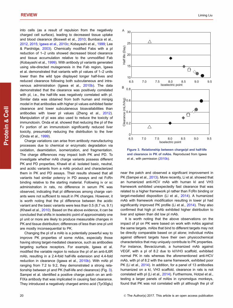
Changing the pI of a mAb is a potentially powerful way toimprove PK properties of some mAbs, especially thosehaving strong target-mediated clearance, such as antibodiestargeting surface receptors. For example, Igawa et al.modified the variable region to reduce the pI of an anti-IL6RmAb, resulting in a 2.4-fold half-life extension and 4.4-foldreduction in clearance (Igawa et al., 2010b). With mAb pIranging from 7.2 to 9.2, they demonstrated a strong rela-tionship between pI and PK (half-life and clearance) (Fig. 3).Sampei et al. identified a positive charge patch on an anti-FIXa antibody that was implicated in causing fast clearance.They introduced a negatively charged amino acid (Tyr30glu)
near the patch and observed a significant improvement inPK (Sampei et al., 2013). More recently, Li et al. showed thatan humanized anti-HCV mAb with human kI and VH3framework exhibited unexpectedly fast clearance that wasrelated to a higher framework pI rather than FcRn binding ortarget-mediated disposition (Li et al., 2014). A humanizedmAb with framework modification resulting in lower pI hadsignificantly improved PK profile (Li et al., 2014). They alsoconfirmed that high pI mAb exhibited higher distribution toliver and spleen than did low pI mAb.
It is worth noting that the above observations on theimpact of pI on PK were based on work with mAbs againstthe same targets. mAbs that bind to different targets may notbe directly comparable based on pI alone; individual mAbsagainst different targets have their own physicochemicalcharacteristics that may uniquely contribute to PK properties.For instance, Bevacizumab, a humanized mAb againstVEGF, with a pI of 8.2 due to kI/VH3 scaffold, exhibitednormal PK in rats whereas the aforementioned anti-HCVmAb, with pI of 8.2 with the same framework, exhibited poorPK (Li et al., 2014). In addition, for a panel of 13 antibodieshumanized on a kI, VH3 scaffold, clearance in rats is notcorrelated with pI (Li et al., 2014). Furthermore, Hotzel et al.,testing a larger panels of mAbs in cynomolgus monkeys,found that PK was not correlated with pI although the pI of
A
B
30
25
20
15
10
0.8
0.6
0.4
0.2
0
6.5 7.0 7.5 8.0 8.5 9.0 9.5
6.5 7.0 7.5 8.0 8.5 9.0 9.5
Isoelectric point
Isoelectric point
Hal
f life
(Day
)C
lear
ance
(mL/
h/kg
)
Figure 3. Relationship between charge/pI and half-life
and clearance in PK of mAbs. Reproduced from Igawa
et al., with permission (2010b).
REVIEW Liming Liu
20 © The Author(s) 2017. This article is an open access publication
Protein
&Cell

the mAbs tested was narrowly clustered, between 7.5 and9.5 (Hotzel et al., 2012a).
Impact of target-mediated drug disposition on mAbPK
Target-mediated drug disposition (TMDD) is a nonlinear PKphenomenon, where drug–target binding (complexation withreceptors, enzymes, or transporters) and subsequent events(dissociation and drug-target complex degradation) result indose-dependent changes in PK (Mager & Jusko, 2001). Incomparison to small molecule drugs, TMDD is more com-mon for mAb drugs, especially for those targeting membranereceptors. It was estimated that half of the marketed mAbsexhibit TMDD (Dirks & Meibohm, 2010; Mould & Sweeney,2007). The actual number is probably higher because theclinical doses at which drugs are studied often saturate thetarget mediated elimination pathway, thereby masking thenonlinear PK.
At low concentrations TMDD accounts for a significantportion of mAb clearance. As mAb concentrations increase,target mediated elimination starts to saturate and clearancedecreases dramatically. At high mAb concentrations, theclearance approaches a first-order process where the FcRnmediated pathway is dominant and the nonlinear pathwaybecomes negligible. To determine whether a mAb undergoesTMDD, a single ascending dose iv PK study in a pharma-cologically relevant species (in which the mAb binds to thetarget of interest) is recommended. A typical characteristic ofTMDD is readily recognizable by plotting clearance or t1/2against dose as depicted in Fig. 4 (Mullamitha et al., 2007).
Nonlinear PK caused by TMDD may have significantconsequences on study design (both pre-clinical and clini-cal), in particular on dose selection, dosing scheme, andsampling times (Dirks & Meibohm, 2010; Grimm, 2009).Preclinical PK studies with mAb for which TMDD is sus-pected must be conducted in pharmacologically relevantspecies (often in NHP) to determine whether the drug issubjected to TMDD. However, the interpretation of theresults may be biased by anti-drug antibodies (ADA) inter-ference. It is preferable to conduct a PK study with PDmeasurement to estimate the exposure and response rela-tionship and to determine whether the PK saturating dose isalso the dose at which the PD is saturated. However, it isimportant to bear in mind that preclinical data can only give arough estimate of the saturating dose of a mAb and thatfindings may not be directly translatable to the clinic. It isgenerally appreciated that preclinical information is a startingpoint for designing a clinical TMDD study.
Although TMDD generally occurs more frequently formAbs that target surface antigens, mAbs against solubleantigens can also exhibit TMDD. For example, Brenner et al.showed that FG-3019, a human mAb against connectivetissue growth factor (CTGF) exhibited fast clearance thatwas determined to be target related. The fast clearance of
the FG-3019-CTGF complex was due to the extremely fastclearance of CTGF itself, which is cleared mainly throughliver uptake (Brenner et al., 2016).
Because of its assumption of linear disposition, the non-compartmental analysis (NCA) method is inappropriate forPK analysis of drugs with TMDD, except perhaps for initialexploration to uncover or confirm TMDD. While steady stateof volume distribution (Vss) can be inferred from bloodconcentration using NCA for small molecules, Vss for mAbmay be underestimated and incorrect if it is derived fromNCA analysis or simple compartmental analysis. This isbecause these models assume linear PK and rapid equilib-rium between blood and tissues (Wang et al., 2008).Therefore, more sophisticated mechanistic models areneeded for the TMDD analysis. A general framework of amechanistic TMDD model was established by Mager andJusko (2001). Following the base model introduction, severalsimilar models have been developed in several variations(Dua et al., 2015; Gibiansky et al., 2008; Grimm, 2009;Mager, 2006). Information on the target pharmacology, in
350
300
250
200
150
100
50
0
8
6
4
2
0
Cle
ranc
e (m
L/D
ay/k
g)H
alf-L
ife (D
ay)
Dose (mg/kg)0 2 4 6 8 10 12
Figure 4. A typical TMDD graph exhibiting the rela-
tionship between clearance or t1/2 and mAb dose.
Anti-αv mAb was infused to cancer patients; clearance or
terminal half-life (t1/2) was plotted against dose (0.1, 0.3, 1,
3, and 10 mg/kg). Adapted from Mullamitha et al., Clin
Cancer Res. (2007).
PK of mAbs and Fc-fusion proteins REVIEW
© The Author(s) 2017. This article is an open access publication 21
Protein
&Cell

particular its expression and turnover, drug-target bindingkinetics, and knowledge of the fate of drug-target complexes,are generally required to build a mechanistic model. How-ever, in most situations, this information is not readily avail-able. For this reason, many analyses use empiricalapproaches for building models that require less informationabout the drug target interaction, receptor numbers, andturnover rates (Dua et al., 2015; Gibiansky et al., 2008;Grimm, 2009; Mager, 2006). Nevertheless, in most situa-tions, at least one of the variant models can be useful inmodeling a specific mAb’s nonlinear behavior.
Based on the model of quasi-steady-state approxima-tions, Grimm thoroughly analyzed the consequences ofTMDD on the disposition of mAb (Grimm, 2009). Severalconclusions were drawn from this analysis. First, saturationof clearance (from PK analysis) signifies the saturation of thetargets when the target is readily accessible. A mAb target-ing soluble antigens in the circulation is one example of thissituation. Second, when the target is only slowly accessibledue to low permeability, saturation of clearance may notimply the saturation of targets, although it may still be usedas a proxy for the saturation of the target. Third, when thesite of target-mediated clearance and the desired site ofaction are not the same, especially when the latter is poorlyaccessible, saturation of clearance no longer implies on-sitetarget saturation. This may occur, for example, when tar-geting a receptor on a solid tumor which is poorly accessible,and the receptor is also expressed on other normal tissues(such as vascular endothelium) (Grimm, 2009). It has beenobserved in many cases that in vitro Kd of a mAb is oftendifferent from the in vivo Kd (or Km) obtained from modeling(Brenner et al., 2016; Ng et al., 2006). The in vitro Kd oftenoverestimates the affinity of the mAb, likely due to the sim-plified assay system. The complex situation in vivo canimpact the apparent Kd differently (Brenner et al., 2016; Nget al., 2006). Other factors such as fast Kint (drug-targetinternalization rate constant), fast target turnover and limi-tation of accessibility or permeability also play importantroles in drug and target interactions (Grimm, 2009). Whenchoosing clinical candidates, what is the optimum affinity fora mAb? Based on Grimm’s analysis, in most cases, opti-mization of an antibody is a matter of optimizing Km. Incertain situations (e.g., Koff ≪ Kint), increasing affinity bydecreasing Koff may yield no benefit because of the fastinternalization of the target (Grimm, 2009).
Impacts of anti-drug antibody (ADA) on mAb PK
Most, if not all, therapeutic proteins including mAbs, areimmunogenic and can induce ADA in humans. The inci-dence and the impacts on PK/PD and efficacy vary fromdrug to drug. Immunogenicity of therapeutic mAbs can causehypersensitivity responses such as anaphylaxis and infusionreactions, and accelerated clearance of drug, resulting indecreased safety and efficacy (Chirmule et al., 2012).
Both humanized and fully human mAbs can induce ADAin humans; there is no evidence that fully human antibodiesare less immunogenic than humanized ones. For example,Humira® (adalimumab), a fully human mAb, induces ADAand impacts PK and efficacy in patients (Bartelds et al.,2011; Chirmule et al., 2012; Harding et al., 2010). Barteldset al. reported that ADA was developed in 28% of 272rheumatoid arthritis (RA) patients who were treated withHumira® for 3 years. RA patients without anti-adalimumabantibodies had much higher adalimumab concentrations(median, 12 mg/L) compared with patients with antibodytiters from 13 to 100 AU/mL (median, 5 mg/L) (Bartelds et al.,2011). There were significantly more treatment failures thatled to the discontinuation of the treatment in patients whodeveloped ADA (Bartelds et al., 2011).
Human mAb or humanized mAb are immunogenic inanimals. Van Meer et al. examined the immunogenicity of 27marketed mAb drugs in NHP and 25 (93%) developed ADAto the administered mAb (van Meer et al., 2013). ADA cancause perturbation of pharmacokinetic profiles, making itdifficult to interpret PK/TK data, particularly when TMDD issuspected. The simplest way to determine the cause ofabnormal PK when both ADA and TMDD are the suspects isto measure ADA levels in the serum or plasma of the dosedanimals. In most cases, clearance likely coincides with thepresence of ADA (Richter et al., 1999). However, theabsence of measurable ADA does not mean the absence ofADA especially when the ADA assay sensitivity is low and/orwhen the drug concentration is above the assay’s drug tol-erance level (Kelley et al., 2013). Figure 5 shows the typicalimpact of ADA on the clearance of an administered mAbdrug.
In most cases, ADA occurs mainly in animals exposed tolower doses rather than higher doses. The reason for thisphenomenon may be related to high dose induced tolerance(high zone tolerance) (Gilliland et al., 1999; Somerfield et al.,2010) or the interference of high concentrations of mAb in
Animal A without ADAAnimal B with ADAADA concentration of animal B
1000
100
10
1
0.1
mA
b co
ncen
tratio
n (μ
g/m
L)
Time (day)0 10 20 30 40 50 60
Figure 5. Hypothetical concentration time curves fol-
lowing iv administration of a mAb in animals with or
without ADA.
REVIEW Liming Liu
22 © The Author(s) 2017. This article is an open access publication
Protein
&Cell

the serum. Since ADA can lower the exposure of the drug intoxicity species, it is important to use sufficiently high doselevels to achieve desired exposure multiples (based on theanticipated clinical dose). It is also important to take intoconsideration the ADA assay performance in terms of drugtolerability before declaring a lack of ADA.
The occurrence of ADA of human mAb can be rapid orslow in preclinical animal species. Typically, ADA mayappear 2–3 weeks after single dose administration and theseare mostly IgG response. In some cases, the occurrence canbe even earlier which most likely is mediated by IgM sub-class. It is important to note that immunogenicity or ADAresponse found in preclinical species does not predict ADAresponse in human (van Meer et al., 2013). It has beenreported that the incidence of ADA formation in NHPs andpatients was comparable in only 59% of the cases (van Meeret al., 2013). mAbs can be very immunogenic in animals andyet show no apparent or negligible immunogenicity inhumans. Keytruda (pembrolizumab), a humanized IgG4 anti-PD1 mAb for the treatment of multiple cancers, is a goodexample of species differences in ADA formation. Keytrudais quite immunogenic in cynomolgus monkeys as a highincidence of ADA was observed. (BLA# 125514Orig1s000Keytruda FDA review package, pharmacology. August 22,2014; Link http://www.accessdata.fda.gov/drugsatfda_docs/nda/2014/125514Orig1s000PharmR.pdf).
However, in clinical studies in patients treated with Key-truda (2 mg/kg every 3 weeks, 200 mg every 3 weeks, or10 mg/kg every 2 or 3 weeks), only 20 (1.7%) of 1,149evaluable patients tested positive for treatment-emergentanti-pembrolizumab antibodies. There was no evidence ofan altered pharmacokinetic profile or increased infusionreactions with anti-pembrolizumab binding antibody devel-opment (Highlights of Prescribing Information of Keytruda;Revised: 08/2016. https://www.merck.com/product/usa/pi_circulars/k/keytruda/keytruda_pi.pdf).
Impacts of off-target binding on mAb PK
Off-target binding can significantly affect the PK, tissue dis-tribution, efficacy, and toxicity of therapeutic antibodies.Although antibodies developed against an antigen are highlyspecific to that antigen, some mAbs can have multi-speci-ficity binding capability for irrelevant antigens (James et al.,2003; Notkins, 2004). To prevent autoimmune diseases,antibodies matured through somatic mutations go throughstringent control processes in vivo to weed out those unfa-vorable mutants that bind non-specifically to self-antigen orinteract broadly with normal tissues. However, antibodiesderived from in vitro selection or maturation lack an in vivo-like control system and rely on the counter screen to ensurespecificity. This can lead to the selection of some non-specifically binding variants. For example, the A4B4 variant,(a prototype of Motavizumab, which is an in vitro affinitymatured anti-RSV mAb derived from palivizumab), demon-strated broad tissue binding and accelerated clearance
in vivo (Wu et al., 2007). Multiple off-target interactions weredemonstrated on protein biochips for adalimumab, whichwas derived in vitro from a cloned human antibody phagelibrary (Feyen et al., 2008).
Recently, Datta-Mannan et al. showed that bispecific IgG-scFv or IgG-ECD antibodies were cleared rapidly throughliver sinusoidal endothelial cells (LSEC) in the liver (Datta-Mannan et al., 2016). The rapid clearance of BsAb was notattributed to TMDD, reduced Fc-FcRn interaction, or poormolecular or biochemical properties. Instead it was due tonon-specific binding or association with LSEC. mAbs mayalso bind specifically to a non-intended target and causealteration of PK/PD and toxicity profiles. Bumbaca et al.reported that a humanized mAb (hLD1.vB) directed againstfibroblast growth factor receptor (FGFR) 4 exhibited specificbinding to mouse complement component 3 (C3), leading toaccelerated clearance and limited efficacy in mice (Bumbacaet al., 2011). Vugmeyster et al. also reported that an anti-monkey A-beta mAb had off-target binding in cynomolgusmonkeys, leading to accelerated clearance (Vugmeysteret al., 2011).
Because the unpredictability of the off-target binding, it isrecommended that mAb candidates go through earlyscreens for non-target bindings in vitro including proteinmicroarrays, light directed peptide synthesis arrays, andtissue cross-reactivity assays (Kelly et al., 2015; Hotzelet al., 2012b). These screens, along with preclinical evalu-ation in rodents and primates, are important tools in theassessment of how well a mAb will behave in the clinic.Hotzel et al. reported a strategy for mitigation of risk forantibodies with fast clearance. It is an assay based on ELISAdetection of non-specific binding to baculovirus particles thatcan identify antibodies having increased risk for fast clear-ance (Hotzel et al., 2012b). This assay can be used duringlead generation or optimization to identify antibodies withincreased risk of having fast clearance in both humans andcynomolgus monkeys, thus increasing the likelihood ofobtaining a suitable drug candidate. Recently high through-put cross-interaction assays were developed to screenmAbs for off-target mediated clearance. These assaysshowed high correlation with clearance rate in mice (Kellyet al., 2015). Others used protein and/or tissue arrays todetermine cross reactivity of mAbs (Lueking et al., 2008;Kijanka et al., 2009).
MAB ABSORPTION
The majority of FDA-approved therapeutic antibodies areadministered intravenously (iv). The iv route allows for rapiddelivery of large amounts of antibodies to the systemic cir-culation with complete systemic availability. In addition, the ivroute allows the administration of larger volumes in com-parison to other parenteral routes of administration. How-ever, iv delivery, often required to be conducted in hospitalsor doctor’s office, is not convenient for patients, andincreases the cost of therapy. In addition, rapid infusion of
PK of mAbs and Fc-fusion proteins REVIEW
© The Author(s) 2017. This article is an open access publication 23
Protein
&Cell

antibody may also induce adverse events such as infusionreactions. Therefore, for some mAbs requiring chronic dos-ing, extravascular routes such as subcutaneous (sc) andintra-muscular (im) administration have been developed. Scor im administration can be performed by a health careprofessional in a patient’s home or even by self-administra-tion (Richter & Jacobsen, 2014). Examples for extravascularadministration of mAbs or Fc fusions are adalimumab (sc),alefacept (im), efalizumab (sc), etanercept (sc), omalizumab(sc), and palivizumab (im).
The systemic absorption of antibodies following sc or imadministration most likely occurs via the lymphatic system.The highly porous lymphatic system allows the transport ofmacromolecules (>20 kDa MW) through the convective flowof the interstitial fluids (Richter et al., 2012; Richter &Jacobsen, 2014). Since the lymph fluid drains slowly into thevascular system, the absorption of antibodies from the site ofadministration usually continues for hours or even days(Richter et al., 2012). In animals and in man, the time tomaximal plasma concentrations (Tmax) of antibody are typi-cally observed 1–8 days following sc or im administration(Lobo et al., 2004). In general, antibodies given by im or scare well absorbed, resulting in bioavailabilities ranging from50% to 100% (Richter & Jacobsen, 2014). There is no cor-relation between MW and the bioavailability of protein ther-apeutics in several animal species and in humans (Richteret al., 2012).
The extent of absorption of mAbs can be variabledepending on the extent of pre-systemic antibody degrada-tion including proteolytic degradation, endocytosis at theinjection site and rates of recycling through interaction withFcRn (Wang et al., 2008). The absorption of therapeuticproteins can also be impacted by formulation and injectionvolumes (Richter et al., 2012; Richter & Jacobsen, 2014). Inaddition, physiologic factors such as age, body weight,movement, heat, and injection site may also serve ascovariates for the bioavailability of therapeutic proteinsincluding mAbs (Richter & Jacobsen, 2014). It was observedthat fat content and physical activities can profoundly impactthe absorption of therapeutic proteins in an animal model(Wang et al., 2012). Sc absorption rates in animals appear tobe more rapid than in humans. Non-human primates tend tooverestimate the sc bioavailability of mAbs and rodent andseveral other preclinical species do not appear to exhibit anyclear patterns in relationship to humans (Richter et al., 2012).Recently the minipig was suggested as a potential translat-able model for mAb pharmacokinetics following iv or scadministration (Zheng et al., 2012).
Large doses may not be feasible for sc or im administrationdue to the relatively limited solubility of IgG (∼100 mg/mL);small volumes can be administered via these routes (2.5 or5 mL for sc and im per site, respectively) (Richter et al., 2012).For mAbs requiring relatively low dose, such as Humira(40 mg/dose), sc administration can be easily achieved at40 mg/0.8 mL injection pen (Humira® PI). However, for mAbsrequiring high doses, such as omalizumab, multiple injection
sites are necessary; a dose of 375 mg is delivered via threeseparate 1 mL sc injections (Xolair PI). More recently, thelimitation of volume in sc administration of mAb appears tohave been ameliorated by including hyaluronidase in theformulation. In the subcutaneous space, the glycosamino-glycan hyaluronan, together with collagen, creates a volumebarrier within the extracellular matrix (Bookbinder et al., 2006).Hyaluronan can be degraded by hyaluronidase and hyalur-onidase derived from animal testes has been used exten-sively to facilitate dispersion of co-injected materials in theclinic (Bookbinder et al., 2006). With co-formulation of hya-luronidase, it is now clinically feasible to administer mAb’s foroncology indications which typically require much largerdoses and volumes (Jackisch et al., 2014; Salar et al., 2014;Shpilberg & Jackisch, 2013). rHuPH20, a recombinant hya-luronidase temporarily cleaving hyaluronan fibers in theinterstitial tissue, and thereby facilitating the sc injection oflarge volumes, has been used to co-formulate severalimportant anti-cancer mAbs. With co-formulation with hyalur-onidae, it is now possible to administer volumes up to 5 mL scin approximately 5 min for 600 mg trastuzumab (Bittner et al.,2012). Rituximab can be delivered in 2–8 min with 4.4–15.0 mL sc (Salar et al., 2014). A fixed dose of 1,400 mg ofrituximab was successfully delivered via sc injection byhyperconcentrating rituximab (12-fold) relative to the IV for-mulation and including the rHuPH20 as an excipient in avolume of 11.7 mL (Salar et al., 2014; Shpilberg & Jackisch,2013). The Ctrough of sc delivered mAb was not inferior to thatdelivered by IV at 375 mg/m2 (Salar et al., 2014). Sc admin-istration is also generally more immunogenic than drug dosedvia the IV route. For example, anti-trastuzumab antibodieswere detected in 8 of the 58 patients who received trastuzu-mab sc compared to zero of the 12 patients who receivedtrastuzumab IV. That said, the presence of anti-trastuzumabantibodies did not correlate with the adverse events andpharmacokinetic profiles in these patients (Wynne et al.,2013). In EU, sc formulation for rituximab was approved forNHL (1,400 mg) in 2014 and for CLL (1,600 mg) in 2016(Roche Media Release, 2016). Herceptin sc formulation(600 mg) for breast cancer was approved in EU in 2013(Roche Media Release, 2013).
MAB DISTRIBUTION
Being large and polar molecules, antibodies move veryslowly across vascular endothelial cells. Convection isbelieved to be the primary mechanism responsible for thetransport of antibody from blood to the interstitial space oftissues (Richter & Jacobsen, 2014). The rate of convectionout of the blood and the rate of antibody catabolism withintissue determine the rate of antibody elimination. In general,antibody concentrations in tissue interstitial fluid are sub-stantially lower than antibody concentrations in plasmabecause of the differences in the rate of convective uptake(which is slow) and elimination of antibody from tissue (whichcould be fast). However, higher antibody concentrations
REVIEW Liming Liu
24 © The Author(s) 2017. This article is an open access publication
Protein
&Cell

have been observed in tissues that are highly perfused andwith relatively leaky vasculature (e.g., bone marrow, spleen,and liver). IgG antibodies show very little distribution to thebrain because of blood brain barrier. Shah and Betts con-ducted analyses of bio-distribution data of non-binding mAbsfrom several different species (mice, rat, monkey, andhuman). Using a physiologically based pharmacokinetic(PBPK) model, they were able to define the relationshipbetween plasma and various tissue concentrations as theantibody biodistribution coefficient (ABC). They found thattypically the concentration of mAb in lung is 15%, heart 10%,kidney 14%, muscle 4%, skin 16%, small intestine 5%, largeintestine 5%, spleen 13%, liver 12%, bone 7%, stomach 5%,lymph node 8%, adipose 5%, brain 0.4%, pancreas 6%,testes 6%, thyroid 68%, and thymus 7% of the plasmaconcentration (Shah & Betts, 2013). These results are con-sistent with the general estimates of tissue to blood ratiorange of 0.1 to 0.5 for mAbs (Lobo et al., 2004). However, itis important to point out that most of the tissue distributiondata were obtained with iodine labeled mAbs and unusuallyhigh distribution to thyroid should be interpreted with cautionbecause of the concentration effect of iodine-labeledmetabolites in the thyroid.
In cases where an antibody shows high-affinity, high-ca-pacity binding to tissue and target-mediated elimination, thetrue Vss may be much greater than the distribution volumeestimated by standard NCA analysis. It was reported that amAb against keratin sulfate exhibited much higher tissueconcentrations than in the plasma with tissue to blood ratiobeing 5.9, 4.3, 3.5, and 2.6 for lung, esophagus, kidney, andliver, respectively (Kairemo et al., 2001). Similarly, somemAbs directed against endothelial antigens also showedvery high tissue to blood ratios. For example, a tissue dis-tribution study showed that the lung-to-blood ratio was 5, 10,and 15 for antibodies of anti-Thy-1.1, anti-ACE, and anti-ICAM-1, respectively (Danilov et al., 2001). It is worth notingthat the non-binding IgG typically has a tissue to blood ratioranging from 0.04 to 0.68 (Shah & Betts, 2013).
In a modeling study comparing antibodies with differentaffinities in distribution to tumors, Fujimori et al. observedthat the average antibody concentration in the tumor doesnot increase linearly with affinity. High antibody affinity at agiven dose tends to decrease antibody percolation or pen-etration to the tumor because the high affinity binding to theedge of the tumor leads to fewer free antibody molecules.This phenomenon was termed “binding site barrier” (Fujimoriet al., 1990). Using mAbs that recognize the same Her2epitope but with different affinities (270, 23, 7.3, and0.56 nmol/L), Rudnick et al. confirmed the “binding sitebarrier” phenomenon in vivo. They reported that moderateaffinity was associated with the highest tumor accumulationat 24 h and 120 h after intravenous injection. High affinitywas found to produce low tumor accumulation (Rudnicket al., 2011) (Fig. 6). Similar results were obtained recently ina tumor distribution study by Glatt et al. with anti-CD44antibodies of different affinities (Glatt et al., 2016).
Zahnd et al. used Darpins, a 14.5 kDa target bindingprotein to systemically evaluate the size and affinity effectson the tumor distribution. Darpins of different size (with orwithout Pegylation) and affinity against HER2 were tested intumor tissue distribution studies. The results showed that forsmall proteins like Darpin that were eliminated rapidly bykidney filtration, improving affinity directly improved tumoruptake. However, for large targeting agents like PegylatedDarpin, the impact of affinity on tumor accumulation isdiminished. The dependence of tumor uptake on bindingaffinity was found to be weak once Kd < 100 nmol/L (Wittrupet al., 2012; Zahnd et al., 2010). scFvs, Fabs, diabodies,darpins, and the like are considered too large for sufficientlyrapid extravasation and too small to escape renal clearance.Whole IgG antibody is considered to be the optimal size fortumor targeting (Wittrup et al., 2012).
mAb accessibility to tissues can also be observed in PDreadouts. In a monkey distribution study with rituximab, thelevels of peripheral blood B cells were already depleted by>94% 2 days after the first dose and 9 days after the seconddose. However, B cell levels in lymph nodes were onlydecreased by 42%–57% at 9 days following the 2nd dose(Mao et al., 2013). These results indicate that solid tissuesare more difficult to penetrate by mAb and the “binding sitebarrier” could restrict the distribution of mAb, leading to adelayed pharmacological effect in the tissues.
HUMAN PK PREDICTION AND FIRST IN HUMANSTARTING DOSE SELECTION
PK parameters of small molecule drugs can be scaledacross species using a power model of the form Y = a*BWb
with reasonable accuracy (Huang & Riviere, 2014; Wang
Anti-Her2 mAbs270 nmol/L23 nmol/L7.3 nmol/L0.56 nmol/L
Per
cent
inje
cted
doe
s pe
r gra
m tu
mor
24 h 120 h
P = 0.0476 P < 0.00019
8
7
6
5
4
3
2
1
0
Figure 6. Tumor distribution of anti-Her2 mAbs with
different affinities. Adapted from Rudnick et al. (2011).
PK of mAbs and Fc-fusion proteins REVIEW
© The Author(s) 2017. This article is an open access publication 25
Protein
&Cell

et al., 2016). This equation is based on the principle ofallometry; Y is the parameter of interest (e.g., clearance orvolume); a is the allometric coefficient; BW is the bodyweight; b is the allometric exponent. For large moleculessuch as mAbs with nonlinear PK, assumptions underlyingallometric scaling, such as the absence of nonlinear phar-macokinetics and species-specific clearance may not becorrect. Nevertheless, in many cases, PK parameters ofmAbs with non-target related elimination pathways or dosesabove the target saturation levels in humans can be rea-sonably predicted using data from cynomolgus monkeyswith an allometric exponent of ∼0.85 (Deng et al., 2011;Dong et al., 2011; Ling et al., 2009; Wang & Prueksaritanont,2010). Different exponents were proposed for reasonablepredictions of human CL and Vss for 24 mAbs targetingeither soluble antigen or membrane receptors (Oitate et al.,2011). By analyzing data from preclinical and clinical studiesof 13 therapeutic mAbs, Deng et al. showed that CL of mAbscan be better predicted based on cynomolgus PK data andan allometric scaling exponent of 0.85. Human concentra-tion–time profiles were also reasonably predicted from thecynomolgus monkey data using species-invariant timemethod with a fixed exponent of 0.85 for CL and 1.0 forvolume of distribution (Deng et al., 2011). A relatively higheraccuracy prediction for pharmacokinetics in humans may beachieved by physiologically based PBPK modeling that hasthe advantage of allowing prediction of antibody levels inmany tissues, including tumors. It also takes into consider-ation the effects of saturable processes (e.g., target binding,FcRn recycling) on antibody PK and the impacts of a varietyof other factors (e.g., antigen expression, antibody affinity)on the tissue selectivity of antibody disposition (Shah &Betts, 2013; Wang et al., 2016). However, PBPK models arecomplex, mathematically difficult to construct, poorly suitedto population analyses, and often limited because of a lack oftissue concentration data, parameter availability, or param-eter identifiability (Shah & Betts, 2013; Wang et al., 2016).Although the drug exposure in humans can be reasonably
predicted for mAbs with linear PK above saturation dose,prediction of drug exposure at low starting doses for mAbs(typically starting dose for dose escalation study) is chal-lenging and generally poor because of nonlinear PK (Donget al., 2011). Further exploration of species differences in thetarget expression level, target-antibody binding and targetkinetics, and in vivo PK studies with relevant dose rangesmay help to solve this issue.
For oncology indications, rule based standard 3 × 3 doseescalation methods have been used for most mAb FIHstudies. This scheme is to ensure safety and tolerability inFIH trials. Several approaches coexisted that are frequentlyused in FIH dose predictions. The classic approach fordetermining the maximum recommended starting dose(MRSD) is based on the no observed adverse effect level(NOAEL) dose determination from toxicity studies and sub-sequent human equivalent dose (HED) estimation accordingto the FDA Guidance (2005). Minimally anticipated biologicaleffect level (MABEL) is EMA’s response to the TGN-1412disaster which was not predicted with the classical method(EMEA, 2007; Muller et al., 2009; UK Expert ScientificGroup, 2006). This approach had been specifically designedfor ‘high-risk medicinal products’ with, for example, novelmechanisms of action or high species specificity. Figure 7graphically depicts the relationship between MABEL,NOAEL, and starting dose. Currently, most clinical devel-opment programs use combinations of several or all avail-able approaches to seek ‘totality of evidence’ to develop aFIH dose selection rationale for a particular mAb-basedtherapeutics. The totality of evidence may include theexposure–response relationships, receptor-occupancy inrelationship to efficacy and biological effects and HED basedon NOAEL. The MOA should be taken into consideration aswell. For example, agonistic or modulator of immune system(high risk) mAbs starting dose should be considered differ-ently from antagonist antibodies. Extreme care should begiven to immune agonist antibodies whereas antagonistscan generally be tolerated relatively higher starting dose
MABEL100%
Effect
MinimalPD effect
Dose or exposure10 100 1000 10000
Therapeutic window Unacceptable toxicityPD
NOAEL
Animal tox
Dose escalation
Figure7. Relationshipbe-
tween MABEL, NOAEL,
therapeutic window and
toxicity in FIH trial.MABEL:
minimally anticipated biologi-
cal effect level; NOAEL: no
observed adverse effect
level. Modified from Muller
et al., Curr Opin Biotechnol.
(2009).
REVIEW Liming Liu
26 © The Author(s) 2017. This article is an open access publication
Protein
&Cell

(Luu et al., 2013; Muller et al., 2009). For mAbs that are notconsidered high-risk biologics, usually antagonist (blocking)mAbs, the MABEL approach may not be required. However,it may still be useful to rationally select the starting humandose from a pharmacological standpoint relative to a toxi-cological standpoint (Luu et al., 2013). Recently, an FDAoncology analysis of immune activating products and first-in-human dose selection by Saber et al. reinforced the con-cepts discussed above on the totality of data for consideringFIH doses of immune activating biologics (Saber et al.,2016). Importantly, the FDA authors pointed out that whileevaluating safe dose is the primary goal of FIH trial, sub-therapeutic doses are not medically justifiable in patientswith cancer; therefore, optimization of the FIH trial designs toallow rapid attainment of active therapeutic doses is alsoimportant (Saber et al., 2016).
Traditionally, doses of therapeutic mAbs are generallychosen based on body weight. Recently, fixed dosing of mAbis gaining popularity because of dosing convenience inmedical practice. The specificity of mAbs, a relatively largetherapeutic window and generally a small contribution ofbody size to pharmacokinetic variability favor fixed dosing ofmAbs (Bai et al., 2012; Wang et al., 2009). Using modelingand simulation to compare the PK variations of body weightbased dosing vs. fixed dosing, Bai et al. demonstrated that,for most mAbs, body weight had little or moderate effect onPK. The difference of variability in exposure between bodyweight-based and fixed dosing was generally less than 20%,which is moderate relative to the variability generallyobserved in pharmacodynamics, efficacy, and safety (Baiet al., 2012). Given the many practical advantages, fixeddosing is generally recommended for FIH studies with mAbs.The dosing strategy in later stages of clinical developmentcould then be determined based on combined knowledge ofthe body weight effect on pharmacokinetics, safety, andefficacy observed in the early clinical trials (Bai et al., 2012;Wang et al., 2009). For those mAbs that exhibit TMDD andnonlinear pharmacokinetics, loading or ‘induction’ dosestrategies may be appropriate to saturate or clear availableantigen targets. For example, Herceptin (trastuzumab) has aloading dose of 4 mg/kg loading dose followed by 2 mg/kgweekly dose (Herceptin PI).
CONCLUSIONS
This review article discusses current understanding of PK oftherapeutic mAb and Fc-fusion proteins. As a large and polarmolecule, mAb and Fc-fusion molecules have PKs that aresubstantially different from that of small molecule drugs.FcRn mediated recycling is the primary determinant of anIgG antibody’s PK properties. Through Fab and/or Fc engi-neering, IgG-FcRn interactions can be used to generate avariety of therapeutic antibodies with significantly enhancedhalf-life or the ability to remove unwanted antigen from cir-culation (sweeping antibody). Glycosylation on mAb or Fc-fusion protein can have a significant impact on the PK of
these molecules. High mannose content is a liability for mAband sialic acids are beneficial to Fc-fusion proteins. mAbcharge can also be important. Variation of pI values by 1–2units is likely to impact PK, with lower pI values beingfavorable. In contrast to small molecule drugs, most mAbsdisplay TMDD. This can have significant consequences onstudy design (both pre-clinical and clinical), in particular ondose selection, dosing scheme, and sampling times. The PKof mAb can also be influenced by anti-drug antibody (ADA)response and off-target binding, which require careful con-sideration during the discovery stage. mAb is primarilyabsorbed through the lymphatic system and can be conve-niently administered by sc dosing. Large doses or volumescan be administered with hyaluronidase co-formulation. mAbslowly distributes to the interstitial space of tissues via con-vection. This results in a tissue to blood ratio ranging from0.1–0.5, although the value can be significantly higher than 1with mAb showing high-affinity and high-capacity binding intissues. PK parameters of mAb with linear PK (above thetarget saturation dose) in humans can be reasonably pre-dicted by using data from cynomolgus monkeys and anallometric exponent of ∼0.85. Combination of several meth-ods such as NOAEL and MABEL should be considered forprediction of FIH starting dose. In some situation, fixed doseis possible in humans if body size and weight do not con-tribute significantly to PK variability.
ACKNOWLEDGEMENTS
I am very grateful for the valuable inputs and discussions provided
by Drs. Raymond Evers, Kapil Mayawala, Brian Topp, Anson
Abraham, Tommy Ruosi Li, and Iain Martin.
ABBREVIATIONS
ABC, antibody biodistribution coefficient; ADA, anti-drug antibody;
ADCC, antibody dependent cell-mediated cytotoxicity; ADCs, anti-
body-drug conjugates; ASGPR, asialoglycoprotein receptor; C3,
complement component 3; CDC, complement dependent cytotoxi-
city; CQA, critical quality attributes; CTGF, connective tissue growth
factor; FcRn, neonatal Fc receptor; FGFR, fibroblast growth factor
receptor; HED, human equivalent dose; im, intra-muscular; LSEC,
liver sinusoidal endothelial cells; mAb, monoclonal antibody;
MABEL, minimally anticipated biological effect level; ManR, man-
nose receptor; MRSD, maximum recommended starting dose; NCA,
non-compartmental analysis; NOAEL, no observed adverse effect
level; PBPK, physiologically based pharmacokinetic; PD, pharma-
codynamics; pI, isoelectric point; PK, pharmacokinetics; sc, subcu-
taneous; Tmax, maximal plasma concentrations; TMDD, target
mediated drug disposition; Vss, steady state of volume distribution
OPEN ACCESS
This article is distributed under the terms of the Creative Commons
Attribution 4.0 International License (http://creativecommons.org/
licenses/by/4.0/), which permits unrestricted use, distribution, and
reproduction in any medium, provided you give appropriate credit to
PK of mAbs and Fc-fusion proteins REVIEW
© The Author(s) 2017. This article is an open access publication 27
Protein
&Cell

the original author(s) and the source, provide a link to the Creative
Commons license, and indicate if changes were made.
REFERENCES
Abuqayyas L, Zhang X, Balthasar JP (2013) Application of knockout
mouse models to investigate the influence of FcgammaR on the
pharmacokinetics and anti-platelet effects of MWReg30, a
monoclonal anti-GPIIb antibody. Int J Pharm 444:185–192Alt N, Zhang TY, Motchnik P, Taticek R, Quarmby V, Schlothauer T,
Beck H, Emrich T, Harris RJ (2016) Determination of critical
quality attributes for monoclonal antibodies using quality by
design principles. Biologicals 44:291–305Arnold JN, Wormald MR, Sim RB, Rudd PM, Dwek RA (2007) The
impact of glycosylation on the biological function and structure of
human immunoglobulins. Annu Rev Immunol 25:21–50Ashwell G, Harford J (1982) Carbohydrate-specific receptors of the
liver. Annu Rev Biochem 51:531–554Bai S, Jorga K, Xin Y, Jin D, Zheng Y, Damico-Beyer LA, Gupta M,
Tang M, Allison DE, Lu D, Zhang Y, Joshi A, Dresser MJ (2012) A
guide to rational dosing of monoclonal antibodies. Clin Pharma-
cokinet 51:119–135Bartelds GM, Krieckaert CL, Nurmohamed MT, van Schouwenburg
PA, Lems WF, Twisk JW, Dijkmans BA, Aarden L, Wolbink GJ
(2011) Development of antidrug antibodies against adalimumab
and association with disease activity and treatment failure during
long-term follow-up. JAMA 305:1460–1468Bertolotti-Ciarlet A, Wang W, Lownes R, Pristatsky P, Fang Y,
McKelvey T, Li Y, Li Y, Drummond J, Prueksaritanont T, Vlasak J
(2009) Impact of methionine oxidation on the binding of human
IgG1 to Fc Rn and Fc gamma receptors. Mol Immunol 46:1878–1882
Bittner B, Richter WF, Hourcade-Potelleret F, McIntyre C, Herting F,
Zepeda ML, Schmidt J (2012) Development of a subcutaneous
formulation for trastuzumab—nonclinical and clinical bridging
approach to the approved intravenous dosing regimen.
Arzneimittelforschung 62:401–409Bookbinder LH, Hofer A, Haller MF, Zepeda ML, Keller GA, Lim JE,
Edgington TS, Shepard HM, Patton JS, Frost GI (2006) A
recombinant human enzyme for enhanced interstitial transport of
therapeutics. J Control Release 114:230–241Boswell CA, Tesar DB, Mukhyala K, Theil FP, Fielder PJ, Khawli LA
(2010) Effects of charge on antibody tissue distribution and
pharmacokinetics. Bioconjug Chem 21:2153–2163Brambell FW (1966) The transmission of immunity from mother to
young and the catabolism of immunoglobulins. Lancet 2:1087–1093
Brambell FW, Hemmings WA, Morris IG (1964) A theoretical model
of gamma-globulin catabolism. Nature 203:1352–1354Brenner MC, Krzyzanski W, Chou JZ, Signore PE, Fung CK,
Guzman D, Li D, Zhang W, Olsen DR, Nguyen VT, Koo CW,
Sternlicht MD, Lipson KE (2016) FG-3019, a human monoclonal
antibody recognizing connective tissue growth factor, is subject to
target-mediated drug disposition. Pharm Res 33:1833–1849Bumbaca D, Wong A, Drake E, Reyes AE, Lin BC, Stephan JP,
Desnoyers L, Shen BQ, Dennis MS (2011) Highly specific off-
target binding identified and eliminated during the humanization
of an antibody against FGF receptor 4. MAbs 3:376–386Bumbaca D, Boswell CA, Fielder PJ, Khawli LA (2012) Physio-
chemical and biochemical factors influencing the pharmacoki-
netics of antibody therapeutics. AAPS J 14:554–558Bumbaca YD, Sharma VK, Boswell CA, Hotzel I, Tesar D, Shang Y,
Ying Y, Fischer SK, Grogan JL, Chiang EY, Urban K, Ulufatu S,
Khawli LA, Prabhu S, Joseph S, Kelley RF (2015) Evaluating the
use of antibody variable region (Fv) charge as a risk assessment
tool for predicting typical cynomolgus monkey pharmacokinetics.
J Biol Chem 290:29732–29741Challa DK, Bussmeyer U, Khan T, Montoyo HP, Bansal P, Ober RJ,
Ward ES (2013) Autoantibody depletion ameliorates disease in
murine experimental autoimmune encephalomyelitis. MAbs
5:655–659Chen X, Liu YD, Flynn GC (2009) The effect of Fc glycan forms on
human IgG2 antibody clearance in humans. Glycobiology
19:240–249Chirmule N, Jawa V, Meibohm B (2012) Immunogenicity to thera-
peutic proteins: impact on PK/PD and efficacy. AAPS J 14:296–302
Clarke S, Gebbie C, Sweeney C, Olszewksi N, Smith J. A phase I,
pharmacokinetic (PK) and preliminary efficacy assessment of
ALD518, a humanized anti-IL-6 antibody, in patients with
advanced cancer. 2009 ASCO poster. 2009. Ref Type: Abstract
Daëron C (2014) Fc receptors as adaptive immunoreceptors. Curr
Top Microbiol Immunol 382:131–164Danilov SM, Gavrilyuk VD, Franke FE, Pauls K, Harshaw DW,
McDonald TD, Miletich DJ, Muzykantov VR (2001) Lung uptake
of antibodies to endothelial antigens: key determinants of
vascular immunotargeting. Am J Physiol Lung Cell Mol Physiol
280:L1335–L1347Datta-Mannan A, Wroblewski VJ (2014) Application of FcRn binding
assays to guide mAb development. Drug Metab Dispos 42:1867–1872
Datta-Mannan A, Witcher DR, Tang Y, Watkins J, Jiang W,
Wroblewski VJ (2007a) Humanized IgG1 variants with differential
binding properties to the neonatal Fc receptor: relationship to
pharmacokinetics in mice and primates. Drug Metab Dispos
35:86–94Datta-Mannan A, Witcher DR, Tang Y, Watkins J, Wroblewski VJ
(2007b) Monoclonal antibody clearance. Impact of modulating
the interaction of IgG with the neonatal Fc receptor. J Biol Chem
282:1709–1717Datta-Mannan A, Chow CK, Dickinson C, Driver D, Lu J, Witcher
DR, Wroblewski VJ (2012a) FcRn affinity-pharmacokinetic rela-
tionship of five human IgG4 antibodies engineered for improved
in vitro FcRn binding properties in cynomolgus monkeys. Drug
Metab Dispos 40:1545–1555Datta-Mannan A, Witcher DR, Lu J, Wroblewski VJ (2012b)
Influence of improved FcRn binding on the subcutaneous
bioavailability of monoclonal antibodies in cynomolgus monkeys.
MAbs 4:267–273Datta-Mannan A, Croy JE, Schirtzinger L, Torgerson S, Breyer M,
Wroblewski VJ (2016) Aberrant bispecific antibody pharmacoki-
netics linked to liver sinusoidal endothelium clearance mecha-
nism in cynomolgus monkeys. MAbs 8:969–982
REVIEW Liming Liu
28 © The Author(s) 2017. This article is an open access publication
Protein
&Cell

Deng R, Loyet KM, Lien S, Iyer S, DeForge LE, Theil FP, Lowman
HB, Fielder PJ, Prabhu S (2010) Pharmacokinetics of humanized
monoclonal anti-tumor necrosis factor-{alpha} antibody and its
neonatal Fc receptor variants in mice and cynomolgus monkeys.
Drug Metab Dispos 38:600–605Deng R, Iyer S, Theil FP, Mortensen DL, Fielder PJ, Prabhu S (2011)
Projecting human pharmacokinetics of therapeutic antibodies
from nonclinical data: what have we learned? MAbs 3:61–66Dirks NL, Meibohm B (2010) Population pharmacokinetics of
therapeutic monoclonal antibodies. Clin Pharmacokinet 49:633–659
Dong JQ, Salinger DH, Endres CJ, Gibbs JP, Hsu CP, Stouch BJ,
Hurh E, Gibbs MA (2011) Quantitative prediction of human
pharmacokinetics for monoclonal antibodies: retrospective anal-
ysis of monkey as a single species for first-in-human prediction.
Clin Pharmacokinet 50:131–142Dua P, Hawkins E, van der Graaf PH (2015) A tutorial on target-
mediated drug disposition (TMDD) models. CPT Pharmacomet
Syst Pharmacol 4:324–337Ecker DM, Jones SD, Levine HL (2015) The therapeutic monoclonal
antibody market. MAbs 7:9–14Elvin JG, Couston RG, van der Walle CF (2013) Therapeutic
antibodies: market considerations, disease targets and biopro-
cessing. Int J Pharm 440:83–98EMEA (2007) Guideline on requirements for first-in-man clinical trials
for potential high-risk medicinal products. Ref Type: Pamphlet
Fasanmade AA, Adedokun OJ, Olson A, Strauss R, Davis HM
(2010) Serum albumin concentration: a predictive factor of
infliximab pharmacokinetics and clinical response in patients
with ulcerative colitis. Int J Clin Pharmacol Ther 48:297–308Feyen O, Lueking A, Kowald A, Stephan C, Meyer HE, Gobel U,
Niehues T (2008) Off-target activity of TNF-alpha inhibitors charac-
terized by protein biochips. Anal Bioanal Chem 391:1713–1720Fujimori K, Covell DG, Fletcher JE, Weinstein JN (1990) A modeling
analysis of monoclonal antibody percolation through tumors: a
binding-site barrier. J Nucl Med 31:1191–1198Gao X, Ji JA, Veeravalli K, Wang YJ, Zhang T, Mcgreevy W, Zheng
K, Kelley RF, Laird MW, Liu J, Cromwell M (2015) Effect of
individual Fc methionine oxidation on FcRn binding: Met252
oxidation impairs FcRn binding more profoundly than Met428
oxidation. J Pharm Sci 104:368–377Ghetie V, Ward ES (1997) FcRn: the MHC class I-related receptor
that is more than an IgG transporter. Immunol Today 18:592–598Gibiansky L, Gibiansky E, Kakkar T, Ma P (2008) Approximations of
the target-mediated drug disposition model and identifiability of
model parameters. J Pharmacokinet Pharmacodyn 35:573–591Gilliland LK, Walsh LA, Frewin MR, Wise MP, Tone M, Hale G,
Kioussis D, Waldmann H (1999) Elimination of the immunogenic-
ity of therapeutic antibodies. J Immunol 162:3663–3671Glatt DM, Beckford Vera DR, Parrott MC, Luft JC, Benhabbour SR,
Mumper RJ (2016) The interplay of antigen affinity, internaliza-
tion, and pharmacokinetics on CD44-positive tumor targeting of
monoclonal antibodies. Mol Pharm 13:1894–1903Goetze AM, Liu YD, Zhang Z, Shah B, Lee E, Bondarenko PV, Flynn
GC (2011) High-mannose glycans on the Fc region of therapeutic
IgG antibodies increase serum clearance in humans. Glycobiol-
ogy 21:949–959
Grimm HP (2009) Gaining insights into the consequences of target-
mediated drug disposition of monoclonal antibodies using quasi-
steady-state approximations. J Pharmacokinet Pharmacodyn
36:407–420Harding FA, Stickler MM, Razo J, DuBridge RB (2010) The
immunogenicity of humanized and fully human antibodies:
residual immunogenicity resides in the CDR regions. MAbs
2:256–265Hinton PR, Johlfs MG, Xiong JM, Hanestad K, Ong KC, Bullock C,
Keller S, Tang MT, Tso JY, Vasquez M, Tsurushita N (2004)
Engineered human IgG antibodies with longer serum half-lives in
primates. J Biol Chem 279:6213–6216Hinton PR, Xiong JM, Johlfs MG, Tang MT, Keller S, Tsurushita N
(2006) An engineered human IgG1 antibody with longer serum
half-life. J Immunol 176:346–356Hotzel I, Theil FP, Bernstein LJ, Prabhu S, Deng R, Quintana L,
Lutman J, Sibia R, Chan P, Bumbaca D, Fielder P, Carter PJ,
Kelley RF (2012) A strategy for risk mitigation of antibodies with
fast clearance. MAbs 4:753–760Huang Q, Riviere JE (2014) The application of allometric scaling
principles to predict pharmacokinetic parameters across species.
Expert Opin Drug Metab Toxicol 10:1241–1253Igawa T, Ishii S, Tachibana T, Maeda A, Higuchi Y, Shimaoka S,
Moriyama C, Watanabe T, Takubo R, Doi Y, Wakabayashi T,
Hayasaka A, Kadono S, Miyazaki T, Haraya K, Sekimori Y,
Kojima T, Nabuchi Y, Aso Y, Kawabe Y, Hattori K (2010a)
Antibody recycling by engineered pH-dependent antigen binding
improves the duration of antigen neutralization. Nat Biotechnol
28:1203–1207Igawa T, Tsunoda H, Tachibana T, Maeda A, Mimoto F, Moriyama C,
Nanami M, Sekimori Y, Nabuchi Y, Aso Y, Hattori K (2010b)
Reduced elimination of IgG antibodies by engineering the
variable region. Protein Eng Des Sel 23:385–392Igawa T, Maeda A, Haraya K, Tachibana T, Iwayanagi Y, Mimoto F,
Higuchi Y, Ishii S, Tamba S, Hironiwa N, Nagano K, Wakabayashi
T, Tsunoda H, Hattori K (2013) Engineered monoclonal antibody
with novel antigen-sweeping activity in vivo. PLoS ONE 8:e63236
Igawa T, Haraya K, Hattori K (2016) Sweeping antibody as a novel
therapeutic antibody modality capable of eliminating soluble
antigens from circulation. Immunol Rev 270:132–151Jackisch C, Muller V, Maintz C, Hell S, Ataseven B (2014)
Subcutaneous administration of monoclonal antibodies in oncol-
ogy. Geburtshilfe Frauenheilkd 74:343–349James LC, Roversi P, Tawfik DS (2003) Antibody multispecificity
mediated by conformational diversity. Science 299:1362–1367Jefferis R (2009a) Glycosylation as a strategy to improve antibody-
based therapeutics. Nat Rev Drug Discov 8:226–234Jefferis R (2009b) Recombinant antibody therapeutics: the impact of
glycosylation on mechanisms of action. Trends Pharmacol Sci
30:356–362Jefferis R, Lund J (2002) Interaction sites on human IgG-Fc for
FcgammaR: current models. Immunol Lett 82:57–65Jones AJ, Papac DI, Chin EH, Keck R, Baughman SA, Lin YS,
Kneer J, Battersby JE (2007) Selective clearance of glycoforms
of a complex glycoprotein pharmaceutical caused by terminal N-
acetylglucosamine is similar in humans and cynomolgus mon-
keys. Glycobiology 17:529–540
PK of mAbs and Fc-fusion proteins REVIEW
© The Author(s) 2017. This article is an open access publication 29
Protein
&Cell
Junghans RP (1997) Finally! The Brambell receptor (FcRB).
Mediator of transmission of immunity and protection from
catabolism for IgG. Immunol Res 16:29–57Kairemo KJ, Lappalainen AK, Kaapa E, Laitinen OM, Hyytinen T,
Karonen SL, Gronblad M (2001) In vivo detection of intervertebral
disk injury using a radiolabeled monoclonal antibody against
keratan sulfate. J Nucl Med 42:476–482Kanda Y, Yamada T, Mori K, Okazaki A, Inoue M, Kitajima-Miyama
K, Kuni-Kamochi R, Nakano R, Yano K, Kakita S, Shitara K,
Satoh M (2007) Comparison of biological activity among nonfu-
cosylated therapeutic IgG1 antibodies with three different N-
linked Fc oligosaccharides: the high-mannose, hybrid, and
complex types. Glycobiology 17:104–118Keck R, Nayak N, Lerner L, Raju S, Ma S, Schreitmueller T,
Chamow S, Moorhouse K, Kotts C, Jones A (2008) Character-
ization of a complex glycoprotein whose variable metabolic
clearance in humans is dependent on terminal N-acetylglu-
cosamine content. Biologicals 36:49–60Kelley M, Ahene AB, Gorovits B, Kamerud J, King LE, McIntosh T,
Yang J (2013) Theoretical considerations and practical
approaches to address the effect of anti-drug antibody (ADA)
on quantification of biotherapeutics in circulation. AAPS J
15:646–658Kelly RL, Sun T, Jain T, Caffry I, Yu Y, Cao Y, Lynaugh H, Brown M,
Vasquez M, Wittrup KD, Xu Y (2015) High throughput cross-
interaction measures for human IgG1 antibodies correlate with
clearance rates in mice. MAbs 7:770–777Khawli LA, Goswami S, Hutchinson R, Kwong ZW, Yang J, Wang X,
Yao Z, Sreedhara A, Cano T, Tesar D, Nijem I, Allison DE, Wong
PY, Kao YH, Quan C, Joshi A, Harris RJ, Motchnik P (2010)
Charge variants in IgG1: isolation, characterization, in vitro
binding properties and pharmacokinetics in rats. MAbs 2:613–624
Kijanka G, Ipcho S, Baars S, Chen H, Hadley K, Beveridge A, Gould
E, Murphy D (2009) Rapid characterization of binding specificity
and cross-reactivity of antibodies using recombinant human
protein arrays. J Immunol Methods 340:132–137Kim JK, Tsen MF, Ghetie V, Ward ES (1994a) Catabolism of the
murine IgG1 molecule: evidence that both CH2–CH3 domain
interfaces are required for persistence of IgG1 in the circulation of
mice. Scand J Immunol 40:457–465Kim JK, Tsen MF, Ghetie V, Ward ES (1994b) Localization of the site
of the murine IgG1 molecule that is involved in binding to the
murine intestinal Fc receptor. Eur J Immunol 24:2429–2434Kobayashi H, Le N, Kim IS, Kim MK, Pie JE, Drumm D, Paik DS,
Waldmann TA, Paik CH, Carrasquillo JA (1999) The pharma-
cokinetic characteristics of glycolated humanized anti-Tac Fabs
are determined by their isoelectric points. Cancer Res 59:422–430
Kohler G, Milstein C (1975) Continuous cultures of fused cells
secreting antibody of predefined specificity. Nature 256:495–497Lee HJ, Pardridge WM (2003) Monoclonal antibody radiopharma-
ceuticals: cationization, pegylation, radiometal chelation, phar-
macokinetics, and tumor imaging. Bioconjug Chem 14:546–553Li B, Tesar D, Boswell CA, Cahaya HS, Wong A, Zhang J, Meng YG,
Eigenbrot C, Pantua H, Diao J, Kapadia SB, Deng R, Kelley RF
(2014) Framework selection can influence pharmacokinetics of a
humanized therapeutic antibody through differences in molecule
charge. MAbs 6:1255–1264Ling J, Zhou H, Jiao Q, Davis HM (2009) Interspecies scaling of
therapeutic monoclonal antibodies: initial look. J Clin Pharmacol
49:1382–1402Liu L (2015) Antibody glycosylation and its impact on the pharma-
cokinetics and pharmacodynamics of monoclonal antibodies and
Fc-fusion proteins. J Pharm Sci 104:1866–1884Liu L, Garcia AM, Santoro H, Zhang Y, McDonnell K, Dumont J,
Bitonti A (2007) Amelioration of experimental autoimmune
myasthenia gravis in rats by neonatal FcR blockade. J Immunol
178:5390–5398Liu L, Stadheim A, Hamuro L, Pittman T, Wang W, Zha D, Hochman
J, Prueksaritanont T (2011) Pharmacokinetics of IgG1 mono-
clonal antibodies produced in humanized Pichia pastoris with
specific glycoforms: a comparative study with CHO produced
materials. Biologicals 39:205–210Liu L, Gomathinayagam S, Hamuro L, Prueksaritanont T, Wang W,
Stadheim TA, Hamilton SR (2013) The impact of glycosylation on
the pharmacokinetics of a TNFR2: Fc fusion protein expressed in
glycoengineered Pichia pastoris. Pharm Res 30:803–812Lobo ED, Hansen RJ, Balthasar JP (2004) Antibody pharmacoki-
netics and pharmacodynamics. J Pharm Sci 93:2645–2668Lueking A, Beator J, Patz E, Mullner S, Mehes G, Amersdorfer P
(2008) Determination and validation of off-target activities of anti-
CD44 variant 6 antibodies using protein biochips and tissue
microarrays. Biotechniques 45:i–vLuu KT, Kraynov E, Kuang B, Vicini P, Zhong WZ (2013) Modeling,
simulation, and translation framework for the preclinical develop-
ment of monoclonal antibodies. AAPS J 15:551–558Mager DE (2006) Target-mediated drug disposition and dynamics.
Biochem Pharmacol 72:1–10Mager DE, Jusko WJ (2001) General pharmacokinetic model for
drugs exhibiting target-mediated drug disposition. J Pharma-
cokinet Pharmacodyn 28:507–532Mao CP, Brovarney MR, Dabbagh K, Birnbock HF, Richter WF,
Del Nagro CJ (2013) Subcutaneous versus intravenous admin-
istration of rituximab: pharmacokinetics, CD20 target coverage
and B-cell depletion in cynomolgus monkeys. PLoS ONE 8:
e80533
Mi Y, Lin A, Fiete D, Steirer L, Baenziger JU (2014) Modulation of
mannose and asialoglycoprotein receptor expression determines
glycoprotein hormone half-life at critical points in the reproductive
cycle. J Biol Chem 289:12157–12167Mould DR, Sweeney KR (2007) The pharmacokinetics and pharma-
codynamics of monoclonal antibodies—mechanistic modeling
applied to drug development. Curr Opin Drug Discov Dev 10:84–96Mullamitha SA, Ton NC, Parker GJ, Jackson A, Julyan PJ, Roberts
C, Buonaccorsi GA, Watson Y, Davies K, Cheung S, Hope L,
Valle JW, Radford JA, Lawrance J, Saunders MP, Munteanu MC,
Nakada MT, Nemeth JA, Davis HM, Jiao Q, Prabhakar U, Lang Z,
Corringham RE, Beckman RA, Jayson GC (2007) Phase I
evaluation of a fully human anti-alphav integrin monoclonal
antibody (CNTO 95) in patients with advanced solid tumors. Clin
Cancer Res 13:2128–2135Muller PY, Milton M, Lloyd P, Sims J, Brennan FR (2009) The
minimum anticipated biological effect level (MABEL) for selection
REVIEW Liming Liu
30 © The Author(s) 2017. This article is an open access publication
Protein
&Cell

of first human dose in clinical trials with monoclonal antibodies.
Curr Opin Biotechnol 20:722–729Ng CM, Stefanich E, Anand BS, Fielder PJ, Vaickus L (2006)
Pharmacokinetics/pharmacodynamics of nondepleting anti-CD4
monoclonal antibody (TRX1) in healthy human volunteers. Pharm
Res 23:95–103Nisen M (2015) The best selling prescription drugs in the world last
year. Quartz. Ref Type: Newspaper
Nose M, Wigzell H (1983) Biological significance of carbohydrate
chains on monoclonal antibodies. Proc Natl Acad Sci USA
80:6632–6636Notkins AL (2004) Polyreactivity of antibody molecules. Trends
Immunol 25:174–179Oitate M, Masubuchi N, Ito T, Yabe Y, Karibe T, Aoki T, Murayama N,
Kurihara A, Okudaira N, Izumi T (2011) Prediction of human
pharmacokinetics of therapeutic monoclonal antibodies from
simple allometry of monkey data. Drug Metab Pharmacokinet
26:423–430Olafsen T (2012) Fc engineering: serum half-life modulation through
FcRn binding. Methods Mol Biol 907:537–556Olafsen T, Kenanova VE, Wu AM (2006) Tunable pharmacokinetics:
modifying the in vivo half-life of antibodies by directed mutage-
nesis of the Fc fragment. Nat Protoc 1:2048–2060Onda M, Kreitman RJ, Vasmatzis G, Lee B, Pastan I (1999)
Reduction of the nonspecific animal toxicity of anti-Tac(Fv)-PE38
by mutations in the framework regions of the Fv which lower the
isoelectric point. J Immunol 163:6072–6077Pan H, Chen K, Chu L, Kinderman F, Apostol I, Huang G (2009)
Methionine oxidation in human IgG2 Fc decreases binding
affinities to protein A and FcRn. Protein Sci 18:424–433Patel DA, Puig-Canto A, Challa DK, Perez MH, Ober RJ, Ward ES
(2011) Neonatal Fc receptor blockade by Fc engineering
ameliorates arthritis in a murine model. J Immunol 187:1015–1022
Pennica D, Lam VT, Weber RF, Kohr WJ, Basa LJ, Spellman MW,
Ashkenazi A, Shire SJ, Goeddel DV (1993) Biochemical charac-
terization of the extracellular domain of the 75-kilodalton tumor
necrosis factor receptor. Biochemistry 32:3131–3138Richter WF, Jacobsen B (2014) Subcutaneous absorption of
biotherapeutics: knowns and unknowns. Drug Metab Dispos
42:1881–1889Richter WF, Gallati H, Schiller CD (1999) Animal pharmacokinetics
of the tumor necrosis factor receptor-immunoglobulin fusion
protein lenercept and their extrapolation to humans. Drug Metab
Dispos 27:21–25Richter WF, Bhansali SG, Morris ME (2012) Mechanistic determi-
nants of biotherapeutics absorption following SC administration.
AAPS J 14:559–570Robbie GJ, Criste R, Dall’Acqua WF, Jensen K, Patel NK, Losonsky
GA, Griffin MP (2013) A novel investigational Fc-modified
humanized monoclonal antibody, motavizumab-YTE, has an
extended half-life in healthy adults. Antimicrob Agents Che-
mother 57:6147–6153Roche Media Release (2013) Herceptin SC formulation approval.
Roche Media Release. Ref Type: Newspaper
Roche Media Release (2016) MABTHERA SC formulation EU
approval. Roche Media Release. Ref Type: Newspaper
Roopenian DC, Akilesh S (2007) FcRn: the neonatal Fc receptor
comes of age. Nat Rev Immunol 7:715–725Rudnick SI, Lou J, Shaller CC, Tang Y, Klein-Szanto AJ, Weiner LM,
Marks JD, Adams GP (2011) Influence of affinity and antigen
internalization on the uptake and penetration of anti-HER2
antibodies in solid tumors. Cancer Res 71:2250–2259Saber H, Gudi R, Manning M, Wearne E, Leighton JK (2016) An
FDA oncology analysis of immune activating products and first-
in-human dose selection. Regul Toxicol Pharmacol 81:448–456Sachs UJ, Socher I, Braeunlich CG, Kroll H, Bein G, Santoso S
(2006) A variable number of tandem repeats polymorphism
influences the transcriptional activity of the neonatal Fc receptor
alpha-chain promoter. Immunology 119:83–89Salar A, Avivi I, Bittner B, Bouabdallah R, Brewster M, Catalani O,
Follows G, Haynes A, Hourcade-Potelleret F, Janikova A,
Larouche JF, McIntyre C, Pedersen M, Pereira J, Sayyed P,
Shpilberg O, Tumyan G (2014) Comparison of subcutaneous
versus intravenous administration of rituximab as maintenance
treatment for follicular lymphoma: results from a two-stage, phase
IB study. J Clin Oncol 32:1782–1791Sampei Z, Igawa T, Soeda T, Okuyama-Nishida Y, Moriyama C,
Wakabayashi T, Tanaka E, Muto A, Kojima T, Kitazawa T,
Yoshihashi K, Harada A, Funaki M, Haraya K, Tachibana T,
Suzuki S, Esaki K, Nabuchi Y, Hattori K (2013) Identification and
multidimensional optimization of an asymmetric bispecific IgG
antibody mimicking the function of factor VIII cofactor activity.
PLoS ONE 8:e57479
Schoch A, Kettenberger H, Mundigl O, Winter G, Engert J, Heinrich
J, Emrich T (2015) Charge-mediated influence of the antibody
variable domain on FcRn-dependent pharmacokinetics. Proc Natl
Acad Sci USA 112:5997–6002Shah DK, Betts AM (2013) Antibody biodistribution coefficients:
inferring tissue concentrations of monoclonal antibodies based
on the plasma concentrations in several preclinical species and
human. MAbs 5:297–305Shpilberg O, Jackisch C (2013) Subcutaneous administration of
rituximab (MabThera) and trastuzumab (Herceptin) using hyalur-
onidase. Br J Cancer 109:1556–1561Simister NE, Mostov KE (1989) An Fc receptor structurally related to
MHC class I antigens. Nature 337:184–187Somerfield J, Hill-Cawthorne GA, Lin A, Zandi MS, McCarthy C,
Jones JL, Willcox M, Shaw D, Thompson SA, Compston AS,
Hale G, Waldmann H, Coles AJ (2010) A novel strategy to reduce
the immunogenicity of biological therapies. J Immunol 185:763–768
Stefanich EG, Ren S, Danilenko DM, Lim A, Song A, Iyer S, Fielder
PJ (2008) Evidence for an asialoglycoprotein receptor on
nonparenchymal cells for O-linked glycoproteins. J Pharmacol
Exp Ther 327:308–315Swiercz R, Chiguru S, Tahmasbi A, Ramezani SM, Hao G, Challa
DK, Lewis MA, Kulkarni PV, Sun X, Ober RJ, Mason RP, Ward
ES (2014) Use of Fc-engineered antibodies as clearing agents to
increase contrast during PET. J Nucl Med 55:1204–1207Tao MH, Morrison SL (1989) Studies of aglycosylated chimeric
mouse-human IgG. Role of carbohydrate in the structure and
effector functions mediated by the human IgG constant region.
J Immunol 143:2595–2601
PK of mAbs and Fc-fusion proteins REVIEW
© The Author(s) 2017. This article is an open access publication 31
Protein
&Cell

Ternant D, Arnoult C, Pugniere M, Dhommee C, Drocourt D,
Perouzel E, Passot C, Baroukh N, Mulleman D, Tiraby G, Watier
H, Paintaud G, Gouilleux-Gruart V (2016) IgG1 allotypes
influence the pharmacokinetics of therapeutic monoclonal anti-
bodies through FcRn binding. J Immunol 196:607–613Tsuchida D, Yamazaki K, Akashi S (2016) Comprehensive charac-
terization of relationship between higher-order structure and
FcRn binding affinity of stress-exposed monoclonal antibodies.
Pharm Res 33:994–1002UK Expert Scientific Group (2006) Expert Scientific Group on Phase
I Clinical Trials. Ref Type: Pamphlet
Vaccaro C, Zhou J, Ober RJ, Ward ES (2005) Engineering the Fc
region of immunoglobulin G to modulate in vivo antibody levels.
Nat Biotechnol 23:1283–1288van Meer PJ, Kooijman M, Brinks V, Gispen-de Wied CC, Silva-Lima
B, Moors EH, Schellekens H (2013) Immunogenicity of mAbs in
non-human primates during nonclinical safety assessment. MAbs
5:810–816Vugmeyster Y, Szklut P, Wensel D, Ross J, Xu X, Awwad M, Gill D,
Tchistiakov L, Warner G (2011) Complex pharmacokinetics of a
humanized antibody against human amyloid beta peptide, anti-
abeta Ab2, in nonclinical species. Pharm Res 28:1696–1706Wang W, Prueksaritanont T (2010) Prediction of human clearance of
therapeutic proteins: simple allometric scaling method revisited.
Biopharm Drug Dispos 31:253–263Wang W, Wang EQ, Balthasar JP (2008) Monoclonal antibody
pharmacokinetics and pharmacodynamics. Clin Pharmacol Ther
84:548–558Wang DD, Zhang S, Zhao H, Men AY, Parivar K (2009) Fixed dosing
versus body size-based dosing of monoclonal antibodies in adult
clinical trials. J Clin Pharmacol 49:1012–1024Wang W, Lu P, Fang Y, Hamuro L, Pittman T, Carr B, Hochman J,
Prueksaritanont T (2011a) Monoclonal antibodies with identical
Fc sequences can bind to FcRn differentially with pharmacoki-
netic consequences. Drug Metab Dispos 39:1469–1477Wang W, Vlasak J, Li Y, Pristatsky P, Fang Y, Pittman T, Roman J,
Wang Y, Prueksaritanont T, Ionescu R (2011b) Impact of
methionine oxidation in human IgG1 Fc on serum half-life of
monoclonal antibodies. Mol Immunol 48:860–866Wang W, Chen N, Shen X, Cunningham P, Fauty S, Michel K, Wang
B, Hong X, Adreani C, Nunes CN, Johnson CV, Yin KC, Groff M,
Zou Y, Liu L, Hamuro L, Prueksaritanont T (2012) Lymphatic
transport and catabolism of therapeutic proteins after subcuta-
neous administration to rats and dogs. Drug Metab Dispos
40:952–962Wang J, Iyer S, Fielder PJ, Davis JD, Deng R (2016) Projecting
human pharmacokinetics of monoclonal antibodies from nonclin-
ical data: comparative evaluation of prediction approaches in
early drug development. Biopharm Drug Dispos 37:51–65
Wittrup KD, Thurber GM, Schmidt MM, Rhoden JJ (2012) Practical
theoretic guidance for the design of tumor-targeting agents.
Methods Enzymol 503:255–268Wright A, Morrison SL (1994) Effect of altered CH2-associated
carbohydrate structure on the functional properties and in vivo
fate of chimeric mouse-human immunoglobulin G1. J Exp Med
180:1087–1096Wright A, Sato Y, Okada T, Chang K, Endo T, Morrison S (2000)
In vivo trafficking and catabolism of IgG1 antibodies with Fc
associated carbohydrates of differing structure. Glycobiology
10:1347–1355Wu H, Pfarr DS, Johnson S, Brewah YA, Woods RM, Patel NK,
White WI, Young JF, Kiener PA (2007) Development of
motavizumab, an ultra-potent antibody for the prevention of
respiratory syncytial virus infection in the upper and lower
respiratory tract. J Mol Biol 368:652–665Wynne C, Harvey V, Schwabe C, Waaka D, McIntyre C, Bittner B
(2013) Comparison of subcutaneous and intravenous adminis-
tration of trastuzumab: a Phase I/Ib trial in healthy male
volunteers and patients with HER2-positive breast cancer.
J Clin Pharmacol. doi:10.1177/0091270012436560
Yeung YA, Leabman MK, Marvin JS, Qiu J, Adams CW, Lien S,
Starovasnik MA, Lowman HB (2009) Engineering human IgG1
affinity to human neonatal Fc receptor: impact of affinity improve-
ment on pharmacokinetics in primates. J Immunol 182:7663–7671Yeung YA, Wu X, Reyes AE, Vernes JM, Lien S, Lowe J, Maia M,
Forrest WF, Meng YG, Damico LA, Ferrara N, Lowman HB (2010)
A therapeutic anti-VEGF antibody with increased potency inde-
pendent of pharmacokinetic half-life. Cancer Res 70:3269–3277Yu M, Brown D, Reed C, Chung S, Lutman J, Stefanich E, Wong A,
Stephan JP, Bayer R (2012) Production, characterization, and
pharmacokinetic properties of antibodies with N-linked mannose-
5 glycans. MAbs 4:475–487Zahnd C, Kawe M, Stumpp MT, de Pasquale C, Tamaskovic R,
Nagy-Davidescu G, Dreier B, Schibli R, Binz HK, Waibel R,
Pluckthun A (2010) Efficient tumor targeting with high-affinity
designed ankyrin repeat proteins: effects of affinity and molecular
size. Cancer Res 70:1595–1605Zheng Y, Tesar DB, Benincosa L, Birnbock H, Boswell CA, Bumbaca
D, Cowan KJ, Danilenko DM, Daugherty AL, Fielder PJ, Grimm
HP, Joshi A, Justies N, Kolaitis G, Lewin-Koh N, Li J, McVay S,
O’Mahony J, Otteneder M, Pantze M, Putnam WS, Qiu ZJ,
Ruppel J, Singer T, Stauch O, Theil FP, Visich J, Yang J, Ying Y,
Khawli LA, Richter WF (2012) Minipig as a potential translat-
able model for monoclonal antibody pharmacokinetics after
intravenous and subcutaneous administration. MAbs 4:243–255
REVIEW Liming Liu
32 © The Author(s) 2017. This article is an open access publication
Protein
&Cell