pglo™ transformation and inquiry kit student manual
TRANSCRIPT
pGLO™ Transformation and Inquiry KitA ThINQ!™ Investigation
Student Manual
Dear Students
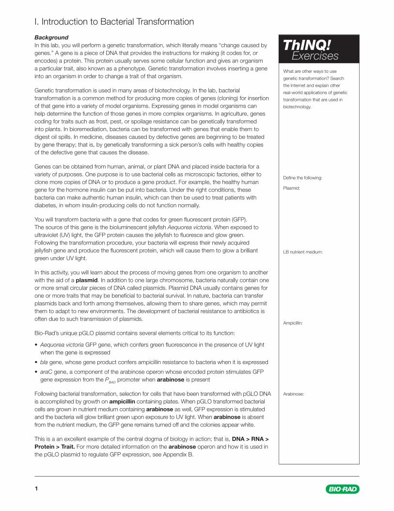
Mice that glow fl uorescent green. Plants that turn red when grown near a land mine. Goats that make milk that can be spun into parachute fabric. Virus-resistant papayas. Cheese puff snacks. Insulin. What do all these things have in common? They are all either genetically modifi ed organisms (GMOs), or produced by GMOs.
GMO stands for genetically modifi ed organism, an organism that has undergone genetic transformation (literally “change caused by genes”). Whether you call it genetic modifi cation, genetic transformation, or genetic engineering, it’s all one and the same — these terms all refer to the process of manipulating the genes of an organism to cause a change to traits of that organism.
Genetic engineering and GMOs are currently debated topics. Opponents of the technology claim, among other things, potential harm to the health of humans and the environment. They contend that the technology needs more testing and much more regulation before it is deemed safe. Meanwhile, supporters of the technology tout its many useful applications: increasing the food supply to accommodate a growing human population, producing medicines and other products more effi ciently, treating or eradicating disease, to name a few. They assert the technology has no demonstrated ill effects on humans, and its utility cannot be ignored.
This instruction manual, and the experiments outlined within it, are designed to help you better understand genetic engineering by creating a GMO of your own — a bacterium that, like the mouse mentioned above, will glow bright green under ultraviolet (UV) light.
The experiments take you through the process of bacterial genetic transformation via a standard protocol used in molecular and cell biology laboratories worldwide. You will begin with a plate of bacteria that appear off-white under normal or UV light conditions, and a tube of DNA containing a gene that codes for green fl uorescent protein (GFP). The source of this gene is the bioluminescent jellyfi sh Aequorea victoria. You will learn all the steps needed to move the DNA into the bacteria, to have the bacteria incorporate the DNA, and to induce the bacteria to produce the fl uorescent protein, which causes them to glow a brilliant bright green under UV light. You will then have the opportunity to expand and explore the process further by designing and performing your own experiments to explore transformation, antibiotic dosage, gene expression, or satellite colony formation. Finally, you’ll end the hands-on experience with a real-world science case study to understand how genetic engineering and bacterial transformation might one day play a major role in the fi ght against malaria.
Throughout these experiments, we hope you ask a lot of questions so that you can widen your understanding of genetic engineering — its promises, its limitations, and the types of careful considerations that should be made before creating or using GMOs for any application. We hope the knowledge you gain in the following investigations will help you develop your own opinions on the complex ethical and practical debates surrounding GMOs and genetic engineering.
Bio-Rad’s Explorer TeamBio-Rad Laboratories6000 James Watson Drive, Hercules, CA [email protected]

I. Introduction and Background ............................................................................................................................ 1Introduction to Bacterial Transformation ..........................................................................................................................................1
II. Investigation #1: pGLO Bacterial Transformation Laboratory (Structured Inquiry) ........................................... 4Workstation Checklist .....................................................................................................................................................................4Protocol ..........................................................................................................................................................................................5Data Collection and Analysis of Results ..........................................................................................................................................9The Interaction between Genes and Environment .........................................................................................................................11Calculation of Transformation Effi ciency ........................................................................................................................................12
III. Investigations #2–5: Guided and Open Inquiry .............................................................................................. 16Investigation #2: Transformation Effi ciency Lab (Guided Inquiry) ....................................................................................................17Investigation #3: Effect of Ampicillin on Bacterial Growth Lab (Open Inquiry) .................................................................................22Investigation #4: Effect of Arabinose on GFP Expression Lab (Open Inquiry) .................................................................................27Investigation #5: Satellite Colonies Lab (Open Inquiry) ...................................................................................................................33Post-Lab Assessment ..................................................................................................................................................................38
IV. Class Discussion ............................................................................................................................................ 40Science Case Study: Can Bacterial Transformation Stop the Spread of Malaria? ..........................................................................40
Appendix A: Serial Dilution and Dilution Instructions ......................................................................................... 48Appendix B: Gene Regulation ............................................................................................................................. 52
explorer.bio-rad.com
Contents
1
What are other ways to use
genetic transformation? Search
the Internet and explain other
real-world applications of genetic
transformation that are used in
biotechnology.
Defi ne the following:
Plasmid:
LB nutrient medium:
Ampicillin:
Arabinose:
ThINQ! Exercises
Background In this lab, you will perform a genetic transformation, which literally means “change caused by genes.” A gene is a piece of DNA that provides the instructions for making (it codes for, or encodes) a protein. This protein usually serves some cellular function and gives an organism a particular trait, also known as a phenotype. Genetic transformation involves inserting a gene into an organism in order to change a trait of that organism.
Genetic transformation is used in many areas of biotechnology. In the lab, bacterial transformation is a common method for producing more copies of genes (cloning) for insertion of that gene into a variety of model organisms. Expressing genes in model organisms can help determine the function of those genes in more complex organisms. In agriculture, genes coding for traits such as frost, pest, or spoilage resistance can be genetically transformed into plants. In bioremediation, bacteria can be transformed with genes that enable them to digest oil spills. In medicine, diseases caused by defective genes are beginning to be treated by gene therapy; that is, by genetically transforming a sick person’s cells with healthy copies of the defective gene that causes the disease.
Genes can be obtained from human, animal, or plant DNA and placed inside bacteria for a variety of purposes. One purpose is to use bacterial cells as microscopic factories, either to clone more copies of DNA or to produce a gene product. For example, the healthy human gene for the hormone insulin can be put into bacteria. Under the right conditions, these bacteria can make authentic human insulin, which can then be used to treat patients with diabetes, in whom insulin-producing cells do not function normally.
You will transform bacteria with a gene that codes for green fl uorescent protein (GFP). The source of this gene is the bioluminescent jellyfi sh Aequorea victoria. When exposed to ultraviolet (UV) light, the GFP protein causes the jellyfi sh to fl uoresce and glow green. Following the transformation procedure, your bacteria will express their newly acquired jellyfi sh gene and produce the fl uorescent protein, which will cause them to glow a brilliant green under UV light.
In this activity, you will learn about the process of moving genes from one organism to another with the aid of a plasmid. In addition to one large chromosome, bacteria naturally contain one or more small circular pieces of DNA called plasmids. Plasmid DNA usually contains genes for one or more traits that may be benefi cial to bacterial survival. In nature, bacteria can transfer plasmids back and forth among themselves, allowing them to share genes, which may permit them to adapt to new environments. The development of bacterial resistance to antibiotics is often due to such transmission of plasmids.
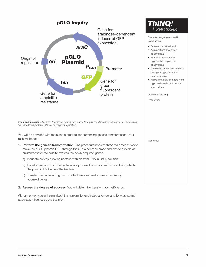
Bio-Rad’s unique pGLO plasmid contains several elements critical to its function:
• Aequorea victoria GFP gene, which confers green fl uorescence in the presence of UV light when the gene is expressed
• bla gene, whose gene product confers ampicillin resistance to bacteria when it is expressed
• araC gene, a component of the arabinose operon whose encoded protein stimulates GFP gene expression from the PBAD promoter when arabinose is present
Following bacterial transformation, selection for cells that have been transformed with pGLO DNA is accomplished by growth on ampicillin containing plates. When pGLO transformed bacterial cells are grown in nutrient medium containing arabinose as well, GFP expression is stimulated and the bacteria will glow brilliant green upon exposure to UV light. When arabinose is absent from the nutrient medium, the GFP gene remains turned off and the colonies appear white.
This is a an excellent example of the central dogma of biology in action; that is, DNA > RNA > Protein > Trait. For more detailed information on the arabinose operon and how it is used in the pGLO plasmid to regulate GFP expression, see Appendix B.
I. Introduction to Bacterial Transformation
2explorer.bio-rad.com
You will be provided with tools and a protocol for performing genetic transformation. Your task will be to:
1. Perform the genetic transformation. The procedure involves three main steps: two to move the pGLO plasmid DNA through the E. coli cell membrane and one to provide an environment for the cells to express the newly acquired genes.
a) Incubate actively growing bacteria with plasmid DNA in CaCl2 solution.
b) Rapidly heat and cool the bacteria in a process known as heat shock during which the plasmid DNA enters the bacteria.
c) Transfer the bacteria to growth media to recover and express their newly acquired genes.
2. Assess the degree of success. You will determine transformation effi ciency.
Along the way, you will learn about the reasons for each step and how and to what extent each step infl uences gene transfer.
The pGLO plasmid. GFP, green fl uorescent protein; araC, gene for arabinose-dependent inducer of GFP expression; bla, gene for ampicillin resistance; ori, origin of replication.
Steps for designing a scientifi c
investigation:
• Observe the natural world
• Ask questions about your
observations
• Formulate a reasonable
hypothesis to explain the
observations
• Create and execute experiments
testing the hypothesis and
generating data
• Analyze the data, compare to the
hypothesis, and communicate
your fi ndings
Defi ne the following:
Phenotype:
Genotype:
ThINQ! Exercises
3
Focus QuestionsScientifi c investigations begin with an observation about the natural world and the formulation of questions about that observation. Below are a few questions for you to ponder as you take on the challenge of performing a genetic transformation lab.
Question 1: Which organism should I choose, and why?1. To genetically transform an entire organism, the new gene must be in every cell in the organism. Considering this, which
organism would be the simplest to work with for total genetic transformation: one composed of many cells, or one composed of a single cell?
2. Scientists often want to know if the genetically transformed organism can pass its new traits on to its offspring and future generations. To get this information, which would be a better candidate for your investigation: an organism that develops each new generation quickly, or one that reproduces more slowly?
3. Safety is another important consideration in choosing an experimental organism. What traits or characteristics should the organism have (or not have) to be sure it will not harm you or the environment?
4. Based on the above considerations, which would be the best choice for a genetic transformation: bacterium, earthworm, fi sh, or mouse? Describe your reasoning.
Question 2: How can I tell if cells have been genetically transformed?The goal of genetic transformation is to change an organism’s traits, also known as its phenotype. Before a change in the phenotype can be detected, however, a thorough examination of its natural (pretransformation) phenotype must be made.
1. Describe how you could use two LB/agar plates, some E. coli, and some ampicillin to determine how E. coli cells are affected by ampicillin.
2. What would you expect your experimental results to indicate about the effect of ampicillin on the E. coli cells?
4explorer.bio-rad.com
In this activity, you will perform a bacterial transformation, transforming a stock E. coli culture with the pGLO plasmid.
� E. coli starter plate 1� Poured nutrient agar plates (1 LB, 2 LB/amp, 1 LB/amp/ara) 4 � Transformation solution (1 ml) 1� LB nutrient broth (1 ml) 1� Inoculation loops (1 pk of 10) 7� Disposable plastic transfer pipets (DPTPs) 4� Foam microcentrifuge tube holder/fl oat 1� Container full of crushed ice (foam/paper cups) 1� Microcentrifuge tubes 2� Marking pen 1
Student workstations Quantity
� Rehydrated pGLO plasmid, vial 1 � 42°C water bath and thermometer 1 � UV pen light 1 � 37°C incubator 1 � Clock or timer for counting seconds 1
Common workstation Quantity
� 2–20 µl adjustable volume micropipet 1 � 2–20 µl micropipet tips, box 1
Optional Quantity
II. Investigation #1: pGLO Bacterial Transformation Laboratory (Structured Inquiry)

5
Protocol1. Label one microcentrifuge tube +pGLO and another –pGLO. Label both tubes with your group’s name. Place them in the foam tube rack.
2. Open the tubes and use a sterile DPTP to transfer 250 ul of transformation solution (50 mM CaCl2) into each tube.
3. Place the tubes on ice.
4. Use a sterile loop to pick 2–4 large colonies of bacteria from the starter plate. Select colonies that are “fat” (1–2 mm in diameter). It is important to take individual colonies
(not a swab of bacteria from the dense portion of the plate), since the bacteria must be actively growing to achieve high transformation effi ciency. Pick up the +pGLO tube and
immerse the loop into the transformation solution in the tube. Spin the loop between your index fi nger and thumb until the colonies are dispersed in the transformation solution (there are no fl oating chunks). Place the tube back in the tube rack in the ice. Using a new sterile loop, repeat for the –pGLO tube.
Collaborate and use outside
resources to answer the
following questions:
Examine the bottle of pGLO plasmid
DNA solution with the UV lamp. What
do you see? Note your observations:
Look at the individual colonies of
E.coli on your starter plates. On
a separate piece of paper list all
observable traits or characteristics
that can be described. For example:
• Color of colonies
• Size of: 1) the largest colony;
2) the smallest colony;
3) the majority of colonies
• Shape of colonies
(both 2-D and 3-D)
• Appearance of the colonies
under regular and UV light
Why do you add the CaCl2
transformation solution?
Why do you place the tubes on ice?
ThINQ! Exercises

6explorer.bio-rad.com
5. Immerse a new sterile loop into the tube of 0.8 x 10⁻¹ µg/µl solution of pGLO plasmid DNA stock tube. Withdraw a loopful (10 µl). You should see a fi lm of plasmid solution across the ring, similar to the soapy fi lm across a ring for blowing soap bubbles. Mix the loop into the cell suspension of the +pGLO tube. Do not add plasmid DNA to the –pGLO tube. Close both the +pGLO and –pGLO tubes and return them to the rack on ice.
6. Incubate the tubes on ice for 10 min. Push the tubes all the way down in the rack so the bottoms of the tubes make contact with the ice.
7. While the tubes are on ice, label the four LB nutrient agar plates on the bottom (not the lid):
• Label one LB/amp plate: +pGLO • Label the LB/amp/ara plate: +pGLO • Label the LB plate: –pGLO • Label the other LB/amp plate: –pGLO
Collaborate and use outside
resources to answer the
following:
Approximately how much volume is
picked up by the loop if the solution
is 0.8 x 10⁻¹ µg/µl and a loopful of
solution contains 0.8 µg of pGLO
plasmid?
Why do you incubate the tubes on
ice for 10 minutes?
Why do you use only four
LB nutrient agar plates, as opposed
to 6 plates (2 LB, 2 LB/amp, and
2 LB/amp/ara)?
What would you expect to grow on
the following LB nutrient agar plates?
+pGLO on LB:
–pGLO on LB/amp/ara:
Why do you heat shock the cells?
Why do you incubate on ice for
2 minutes?
ThINQ! Exercises
7
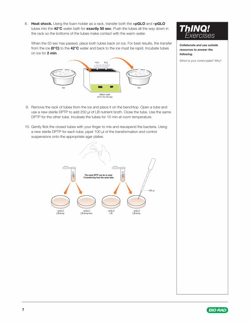
8. Heat shock. Using the foam holder as a rack, transfer both the +pGLO and –pGLO tubes into the 42°C water bath for exactly 50 sec. Push the tubes all the way down in the rack so the bottoms of the tubes make contact with the warm water.
When the 50 sec has passed, place both tubes back on ice. For best results, the transfer from the ice (0°C) to the 42°C water and back to the ice must be rapid. Incubate tubes on ice for 2 min.
9. Remove the rack of tubes from the ice and place it on the benchtop. Open a tube and use a new sterile DPTP to add 250 µl of LB nutrient broth. Close the tube. Use the same DPTP for the other tube. Incubate the tubes for 10 min at room temperature.
10. Gently fl ick the closed tubes with your fi nger to mix and resuspend the bacteria. Using a new sterile DPTP for each tube, pipet 100 µl of the transformation and control suspensions onto the appropriate agar plates.
Collaborate and use outside
resources to answer the
following.
Which is your control plate? Why?
ThINQ! Exercises
8explorer.bio-rad.com
Collaborate and use outside
resources to answer the
following questions:
Why do you need to place the stack
of plates upside down?
Alternatively, you could incubate
your plates at room temperature.
What difference would you expect
if plates were incubated at 37°C vs.
room temperature (22°C)?
Review Questions:
On which of the plates would you
expect to fi nd bacteria most like
the original untransformed E. coli
colonies you initially observed?
Explain your prediction.
If there are any transformed bacterial
cells, on which plate(s) would they
most likely be located? Explain your
prediction.
Which plates should be compared
to determine if any genetic
transformation has occurred? Why?
ThINQ! Exercises
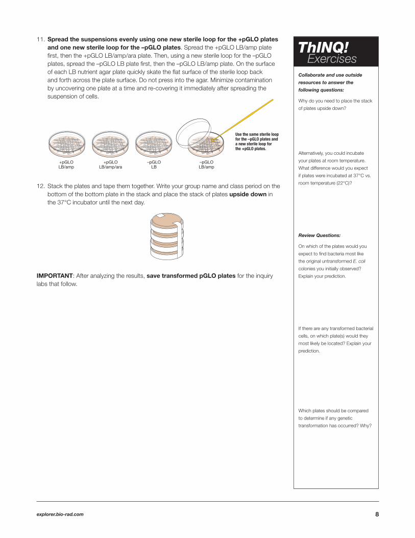
11. Spread the suspensions evenly using one new sterile loop for the +pGLO plates and one new sterile loop for the –pGLO plates. Spread the +pGLO LB/amp plate fi rst, then the +pGLO LB/amp/ara plate. Then, using a new sterile loop for the –pGLO plates, spread the –pGLO LB plate fi rst, then the –pGLO LB/amp plate. On the surface of each LB nutrient agar plate quickly skate the fl at surface of the sterile loop back and forth across the plate surface. Do not press into the agar. Minimize contamination by uncovering one plate at a time and re-covering it immediately after spreading the suspension of cells.
12. Stack the plates and tape them together. Write your group name and class period on the
bottom of the bottom plate in the stack and place the stack of plates upside down in the 37°C incubator until the next day.
IMPORTANT: After analyzing the results, save transformed pGLO plates for the inquiry labs that follow.

9
Observations
+pGLO LB/amp
+pGLO LB/amp/ara
Observations
–pGLO LB/amp
–pGLO LB
Con
trol
pla
tes
Tran
sfor
mat
ion
pla
tes
Data CollectionObserve the results you obtained from the transformation lab under normal room lighting. Then turn out the lights and hold the ultraviolet (UV) light over the plates.
Carefully observe and draw what you see on each of the four plates. Put your drawings in the data table below. Record your data to allow you to compare your observations of the +pGLO cells with your observations of the non-transformed E. coli. Write down the following observations for each plate.
1. How much bacterial growth do you see on each plate, relatively speaking?
2. What color are the bacteria under normal light and UV light conditions?
3. How many bacterial colonies are on each plate (count the colonies you see).

10explorer.bio-rad.com
Analysis of Results The goal of this analysis is to determine whether genetic transformation has occurred.
1. How did the traits you originally observed for E. coli alter?
2. If the transformed cells have acquired the ability to grow in the presence of ampicillin, then what might be inferred about their ability to glow bright green under UV light?
3. From the results that you obtained, how could you provide evidence to support your hypothesis/argument that the changes that occurred were due to the procedure that you performed?

11
The Interaction between Genes and EnvironmentLook again at the four plates. Do you observe some E. coli growing on the LB plate that does not contain ampicillin or arabinose?
1. From your results, can you tell if these bacteria are ampicillin resistant by looking at them on the LB plate? Explain your answer.
2. What might happen to these bacteria if you moved them to plates containing ampicillin?
3. Often an organism’s traits are caused by a combination of its genes and its environment. Think about the green color you saw in the genetically transformed bacteria as you consider these questions:
a) What two factors must be present in the bacteria’s environment for you to see the green color?
b) Provide another example of a change in the environment causing expression of a different trait.
c) What advantage would there be for an organism to be able to turn on or off particular genes in response to certain conditions?

12explorer.bio-rad.com
Calculation of Transformation Effi ciencyThe next task in this investigation will be to determine how many of the E. coli cells were transformed. This quantitative measurement is referred to as transformation effi ciency.
In many experiments, it is important to transform as many cells as possible. For example, in some types of gene therapy, cells are collected from a patient, transformed in the laboratory, and then put back into the patient. The more cells that are transformed to produce the needed protein, the more likely the therapy will work. Transformation effi ciency helps scientists determine how well transformation is working.
The TaskYou are about to calculate the transformation effi ciency for this experiment, which indicates how effective you were in getting new DNA molecules into bacterial cells.
Transformation effi ciency is a number: the total number of colonies growing on the plate divided by the amount of DNA spread on the plate. It represents the total number of bacterial cells transformed using one microgram of DNA. Each colony on the plate can be assumed to derive from a single cell. As individual cells reproduce, more and more cells accumulate, developing into a colony. The most direct way to determine the total number of bacteria that were transformed with the pGLO plasmid is to count the number of colonies on the plate. Transformation effi ciency is calculated using the following formula:
Therefore, before you can calculate the effi ciency of your transformation, you will need two pieces of information:
1. The total number of green fl uorescent colonies growing on your LB/amp/ara plate.2. The total amount of pGLO plasmid DNA used for bacterial transformation that was spread on the LB/amp/ara plate. 1. Determining the Total Number of Transformed Green Fluorescent Colonies Place the LB/amp/ara plate near a UV light source. Count the number of green fl uorescent colonies that glow under UV light on the plate.
2. Determining the Amount of pGLO DNA in the Cells Spread on the LB/Amp/Ara Plate You need two pieces of information to determine the amount of pGLO DNA in the bacterial cells spread on the LB/amp/ara
plate in this experiment: (a) the total amount of DNA we began the experiment with, and (b) the fraction of the DNA (in the bacteria) that was spread onto the LB/amp/ara plates.
Once you calculate these data, you multiply the total amount of pGLO DNA used in this experiment by the fraction of DNA you spread on the LB/amp/ara plate. This will tell you the amount of pGLO DNA in the bacterial cells that were spread on the LB/amp/ara plate.
A. Determining the total amount of pGLO plasmid DNA The total amount of DNA we began with is equal to the product of the concentration and the total volume used, or
In this experiment you used 10 µl of pGLO at a concentration of 0.08 µg/µl. This means that each microliter of solution contained 0.08 µg of pGLO DNA. Calculate the total amount of DNA used in this experiment
Transformation efficiency = Total number of colonies growing on the agar plate
Amount of DNA spread on the agar plate (in µg)
Enter that number here Total number of colonies =
(DNA in µg) = (concentration of DNA in µg/µl) x (volume of DNA in µl)
Enter that number here Total amount of pGLO DNA, µg
used in this experiment =
13
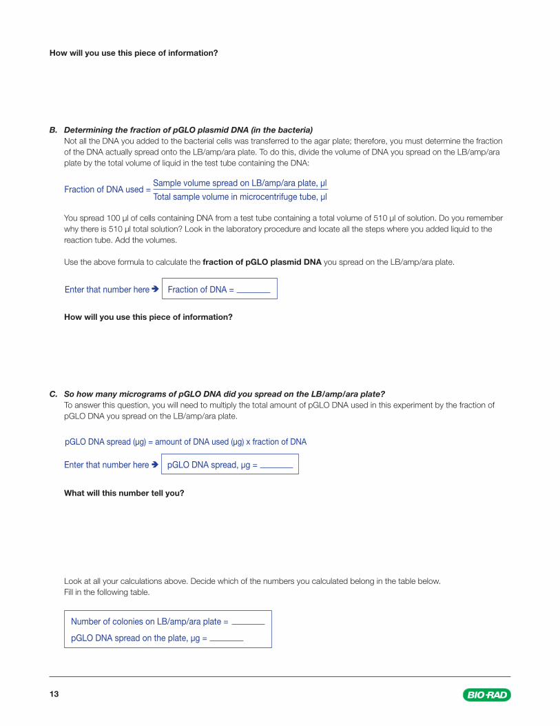
How will you use this piece of information?
B. Determining the fraction of pGLO plasmid DNA (in the bacteria) Not all the DNA you added to the bacterial cells was transferred to the agar plate; therefore, you must determine the fraction
of the DNA actually spread onto the LB/amp/ara plate. To do this, divide the volume of DNA you spread on the LB/amp/ara plate by the total volume of liquid in the test tube containing the DNA:
You spread 100 µl of cells containing DNA from a test tube containing a total volume of 510 µl of solution. Do you remember why there is 510 µl total solution? Look in the laboratory procedure and locate all the steps where you added liquid to the reaction tube. Add the volumes.
Use the above formula to calculate the fraction of pGLO plasmid DNA you spread on the LB/amp/ara plate.
How will you use this piece of information?
C. So how many micrograms of pGLO DNA did you spread on the LB/amp/ara plate? To answer this question, you will need to multiply the total amount of pGLO DNA used in this experiment by the fraction of
pGLO DNA you spread on the LB/amp/ara plate.
What will this number tell you?
Look at all your calculations above. Decide which of the numbers you calculated belong in the table below. Fill in the following table.
Fraction of DNA used = Sample volume spread on LB/amp/ara plate, µl
Total sample volume in microcentrifuge tube, µl
Enter that number here Fraction of DNA =
Enter that number here pGLO DNA spread, µg =
Number of colonies on LB/amp/ara plate =
pGLO DNA spread on the plate, µg =
pGLO DNA spread (µg) = amount of DNA used (µg) x fraction of DNA
14explorer.bio-rad.com
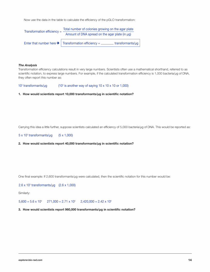
Now use the data in the table to calculate the effi ciency of the pGLO transformation:
The AnalysisTransformation effi ciency calculations result in very large numbers. Scientists often use a mathematical shorthand, referred to as scientifi c notation, to express large numbers. For example, if the calculated transformation effi ciency is 1,000 bacteria/µg of DNA, they often report this number as:
1. How would scientists report 10,000 transformants/µg in scientifi c notation?
Carrying this idea a little further, suppose scientists calculated an effi ciency of 5,000 bacteria/µg of DNA. This would be reported as:
2. How would scientists report 40,000 transformants/µg in scientifi c notation?
One fi nal example: If 2,600 transformants/µg were calculated, then the scientifi c notation for this number would be:
Similarly:
3. How would scientists report 960,000 transformants/µg in scientifi c notation?
Enter that number here Transformation efficiency = transformants/µg
103 transformants/µg (103 is another way of saying 10 x 10 x 10 or 1,000)
2.6 x 10³ transformants/µg (2.6 x 1,000)
5,600 = 5.6 x 10³ 271,000 = 2.71 x 10⁵ 2,420,000 = 2.42 x 10⁶
5 x 10³ transformants/µg (5 x 1,000)
Transformation efficiency = Total number of colonies growing on the agar plate
Amount of DNA spread on the agar plate (in µg)

15
4. Report your calculated transformation effi ciency in scientifi c notation.
5. Use a sentence or two to explain what your calculation of transformation effi ciency means.
Biotechnologists generally agree that the transformation protocol you just completed usually has a transformation effi ciency of between 8.0 x 10² and 7.0 x 10³ transformants per microgram of DNA.
6. How does your transformation effi ciency compare with the above?
7. In the table below, report the transformation effi ciency of several of the teams in the class.
IMPORTANT: Save the transformed pGLO plates for the inquiry labs that follow.
Team Effi ciency
How does your transformation effi ciency compare to theirs?
16explorer.bio-rad.com
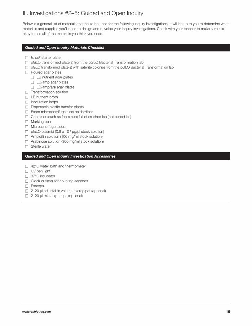
Below is a general list of materials that could be used for the following inquiry investigations. It will be up to you to determine what materials and supplies you’ll need to design and develop your inquiry investigations. Check with your teacher to make sure it is okay to use all of the materials you think you need.
III. Investigations #2–5: Guided and Open Inquiry
Guided and Open Inquiry Materials Checklist
� E. coli starter plate � pGLO transformed plate(s) from the pGLO Bacterial Transformation lab� pGLO transformed plate(s) with satellite colonies from the pGLO Bacterial Transformation lab� Poured agar plates � LB nutrient agar plates � LB/amp agar plates � LB/amp/ara agar plates � Transformation solution � LB nutrient broth � Inoculation loops � Disposable plastic transfer pipets � Foam microcentrifuge tube holder/fl oat � Container (such as foam cup) full of crushed ice (not cubed ice) � Marking pen � Microcentrifuge tubes � pGLO plasmid (0.8 x 10⁻¹ µg/µl stock solution) � Ampicillin solution (100 mg/ml stock solution) � Arabinose solution (300 mg/ml stock solution)� Sterile water
Guided and Open Inquiry Investigation Accessories
� 42°C water bath and thermometer � UV pen light � 37°C incubator � Clock or timer for counting seconds � Forceps� 2–20 µl adjustable volume micropipet (optional)� 2–20 µl micropipet tips (optional)
17
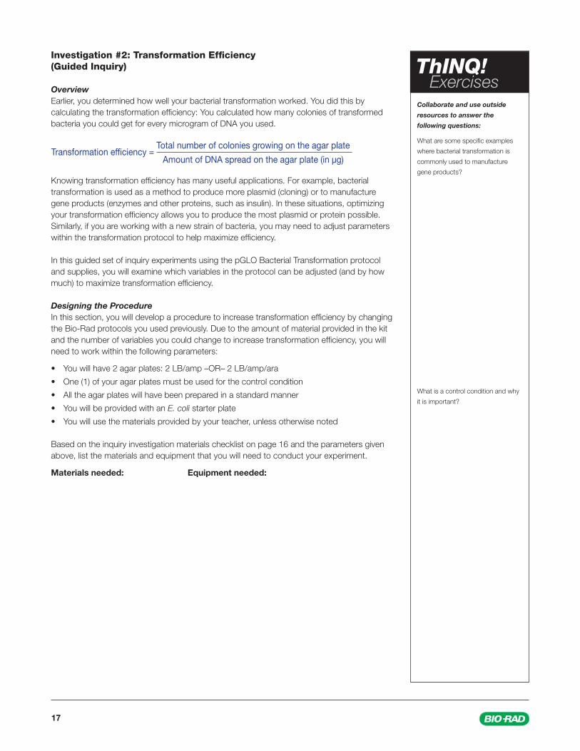
Investigation #2: Transformation Effi ciency (Guided Inquiry)
Overview Earlier, you determined how well your bacterial transformation worked. You did this by calculating the transformation effi ciency: You calculated how many colonies of transformed bacteria you could get for every microgram of DNA you used.
Knowing transformation effi ciency has many useful applications. For example, bacterial transformation is used as a method to produce more plasmid (cloning) or to manufacture gene products (enzymes and other proteins, such as insulin). In these situations, optimizing your transformation effi ciency allows you to produce the most plasmid or protein possible. Similarly, if you are working with a new strain of bacteria, you may need to adjust parameters within the transformation protocol to help maximize effi ciency.
In this guided set of inquiry experiments using the pGLO Bacterial Transformation protocol and supplies, you will examine which variables in the protocol can be adjusted (and by how much) to maximize transformation effi ciency.
Designing the Procedure In this section, you will develop a procedure to increase transformation effi ciency by changing the Bio-Rad protocols you used previously. Due to the amount of material provided in the kit and the number of variables you could change to increase transformation effi ciency, you will need to work within the following parameters:
• You will have 2 agar plates: 2 LB/amp –OR– 2 LB/amp/ara
• One (1) of your agar plates must be used for the control condition
• All the agar plates will have been prepared in a standard manner
• You will be provided with an E. coli starter plate
• You will use the materials provided by your teacher, unless otherwise noted
Based on the inquiry investigation materials checklist on page 16 and the parameters given above, list the materials and equipment that you will need to conduct your experiment.
Materials needed: Equipment needed:
Transformation efficiency = Total number of colonies growing on the agar plate
Amount of DNA spread on the agar plate (in µg)
Collaborate and use outside
resources to answer the
following questions:
What are some specifi c examples
where bacterial transformation is
commonly used to manufacture
gene products?
What is a control condition and why
it is important?
ThINQ! Exercises
18explorer.bio-rad.com
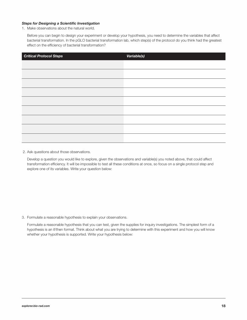
Steps for Designing a Scientifi c Investigation 1. Make observations about the natural world.
Before you can begin to design your experiment or develop your hypothesis, you need to determine the variables that affect bacterial transformation. In the pGLO bacterial transformation lab, which step(s) of the protocol do you think had the greatest effect on the effi ciency of bacterial transformation?
2. Ask questions about those observations.
Develop a question you would like to explore, given the observations and variable(s) you noted above, that could affect transformation effi ciency. It will be impossible to test all these conditions at once, so focus on a single protocol step and explore one of its variables. Write your question below:
3. Formulate a reasonable hypothesis to explain your observations.
Formulate a reasonable hypothesis that you can test, given the supplies for inquiry investigations. The simplest form of a hypothesis is an if/then format. Think about what you are trying to determine with this experiment and how you will know whether your hypothesis is supported. Write your hypothesis below:
Critical Protocol Steps Variable(s)

19
4. Create and execute experiments to test the hypothesis and generate data.
Design your experiment. What variables will you manipulate and what variables should you keep constant to test your hypothesis? (Hint: Don’t forget to describe what controls you have designed for the experiment.)
Consider your hypothesis and your experiment and predict what you think you will observe.
5. Analyze your data, compare to the hypothesis, and communicate your fi ndings. To be able to illustrate results graphically and make comparisons, you will need to interpret your results quantitatively
(numerically) as well as qualitatively (with words). Please explain how you will quantify your results (that is, measure them).
20explorer.bio-rad.com
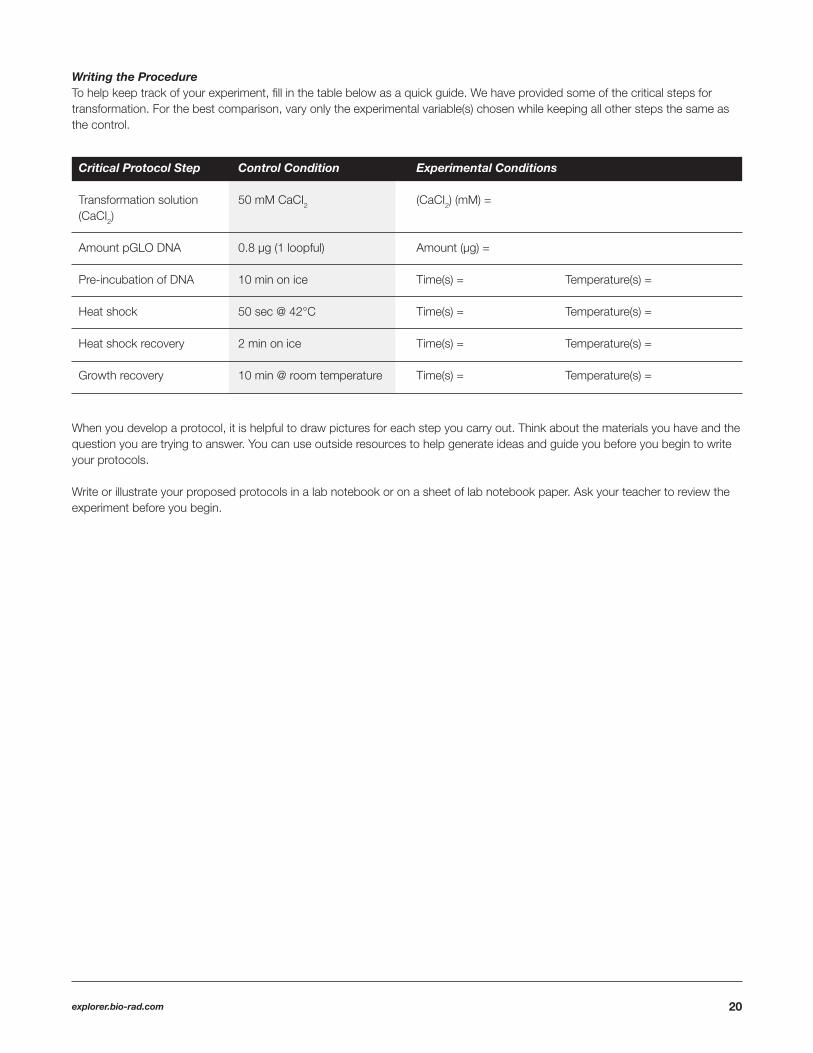
Writing the Procedure To help keep track of your experiment, fi ll in the table below as a quick guide. We have provided some of the critical steps for transformation. For the best comparison, vary only the experimental variable(s) chosen while keeping all other steps the same as the control.
When you develop a protocol, it is helpful to draw pictures for each step you carry out. Think about the materials you have and the question you are trying to answer. You can use outside resources to help generate ideas and guide you before you begin to write your protocols.
Write or illustrate your proposed protocols in a lab notebook or on a sheet of lab notebook paper. Ask your teacher to review the experiment before you begin.
Critical Protocol Step Control Condition Experimental Conditions
Transformation solution 50 mM CaCI2 (CaCI2) (mM) = (CaCI2)
Amount pGLO DNA 0.8 µg (1 loopful) Amount (µg) =
Pre-incubation of DNA 10 min on ice Time(s) = Temperature(s) =
Heat shock 50 sec @ 42°C Time(s) = Temperature(s) =
Heat shock recovery 2 min on ice Time(s) = Temperature(s) =
Growth recovery 10 min @ room temperature Time(s) = Temperature(s) =

21
Results Use this page to restate your hypothesis and summarize your results.
Hypothesis
ResultsWhat did you do to test the hypothesis?
What were the results? Calculate and compare the transformation effi ciency for each of the conditions you tested.
Do the data support your hypothesis?
List any observations relevant to your experiment.
List any ideas you have for refi ning your hypothesis and testing your experiment.
22explorer.bio-rad.com
Investigation #3: Effect of Ampicillin on Bacterial Growth (Open Inquiry)
Overview Imagine you are planning to work with a new strain of bacteria. How would you know which antibiotic to use, and how much of it, for selection in your transformation experiments? In this set of experiments, you will use the supplies in the pGLO Transformation and Inquiry Kit and some extra information to design an experiment that tests the effects of ampicillin on bacterial growth.
Ampicillin is an antibiotic used to treat a number of bacterial infections, such as bronchitis, sinus and ear infections. Ampicillin reduces the growth of bacteria by acting as an irreversible inhibitor of the enzyme transpeptidase, which is needed by bacteria to make cell walls. Though ampicillin is often a fi rst line of treatment for these common infections, some strains of bacteria have developed resistance to it. Antibiotics like ampicillin are also used routinely in bacterial transformation experiments to help select for transformants.
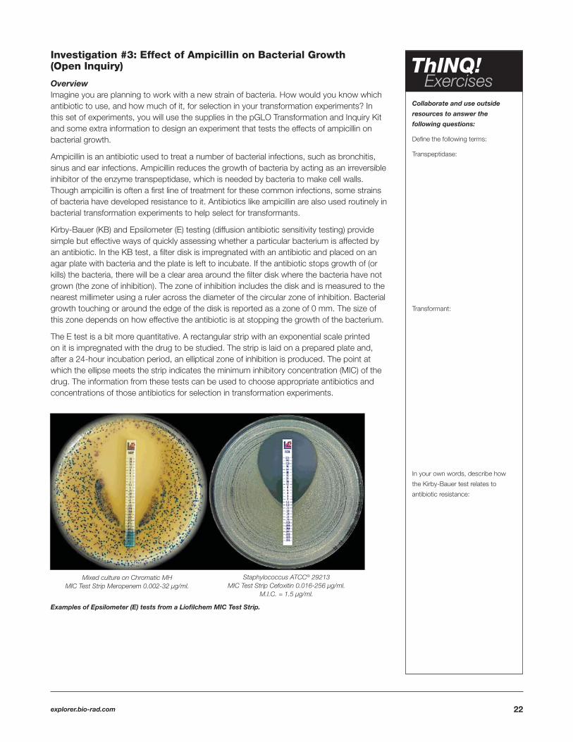
Kirby-Bauer (KB) and Epsilometer (E) testing (diffusion antibiotic sensitivity testing) provide simple but effective ways of quickly assessing whether a particular bacterium is affected by an antibiotic. In the KB test, a fi lter disk is impregnated with an antibiotic and placed on an agar plate with bacteria and the plate is left to incubate. If the antibiotic stops growth of (or kills) the bacteria, there will be a clear area around the fi lter disk where the bacteria have not grown (the zone of inhibition). The zone of inhibition includes the disk and is measured to the nearest millimeter using a ruler across the diameter of the circular zone of inhibition. Bacterial growth touching or around the edge of the disk is reported as a zone of 0 mm. The size of this zone depends on how effective the antibiotic is at stopping the growth of the bacterium.
The E test is a bit more quantitative. A rectangular strip with an exponential scale printed on it is impregnated with the drug to be studied. The strip is laid on a prepared plate and, after a 24-hour incubation period, an elliptical zone of inhibition is produced. The point at which the ellipse meets the strip indicates the minimum inhibitory concentration (MIC) of the drug. The information from these tests can be used to choose appropriate antibiotics and concentrations of those antibiotics for selection in transformation experiments.
Examples of Epsilometer (E) tests from a Liofi lchem MIC Test Strip.
Collaborate and use outside
resources to answer the
following questions:
Defi ne the following terms:
Transpeptidase:
Transformant:
In your own words, describe how
the Kirby-Bauer test relates to
antibiotic resistance:
ThINQ! Exercises
Mixed culture on Chromatic MHMIC Test Strip Meropenem 0.002-32 µg/ml.
Staphylococcus ATCC® 29213MIC Test Strip Cefoxitin 0.016-256 µg/ml.
M.I.C. = 1.5 µg/ml.

23
Designing the Procedure In this section, you will formulate a hypothesis and develop a procedure to test the effect of ampicillin on bacterial growth. Due to the amount of material provided in the kit, you will need to work within the following parameters:
• You will have 1 LB agar plate
• You will be provided with an E. coli starter plate
• You will need to use the materials provided by your teacher, unless otherwise noted
Based on the inquiry investigation material checklist on page 16 and the parameters given above, list the materials and equipment that you will need to conduct your experiment.
Materials needed: Equipment needed:

24explorer.bio-rad.com
Steps for Designing a Scientifi c Investigation
1. Make observations about the natural world.
Think about what you have read about antibiotic resistance, the Kirby-Bauer test, and the E test. Make some observations about the natural world and the pGLO experiment that may relate to antibiotic dosage and resistance.
2. Ask questions about those observations.
Develop a question you would like to explore, given the observations you have made and components of the pGLO bacterial transformation kit. Write your question below:
3. Formulate a reasonable hypothesis to explain your observations.
Formulate a reasonable hypothesis that you could test, given the supplies for inquiry investigations. The simplest form of a hypothesis is an if/then format. Think about what you are trying to determine with this experiment and how you will know whether your hypothesis is supported. Write your hypothesis below:
4. Create and execute experiments to test the hypothesis and generate data.
Design your experiment. What variables will you manipulate and what variables should you keep constant to test your hypothesis?
5. Analyze your data, compare to the hypothesis, and communicate your fi ndings.
Consider your hypothesis and your experiment and predict what you think you will observe.
To illustrate results graphically and make comparisons, you will need to interpret your results quantitatively (numerically) as well as qualitatively (with words). Please explain how you will quantify your results (that is, measure them).

25
Writing the Procedure When you develop a protocol, it is helpful to draw pictures for each step you carry out. Think about the materials you have and the question you are trying to answer. You can use outside resources to help generate ideas and guide you before you begin to write your protocols.
Write or illustrate your proposed protocols in a lab notebook or on a sheet of lab notebook paper. Ask your teacher to review the experiment before you begin.

26explorer.bio-rad.com
Results Use this page to restate your hypothesis and summarize your results.
Hypothesis
ResultsWhat did you do to test the hypothesis?
What were the results?
Do the data support your hypothesis?
List any observations relevant to your experiment.
List any ideas you have for further refi ning your hypothesis and testing your experiment.
27
Investigation #4: Effect of Arabinose on GFP Expression (Open Inquiry)
Overview
Why Is Gene Expression Regulated? Gene expression is carefully regulated in part to allow organisms to adapt to different environmental conditions. Genes can be turned on when particular proteins are needed and off when they are no longer necessary, preventing wasted effort and overproduction of proteins.
For bacteria, the genes involved in the breakdown of different food sources are often highly regulated. The simple plant sugar arabinose, for example, is a source of both energy and carbon for bacteria. The bacterial genes that encode digestive enzymes that break down arabinose for food are not expressed when arabinose is not in the environment. When arabinose is present, however, these genes are turned on, and when the arabinose runs out, the genes are turned off again. Arabinose initiates transcription of these genes by promoting the binding of RNA polymerase.
In the genetically engineered pGLO plasmid, some of the genes involved in arabinose breakdown have been replaced by the jellyfi sh gene that encodes GFP. Therefore, the expression of GFP is induced by arabinose. When bacteria transformed with the pGLO plasmid are grown in the presence of arabinose, expression of the GFP gene is turned on, and the bacteria glow brilliant green when exposed to UV light.
Why Regulate Expression of a Gene We Transform into Bacteria? In the wild, regulation of gene expression is clearly benefi cial. But why would it be necessary in the lab, when we are trying to get our bacteria to glow green?
One reason is that certain proteins, when overexpressed, may be toxic to the bacteria. If production of these proteins is not regulated, the toxic proteins would quickly accumulate and kill the bacteria that are producing them.
Another reason is that bacteria expend signifi cant amounts of energy and environmental resources to grow and divide. Consumption of these energy and resource pools by constant production of a large amount of a foreign protein may interfere with the bacteria’s ability to grow and divide. In this situation, bacteria would overexpress the foreign protein at the expense of their ability to make other proteins needed for growth and population expansion, resulting in a smaller bacterial population.
Collaborate and use outside
resources to answer the
following questions:
Defi ne the following terms:
Transcription:
RNA polymerase:
What are clinical trials?
What is the FDA?
What is the FDA’s role in clinical
trials?
Why are FDA-approved clinical trials
important?
ThINQ! Exercises

28explorer.bio-rad.com
Why Does It Matter if Bacterial Growth or Survival Is Negatively Affected by Expression of a Foreign Protein? Imagine a scenario in which you have transformed bacteria with plasmid DNA that encodes a new and potentially powerful drug (for example, insulin). You will need to make large amounts of this peptide for FDA-regulated clinical trials as a potential new treatment.
If it is diffi cult to isolate large amounts of this peptide and costly to synthesize large amounts of it chemically, you may wish to express large quantities of it in bacteria. However, if constant expression of this peptide either is toxic to the bacteria or prevents the bacteria from growing and dividing, you would not be able to grow enough bacteria to make enough of the peptide to carry out your FDA-regulated clinical trials.
But what if peptide production could be regulated? You could grow very large numbers of bacteria, and then activate gene expression for the plasmid-encoded peptide, which would allow you to obtain massive amounts of the peptide without worrying about its negative effects on bacteria growth or survival.
Examples of GFP regulation with varied arabinose dosage.

29
Designing the Procedure In this section, you will develop a procedure to test your question and hypothesis about GFP gene regulation and arabinose dosage. Due to the amount of material provided in the kit, you will need to work within the following parameters:
• You will have 1 LB/amp agar plate
• You will be provided with a plate containing pGLO transformed E. coli
• You will need to use the materials provided by your teacher, unless otherwise noted
Based on the inquiry investigation material checklist on page 16 and the parameters given above, list the materials and equipment that you will need to conduct your experiment.
Materials needed: Equipment needed:

30explorer.bio-rad.com
Steps for Designing a Scientifi c Investigation 1. Make observations about the natural world.
Think about what you have read about gene expression regulation and similar experiments that test antibiotic concentration dosage. Make observations about the natural world and the pGLO experiment that may relate to gene expression regulation.
2. Ask questions about those observations.
Develop a question you would like to explore given the observations you have made and materials available in the pGLO Transformation and Inquiry Kit. Write your question below:
3. Formulate a reasonable hypothesis to explain your observations.
Formulate a reasonable hypothesis that you could test given the supplies for inquiry investigations. The simplest form of a hypothesis is an if/then format. Think about what you are trying to determine with this experiment and how you will know whether your hypothesis is supported. Write your hypothesis below:
4. Create and execute experiments testing the hypothesis and generating data.
Design your experiment. What variables will you manipulate and what variables should you keep constant to test your hypothesis?
5. Analyze your data, compare to the hypothesis, and communicate your fi ndings.
Consider your hypothesis and your experiment and predict what you think you will observe.
In order to illustrate results graphically and make comparisons, you will need to interpret your results quantitatively (numerically) as well as qualitatively (with words). Please explain how you will quantify your results (that is, measure them).

31
Writing the Procedure When you develop a protocol, it is helpful to draw pictures for each step you carry out. Think about the materials you have and the question you are trying to answer. You can use outside resources to help generate ideas and guide you before you begin to write your protocols.
Write or illustrate your proposed protocols in a lab notebook or on a sheet of lab notebook paper. Ask your teacher to review the experiment before you begin.

32explorer.bio-rad.com
Results Use this page to restate your hypothesis and summarize your results.
Hypothesis
ResultsWhat did you do to test the hypothesis?
What were the results?
Do the data support your hypothesis?
List any observations relevant to your experiment.
List any ideas you have for further refi ning your hypothesis and testing your experiment.

33
Investigation #5: Satellite Colonies (Open Inquiry)
Recall that the pGLO plasmid DNA contains bla, a gene that confers resistance to the antibiotic ampicillin. Ampicillin was included in the growth medium following transformation so that only transformed bacteria would grow. This inclusion of antibiotic applies selective pressure to the bacterial population; it makes the environment on the plate inhospitable to any bacteria that cannot express genes that encode antibiotic resistance. Without this selection strategy, all bacteria would grow on the plate, and it would be diffi cult to determine which bacteria were transformed.
Over time, the amount of antibiotic present in the growth medium may become depleted for a number of reasons. When this happens, the selective pressure on the bacterial population is removed, and bacteria that are not antibiotic resistant may grow on the plate.
You may have noticed that your LB/amp/ara plates contain different types of colonies: the primary (larger) colonies of transformed bacteria and smaller “satellite” colonies surrounding them. This occurs especially if the plates are kept at room temperature for several days. Why might that be?
Just as a scientist in a lab producing a powerful drug like insulin would, you need to investigate what the properties of these satellite colonies are. Develop a question and design an experiment to understand what these foreign satellite colonies are and why they grew on your transformed pGLO colony plates.
Plate containing pGLO transformants and satellite colonies. In normal light (left), satellite colonies appear as noticeably smaller colonies surrounding larger colonies. Under UV light (right), the larger colonies glow bright green, whereas the satellite colonies do not.
Collaborate and use outside
resources to answer the
following questions:
Can you see different types of
colonies on your LB/amp/ara plates?
Compare and contrast the physical
characteristics of these different
types of colonies.
What is selective pressure?
What are some reasons the
antibiotic in the growth medium
might become depleted over time?
How does the pGLO plasmid give
rise to ampicillin resistance? Refer
back to the plasmid map and the
introductory information in the pGLO
Bacterial Transformation exercise.
Why would you expect to see
satellite colonies if your growth
plate contains a large number of
transformed colonies on a plate
containing ampicillin?
ThINQ! Exercises

34explorer.bio-rad.com
Designing the Procedure In this section, you will develop your own hypothesis about satellite colony formation. You can ask questions about whether satellite colonies contain a plasmid, or if they are resistant to an antibiotic. Try to think outside the box, but keep in mind you will need to work within the following parameters:
• You will have 2 agar plates: 1 LB/amp/ara and 1 LB
• You will be using an already prepared plate that contains satellite colonies
• You will need to use the materials provided by your teacher, unless otherwise noted
Based on the inquiry investigation material checklist on page 16 and the parameters given above, list the materials and equipment that you will need to conduct your experiment.
Materials needed: Equipment needed:
35
Steps for Designing a Scientifi c Investigation 1. Make observations about the natural world.
Think about what you have read and researched about satellite colonies and what you might need to explore about these unexpected colonies. Make observations about the natural world and the pGLO experiment that may relate to satellite colonies.
2. Ask questions about those observations.
Develop a question you would like to explore given the observations you have made and materials available in the pGLO Transformation and Inquiry Kit. Write your question below:
3. Formulate a reasonable hypothesis to explain your observations.
Formulate a reasonable hypothesis that you could test given the supplies for inquiry investigations. The simplest form of a hypothesis is an if/then format. Think about what you are trying to determine with this experiment and how you will know whether your hypothesis is supported. Write your hypothesis below:
4. Create and execute experiments testing the hypothesis and generating data.
Design your experiment. What variables will you manipulate and what variables should you keep constant to test your hypothesis?
5. Analyze your data, compare to the hypothesis, and communicate your fi ndings.
Consider your hypothesis and your experiment and predict what you think you will observe.
In order to illustrate results graphically and make comparisons, you will need to interpret your results quantitatively (numerically) as well as qualitatively (with words). Please explain how you will quantify your results (that is, measure them).

36explorer.bio-rad.com
Writing the Procedure When you develop a protocol, it is helpful to draw pictures for each step you carry out. Think about the materials you have and the question you are trying to answer. You can use outside resources to help generate ideas and guide you before you begin to write your protocols.
Write or illustrate your proposed protocols in a lab notebook or on a sheet of lab notebook paper. Ask your teacher to review the experiment before you begin.

37
Results Use this page to restate your hypothesis and summarize your results.
Hypothesis
ResultsWhat did you do to test the hypothesis?
What were the results?
Do the data support your hypothesis?
List any observations relevant to your experiment.
List any ideas you have for further refi ning your hypothesis and testing your experiment:
38explorer.bio-rad.com
Post-Lab Assessment
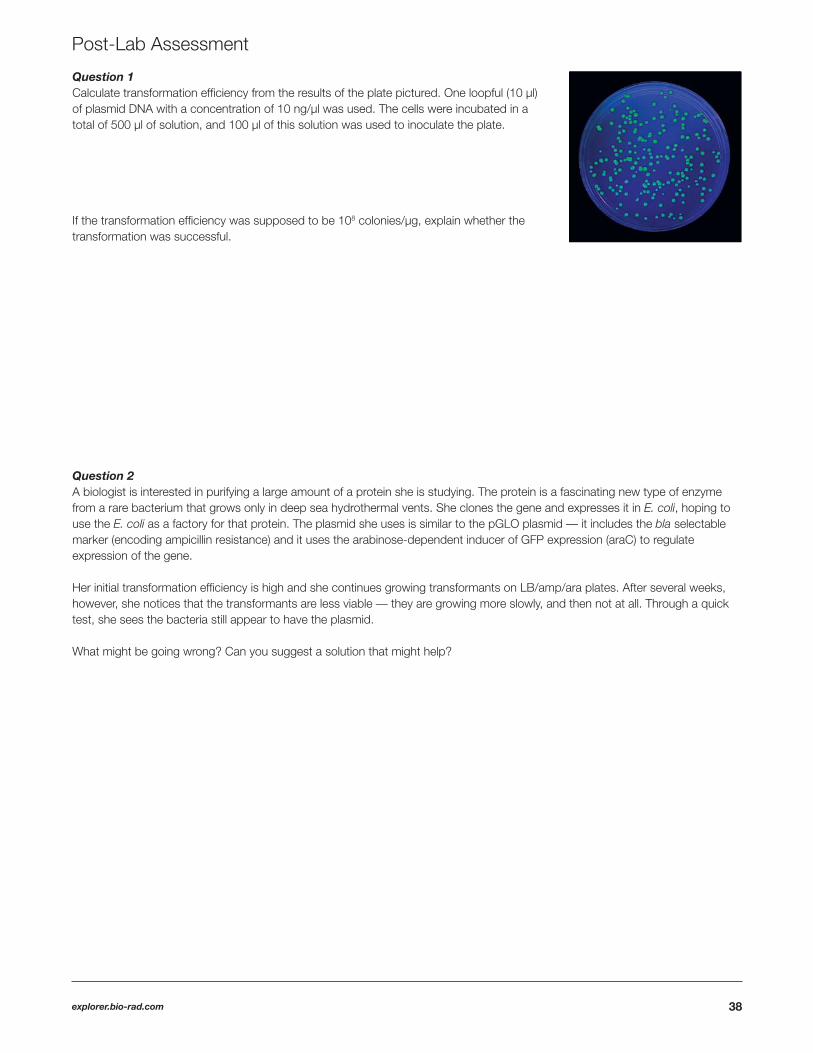
Question 1 Calculate transformation effi ciency from the results of the plate pictured. One loopful (10 µl) of plasmid DNA with a concentration of 10 ng/µl was used. The cells were incubated in a total of 500 µl of solution, and 100 µl of this solution was used to inoculate the plate.
If the transformation effi ciency was supposed to be 108 colonies/µg, explain whether the transformation was successful.
Question 2A biologist is interested in purifying a large amount of a protein she is studying. The protein is a fascinating new type of enzyme from a rare bacterium that grows only in deep sea hydrothermal vents. She clones the gene and expresses it in E. coli, hoping to use the E. coli as a factory for that protein. The plasmid she uses is similar to the pGLO plasmid — it includes the bla selectable marker (encoding ampicillin resistance) and it uses the arabinose-dependent inducer of GFP expression (araC) to regulate expression of the gene.
Her initial transformation effi ciency is high and she continues growing transformants on LB/amp/ara plates. After several weeks, however, she notices that the transformants are less viable — they are growing more slowly, and then not at all. Through a quick test, she sees the bacteria still appear to have the plasmid. What might be going wrong? Can you suggest a solution that might help?

39
Question 3Olivia Hamilton, a clinical lab technician, is testing a panel of antibiotics against a new strain of bacteria that is causing infections in the local area. The goal is to help fi nd the best treatment strategy. She performs a Kirby-Bauer test by growing lawns of the bacterial strain on agar plates with four paper disks impregnated with the same concentration of three antibiotics (A, B, and C) and a control with sterile water. A clear zone forms around the paper disks if the antibiotic inhibits bacterial growth.
Why would it be useful to have more than one antibiotic to treat these infections?
Rank the bacterial resistance to these antibiotics from highest to lowest resistance.
Question 4Of the following potential applications for genetic engineering, which one is not yet done regularly and why do you think that is?
• Production of tomatoes in which ripening is delayed, making them easier to ship• Production of hormones for treating diabetes• Genetic testing for harmful alleles in adults (such as alleles associated with diseases like breast cancer)• Genetic testing for harmful alleles in unborn infants (prenatal genetic testing)• Transformation of engineered genes into human gametes (germ-line gene therapy)

40explorer.bio-rad.com
IV. Class Discussion
Science Case Study: Can Bacterial Transformation Stop the Spread of Malaria?
I. The Global Impact of Malaria
Lerato sits in the clinic, watching her son Baruti, age 4, writhe with fever. He is sleeping for the fi rst time in days, and Lerato is anxious for signs that the medication the doctor had given her son is working. She worries she may have waited too long to bring Baruti to the clinic.
Baruti had fallen ill a week before with fever, chills, and body aches, all vague fl u-like symptoms that Lerato had assumed would clear in a few days, as so many illnesses had in the past. Lerato and her family live an hour’s walk away from the medical clinic in Seronga, a remote village in Okavango, Botswana. Making that long trip with a sick child is diffi cult, but it was a trip she had to make when her son’s symptoms grew more severe. He was now in a clinic bed, suffering from extreme anemia secondary to (brought on by) malaria.
Malaria is spread by mosquitoes, and like so many others in the Okavanago region, Lerato’s family had been issued mosquito nets for their beds. The nets are covered with insecticidal chemicals and are an effective and relatively inexpensive method for controlling mosquitoes. The nets, though, do not allow much air circulation and so are very hot to sleep under, and little Baruti tends to kick them away while he sleeps, exposing his limbs to the bites of mosquitoes.
Questions1. Malaria is the third leading cause of infectious disease death in the world, after tuberculosis and AIDS. According to the World Health Organization, 3.4 billion people — nearly half the global population — are currently at risk for malaria. Most prevalent in African or tropical Asian countries, malaria is often considered a “disease of the developing world.” Though vaccines are not yet available, it can be cured if diagnosed and treated promptly.
Given this information, what might the biggest hurdles be in fi ghting malaria? Consider the regions the disease affects and the challenges faced by the people living there.
2. Malaria can be spread only through the bite of a mosquito, and it was nearly eliminated in the U.S. back in the 1950s. Despite this, as many as 1,500–2,000 new cases of malaria are reported in the U.S. annually. How can this be? How might malaria be coming into the country?
3. Considering that malaria can be spread only from infected blood and through mosquito bites, how might malaria eventually be eradicated in a particular region, such as the U.S.?
41
II. Mosquitoes — Flying Factories of Malaria
In order to expand the discussion of malaria and possible methods for its treatment, control, and eradication, it is important to understand the biology behind the disease.
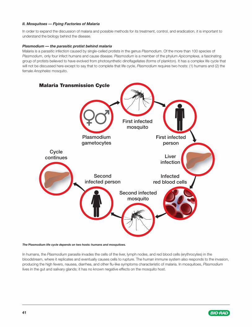
Plasmodium — the parasitic protist behind malariaMalaria is a parasitic infection caused by single-celled protists in the genus Plasmodium. Of the more than 100 species of Plasmodium, only four infect humans and cause disease. Plasmodium is a member of the phylum Apicomplexa, a fascinating group of protists believed to have evolved from photosynthetic dinofl agellates (forms of plankton). It has a complex life cycle that will not be discussed here except to say that to complete that life cycle, Plasmodium requires two hosts: (1) humans and (2) the female Anopheles mosquito.
The Plasmodium life cycle depends on two hosts: humans and mosquitoes.
In humans, the Plasmodium parasite invades the cells of the liver, lymph nodes, and red blood cells (erythrocytes) in the bloodstream, where it replicates and eventually causes cells to rupture. The human immune system also responds to the invasion, producing the high fevers, nausea, diarrhea, and other fl u-like symptoms characteristic of malaria. In mosquitoes, Plasmodium lives in the gut and salivary glands; it has no known negative effects on the mosquito host.

42explorer.bio-rad.com
As mentioned, Plasmodium is mainly transmitted between infected humans by mosquitoes. Specifi cally, it is spread only by female mosquitoes of the genus Anopheles. This is because only female mosquitoes bite humans. They ingest human blood to obtain the proteins necessary for egg development. Most female Anopheles mosquitoes are nocturnal feeders (they bite only at night).
When a female mosquito bites and takes blood from a person infected with Plasmodium, the microscopic parasite moves along with the human’s red blood cells to the mosquito’s gut, where it continues through its life cycle. It then moves to the mosquito’s salivary gland; when the mosquito takes a bite from another human, Plasmodium is injected along with the mosquito’s saliva. (It is the proteins in mosquito saliva that trigger an immune response from your body, causing bites to itch.) The parasites can then be ingested by another mosquito, completing the life cycle and transmitting the disease from human to human to human.
Malaria is passed from person to person through mosquito bites.
Blood smear showing the presence of the Plasmodium parasite (crescent shapes). In the absence of more sophisticated tests, microscopic analysis of blood samples is a common diagnostic approach for malaria in clinics.
43
Questions1. Malaria is most often transmitted by mosquitoes, but considering Plasmodium lives in erythrocytes, what are other ways in which the disease might spread from human to human?
2. Understanding the biology of malaria, it is not surprising that the most common methods of reducing outbreaks involve mosquito control. Relatively inexpensive and simple to perform, indoor spraying with insecticides kills mosquitoes for 3–6 months, and insecticidal bed nets provide additional protection from bites, with the insecticidal qualities of the nets lasting 3–5 years. What are the main benefi ts and drawbacks of both these approaches?
Indoor spraying
Benefi ts:
Drawbacks:
Bed nets
Benefi ts:
Drawbacks:

44explorer.bio-rad.com
3. Malaria is only one of many parasite-mediated, mosquito-borne illnesses affecting the world’s population. For this reason, some researchers advocate mosquito elimination — completely killing off mosquito populations in affected regions. The benefi ts of elimination are obvious (many of us would love to see mosquito-free lakes, streams, and ponds). However, what might some of the concerns be in eliminating a species from a habitat? Do we know what roles mosquitoes play in the environment? Does it make a difference if we know or don’t know?
4. Many regions in Africa are reporting increasing populations of Anopheles mosquito species that show resistance to insecticides. Even more troubling are fi ndings that climate change is expanding the habitat of the mosquitoes into regions where malaria had not been a health concern. How does your answer to the previous question change if the mosquito in question is an invasive species (for example, in the spread of dengue fever, the mosquito vector in question is often an invasive species new to habitats in North America)?
45
III. A Novel Approach Involves Bacterial Transformation
Biotechnology and genetic engineering methods are also being investigated as mechanisms for eliminating malaria. Theoretically, any of the species involved — the Plasmodium parasite or the human or mosquito host — can be the target of genetic modifi cations that disrupt either the life cycle or transmission of the parasite. Practically, however, these systems have their drawbacks in terms of their ability to be either cultured or manipulated genetically. Therefore, researchers have turned their attention to a more familiar subject: bacteria.
How can DNA “transform” bacteria? A background to bacterial transformationIn the 1920s, scientists demonstrated how to turn a harmless strain of bacteria into a virulent strain, just by mixing the two strains together (Griffi th 1928). What is truly incredible about this experiment is that the virulent strain had been killed prior to mixing, so something in the dead bacteria could “transform” the harmless bacteria, making them virulent.
It wasn’t until the 1940s that scientists understood the chemical basis for this transformation. A team of scientists led by Oswald Avery at the Rockefeller Institute found that an extract of the bacteria was unaffected by treatment with protein-digesting enzymes, but was destroyed by a DNA-digesting enzyme. This showed that the agent that transformed the harmless bacteria was DNA (Avery et al. 1944).
Today, we understand that genes within DNA encode proteins that give rise to certain traits. We also know how to exploit the fact that many bacteria can acquire new genes by taking up DNA molecules encoding those genes (for instance, a plasmid) from their surroundings. The process is optimized by adding salts to the transformation medium and using a heat shock step, steps we use deliberately to transform bacteria and other microorganisms. The ability to transform the bacterium E. coli, for example, has made possible the cloning of genes, the cornerstone of many modern advances in sciences and of the biotechnology industry.
So how does bacterial transformation relate to our battle against malaria?
Mosquitoes also have gut microbiota?It is surprising to many that, like humans, mosquitoes harbor a number of symbiotic bacteria within their gut. These symbiotic bacteria can be engineered, using procedures like those you used to transform E. coli bacteria, to produce proteins. However, in this case the symbiotic bacteria can be engineered to produce and secrete proteins that interfere with the life cycle of the Plasmodium parasite.
In one experiment (Wang et al. 2012), researchers used a bacterium called Pantoea agglomerans, which grows abundantly inside Anopheles mosquitoes. P. agglomerans can be grown and transformed using the same culturing and transformation techniques used with other more common bacteria, like E. coli. Researchers used these techniques to engineer P. agglomerans to express the genes of the hemolysin (hly) A system of E. coli bacteria, three proteins that cause red blood cells to lyse. The transformed bacteria were fed to mosquitoes through sugar solutions. The idea was that when the transformed bacteria colonized the mosquito gut, they would produce the toxic proteins. If that host mosquito then fed upon a human infected with malaria, the toxins produced by the transformed bacteria would cause the red blood cells (from the human blood) to burst. This would halt the life cycle of the Plasmodium parasite and stop the spread of malaria.
However, the researchers also hypothesized that transformation and expression of foreign genes might affect the ability of the transformed P. agglomerans bacteria to grow or colonize the mosquito gut (in other words, their fi tness for that environment might be reduced). This could jeopardize the effectiveness of this strategy for fi ghting malaria in the wild. So they carried out another experiment: they transformed the bacteria with a plasmid that contains a green fl uorescent protein (GFP), derived from the jellyfi sh Aequorea victoria, and fed the transformed bacteria to mosquitoes. The researchers then monitored how much fl uorescence came from the mosquito gut. They found that after the host mosquitoes were given a blood meal, the GFP fl uorescence in their guts increased, indicating the number of transformed bacteria there had rapidly increased. This demonstrated that transformed P. agglomerans could grow in the mosquito gut and, more importantly, replicate quickly when the mosquito ingested a blood meal. This meant the bacteria would also likely produce more hly A proteins when the host mosquito ingested potentially infected blood cells. In terms of the effi cacy of the transformed bacteria against the Plasmodium parasite, when mosquitoes with the transformed bacteria were fed a blood meal containing the Plasmodium parasite, the development of the parasite was inhibited by nearly 98% (Wang et al. 2012).

46explorer.bio-rad.com
Questions1. Scientists have also demonstrated that it is possible to genetically modify the Anopheles mosquito to produce a substance in their gut that kills off Plasmodium. Why might symbiotic bacteria be a more suitable subject for transformation than the Anopheles mosquito?
2. As in the human gut, many different bacterial species inhabit the Anopheles gut. If you were the researcher, how would you pick the best species for use in this transformation experiment? What factors should you consider?
3. Why was GFP used in the bacterial transformation experiment?
4. E. coli was also used in this experiment for plasmid production (to grow more copies of the plasmids), and the plasmid also contained genes for antibiotic resistance. After transformation, the bacteria were grown on plates with antibiotic in them. Why do you think this is a common step in bacterial transformation?
5. The experiment demonstrated that in the lab the transformed bacteria could survive and proliferate within the mosquito gut after transformation and that they could inhibit Plasmodium growth and development by nearly 98%. What other experiments might be needed to demonstrate this is a viable option for malaria elimination in the wild? Consider the life cycles and roles of all the key players in the spread of disease.
6. The hly A system used in the experiment described causes lysis of red blood cells. Considering this, would you have concerns about releasing these transformed bacteria into the environment? Under what conditions might these concerns be alleviated?
7. As a fi nal thought, what do you think the greatest hurdles will be to successfully implement this bacteria-based approach in the wild? What are the technical, ethical, or regulatory challenges, and how might they be handled?
47
IV: Prognosis
Baruti opens his eyes and sees his mother sitting at his side. She takes his hand and tells him she loves him. She has waited for this moment for three days. She is exhausted from the sleepless nights and constant worry.
The doctors come into the room to check on his progress. They are cautiously optimistic that the mixture of antimalarial drugs is working, and they tell Lerato to be patient. They tell her that her boy was very ill and that treatment takes time, but they are encouraged by the progress he is making. They expect him to recover.
Questions1. Most antimalarial drugs target the red blood cell (erythrocytic) stage of malaria infection, which is the phase of infection that causes symptomatic illness. Why might it be important to research other medications targeting other stages (for example, the liver stage) of the life cycle? Refer to the fi gure describing the life cycle of Plasmodium.
2. When Plasmodium becomes resistant to antimalarial drugs, this results in a delayed or incomplete clearance of the parasite from the patient’s blood. How might an organism develop resistance to a chemical that can otherwise kill it?
3. The problem of antimalarial drug resistance can be compounded by cross-resistance, in which resistance to one drug confers resistance to other drugs that belong to the same chemical family or have similar modes of action. How and why do you think this might happen?
4. Current practice in treating cases of malaria is based on combination therapy, in which several different classes of drugs are combined. What might some advantages of this approach be?
5. Many people take antibiotics to treat bacterially mediated illnesses like strep throat or sinus infections. When you take antibiotics, you are told you must take the entire course of the medication in order to reduce the risk of developing antibiotic resistance. Why is a full course needed?

48explorer.bio-rad.com
Appendix A: Serial Dilution and Dilution Instructions
How to Perform Serial Dilutions
A serial dilution is a series or chain of dilutions of a substance in a solution, typically distilled or sterile water. Serial dilutions are used to create accurate, highly diluted solutions intended to result in a concentration change. Many of the serial dilutions that are needed in the pGLO Transformation and Inquiry Kit will start from an original stock solution and will be successively diluted to create the desired concentration(s).
Serial dilutions require a degree of precision that is diffi cult to obtain using a DPTP, but not impossible. The dilution series depicted here can be done with a DPTP using the following gradient patterns.
Inquiry Investigation #1: Transformation Effi ciency
Transformation solution (CaCl2 ) concentrationIf you have chosen to vary the concentration of CaCl2 transformation solution, you will need to make either a dilution of transformation solution using sterile water or a 0 mM solution using LB nutrient broth.
To make experimental dilutions of CaCl2 you will use transformation solution and LB nutrient broth.
a. For a 25 mM dilution of CaCl2 use a sterile DPTP to transfer 125 µl of 50 mM CaCl2 into a tube of 125 µl of LB broth.
b. For a 0 mM dilution of CaCl2 use a sterile DPTP to transfer 250 µl of LB broth into a tube.
Amount of pGLO plasmid DNA
Transforming with less pGLO plasmid DNATo make experimental dilutions of the pGLO plasmid DNA use the pGLO stock (0.8 x 10⁻¹ µg/µl) and transformation solution. A sample dilution series is illustrated.
a. Use the rehydrated stock pGLO plasmid DNA (0.8 x 10⁻¹ µg/µl)
b. Label blank microcentrifuge tubes with the expected concentrations.
c. Using a sterile DPTP, transfer 225 µl of transformation solution (the diluent for pGLO plasmid DNA) to the tubes.
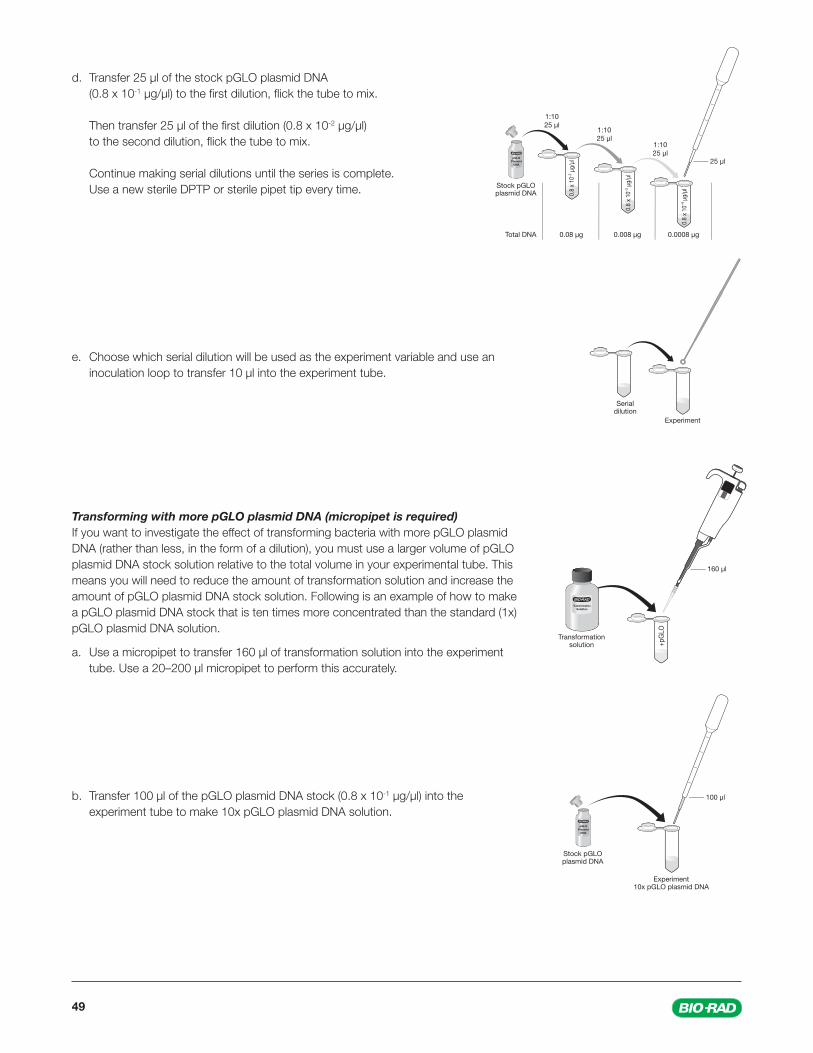
49
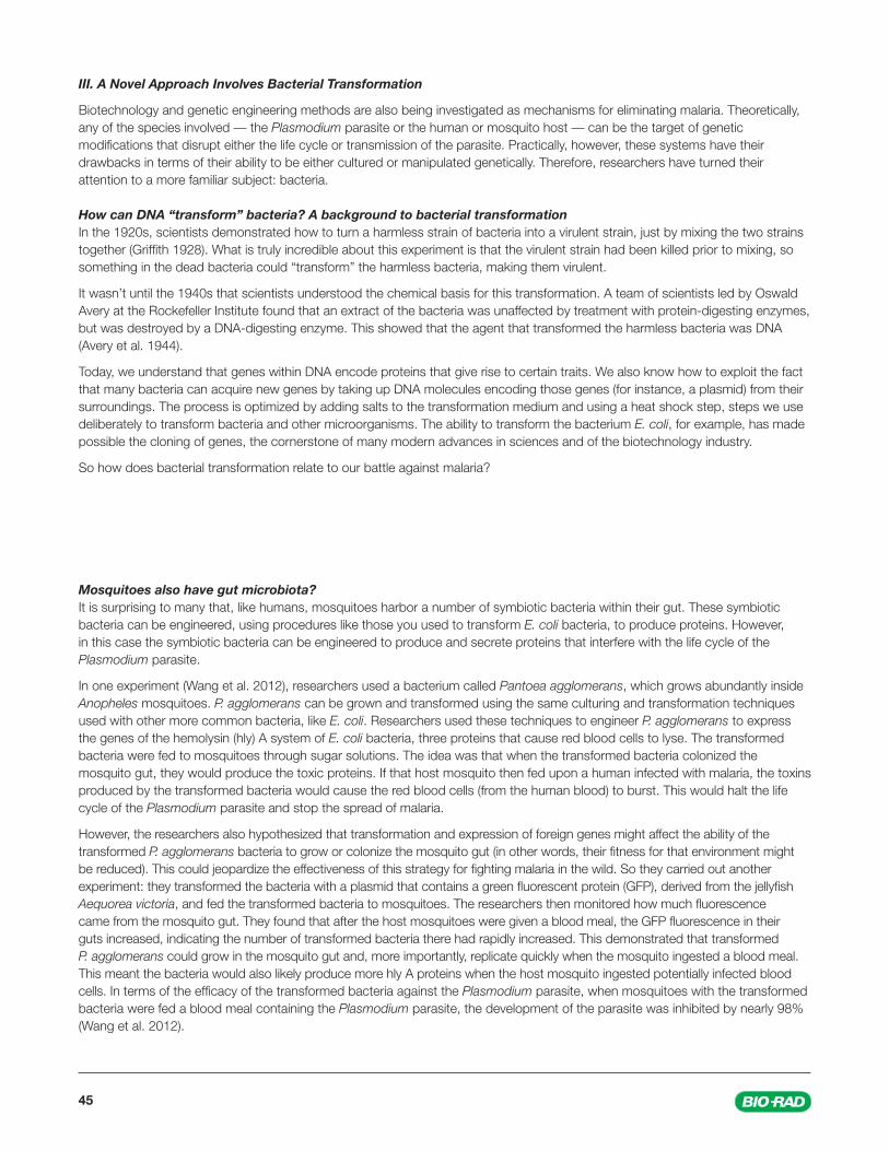
d. Transfer 25 µl of the stock pGLO plasmid DNA (0.8 x 10⁻¹ µg/µl) to the fi rst dilution, fl ick the tube to mix.
Then transfer 25 µl of the fi rst dilution (0.8 x 10⁻² µg/µl) to the second dilution, fl ick the tube to mix.
Continue making serial dilutions until the series is complete. Use a new sterile DPTP or sterile pipet tip every time.
e. Choose which serial dilution will be used as the experiment variable and use an inoculation loop to transfer 10 µl into the experiment tube.
Transforming with more pGLO plasmid DNA (micropipet is required)If you want to investigate the effect of transforming bacteria with more pGLO plasmid DNA (rather than less, in the form of a dilution), you must use a larger volume of pGLO plasmid DNA stock solution relative to the total volume in your experimental tube. This means you will need to reduce the amount of transformation solution and increase the amount of pGLO plasmid DNA stock solution. Following is an example of how to make a pGLO plasmid DNA stock that is ten times more concentrated than the standard (1x) pGLO plasmid DNA solution.
a. Use a micropipet to transfer 160 µl of transformation solution into the experiment tube. Use a 20–200 µl micropipet to perform this accurately.
b. Transfer 100 µl of the pGLO plasmid DNA stock (0.8 x 10⁻¹ µg/µl) into the experiment tube to make 10x pGLO plasmid DNA solution.
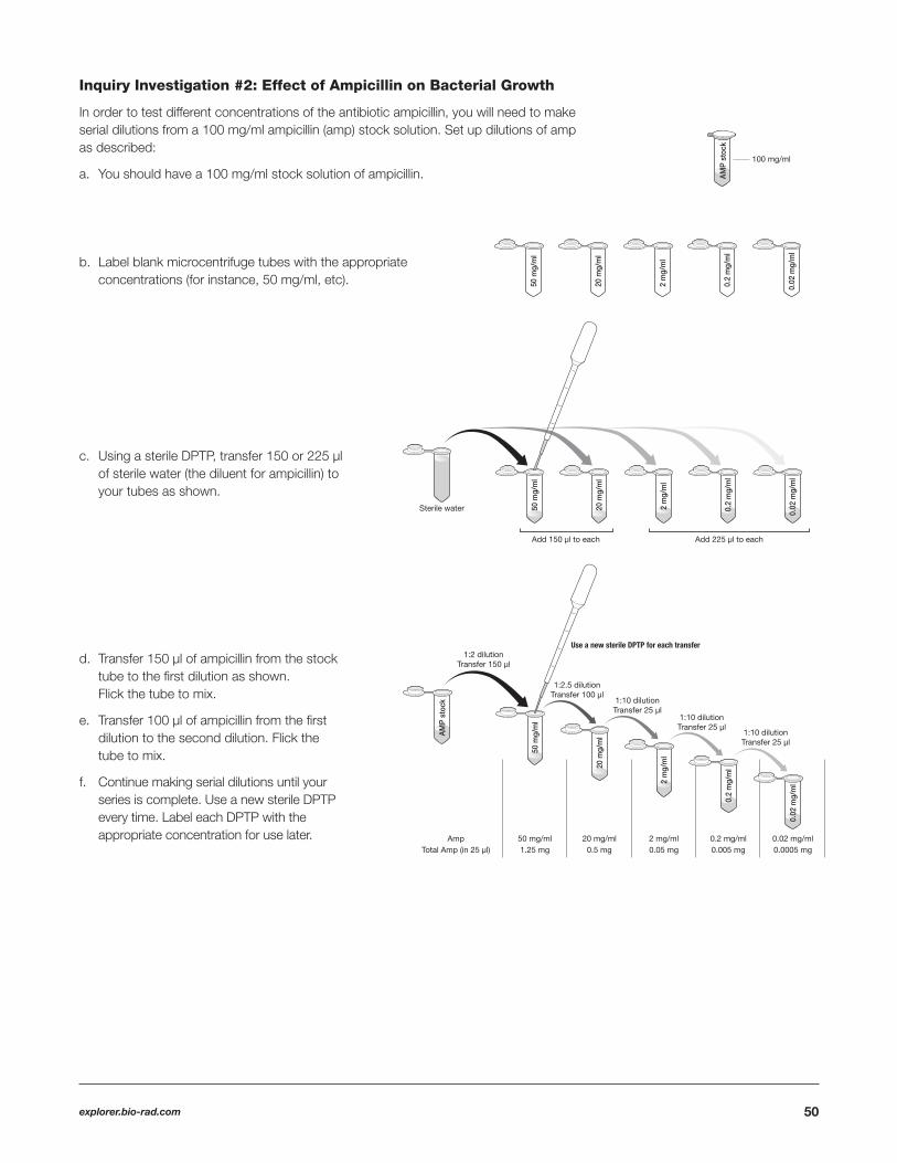
50explorer.bio-rad.com
Inquiry Investigation #2: Effect of Ampicillin on Bacterial Growth
In order to test different concentrations of the antibiotic ampicillin, you will need to make serial dilutions from a 100 mg/ml ampicillin (amp) stock solution. Set up dilutions of amp as described:
a. You should have a 100 mg/ml stock solution of ampicillin.
b. Label blank microcentrifuge tubes with the appropriate concentrations (for instance, 50 mg/ml, etc).
c. Using a sterile DPTP, transfer 150 or 225 µl of sterile water (the diluent for ampicillin) to your tubes as shown.
d. Transfer 150 µl of ampicillin from the stock tube to the fi rst dilution as shown. Flick the tube to mix.
e. Transfer 100 µl of ampicillin from the fi rst dilution to the second dilution. Flick the tube to mix.
f. Continue making serial dilutions until your series is complete. Use a new sterile DPTP every time. Label each DPTP with the appropriate concentration for use later.
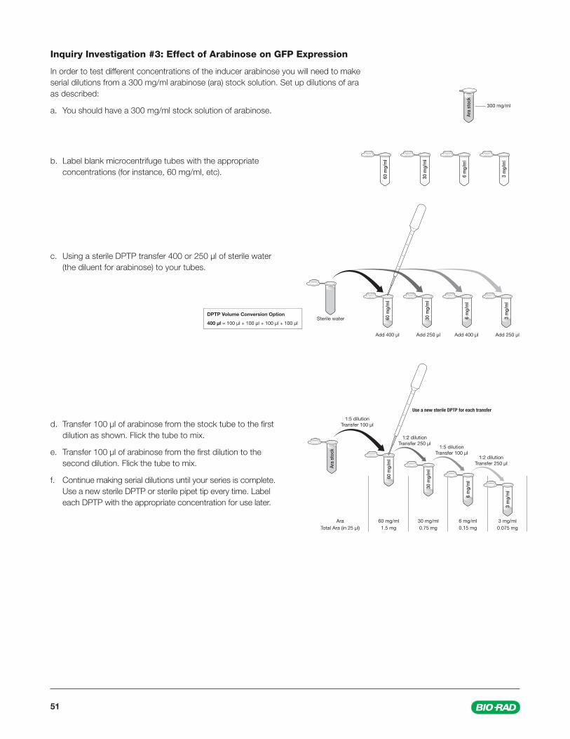
51
Inquiry Investigation #3: Effect of Arabinose on GFP Expression
In order to test different concentrations of the inducer arabinose you will need to make serial dilutions from a 300 mg/ml arabinose (ara) stock solution. Set up dilutions of ara as described:
a. You should have a 300 mg/ml stock solution of arabinose.
b. Label blank microcentrifuge tubes with the appropriate concentrations (for instance, 60 mg/ml, etc).
c. Using a sterile DPTP transfer 400 or 250 µl of sterile water (the diluent for arabinose) to your tubes.
d. Transfer 100 µl of arabinose from the stock tube to the fi rst dilution as shown. Flick the tube to mix.
e. Transfer 100 µl of arabinose from the fi rst dilution to the second dilution. Flick the tube to mix.
f. Continue making serial dilutions until your series is complete. Use a new sterile DPTP or sterile pipet tip every time. Label each DPTP with the appropriate concentration for use later.
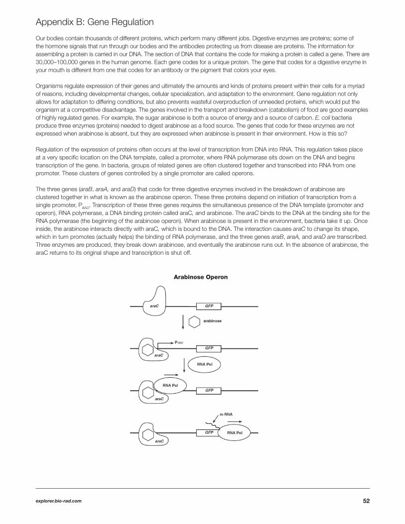
52explorer.bio-rad.com
Our bodies contain thousands of different proteins, which perform many different jobs. Digestive enzymes are proteins; some of the hormone signals that run through our bodies and the antibodies protecting us from disease are proteins. The information for assembling a protein is carried in our DNA. The section of DNA that contains the code for making a protein is called a gene. There are 30,000–100,000 genes in the human genome. Each gene codes for a unique protein. The gene that codes for a digestive enzyme in your mouth is different from one that codes for an antibody or the pigment that colors your eyes.
Organisms regulate expression of their genes and ultimately the amounts and kinds of proteins present within their cells for a myriad of reasons, including developmental changes, cellular specialization, and adaptation to the environment. Gene regulation not only allows for adaptation to differing conditions, but also prevents wasteful overproduction of unneeded proteins, which would put the organism at a competitive disadvantage. The genes involved in the transport and breakdown (catabolism) of food are good examples of highly regulated genes. For example, the sugar arabinose is both a source of energy and a source of carbon. E. coli bacteria produce three enzymes (proteins) needed to digest arabinose as a food source. The genes that code for these enzymes are not expressed when arabinose is absent, but they are expressed when arabinose is present in their environment. How is this so?
Regulation of the expression of proteins often occurs at the level of transcription from DNA into RNA. This regulation takes place at a very specifi c location on the DNA template, called a promoter, where RNA polymerase sits down on the DNA and begins transcription of the gene. In bacteria, groups of related genes are often clustered together and transcribed into RNA from one promoter. These clusters of genes controlled by a single promoter are called operons.
The three genes (araB, araA, and araD) that code for three digestive enzymes involved in the breakdown of arabinose are clustered together in what is known as the arabinose operon. These three proteins depend on initiation of transcription from a single promoter, PBAD. Transcription of these three genes requires the simultaneous presence of the DNA template (promoter and operon), RNA polymerase, a DNA binding protein called araC, and arabinose. The araC binds to the DNA at the binding site for the RNA polymerase (the beginning of the arabinose operon). When arabinose is present in the environment, bacteria take it up. Once inside, the arabinose interacts directly with araC, which is bound to the DNA. The interaction causes araC to change its shape, which in turn promotes (actually helps) the binding of RNA polymerase, and the three genes araB, araA, and araD are transcribed. Three enzymes are produced, they break down arabinose, and eventually the arabinose runs out. In the absence of arabinose, the araC returns to its original shape and transcription is shut off.
Appendix B: Gene Regulation
Arabinose Operon
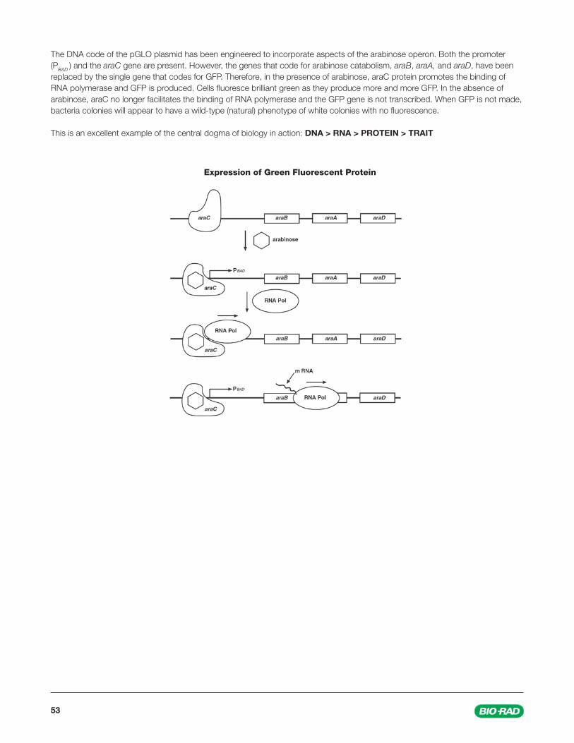
53
Expression of Green Fluorescent Protein
The DNA code of the pGLO plasmid has been engineered to incorporate aspects of the arabinose operon. Both the promoter (PBAD ) and the araC gene are present. However, the genes that code for arabinose catabolism, araB, araA, and araD, have been replaced by the single gene that codes for GFP. Therefore, in the presence of arabinose, araC protein promotes the binding of RNA polymerase and GFP is produced. Cells fl uoresce brilliant green as they produce more and more GFP. In the absence of arabinose, araC no longer facilitates the binding of RNA polymerase and the GFP gene is not transcribed. When GFP is not made, bacteria colonies will appear to have a wild-type (natural) phenotype of white colonies with no fl uorescence.
This is an excellent example of the central dogma of biology in action: DNA > RNA > PROTEIN > TRAIT
Sig 121410048976 Rev A US/EG
Web site www.bio-rad.com USA 800 424 6723 Australia 61 2 9914 2800 Austria 43 1 877 89 01 Belgium 03 710 53 00 Brazil 55 11 3065 7550 Canada 905 364 3435 China 86 21 6169 8500 Czech Republic 420 241 430 532 Denmark 44 52 10 00 Finland 09 804 22 00 France 01 47 95 69 65 Germany 49 89 31 884 0 Greece 30 210 9532 220 Hong Kong 852 2789 3300 Hungary 36 1 459 6100 India 91 124 4029300 Israel 03 963 6050 Italy 39 02 216091 Japan 81 3 6361 7000 Korea 82 2 3473 4460 Mexico 52 555 488 7670 The Netherlands 0318 540666 New Zealand 64 9 415 2280 Norway 23 38 41 30 Poland 48 22 331 99 99 Portugal 351 21 472 7700 Russia 7 495 721 14 04 Singapore 65 6415 3188 South Africa 27 (0) 861 246 723 Spain 34 91 590 5200 Sweden 08 555 12700 Switzerland 026 674 55 05 Taiwan 886 2 2578 7189 Thailand 1800 88 22 88 United Kingdom 020 8328 2000
Bio-Rad Laboratories, Inc.
Life ScienceGroup
References
Avery OT et al. (1944). Studies on the chemical nature of the substance inducing transformation of pneumococcal types: Induction of transformation by a desoxyribonucleic acid fraction isolated from Pneumococcus Type III. J Exp Med 79, 137–158.
Griffi th F (1928). The signifi cance of pneumococcal types. J Hyg 27, 113–159.
Wang S et al. (2012). Fighting malaria with engineered symbiotic bacteria from vector mosquitoes. Proc Natl Acad Sci 109, 12734-12739.
Suggested Reading, Listening
Kill ‘Em All (NPR Radiolab podcast) http://www.radiolab.org/story/kill-em-all/
What if We Don’t Kill ‘Em All (NPR Radiolab podcast) http://www.radiolab.org/story/what-if-we-dont-kill-em-all/
Legal Notice
Liofi lchem is a trademark of Liofi lchem S.R.L., LLC.