personal genetics - massbioed · personal genetics: pcr determination of ptc tasters introduction...
TRANSCRIPT

Personal Genetics: PCR Determination of PTC Tasters
Student Materials
Introduction ................................................................................................................. 2
Pre-Lab Questions ........................................................................................................ 4
Lab Protocol ................................................................................................................. 8
Data Collection Worksheet ........................................................................................... 15
Post-Lab Questions and Analysis ................................................................................... 17
Last updated: August 7, 2017
2
Personal Genetics: PCR Determination of PTC Tasters Introduction
Look around. Would you say that individuals look the same or different? Most of us would agree that individuals look different. However, if you only look at the DNA of individuals you might say that different people actually look the same! The human genome contains approximately 3 billion nucleotides (A, T, C, and G) linked together in a specific order on long DNA molecules called chromosomes. The human genome is 99.9% identical from person to person.
What is considered the normal number of chromosomes for human body cells?
What is considered the normal number of chromosomes for human gametes?
Although we are almost identical at the DNA level, it is the small difference between individuals that make each of us unique. These differences define our personal genetics and determine many aspects of our individual biology. They specify hair and eye color, food allergies, reactions to certain medications, and the risk for particular health problems such as high blood pressure or diabetes. The DNA differences between people are also the basis of DNA fingerprinting.
Scientists have developed a number of methods to determine the nucleotide differences between individuals in order to predict and prevent unwanted health problems. This emerging field of personalized medicine strives to tailor medical procedures, practices, and/or products to the individual patients. Through the use of detailed genetic information, medical professionals have the potential to improve healthcare outcomes. Today we will use the Polymerase Chain Reaction (PCR) to test your genotype for a specific trait, the ability to taste a bitter chemical.
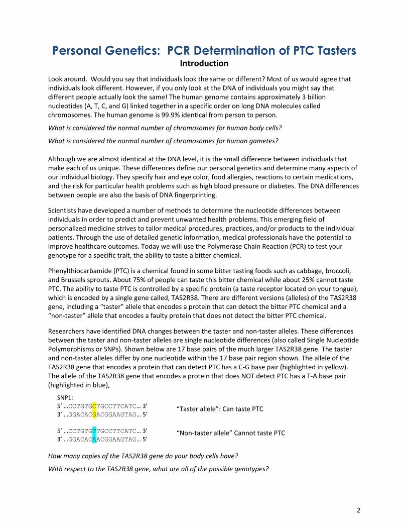
Phenylthiocarbamide (PTC) is a chemical found in some bitter tasting foods such as cabbage, broccoli, and Brussels sprouts. About 75% of people can taste this bitter chemical while about 25% cannot taste PTC. The ability to taste PTC is controlled by a specific protein (a taste receptor located on your tongue), which is encoded by a single gene called, TAS2R38. There are different versions (alleles) of the TAS2R38 gene, including a “taster” allele that encodes a protein that can detect the bitter PTC chemical and a “non-taster” allele that encodes a faulty protein that does not detect the bitter PTC chemical.
Researchers have identified DNA changes between the taster and non-taster alleles. These differences between the taster and non-taster alleles are single nucleotide differences (also called Single Nucleotide Polymorphisms or SNPs). Shown below are 17 base pairs of the much larger TAS2R38 gene. The taster and non-taster alleles differ by one nucleotide within the 17 base pair region shown. The allele of the TAS2R38 gene that encodes a protein that can detect PTC has a C-G base pair (highlighted in yellow). The allele of the TAS2R38 gene that encodes a protein that does NOT detect PTC has a T-A base pair (highlighted in blue),
SNP1:
5’ …CCTGTGCTGCCTTCATC… 3’
3’ …GGACACGACGGAAGTAG… 5’
5’ …CCTGTGTTGCCTTCATC… 3’
3’ …GGACACAACGGAAGTAG… 5’
How many copies of the TAS2R38 gene do your body cells have?
With respect to the TAS2R38 gene, what are all of the possible genotypes?
“Taster allele”: Can taste PTC
“Non-taster allele” Cannot taste PTC

3
In some cases, restriction enzymes can be used to identify the differences between alleles of SNPs. Restriction enzymes recognize a specific DNA sequence and cut the DNA at that site. The restriction enzymes are so specific that a single nucleotide difference from the recognized sequence will prevent the restriction enzyme from cutting the DNA. For example, an enzyme called Fnu4HI recognizes and cuts within the DNA sequence:
5’ …GCTGC… 3’ but Fnu4HI will not recognize and cut 5’ …GTTGC… 3’ 3’ …CGACG… 5’ 3’ …CAACG… 5’
SNP1 of the TAS2R38 gene falls within an Fnu4HI recognition site. The result is that Fnu4HI will cut the DNA sequence at SNP1 of the taster allele, but it will not cut the DNA sequence at SNP1 of the non-taster allele.
SNP1, taster allele:
5’ …CCTGTGCTGCCTTCATC… 3’
3’ …GGACACGACGGAAGTAG… 5’
SNP1, non-taster allele: 5’ …CCTGTGTTGCCTTCATC… 3’
3’ …GGACACAACGGAAGTAG… 5’
Within the TAS2R38 gene there is a second site, SNP2, where the DNA differs between the taster and non-taster alleles. SNP2 affects the recognition sequence for the restriction enzyme, Cac8I, which cuts at: 5’ …GCAGGC… 3’
3’ …CGACCG… 5’
SNP2, taster allele:
5’ …AGAGGCAGCCACT… 3’
3’ …TCTCCGTCGGTGA… 5’
SNP2, non-taster allele:
5’ …AGAGGCAGGCACT… 3’
3’ …TCTCCGTCCGTGA… 5’
Are you a taster or a non-taster? Today you will learn your TAS2R38 genotype DNA. You will then test your ability to taste PTC using special paper and determine whether your PTC taster genotype matches your PTC taster phenotype.
Given your food preferences, make a guess as to whether you are a taster or a non-taster.
Can taste PTC and DNA is cut by Fnu4HI at SNP1
Cannot taste PTC and DNA is NOT cut by Fnu4HI at SNP1
Can taste PTC and DNA is not cleaved by Cac8I at SNP2
Cannot taste PTC and DNA is cleaved by Cac8I at SNP2
4
Personal Genetics Pre-Lab Questions
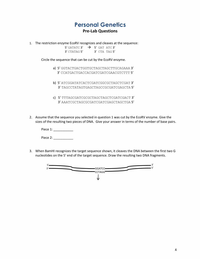
1. The restriction enzyme EcoRV recognizes and cleaves at the sequence:
5’ GATATC 3’ 5’ GAT ATC 3’
3’ CTATAG 5’ 3’ CTA TAG 5’
Circle the sequence that can be cut by the EcoRV enzyme.
a) 5’ GGTACTGACTGGTGCTAGCTAGCTTGCAGAAA 3’ 3’ CCATGACTGACCACGATCGATCGAACGTCTTT 5’
b) 5’ ATCGGATATCACTCGATCGGCGCTAGCTCGAT 3’
3’ TAGCCTATAGTGAGCTAGCCGCGATCGAGCTA 5’
c) 5’ TTTAGCGATCGCGCTAGCTAGCTCGATCGACT 3’ 3’ AAATCGCTAGCGCGATCGATCGAGCTAGCTGA 5’
2. Assume that the sequence you selected in question 1 was cut by the EcoRV enzyme. Give the sizes of the resulting two pieces of DNA. Give your answer in terms of the number of base pairs.
Piece 1: ___________
Piece 2: ___________
3. When BamHI recognizes the target sequence shown, it cleaves the DNA between the first two G nucleotides on the 5’ end of the target sequence. Draw the resulting two DNA fragments.
GGATCC CCTAGG
5’ 3’ 5’
3’

5
4. In gel electrophoresis, an applied electrical current causes the DNA pieces to move through the
gel matrix. In the diagram below, indicate in which direction the DNA will migrate through the gel during electrophoresis. Draw the chemical structure of a DNA nucleotide and use that drawing to explain why DNA will migrate through the gel in the manner you have indicated.
5. During gel electrophoresis, smaller pieces of DNA will travel faster than larger pieces of DNA.
This is because smaller pieces of DNA can move easily or migrate through the “obstacle course” gel matrix. In the gel below, indicate which band represents the larger pieces of DNA.
DNA
Negative Electrode
Positive Electrode
Lane 1 Negative Electrode
Positive Electrode
band 1
band 2
well
6
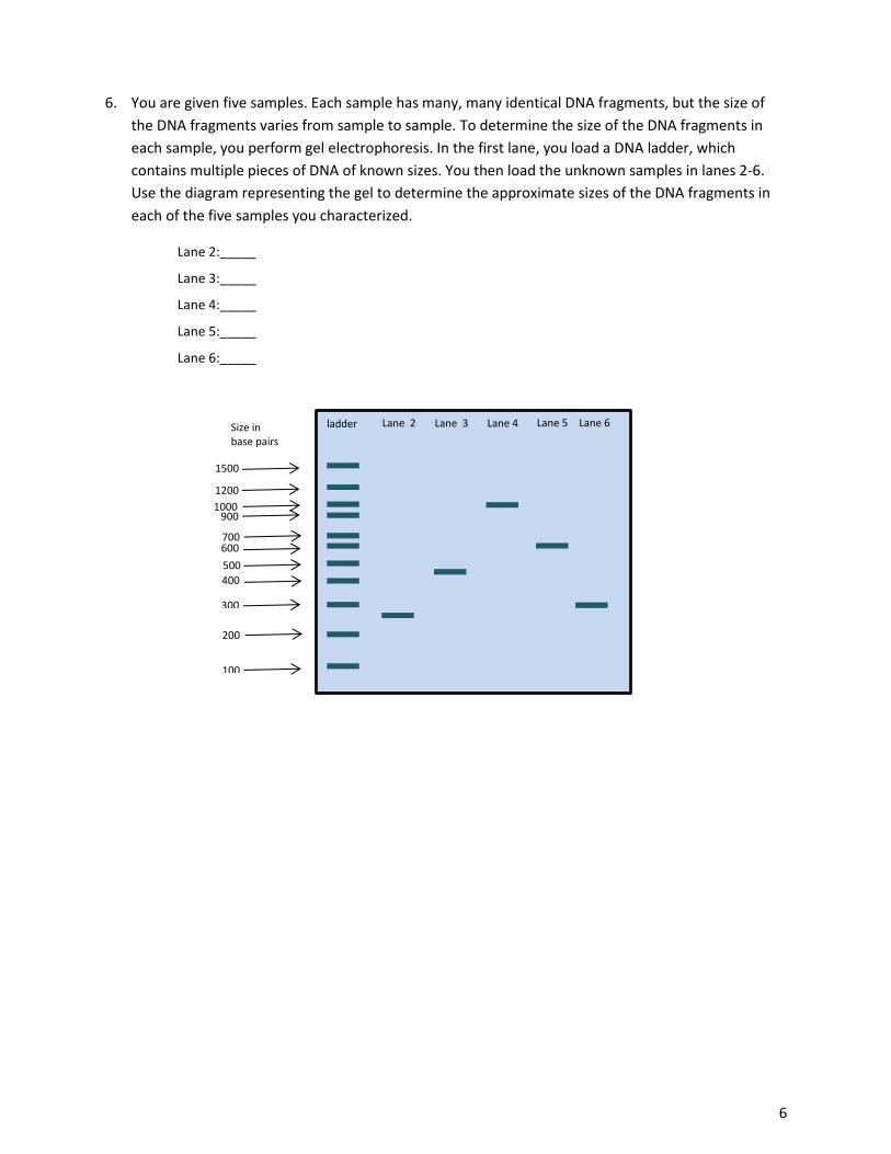
6. You are given five samples. Each sample has many, many identical DNA fragments, but the size of
the DNA fragments varies from sample to sample. To determine the size of the DNA fragments in
each sample, you perform gel electrophoresis. In the first lane, you load a DNA ladder, which
contains multiple pieces of DNA of known sizes. You then load the unknown samples in lanes 2-6.
Use the diagram representing the gel to determine the approximate sizes of the DNA fragments in
each of the five samples you characterized.
Lane 2:_____
Lane 3:_____
Lane 4:_____
Lane 5:_____
Lane 6:_____
400 500
300
200
100
900
1500
700 600
1200 1000
ladder Lane 2 Lane 3 Lane 4 Lane 5 Lane 6 Size in base pairs

7
7. Sickle-cell trait and sickle-cell anemia are associated with a single nucleotide polymorphism (SNP) that changes an A/T base pair at nucleotide 17 in the beta-globin gene to a T/A base pair. You have developed an assay to distinguish between the two variants. In alleles with the A/T variant, BamHI can recognize and cleave the DNA at this SNP. BamHI cannot cleave the T/A variant of the beta-globin gene at the SNP. Individuals with one copy of the T/A variant and one copy of the A/T variant have sickle-cell trait. Individuals with two copies of the T/A variant (homozygous for the mutant allele) have sickle-cell anemia. Individuals homozygous for the A/T variant do not have sickle-cell trait or anemia.
The schematic below shows a region of the beta-globin gene that was amplified using PCR and that includes the BamHI restriction site present in the T variant. This BamHI restriction site is not present in the A variant.
a. For people with sickle-cell anemia, PCR amplification of genomic DNA from this region of
the beta-globin gene and digestion of the PCR product with BamHI will result in a DNA
piece or DNA pieces of what size?
b. For people who do not have sickle-cell trait or anemia, PCR amplification of genomic DNA
from this region of the beta-globin gene and digestion of the PCR product with BamHI
will result in a DNA piece or DNA pieces of what size?
c. For people with sickle-cell trait, PCR amplification of genomic DNA from this region of the
beta-globin gene and digestion of the PCR product with BamHI will result in a DNA piece
or DNA pieces of what size?
d. You isolate DNA from five people and amplify the region of the beta-globin gene that contains the SNP. You then cut the amplified DNA molecules with the BamHI enzyme and separate the resulting fragments using gel electrophoresis. You find that the patient represented in lane 2 has sickle-cell anemia, the patient represented in lane 4 has sickle-cell trait and the other three are normal. Draw the expected bands on the gel below.
400
200
100
1000
800
1200
ladder Lane 2 Lane 3 Lane 4 Lane 5 Lane 6 Size in base pairs
600
BamHI
400 base pairs 600 base pairs

8
Personal Genetics
Lab Protocol
Overview: The first step in determining your personal genetics is to collect some of your cells. You will use your cheek cells as they can be gently scraped off with a toothpick or wooden stick.
You will then isolate the DNA from these cells by exposing them to sodium hydroxide (NaOH), which will break open the cell membrane so that your DNA will be released into solution.
Although you will have DNA from many cells, there will not be enough DNA to study the TAS2R38 gene, so the next step is to make many copies of the two regions of the TAS2R38 gene that contain SNP1 and SNP2 for further analysis. You will use a powerful technique called polymerase chain reaction (PCR) to amplify the two DNA regions.
PCR will produce millions of identical DNA molecules that will include SNP1. All of the molecules produced by PCR will be the same size, whether or not you are a taster. But remember that the restriction enzyme Fnu4HI will cut the DNA molecules if you have the taster allele, but will not cut the molecules if you are a non-taster.
Likewise, a second PCR reaction will produce millions of identical DNA molecules that will include SNP2. All of the molecules produced by this PCR will be the same size, but the restriction enzyme Cac8I will NOT cut the DNA molecules if you have the taster allele, but will cut the molecules if you are a non-taster.
Lastly, you will visualize the sizes of the DNA fragments generated by restriction enzyme digest by performing gel electrophoresis. Gel electrophoresis allows us to separate the DNA fragments based on size. By analyzing the size of the DNA pieces, you will be able to determine which alleles of the TAS2R38 gene you have in your DNA.
Experiment Flowchart:
9
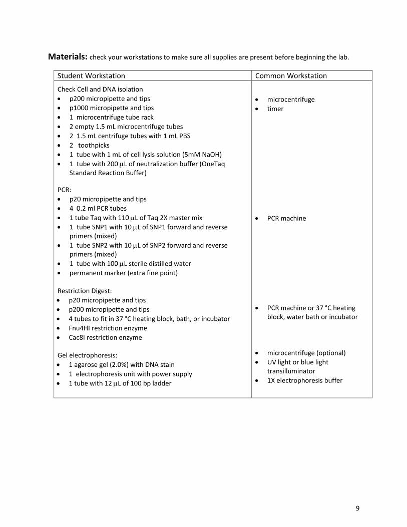
Materials: check your workstations to make sure all supplies are present before beginning the lab.
Student Workstation Common Workstation
Check Cell and DNA isolation
p200 micropipette and tips
p1000 micropipette and tips
1 microcentrifuge tube rack
2 empty 1.5 mL microcentrifuge tubes
2 1.5 mL centrifuge tubes with 1 mL PBS
2 toothpicks
1 tube with 1 mL of cell lysis solution (5mM NaOH)
1 tube with 200 L of neutralization buffer (OneTaq Standard Reaction Buffer)
PCR:
p20 micropipette and tips
4 0.2 ml PCR tubes
1 tube Taq with 110 L of Taq 2X master mix
1 tube SNP1 with 10 L of SNP1 forward and reverse primers (mixed)
1 tube SNP2 with 10 L of SNP2 forward and reverse primers (mixed)
1 tube with 100 L sterile distilled water
permanent marker (extra fine point) Restriction Digest:
p20 micropipette and tips
p200 micropipette and tips
4 tubes to fit in 37 °C heating block, bath, or incubator
Fnu4HI restriction enzyme
Cac8I restriction enzyme Gel electrophoresis:
1 agarose gel (2.0%) with DNA stain
1 electrophoresis unit with power supply
1 tube with 12 L of 100 bp ladder
microcentrifuge
timer
PCR machine
PCR machine or 37 °C heating block, water bath or incubator
microcentrifuge (optional)
UV light or blue light transilluminator
1X electrophoresis buffer
10
Procedure:
Cheek Cell Isolation: (Each student performs all steps)
1. Avoid eating food immediately prior to cheek swab. If you have recently eaten rinse your mouth vigorously with water.
2. Collect tissue sample: a. Insert toothpick in mouth and gently scrape inner cheek for 10-15 seconds. Do not
scrape too vigorously (this should not be painful). b. Insert toothpick containing cheek cells into the PBS solution in the microcentrifuge tube.
Make sure the toothpick is oriented so that the cheek cells are immersed in the PBS solution. Stir the PBS with the stick and let sit in tube for 2-3 minutes. Gently shake stick to dislodge attached cells and remove stick from tube.
c. Dispose of toothpick in biohazard trash. Do not reuse toothpick.
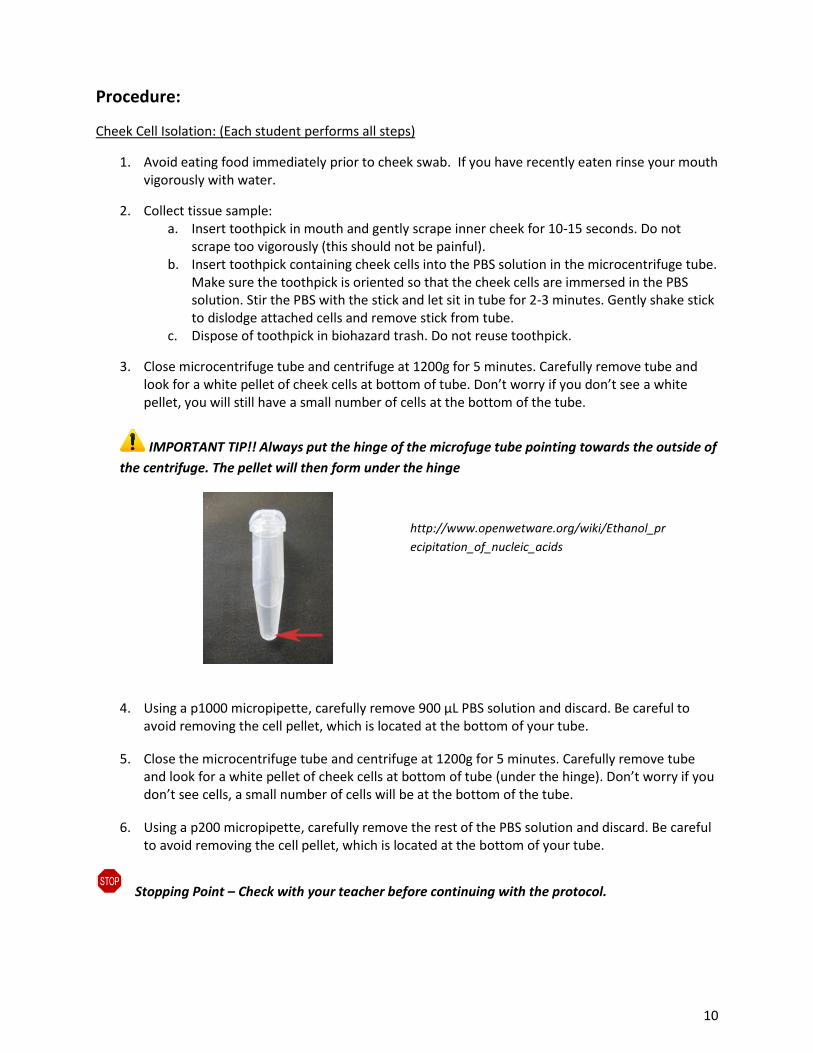
3. Close microcentrifuge tube and centrifuge at 1200g for 5 minutes. Carefully remove tube and look for a white pellet of cheek cells at bottom of tube. Don’t worry if you don’t see a white pellet, you will still have a small number of cells at the bottom of the tube.
IMPORTANT TIP!! Always put the hinge of the microfuge tube pointing towards the outside of
the centrifuge. The pellet will then form under the hinge
4. Using a p1000 micropipette, carefully remove 900 µL PBS solution and discard. Be careful to
avoid removing the cell pellet, which is located at the bottom of your tube.
5. Close the microcentrifuge tube and centrifuge at 1200g for 5 minutes. Carefully remove tube and look for a white pellet of cheek cells at bottom of tube (under the hinge). Don’t worry if you don’t see cells, a small number of cells will be at the bottom of the tube.
6. Using a p200 micropipette, carefully remove the rest of the PBS solution and discard. Be careful to avoid removing the cell pellet, which is located at the bottom of your tube.
Stopping Point – Check with your teacher before continuing with the protocol.
http://www.openwetware.org/wiki/Ethanol_pr
ecipitation_of_nucleic_acids

11
DNA Isolation: (Each student performs all steps)
7. Add 240 µL of Cell Lysis Solution (5mM NaOH) to the cheek cells in the tube. Pipette the cells up and down in the solution until the cells disappear (5-10 times).
8. Incubate the cells in the Cell Lysis Solution at room temperature (~25C) for 10 minutes.
9. Using a p200 micropipette, add 60 µL of neutralization buffer (Neut Buffer) to cells.
10. Centrifuge the tube at 1200g (the speed is not critical) for 1 minute to pellet the cellular debris.
11. Using a p200, carefully remove 150 µL of the solution from the top of the tube (do not disturb the pellet) and place it into a clean microcentrifuge tube labeled with “DNA” and your initials. This is now the DNA sample you will use for PCR.
Stopping Point – Check with your teacher before continuing with the protocol.
PCR: (Each student performs all steps)
IMPORTANT TIP!! Keep tubes on ice until placed in the PCR machine.
12. Label the side of one PCR tube with your initials and SNP1.
13. Label the side of another PCR tube with your initials and SNP2.
14. Using a p200 micropipette, add 25 µL of Taq mix to each PCR tube. The Taq solution contains Taq polymerase, dNTPs, and loading dye. The Taq mix is green.
15. Using a p20 micropipette, add 19 µL of sterile water to each PCR tube. Be sure to use a fresh tip
for each tube!
16. Using a p20 micropipette, add 3 µL of your DNA sample to each PCR tube (3 µL for each tube). Be sure to use a fresh tip for each tube!
17. Using a p20 micropipette, add 3 µL of Primer Mix SNP1 to the PCR tube labeled SNP1. Be sure to use a fresh tip for each tube!
18. Using a p20 micropipette, add 3µL of Primer Mix SNP2 to the PCR tube labeled SNP2. Be sure to use a fresh tip for each tube!
19. Place PCR tubes in PCR machine and run the following PCR program:
# of Cycles Temperature Time
1 94 °C 3 minutes
40
94 °C 15 seconds
60 °C 15 seconds
70 °C 30 seconds
1 70°C 3 minutes
Stopping Point – Check with your teacher before continuing with the protocol.

12
Restriction Digest: (Each student performs all steps)
20. Label the side of 1 tube* with your initials and SNP1 + Fnu4HI. Label the side of another tube* with your initials and SNP2 + Cac8I. (*check that the tubes fit into the 37 °C heating block, bath, or incubator)
21. Using a p200 micropipette, remove 25 µL of PCR reaction from SNP1 and add it to the bottom of the tube labeled SNP1 + Fnu4HI. Be sure to use a fresh tip for each tube!
22. Obtain Fnu4HI enzyme from teacher. Using a p20 micropipette, remove 1 µL of Fnu4HI
restriction enzyme and add it to the solution in the SNP1 + Fnu4HI tube. Pipette up and down a couple of times. Be sure to use a fresh tip for each tube!
23. Using a p200 micropipette, remove 25 µL of PCR reaction from SNP2 and add it to the bottom of
the tube labeled SNP2 + Cac8I. Be sure to use a fresh tip for each tube!
24. Obtain Cac8I enzyme from teacher. Using a p20 micropipette, remove 1 µL of Cac8I restriction enzyme and add it to the solution in the SNP2 + Cac8I tube. Pipette up and down a couple of times. Be sure to use a fresh tip for each tube!
25. Incubate restriction digest tubes at 37 °C for 1hr.
Stopping Point – Check with your teacher before continuing with the protocol.
Gel Electrophoresis: (Students work in pairs- 1 gel per 2 students)
26. You and your partner will need to obtain a 2.0% agarose gel with at least 9 lanes (or wells).
27. Load, run, and examine the gel: Coordinate with your partner to load and run the gel.
a. Using a p20, load 10 µL of 100bp ladder to lane 5 of your gel. The wells to the right of lane 5 will hold one partner’s samples. The wells to the left of lane 5 will hold the other partner’s samples.
b. Using a p20, load 15 µL of one partner’s sample into the gel wells 1-4 in the following order. Then load 15 µL of the other partner’s sample into the gel wells 6-9. Be sure to use a fresh tip for each tube!
c. Run the gel as instructed by your teacher.
d. Place your gel on the illuminator and examine your results. Be sure to take a photograph of your gel or sketch your results on the template provided.
28. Clean up your lab bench, pour the gel running buffer down the drain and dispose of your gel.
Student 1 Student 2
SNP1 uncut
SNP1+ Fnu4HI
SNP2 uncut
SNP2+ Cac8I
100 bp Ladder
SNP1 uncut
SNP1 + Fnu4HI
SNP2 uncut
SNP2 + Cac8I
13
Note: you do not need to add loading dye to your DNA samples because the OneTaq 2X Master Mix (used in step #14) and 100 bp ladder already include loading dye.
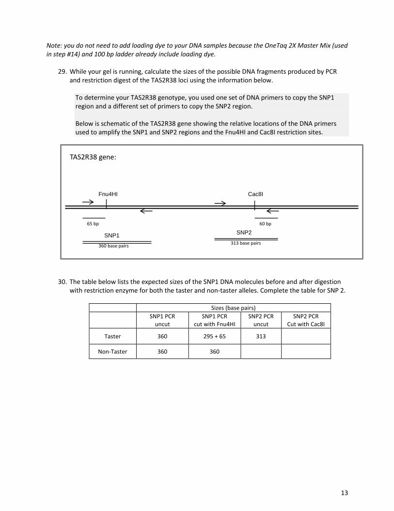
29. While your gel is running, calculate the sizes of the possible DNA fragments produced by PCR and restriction digest of the TAS2R38 loci using the information below.
To determine your TAS2R38 genotype, you used one set of DNA primers to copy the SNP1 region and a different set of primers to copy the SNP2 region. Below is schematic of the TAS2R38 gene showing the relative locations of the DNA primers used to amplify the SNP1 and SNP2 regions and the Fnu4HI and Cac8I restriction sites.
30. The table below lists the expected sizes of the SNP1 DNA molecules before and after digestion with restriction enzyme for both the taster and non-taster alleles. Complete the table for SNP 2.
Sizes (base pairs)
SNP1 PCR uncut
SNP1 PCR cut with Fnu4HI
SNP2 PCR uncut
SNP2 PCR Cut with Cac8I
Taster 360 295 + 65 313
Non-Taster 360 360
SNP1 SNP2
360 base pairs 313 base pairs
65 bp 60 bp
Fnu4HI Cac8I
TAS2R38 gene:

14
31. Use the gel diagram below to predict the expected results for each of the three possible
genotypes. Draw a band to indicate where the DNA bands would appear. Use the ladder on the
left to judge distance traveled.

15
Personal Genetics Data Collection Worksheet
Directions: After completing the Personal Genetics lab, answer the questions below.
1. Sketch what your gel looks like on the template below. Use a ruler to draw your results on gel the template below as accurately as possible. If possible, take a photograph of your gel and attach it to this sheet.
2. Using the fragment sizes of the 100 bp DNA ladder as indicated below, estimate the sizes of the restriction fragments you observed on your gel. List the estimated sizes in the table below:
Student 1 Student 2
1 2 3 4 6 7 8 9
La
dd
er
1. SNP1 PCR uncut
2. SNP1 PCR + Fnu4HI
3. SNP2 PCR uncut
4. SNP2 PCR + Cac8I
5. 100bp DNA Ladder
6. SNP1 PCR uncut
7. SNP1 PCR + Fnu4HI
8. SNP2 PCR uncut
9. SNP2 PCR + Cac8I

16
Sizes (base pairs)
SNP1 PCR uncut
SNP1 PCR cut with Fnu4HI
SNP2 PCR uncut
SNP2 PCR Cut with Cac8I
Taster
Non-Taster
3. Compare your genotype to your phenotype
Obtain two pieces of taster paper, one is PTC paper, the other is a control.
Place the control paper on your tongue. Record what you experienced below:
Place the PTC paper on your tongue. Record what you experienced below:
a) Does your ability to taste PTC correlate with your genotype as determined by DNA fingerprinting?
b) Does your ability to taste PTC correlate with the prediction you made on page 3?
400 nucleotides 500 nucleotides
300 nucleotides
200 nucleotides
100 nucleotides
a) Were there any differences between the size of the SNP1
PCR product with and without enzyme? Explain why you
obtained this result.
c) What is your genotype?
b) Were there any differences between the size of the SNP2
PCR product with and without enzyme?

17
Personal Genetics Post-Lab Questions and Analysis
Directions: After completing the Personal Genetics lab, answer the questions below.
1. Did your experiment allow you to identify your genotype? If your experiment did not allow you to
identify your genotype, explain what happened that prevented you from obtaining the needed data
and suggest changes you could make if you were to repeat the experiment.
2. How did your ability to taste PTC align with your TAS2R38 genotype? Did everyone’s PTC taster phenotype align with his or her genotype? If not, can you come up with a reason why not?
3. What does SNP stand for? Briefly explain how SNPs are biologically important in nature and in medicine.
4. What was the purpose of performing the PCR reaction in this experiment?
5. How did you use restriction enzymes to distinguish between people with PTC taster and non-taster alleles in this lab?
6. What are some ways in which scientists use personalized medicine to provide better health care?