peroxisome proliferator-activated receptor-gamma agonist troglitazone protects against nondiabetic...
TRANSCRIPT
Kidney International, Vol. 59 (2001), pp. 1899–1910
Peroxisome proliferator-activated receptor-g agonisttroglitazone protects against nondiabeticglomerulosclerosis in rats
LI-JUN MA, CARMELITA MARCANTONI, MACRAE F. LINTON, SERGIO FAZIO, and AGNES B. FOGO
Department of Pathology and Department of Medicine, Vanderbilt University Medical Center, Nashville, Tennessee, USA
PAI-1 mRNA by in situ hybridization. Glomerular and tubularPeroxisome proliferator-activated receptor-g agonist troglita-transforming growth factor-b (TGF-b) mRNA expression waszone protects against nondiabetic glomerulosclerosis in rats.decreased with TGL treatment. Glomerular macrophages,Background. Peroxisome proliferator-activated receptor-gpresent in CONT and TRX rats, did not express PPARg, in(PPARg) is a member of the nuclear receptor superfamily ofcontrast to PPARg 1 macrophages in control carotid arteryligand-dependent transcriptional factors with beneficial effects
in diabetes mediated by improved insulin sensitivity and lipid plaque. PPARg was expressed in resident cells.metabolism, but potential adverse effects in atherosclerosis by Conclusions. Our results demonstrate in vivo that the PPARgpromoting in vitro foam cell formation. We explored whether ligand TGL ameliorates the progression of glomerulosclerosisa PPARg agonist, troglitazone (TGL), affects sclerosis by in a nondiabetic model. Macrophages show phenotypic diver-mechanisms unrelated to insulin and lipid effects in a model sity in glomerular versus vascular sclerosis, with macrophageof nondiabetic glomerulosclerosis. PPARg expression in only the latter. PPARg beneficial effects
Methods. Adult male Sprague Dawley rats underwent 5/6 are independent of insulin/glucose effects and are associatednephrectomy and were treated for 12 weeks as follows: control with regulation of glomerular cell proliferation, hypertrophy,(CONT), no further treatment; triple antihypertensive therapy and decreased PAI-1 and TGF-b expression.(TRX); and TGL or TGL 1 TRX. Functional, morphological,and molecular analyses were performed.
Results. Systolic blood pressure (SBP) was increased inInsulin resistance has been highlighted as a commonCONT and TGL groups (161 6 1 and 160 6 3 mm Hg), but
not in TGL 1 TRX and TRX (120 6 3 vs. 126 6 1 mm Hg, causal factor for hypertension, diabetes mellitus, andP , 0.0001 vs. non-TRX). Serum triglyceride and cholesterol obesity and is the underlying defect in a distinct clinicallevels in all groups remained normal except for slightly higher entity defined as metabolic syndrome X [1, 2]. Treatmentserum cholesterol levels in TRX group. TGL groups had re-
of this insulin resistance has great therapeutic potentialduced proteinuria, serum creatinine, and glomerulosclerosisfor the control of type II diabetes. Troglitazone (TGL)versus CONT, in contrast to no significant effect with TRX
alone (sclerosis index, 0 to 41 scale: CONT 1.99 6 0.42, TGL was the first marketed example of a novel therapeutic0.85 6 0.12, TGL 1 TRX 0.56 6 0.14, TRX 1.30 6 0.21; TGL, class of antidiabetic agents, the thiazolidinedionesP , 0.05; TGL 1 TRX, P 5 0.01 vs. CONT). Glomerular (TZDs), for the treatment of type 2 diabetes. As a class,cell proliferation, assessed by proliferating cell nuclear antigen
TZDs (TGL, rosiglitazone, and pioglitazone) have been(PCNA), was decreased after treatment with TGL or TGL 1shown to exert antihyperglycemic and antihyperinsulin-TRX, in parallel with decreases in glomerular p21 mRNA and
p27 protein compared with CONT and TRX (PCNA 1 cells/ emic actions in laboratory animal models of insulin resis-glomerulus: CONT 2.04 6 0.64, TGL 0.84 6 0.21, TGL 1 TRX tance and diabetes [3–7] and in humans [8–10]. Clinically,0.30 6 0.07, TRX 1.38 6 0.37; TGL, P , 0.05, TGL 1 TRX, P ,
these actions result in a reduction in fasting hyperglyce-0.01 vs. CONT). Glomerular plasminogen activator inhibitor-1mia without an increase in plasma insulin levels. In addi-(PAI-1) immunostaining was decreased in TGL or TGL 1
TRX groups (0 to 41 scale, CONT 2.42 6 0.32, TGL 1.40 6 tion to their antihyperglycemic effect, TZDs also reduce0.24, TGL 1 TRX 1.24 6 0.17, TRX 2.53 6 0.24; TGL or circulating levels of triglycerides (TGs) and free fattyTGL 1 TRX vs. CONT, P , 0.05), with a parallel decrease in acids (FFAs) [7].
Recent evidence indicates that the peroxisome prolif-erator-activated receptor-g (PPARg) is the molecularKey words: cell turnover, plasminogen activator inhibitor-1, macro-
phage, p21, p27. target of the TZDs [11, 12]. PPARg is a member of thenuclear receptor superfamily of ligand-activated tran-
Received for publication June 30, 2000scriptional factors that also includes receptors for steroidand in revised form December 5, 2000
Accepted for publication December 11, 2000 hormones, thyroid hormone, retinoids, and vitamin D[13]. PPARg can also be activated by naturally occurring 2001 by the International Society of Nephrology
1899

Ma et al: Troglitazone in nondiabetic glomerulosclerosis1900
arachidonic acid metabolites derived from the cyclooxy- glomerulosclerosis in a model of nondiabetic vascularinjury. Furthermore, we have linked this protective effectgenase pathway, such as 15-deoxy-D12,14-prostaglandin J2
(15-deoxy PGJ2) [14, 15]. Certain nonsteroidal anti- in sclerosis to decreased macrophage infiltration, de-creased PAI-1 and transforming growth factor-b (TGF-b),inflammatory drugs (NSAIDs) are also identified as
PPARg ligands [16]. Following activation, PPARg heter- and regulation of glomerular cell growth.odimerizes with retinoic X receptor (RXR) and bindsto peroxisome proliferator response elements (PPREs)
METHODSin the promoter region of target genes, thereby regulat-
Experimental design and animalsing their transcription [17].Recent advances in the understanding of the biology Adult male Sprague Dawley rats (250 to 300 g; Charles
River, Nashville, TN, USA) were studied. Rats wereof the PPARs suggest a major role for PPARg in theregulation of adipocyte differentiation and the expres- housed under normal conditions with a 12-hour light/
dark cycle at 708F with 40% humidity and 12 air ex-sion of adipocyte-specific genes and of key regulatorygenes involved in carbohydrate and lipid metabolism changes per hour and received standard rat powdered
diet (Purina Rodent “5001” meal, 23.4% protein, 4.5%[18–20]. Although adipose tissues have been recognizedas a principal site of expression of PPARg, these recep- fat, 6.0% fiber, 0.40% sodium; Tusculum Feed Center,
Nashville, TN, USA) and water. Rats underwent 5/6tors are also expressed at low levels in many other tissuesand cell types and may play important roles in nonadi- nephrectomy (Nx) by right Nx and ligation of two or
three main branches of the left renal artery by silk liga-pose sites [20, 21].One of the earliest steps in the formation of atheroscle- ture to remove approximately 5/6 renal mass. The sur-
gery was performed under anesthesia with sodium pento-rotic lesions is the conversion of macrophages to choles-terol-engorged foam cells [22]. The formation of foam barbital (50 mg/kg body wt, IP). The Nx rats were then
divided into four groups as follows: The control groupcells is characterized by dramatic changes in the cells’lipid metabolism, including increased expression of scav- (CONT, N 5 7) received normal rat chow powder as
described previously in this article. The triple-therapyenger receptors and uptake of oxidized low-density lipo-protein (LDL). In vitro, PPARg promoted monocyte to group received antihypertensive drugs [TRX, N 5 7,
reserpine 5 mg/L drinking water (DW), hydralazine 80foam cell transformation and uptake of oxidized LDLthrough transcriptional induction of scavenger receptors mg/L DW, and hydrochlorothiazide 25 mg/L DW]. This
dose, although normalizing systemic blood pressure inCD36 [23, 24]. Furthermore, in vivo PPARg was ex-pressed at high levels in foam cells of human atheroscle- previous studies in this model, does not protect against
glomerulosclerosis [30, 31]. The TGL group receivedrosis lesions [25]. These observations suggested thatPPARg has potential adverse effects in atherosclerosis. TGL mixed with powdered chow (TGL, 500 mg/kg/day
body wt, N 5 7; kindly provided by Parke Davis, MorrisPeroxisome proliferator-activated receptor-g activa-tion also can regulate other mediators of vascular lesions, Plains, NJ, USA). The dose of TGL was chosen based
on this dose correcting abnormal carbohydrate and lipidsuch as plasminogen activator inhibitor-1 (PAI-1). Vari-able response of PAI-1 to PPARg activation was ob- metabolism in rats and mice with diabetes [7, 32, 33]. In
addition, these studies have shown that this dose resultsserved in in vitro studies in endothelial cells [26, 27].Recent studies in mesangial cells in culture showed that in a plasma concentration of TGL comparable with that
achieved in clinical studies. The appropriate amount ofPAI-1 mRNA induction was decreased by PPARg ago-nist (communication with S. Nicholas). TGL in chow in this study was regularly adjusted on the
basis of body weight. The last group received a combina-In vivo, beneficial effects of PPARg were demon-strated in diabetic nephropathy and in the WHHL rabbit tion of TGL and antihypertensive therapy as discussed
previously in this article (TGL 1 TRX, N 5 7). Animalsmodel of atherosclerosis [28, 29]. Rosiglitazone, a PPARagonist, ameliorated development of injury in the Zucker were monitored (discussed later in this article) and sacri-
ficed at 12 weeks. Kidneys were harvested for analysisfatty rat model of type 2 diabetes. This effect was associ-ated with reduced plasma glucose, total cholesterol, and of morphologic and molecular parameters.TG levels [28]. The protective metabolic effects could,
Analysis of kidney functionhowever, mask potentially adverse effects on vascularinjury suggested by the previously mentioned studies. Systolic blood pressure (SBP) and 24-hour urinary
protein were assessed at weeks 4, 8, and 12. SBP wasWe thus hypothesized that analogous to the manifoldactions of angiotensin now recognized beyond vasocon- measured using tail-cuff plethysmography in unanesthe-
tized prewarmed trained rats at ambient temperature ofstriction, PPARg could have multiple effects beyondlipid metabolism and insulin sensitization due to its ef- 298C. Animals were placed in metabolic cages for 24
hours for urine collection, and urine protein was mea-fects on transcription factors. Our results indeed showthat PPARg agonist protects against development of sured by Bio-Rad Protein Assay Kit (Bio-Rad Labora-

Ma et al: Troglitazone in nondiabetic glomerulosclerosis 1901
tories, Hercules, CA, USA). Serum creatinine was mea- goat anti-rabbit Ig (BioGenex Laboratories) for 45 min-sured by Vitros CREA slides (Johnson & Johnson utes, followed by peroxidase-conjugated streptavidin forClinical Diagnostics Inc., Rochester, NY, USA). 45 minutes. After rinsing with PBS, diaminobenzidene
was added as a chromagen. Slides were counterstainedStructural analyses with hematoxylin. The number of PCNA or p27-positive
Kidney tissue from rats was immersion fixed in 4% nuclei per glomerulus was counted. PAI-1 glomerularparaformaldehyde/phosphate-buffered saline (PBS) so- expression was evaluated by a semiquantitative score.lution and routinely processed, and 4 mm sections were Scores of 0 to 4 represent negative, trace, ,10%, 10 tostained with periodic acid-Schiff and Masson’s tri- 25%, and .25% staining, respectively, in each glomeru-chrome. Immunohistochemistry and in situ hybridization lus. Control slides treated with nonspecific antisera in-studies were performed (discussed later in this article). stead of primary antibody showed no staining. All sec-
A semiquantitative score [sclerosis index (SI)] was tions were examined without the knowledge of theused to evaluate the degree of glomerular sclerosis. The treatment protocol.severity of sclerosis for each glomerulus was graded from0 to 41 as follows: 0 represents no lesion; 11 represents Reverse transcriptase-polymerase chain reactionsclerosis of ,25% of the glomerulus, while 21, 31, and Rat PPARg cDNA fragment comprising part of the41 represent sclerosis of 25 to 50%, 50 to 75%, and A/B and DNA binding domains was generated by re-.75% of the glomerulus, respectively. A whole kidney verse transcriptase-polymerase chain reaction (RT-average SI was obtained by averaging scores from all PCR) using 10 mg of total RNA from 5/6 Nx rat kidneyglomeruli on one section. Tubulointerstitial fibrosis was tissue. The specific primers were directed against the ratevaluated qualitatively on Masson’s trichrome-stained sequences for PPARg: 59-GAG ATG GAA TTC TGGsection. All sections were examined without knowledge CCC ACC AAC TTC GG-39 (sense) and 59-TAT CATof the treatment protocol. AAA TAA GCT TCA ATC GGA TGG TTC-39 (anti-
sense). Total RNA was extracted by RNAzole B methodSerum cholesterol and triglyceride analysis(Tel-Test, Inc., Friendswood, TX, USA) and reverse
Serum cholesterol levels were determined using transcribed to single-stranded cDNAs using Super-Raichem reagent no. 80035 (Raichem, San Diego, CA, scripte II reverse transcriptase (GIBCO BRL, Gaithers-USA), adapted for a microtiter plate assay [34]. Briefly, burg, MD, USA) and 100 mmol/L oligo dT according100 mL of a 1:100 dilution of serum were mixed with 100 to the manufacturer’s instructions (GIBCO BRL). ThemL of reagent and incubated at 378C for 10 minutes in
cDNA was then amplified using PPARg-selective prim-a microtiter plate. The 490 nm absorbance was read
ers. PCR reactions were carried out in 10 mmol/L TrisCl,on a Molecular Devices microplate reader (Molecular50 mmol/L KCl, 2.5 mmol/L MgCl2, 0.2 mmol/L dNTPs,Devices, Menlo Park, CA, USA). Serum TG levels wereand 1 mmol/L primers at 958C for one minute, 558C fordetermined using Sigma kit no. 339 (Sigma, St. Louis,two minutes, and 728C for one minute for 35 cycles in aMO, USA) similarly adapted for microtiter plate assay.thermal cycler followed by an extension step at 728C forseven minutes. After amplification, a 403 bp long PPARgImmunostaining of PCNA, p27, and PAI-1cDNA was sequenced and subcloned in PCRII vectorFour micrometer sections from tissue fixed overnight(Invitrogen, Carlsbad, CA, USA). The first-strand cDNAat 48C with paraformaldehyde were dewaxed, dehydrated,of human p27 was made from 10 mg of total RNA fromtreated by 3% hydrogen peroxidase for 10 minutes, andnormal human kidney tissue. The reverse transcriptionexposed to Power Block (BioGenex Laboratories, Sanprocess followed the same protocol as described pre-Ramon, CA, USA) for 45 minutes. Proliferating cellsviously in this article. The gene-specific primers for hu-were identified in rat kidney by proliferating cell nuclearman p27 were 59-CCT CTT CGG CCC GGT GGA C-39antigen (PCNA) cyclin polypeptide immunohistochem-(sense, in the coding region from nucleotide 93 to 111)istry. For PCNA staining, monoclonal mouse anti-humanand 59-TCT GCT CCA CAG AAC CGG C-39 (anti-PCNA (Dako Corporation, Carpinteria, CA, USA) wassense, in the coding region from nucleotide 541 to 559)applied and incubated for 30 minutes at room tempera-[36]. PCR reactions were carried out at 958C for oneture followed by rabbit anti-mouse antibody (Dako) forminute, 628C for two minutes, and 728C for one minute30 minutes. For detection of p27, goat anti-rat p27 poly-for 35 cycles in a thermal cycler followed by an extensionclonal antibody (Santa Cruz Biotechnology Inc., Santastep at 728C for seven minutes. After amplification, aCruz, CA, USA) was added, incubated for 60 minutes,467 bp long p27 cDNA was sequenced and subclonedfollowed by rabbit anti-goat antibody (Dako) for 30 min-in PCRII vector (Invitrogen). A 420 bp length cDNAutes. For PAI-1 staining, sections were incubated withfragment specific for human p21 in the coding regionrabbit anti-rat PAI-1 antibody (American Diagnostica
Inc., Greenwich, CT, USA) overnight [35], biotinylated was amplified by RT-PCR and subcloned into a vector
Ma et al: Troglitazone in nondiabetic glomerulosclerosis1902
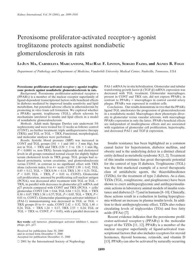
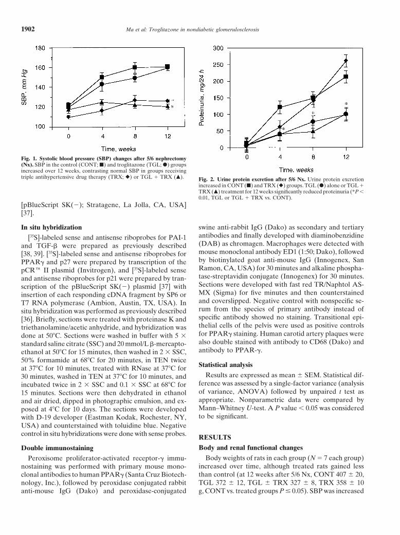
Fig. 1. Systolic blood pressure (SBP) changes after 5/6 nephrectomy(Nx). SBP in the control (CONT; j) and troglitazone (TGL; d) groupsincreased over 12 weeks, contrasting normal SBP in groups receivingtriple antihypertensive drug therapy (TRX; r) or TGL 1 TRX (m). Fig. 2. Urine protein excretion after 5/6 Nx. Urine protein excretion
increased in CONT (j) and TRX (r) groups. TGL (d) alone or TGL 1TRX (m) treatment for 12 weeks significantly reduced proteinuria (*P ,0.01, TGL or TGL 1 TRX vs. CONT).
[pBlueScript SK(2); Stratagene, La Jolla, CA, USA][37].
swine anti-rabbit IgG (Dako) as secondary and tertiaryIn situ hybridizationantibodies and finally developed with diaminobenzidine[35S]-labeled sense and antisense riboprobes for PAI-1(DAB) as chromagen. Macrophages were detected withand TGF-b were prepared as previously describedmouse monoclonal antibody ED1 (1:50; Dako), followed[38, 39]. [35S]-labeled sense and antisense riboprobes forby biotinylated goat anti-mouse IgG (Innogenex, SanPPARg and p27 were prepared by transcription of theRamon, CA, USA) for 30 minutes and alkaline phospha-pCRe II plasmid (Invitrogen), and [35S]-labeled sensetase-streptavidin conjugate (Innogenex) for 30 minutes.and antisense riboprobes for p21 were prepared by tran-Sections were developed with fast red TR/Naphtol AS-scription of the pBlueScript SK(2) plasmid [37] withMX (Sigma) for five minutes and then counterstainedinsertion of each responding cDNA fragment by SP6 orand coverslipped. Negative control with nonspecific se-T7 RNA polymerase (Ambion, Austin, TX, USA). Inrum from the species of primary antibody instead ofsitu hybridization was performed as previously describedspecific antibody showed no staining. Transitional epi-[36]. Briefly, sections were treated with proteinase K andthelial cells of the pelvis were used as positive controlstriethanolamine/acetic anhydride, and hybridization wasfor PPARg staining. Human carotid artery plaques weredone at 508C. Sections were washed in buffer with 5 3also double stained with antibody to CD68 (Dako) andstandard saline citrate (SSC) and 20 mmol/L b-mercapto-antibody to PPAR-g.ethanol at 508C for 15 minutes, then washed in 2 3 SSC,
50% formamide at 688C for 20 minutes, in TEN twiceStatistical analysisat 378C for 10 minutes, treated with RNase at 378C for
Results are expressed as mean 6 SEM. Statistical dif-30 minutes, washed in TEN at 378C for 10 minutes, andference was assessed by a single-factor variance (analysisincubated twice in 2 3 SSC and 0.1 3 SSC at 688C forof variance, ANOVA) followed by unpaired t test as15 minutes. Sections were then dehydrated in ethanolappropriate. Nonparametric data were compared byand air dried, dipped in photographic emulsion, and ex-Mann–Whitney U-test. A P value , 0.05 was consideredposed at 48C for 10 days. The sections were developedto be significant.with D-19 developer (Eastman Kodak, Rochester, NY,
USA) and counterstained with toluidine blue. Negativecontrol in situ hybridizations were done with sense probes. RESULTS
Body and renal functional changesDouble immunostainingBody weights of rats in each group (N 5 7 each group)Peroxisome proliferator-activated receptor-g immu-
increased over time, although treated rats gained lessnostaining was performed with primary mouse mono-than control (at 12 weeks after 5/6 Nx, CONT 407 6 20,clonal antibodies to human PPARg (Santa Cruz Biotech-TGL 372 6 12, TGL 6 TRX 327 6 8, TRX 358 6 10nology, Inc.), followed by peroxidase conjugated rabbit
anti-mouse IgG (Dako) and peroxidase-conjugated g, CONT vs. treated groups P # 0.05). SBP was increased

Ma et al: Troglitazone in nondiabetic glomerulosclerosis 1903
Fig. 3. Renal injury at 12 weeks after 5/6 Nx. TGL alone or TGL 1 TRX treatment for 12 weeks significantly decreased glomerulosclerosis versuscontrol (E). Glomerulosclerosis, tubular atrophy, and interstitial fibrosis were present in CONT (A) and TRX (B) rats; treatment with TGL (C)or TGL 1 TRX (D) for 12 weeks decreased glomerulosclerosis (PAS stain, 3100).
significantly in untreated control rats by four weeks after Lipid levels5/6 Nx (150 6 5 vs. baseline 122 6 3 mm Hg, P , 0.0001) At 12 weeks after 5/6 Nx, serum TG and cholesteroland continued to increase slightly until 12 weeks (Fig. levels in all groups were comparable and remained in1). Interestingly, TGL did not reduce SBP (at 12 weeks the normal range except for a significantly higher serum160 6 3 vs. CONT 161 6 1 mm Hg, P 5 NS). SBP in cholesterol level in the TRX group (serum TG, CONTTRX and TGL 1 TRX groups remained normal (TRX 89 6 9, TGL 134.1 6 10.4, TGL 6 TRX 110.5 6 17.8,126 6 1, TGL 1 TRX 120 6 3 mm Hg, P , 0.0001 vs. TRX 112.5 6 31.6 mg/dL, P 5 NS; serum cholesterol,CONT or TGL, respectively; Fig. 1). Urinary protein CONT 122.9 6 5.4, TGL 130.1 6 6.7, TGL 6 TRX 129.4 6excretion was dramatically increased in CONT rats by 13.1, TRX 161.5 6 14.6 mg/dL, TRX vs. CONT, P # 0.05).four weeks after 5/6 Nx (120.9 6 15.3 vs. baseline 12.0 6
Cellular proliferation1.4 mg/24 h, P , 0.0001) and continued to increase (at12 weeks 213.4 6 29.1 mg/24 h, P , 0.0001 compared Immunostaining for PCNA was negative in the normalwith baseline). TRX did not affect the course of protein- rat glomerulus (data not shown). In CONT rats, PCNA-uria (P 5 NS vs. CONT). In contrast, TGL alone or in positive cells were present in glomeruli and tubules. Glo-combination with TRX significantly reduced proteinuria merular visceral and parietal epithelial cells, mesangial,at 4, 8, and 12 weeks (Fig. 2). At 12 weeks after 5/6 and endothelial cells showed occasional PCNA staining.Nx, serum creatinine was increased in CONT and TRX Glomerular PCNA positivity was dramatically decreasedgroups; TGL and TGL 1 TRX treatment decreased after treatment with TGL or TGL 1 TRX, but not afterserum creatinine levels compared with CONT (serum TRX alone (Fig. 4).creatinine: CONT 3.0 6 0.7, TGL 1.6 6 0.2, TGL 1
p21 and p27 expressionTRX 1.0 6 0.04, TRX 2.0 6 0.4 mg/dL; TGL or TGL 1TRX vs. CONT, P , 0.05). Glomerular p21 mRNA expression, primarily in vis-
ceral epithelial cells, was increased in CONT (Fig. 5A)Renal morphological changes and TRX rats (data not shown) compared with baseline
In untreated CONT rats and TRX rats, glomeruloscle- levels in TGL (Fig. 5B). Glomerular p27 mRNA wasrosis, tubular atrophy, dilation, and interstitial fibrosis expressed at a diffuse, low level, similar in all groupswere present. TRX, as expected, had no significant effect (Fig. 5 C, D). p27 protein expression was up-regulatedon glomerulosclerosis (SI, 0 to 4 scale: CONT 1.99 6 in glomeruli in CONT (Fig. 5E) and TRX rats (data not0.42 vs. TRX 1.30 6 0.21, P 5 NS) [30, 31]. Both TGL shown); treatment with TGL (Fig. 5F) or TGL 1 TRXalone and TGL 1 TRX significantly ameliorated the (data not shown) decreased the p27 protein expression.development of glomerulosclerosis compared with con-
PAI-1 mRNA and protein expressiontrol (CONT 1.99 6 0.42 vs. TGL 0.85 6 0.12, P , 0.05,vs. TGL 1 TRX 0.56 6 0.14, P 5 0.01; Fig. 3 A, B). In situ hybridization study revealed high-level PAI-1Tubulointerstitial fibrosis was reduced in parallel with mRNA expression in CONT and TRX kidneys (Fig.
6 A, B). Strong PAI-1 mRNA signals were detectedglomerulosclerosis after treatment with TGL.

Ma et al: Troglitazone in nondiabetic glomerulosclerosis1904
Fig. 4. Glomerular cell proliferation. TGL alone or TGL 1 TRX treatment for 12 weeks significantly decreased glomerular cell proliferationversus control (E). Staining for PCNA was not detected in the normal rat glomerulus (data not shown). There was a dramatic increase in PCNAstaining in the glomeruli in CONT (A) and TRX-treated rats (B) at 12 weeks after 5/6 Nx. TGL alone (C) or TGL 1 TRX treatment (D) markedlyreduced the PCNA-positive cells in glomeruli.
Fig. 5. p21 and p27 expression. p21 mRNA expression in glomeruli inCONT (A, arrow) and TGL rats (B). p27 mRNA was expressed at adiffuse, low level in glomeruli in CONT (C ) and TGL (D) rats. p27protein was highly expressed in glomeruli in CONT (E ) and was de-creased in TGL (F ). (G) TGL alone or TGL 1 TRX treatment for 12weeks significantly decreased glomerular p27 protein expression versuscontrol.
primarily in sclerotic glomeruli and some tubules. Glo- or TGL 1 TRX vs. CONT, P , 0.05 and P , 0.01,respectively; Fig. 7E). PAI-1 protein expression showedmerular visceral and parietal epithelial, mesangial, and
endothelial cells showed signal. PAI-1 mRNA expres- the same pattern as mRNA (Fig. 7 A-D).sion was markedly reduced in TGL (Fig. 6C) and TGL 1
TGF-b1 mRNA expressionTRX-treated (Fig. 6D) rats compared with untreatedCONT rats. In situ hybridizations with sense probes In situ hybridization showed up-regulated TGF-b
mRNA in untreated CONT rat kidneys (both glomerulishowed no signal. PAI-1 protein expression in TGL andTGL 1 TRX groups was also decreased compared with and tubules). The TGF-b signal appeared attenuated
after TGL treatment for 12 weeks (Fig. 8). Sense probesCONT (PAI-1 scoring: CONT 2.42 6 0.32, TRX 2.53 60.24, TGL 1.40 6 0.24, TGL 1 TRX 1.24 6 0.17; TGL showed no specific signal.

Ma et al: Troglitazone in nondiabetic glomerulosclerosis 1905
Fig. 8. Transforming growth factor-b (TGF-b) mRNA expression byFig. 6. Plasminogen activator inhibitor-1 (PAI-1) mRNA expression.in situ hybridization. TGF-b mRNA was up-regulated in glomerularIn situ hybridization for PAI-1 mRNA showed high expression in glo-cells (A) and tubular epithelial cells (C) in CONT. TGF-b mRNAmeruli in CONT (A) and in TRX-treated rats (B), with signals detectedexpression was attenuated after treatment with TGL for 12 weeks (Bprimarily in sclerotic glomeruli and some cortical tubules. PAI-1 mRNAand D).expression was markedly diminished in rats treated with TGL alone
for 12 weeks (C) with trace signal in parietal epithelial cells. No PAI-1mRNA was expressed in glomeruli in TGL 1 TRX-treated rats (D).
were also PPARg positive. In situ hybridization of ratkidneys showed PPARg mRNA expression with the same
Modulation of glomerular macrophages distribution pattern as PPARg protein (Fig. 10 B, D, F).At 12 weeks after 5/6 Nx, macrophages were present
in glomeruli of CONT and TRX-treated rats (macro-DISCUSSIONphages/glomerulus: CONT 3.84 6 1.24, TRX 2.60 6 1.07,
P 5 NS; Fig. 9). Treatment with TGL alone or TGL 1 Progressive deterioration of the kidney is common toTRX significantly reduced glomerular infiltrating macro- many renal diseases. The structural injury that leads tophages (TGL 0.30 6 0.28, TGL 1 TRX 0.05 6 0.03, this progressive loss of function consists of glomerulo-TGL or TGL 1 TRX vs. CONT, P , 0.05 and P , 0.01, sclerosis and tubulointerstitial fibrosis and atrophy.respectively; Fig. 9). Many intervention strategies have been explored to slow
down or even reverse the progression of glomeruloscle-PPARg expression rosis [40, 41]. In this study, to our knowledge for the first
time, we have demonstrated that PPARg agonist aloneImmunohistochemistry of rat tissue sections demon-strated that PPARg was intensely expressed in the transi- can ameliorate the progression of glomerulosclerosis in
a nondiabetic, hypertensive glomerulosclerosis.tional epithelium of the ureter in all groups as previouslydescribed (Fig. 10A) [21]. This staining thus served as Many studies have shown efficacy of TZD class agents
in diabetic vascular and renal lesions, linked to improveda positive internal control. PPARg was also expresseddiffusely in tubules in all rats. Double immunostaining glucose and lipid metabolism [7, 28, 32, 33]. However,
so far, studies have not examined PPARg agonist effectsfor both macrophages and PPARg demonstrated thatglomerular infiltrating macrophages did not express on nondiabetic glomerulosclerosis. The in vitro evidence
showing promotion of foamy macrophage transforma-PPARg, although resident glomerular cells (includingvisceral, parietal epithelial cells, and mesangial cells) ex- tion by TGL suggested that this class of drugs might be
a dual-edged sword with underlying adverse effects topressed PPARg in CONT rats (Fig. 10C). Glomerularvisceral epithelial cells and tubular epithelial cells in promote atherosclerosis. Our current study shows the
efficacy of PPARg agonist to ameliorate the progressionCONT rats were PPARg positive, and these cell typeswere also PCNA positive. Neither PPARg mRNA nor of glomerulosclerosis. The effect is independent of insu-
lin effects and could only be partially due to lipid effects,protein was detected in glomeruli of TGL or TGL 1TRX-treated rats. Tubular PPARg expression was also as TGL treatment eliminated the significant but small
increase in cholesterol observed in the TRX group, andreduced in rats treated with TGL. TRX-treated rats ex-pressed low levels of PPARg. In TGL or TGL 1 TRX plasma TG levels remained normal in all groups of rats.
Although blood pressure is an important factor associ-rats, there were no macrophages in glomeruli (Fig. 10E).In contrast, macrophages in human carotid artery plaque, ated with progression [42], in this study, we observed
renoprotection with TGL that was independent of effectsstained as control, were identified by CD68 positivity and

Ma et al: Troglitazone in nondiabetic glomerulosclerosis1906
a return to baseline levels for p27 and sustained increasefor p21 [53]. Immune-mediated injury in the passive Hey-mann nephritis model of membranous nephropathy isassociated with an increase in p21 and p27 levels, whichcoincide with little if any proliferation [54]. However,p21 has divergent functions. p21 can act as an assemblyfactor of cdk4/cyclin D complexes or as an inhibitor ofCDK/cyclin [55, 56]. In mesangial cells in vitro, platelet-derived growth factor (PDGF) causes a marked increaseof p21 protein [57]. The induction of p21 protein byPDGF was also reported in p53-deficient as well as innormal mouse fibroblasts, suggesting p53-independentup-regulation of p21 by PDGF. Megyesi et al has recentlyshown that lack of a functional p21 gene amelioratesFig. 9. Modulation of remnant glomerular macrophages. Treatment
with TGL alone or TGL 1 TRX significantly decreased glomerular progression to chronic renal failure [58]. They speculatedmacrophage infiltration versus CONT. that p21 regulates the balance between hyperplasia and
hypertrophy after renal ablation, promoting hypertrophyand sclerosis. We found highly expressed p21 and p27in the glomeruli of untreated 5/6 Nx rats, thus suggestingon systemic hypertension. This indicates that additionalthat there is active ongoing hypertrophy in these scleroticnonhemodynamic factors contribute to sclerosis, as wekidneys. The decreased p21 mRNA expression and p27have extensively investigated previously [43–46].protein in response to TGL indicate that these hypertro-These renal protective effects of PPARg agonist sug-phic, prosclerotic mechanisms were dampened by TGL.gest that PPARg agonists may provide a novel interven-Our results thus support a role for TGL, via PPARgtion strategy to prevent vascular and glomerular sclerosis.activation, on cell cycle events and balance of prolifera-We hypothesize that the multiplicity of PPARg actionstion and hypertrophy.may be due to its effects on several transcriptional fac-
Our study also showed that amelioration of glome-tors. These effects include cell cycle regulation, de-rulosclerosis by PPARg agonist was linked to down-creased PAI-1 and TGF-b, and decreased macrophageregulated PAI-1 expression. PAI-1, a member of theinfiltration.superfamily of serine protease inhibitors, is a major phys-Abnormal cell growth with associated increased ma-iological inhibitor of tissue-type plasminogen activatortrix accumulation is one of the important factors contrib-(t-PA) and urokinase-type plasminogen activator (u-PA).
uting to the development of glomerulosclerosis. Cell pro-These PAs activate plasminogen to plasmin and promote
liferation versus hypertrophy is controlled at the nuclearfibrinolysis as well as proteolysis [59, 60]. Up-regulation
level by cyclin-dependent kinases (CDKs) and cyclin of PAI-1 can thus inhibit proteolysis of extracellular ma-kinase inhibitors (CKIs) [47]. The latter inhibit cell pro- trix, leading to matrix accumulation and sclerosis. PAI-1liferation by causing cell cycle arrest. p21CIP1/WAF1 and is linked to vascular sclerosis in both animal models andp27kip1 belong to the CIP/Kip family of CKIs [48]. humans and is induced by angiotensin [40, 41]. PAI-1 is
One of the possible mechanisms for the beneficial ef- also increased in plasma of patients with insulin resis-fects of PPARg agonist on sclerosis is the observed inhi- tance and is correlated with the severity of atherosclero-bition of increased glomerular cell proliferation, which sis in such patients [61, 62]. In vitro, it was shown thatwas associated with a low-level expression of p21 and TZDs decreased basal and tumor necrosis factor-a–p27. Previous studies have suggested a role for PPARg stimulated PAI-1 expression in human umbilical veinreceptors in the control of cell proliferation, especially endothelial cells [27]. Recent data suggest that PPARgin vascular smooth muscle cell (VSMC) proliferation and agonist inhibited gene transcription by antagonizing themigration, which are critical events in the development activities of the transcription factors (TFs) activator pro-of atherosclerosis and restenosis [49–51]. In the aortic tein-1 (AP-1) and nuclear factor-kB (NF-kB) [63]. Theballoon injury model in rats, TGL resulted in a signifi- PAI-1 promoter contains motifs for both these TFscantly less neointimal/media area ratio compared with [64, 65]. Thus, we speculate that down-regulation of PAI-1control [52], which may relate to PPARg inhibition of mRNA expression may be mediated via a PPARg effectearly inflammatory/proliferation responses. on AP-1 and/or NF-kB–mediated transcriptional activity
Recent studies have shown that mesangial cell prolif- of PAI-1.eration in the Thy-1 experimental glomerulonephritis Transforming growth factor-b is one of the key factorsmodel is associated with decreased p27 levels; the resolu- contributing to extracellular matrix accumulation and
glomerulosclerosis [66], and its inhibition can decreasetion of mesangial cell proliferation was associated with

Ma et al: Troglitazone in nondiabetic glomerulosclerosis 1907
Fig. 7. PAI-1 expression detected by immunostaining. TGL alone or TGL 1 TRX treatment for 12 weeks significantly reduced glomerular PAI-1expression versus control (E ). PAI-1 was highly expressed in glomeruli in CONT (A) and in TRX (B), associated with sclerotic lesions; PAI-1protein was only weakly expressed in some glomerular parietal epithelial cells in TGL group (C). There was only trace PAI-1 expression inglomeruli in the TGL 1 TRX (D) group.
matrix accumulation [67]. In this study, we observedup-regulated TGF-b mRNA in CONT rats, and TGLdiminished this TGF-b mRNA expression. These resultssuggest that beneficial effects of TGL on sclerosis mayoccur through inhibition of both PAI-1 and TGF-b, thusaugmenting matrix turnover and decreasing matrix syn-thesis.
An additional finding in this study was the lack ofPPARg expression in macrophages infiltrating glomer-uli. Monocytes and macrophages play an important rolein immune and nonimmune chronic sclerosing renal dis-eases [68–70]. Activated macrophages are a rich sourceof growth factors, cytokines, vasoactive substances, pro-teolytic enzymes, and reactive oxygen species (ROS) [71].Decreased ROS-related injury caused by decreased mac-rophages may thus have contributed to amelioration ofsclerosis by TGL. Additionally, the antioxidant moiety ofTGL may have decreased oxidant injury directly [72, 73].
These infiltrating macrophages did not express PPARg,although glomerular-resident cells (glomerular visceral,parietal epithelial cells, and mesangial cells) did showincreased PPARg expression in the untreated 5/6 Nxrats. Our results indicate that there is a phenotypic diver-sity of macrophages in glomerular versus vascular sclero-sis. Macrophages in human atherosclerotic carotid arteryplaques did express PPARg and are thought to promote
Fig. 10. Detection of PPARg and macrophages. Transitional epithelial foam cell transformation and plaque formation [74]. Be-cells of pelvis, which express PPARg, served as a positive control (A).cause glomerular resident cells contribute actively to glo-Carotid artery plaque, another positive control, showed PPARg-posi-
tive, CD68-positive macrophages (data not shown). Double immuno- merulosclerosis [41], our results also suggest that glomer-staining showed that glomerular infiltrating macrophages (C, red, arrow- ular intrinsic cells are a target of PPARg agonist. PPARghead) did not express PPARg, although glomerular cells did show
expressed in these glomerular cells may act to counter-increased PPARg expression (C, brown, arrow). There was no detect-able PPARg in glomeruli in TGL rats (E ). In situ hybridization showed balance the existing overwhelming proinflammatory andthat PPARg mRNA expression had similar distribution as PPARg prosclerotic effects. A reduction of glomerular macro-receptor protein expression [(D), CONT and (F ), TGL]. Ureteral epi-thelium, which expresses PPARg, served as a positive control (B). phages by TGL treatment in our study could reflect

Ma et al: Troglitazone in nondiabetic glomerulosclerosis1908
retinoic X receptor; SI, sclerosis index; SPB, systolic blood pressure;TGL’s effects in inflammation regulation. Possible mech-TF, transcription factor; TGF-b, transforming growth factor-b; TGL,
anisms include TGL inhibition of expression of vascular troglitazone group; TNF-a, tumor necrosis factor-a; t-PA tissue-typecell adhesion molecule-1 (VCAM-1) and intercellular plasminogen activator; TRX, triple antihypertensive drug therapy group;
TZDs, thiazolidinediones; VCAM-1, vascular cell adhesion molecule-1;adhesion molecule-1 (ICAM-1) in endothelial cells, re-VSMC, vascular smooth muscle cell; u-PA, urokinase-type plasmino-sulting in significantly reduced monocyte/macrophage gen activator.
homing to atherosclerotic plaques [75]. PPARg ligandsalso inhibited the inflammatory response and reduced REFERENCEScolonic epithelial injury in a mouse model of colitis with
1. Reaven GM: Role of insulin resistance in human disease. DiabetesPPARg expression primarily in the colonic epithelium 37:1595–1607, 1988[76]. Interestingly, in our study, glomerular PPARg was 2. Itoh H, Doi K, Tanaka T, et al: Hypertension and insulin resis-
tance: Role of peroxisome proliferator-activated receptor gamma.undetectable by in situ hybridization in TGL-treated ani-Clin Exp Pharmacol Physiol 26:558–560, 1999mals. We speculate that this finding could represent li-
3. Fujita T, Sugiyama Y, Taketomi S, et al: Reduction of insulingand-mediated receptor down-regulation, which has been resistance in obese and/or diabetic animals by 5-[4-(1-methylcyclo-
hexylmethoxy) benzyl]-thiazolidine-2,4-dione (ADD-3878, U-63,287,demonstrated for other cytoplasmic and nuclear recep-ciglitazone), a new antidiabetic agent. Diabetes 32:804–810, 1983tors [77, 78].
4. Fujiwara T, Yoshioka S, Yoshioka T, et al: Characterization ofIn summary, this study shows that PPARg activation new oral antidiabetic agent CS-045: Studies in KK and ob/ob mice
and Zucker fatty rats. Diabetes 37:1549–1558, 1988in vivo in a nondiabetic, glomerulosclerotic model leads5. Sreenan S, Sturis J, Pugh W, et al: Prevention of hyperglycemiato beneficial effects, disproving the hypothesis suggested
in the Zucker diabetic fatty rat by treatment with metformin orby in vitro studies that PPARg activation promoting troglitazone. Am J Physiol 271:E742–E747, 1996
6. Kemnitz JW, Elson DF, Roecker EB, et al: Pioglitazone increasesmacrophage foam cell transformation results in in-insulin sensitivity, reduces blood glucose, insulin, and lipid levels,creased vascular sclerosis. PPARg’s beneficial effects ap-and lowers blood pressure in obese, insulin-resistant rhesus mon-
pear to be related to the regulation of glomerular cell keys. Diabetes 43:204–211, 19947. Brown KK, Henke BR, Blanchard SG, et al: A novel N-arylproliferation and hypertrophy, decreased macrophage
tyrosine activator of peroxisome proliferator-activated receptor-infiltration, and decreased PAI-1 and TGF-b and aregamma reverses the diabetic phenotype of the Zucker diabetic
not mediated by improving insulin sensitivity. Our results fatty rat. Diabetes 48:1415–1424, 19998. Iwamoto Y, Kuzuya T, Matsuda H, et al: Effect of new oralfurther indicate that there is a phenotypic diversity of
antidiabetic agent CS-045 on glucose tolerance and insulin secre-macrophages in glomerular versus vascular sclerosis,tion in patients with NIDDM. Diabetes Care 14:1083–1086, 1991
with PPARg expression in macrophages occurring only 9. Yamasaki Y, Kawamori R, Wasada T, et al: Pioglitazone (AD-4833) ameliorates insulin resistance in patients with NIDDM: AD-in the latter. We speculate that amelioration of develop-4833 Glucose Clamp Study Group, Japan. Tohoku J Exp Medment of glomerulosclerosis could be mediated via a183:173–183, 1997
PPARg effect on AP-1 and/or NF-kB–mediated tran- 10. Saltiel AR, Olefsky JM: Thiazolidinediones in the treatment ofinsulin resistance and type II diabetes. Diabetes 45:1661–1669, 1996scriptional activity of PAI-1. Our results also suggest
11. Lehmann JM, Moore LB, Smith-Oliver TA, et al: An antidiabeticthat PPARg agonists will provide a novel approach withthiazolidinedione is a high affinity ligand for peroxisome prolifera-
therapeutic potential in nondiabetic glomerulosclerosis. tor-activated receptor gamma (PPAR gamma). J Biol Chem 270:12953–12956, 1995
12. Willson TM, Cobb JE, Cowan DJ, et al: The structure-activityACKNOWLEDGMENTSrelationship between peroxisome proliferator-activated receptorgamma agonism and the antihyperglycemic activity of thiazoli-These studies were supported by National Institutes of Healthdinediones. J Med Chem 39:665–668, 1996Grants DK 39261 and DK 52104 (A.B.F.), and was supported in part
13. Mangelsdorf DJ, Thummel C, Beato M, et al: The nuclear recep-by AHA Grant-in-Aid #95011450 (S.F.) and by National Institutes oftor superfamily: The second dacade. Cell 83:835–839, 1995Health grants HL53989 (M.F.L.), HL57986 (S.F.). Drs. Fazio and Lin-
14. Forman BM, Tontonoz P, Chen J, et al: 15-Deoxy-delta 12, 14-ton are Established Investigators of the American Heart Association.prostaglandin J2 is a ligand for the adipocyte determination factorPPAR gamma. Cell 83:803–812, 1995Reprint requests to Agnes B. Fogo, M.D., MCN C3310, Department
15. Kliewer SA, Lenhard JM, Willson TM, et al: A prostaglandinof Pathology, Vanderbilt University Medical Center, 21st and GarlandJ2 metabolite binds peroxisome proliferator-activated receptorAvenue, Nashville, Tennessee 37232-2561, USA.gamma and promotes adipocyte differentiation. Cell 83:813–819,E-mail: [email protected]
16. Lehmann JM, Lenhard JM, Oliver BB, et al: Peroxisome prolifer-ator-activated receptors alpha and gamma are activated by indo-APPENDIXmethacin and other non-steroidal anti-inflammatory drugs. J BiolChem 272:3406–3410, 1997Abbreviations used in this study are: AP-1, activated protein-1;
17. Varanasi U, Chu R, Huang Q, et al: Identification of a peroxisomeCDK, cyclin-dependent kinase; CKI, cyclin kinase inhibitor; CONT,proliferator-responsive element upstream of the human peroxi-control; DAB, diaminobenzidine; 15-deoxy PGJ2, 15-deoxy-D12,14-pros-somal fatty acyl coenzyme A oxidase gene. J Biol Chem 271:2147–taglandin J2; FFAs, free fatty acids; HUVEC, human umblical vein2155, 1996endothelial cell; ICAM-1, intercellular adhesion molecule-1; LDL, low
18. Tontonoz P, Hu E, Spiegelman BM: Stimulation of adipogenesisdensity lipoprotein; NF-kB, nuclear factor-kB; NSAIDs, nonsteroidalin fibroblasts by PPAR gamma 2, a lipid-activated transcriptionanti-inflammatory drugs; Nx, nephrectomy; PAI-1, plasminogen activa-factor. Cell 79:1147–1156, 1994tor inhibitor-1; PCNA, proliferating cell nuclear antigen; PDGF, plate-
19. Brun RP, Kim JB, Hu E, et al: Adipocyte differentiation: A tran-let-derived growth factor; PPARg, peroxisome proliferator-activatedreceptor-g; PPREs, peroxisome proliferator response elements; RXR, scriptional regulatory cascade. Curr Opin Cell Biol 8:826–832, 1996

Ma et al: Troglitazone in nondiabetic glomerulosclerosis 1909
20. Spiegelman BM: PPAR-g: Adipogenic regulator and thiazoli- of glomerular function in two rat models of glomerular sclerosis.J Clin Invest 82:322–330, 1988dinedione receptor. Diabetes 47:507–514, 1998
21. Guan YF, Zhang YH, Davis L, et al: Expression of peroxisome 44. Yoshioka T, Shiraga S, Yoshida Y, et al: “Intact nephrons” asthe primary origin of proteinuria in chronic renal disease: Studyproliferator-activated receptors in urinary tract of rabbits and hu-
mans. Am J Physiol 273:F1013–F1022, 1997 in the rat model of subtotal nephrectomy. J Clin Invest 82:1614–1623, 198822. Steinberg D, Parthasarathy S, Carew TE, et al: Beyond choles-
terol. N Engl J Med 320:915–924, 1989 45. Yoshida Y, Fogo A, Ichikawa I: Glomerular hemodynamicchanges vs. hypertrophy in experimental glomerular sclerosis. Kid-23. Tontonoz P, Nagy L, Alvarez JGA, et al: PPARg promotes
monocyte/macrophage differentiation and uptake of oxidized ney Int 35:654–660, 198946. Ikoma M, Kawamura T, Fogo A, et al: Cause of variable therapeu-LDL. Cell 93:246–252, 1998
24. Nagy L, Tontonoz P, Alvarez J, et al: Oxidized LDL regulates tic efficiency of angiotensin-converting enzyme inhibitor on theglomerular mesangial lesions. Kidney Int 40:195–202, 1991macrophage gene expression through ligand activation of PPARg.
Cell 93:229–240, 1998 47. Pardee AB: G1 events and regulation of cell proliferation. Science246:603–608, 198925. Ricote M, Huang J, Fajas L, et al: Expression of the peroxisome
proliferator-activated receptor gamma (PPARgamma) in human 48. Shankland S: Cell cycle regulatory proteins in glomerular disease.Kidney Int 56:1208–1215, 1999atherosclerosis and regulation in macrophages by colony stimulat-
ing factors and oxidized low density lipoprotein. Proc Natl Acad 49. Ross R: The pathogenesis of atherosclerosis: A perspective forthe 1990s. Nature 362:801–809, 1993Sci USA 95:7614–7619, 1998
26. Marx N, Bourcier T, Sukhova GK, et al: PPARg activation in 50. Dubey RK, Zhang HY, Reddy SR, et al: Pioglitazone attenuateshypertension and inhibits growth of renal arteriolar smooth musclehuman endothelial cells increases plasminogen activator inhibitor
type-1 expression. Arterioscler Thromb Vasc Biol 19:546–551, 1999 in rats. Am J Physiol 265:R726–R732, 199351. Marx N, Schonbeck U, Lazar MA, et al: Peroxisome proliferator-27. Kato K, Satoh H, Endo Y, et al: Thiazolidinediones down-regulate
plasminogen activator inhibitor type 1 expression in human vascu- activated receptor gamma activators inhibit gene expression andmigration in human vascular smooth muscle cells. Circ Res 83:1097–lar endothelial cells: A possible role for PPARgamma in endothe-
lial function. Biochem Biophys Res Commun 258:431–435, 1999 1103, 199852. Law R, Meehan W, Xi X, et al: Troglitazone inhibits vascular28. Buckingham RE, Al-Barazanji KA, Toseland CD, et al: Peroxi-
some proliferator-activated receptor-gamma agonist, rosiglitazone, smooth muscle cell growth and intimal hyperplasia. J Clin Invest98:1897–1905, 1996protects against nephropathy and pancreatic islet abnormalities in
Zucker fatty rats. Diabetes 47:1326–1334, 1998 53. Shankland SJ, Hugo C, Coats SR, et al: Changes in cell-cycleprotein expression during experimental mesangial proliferative29. Shiomi M, Ito T, Tsukada T, et al: Combination treatment with
troglitazone, an insulin action enhancer, and pravastatin, an inhibi- glomerulonephritis. Kidney Int 50:1230–1239, 199654. Shankland SJ, Floege J, Thomas SE, et al: Cyclin kinase inhibitorstor of HMG-CoA reductase, shows a synergistic effect on athero-
sclerosis of WHHL rabbits. Atherosclerosis 142:345–353, 1999 are increased during experimental membranous nephropathy: Po-tential role in limiting glomerular epithelial cell proliferation in30. Anderson S, Rennke H, Brenner BM: Therapeutic advantage of
converting enzyme inhibitors in arresting progressive renal disease vivo. Kidney Int 52:404–413, 199755. Funk JO, Galloway DA: Inhibiting CDK inhibitors: New lessonsassociated with systemic hypertension in the rat. J Clin Invest
77:1993–2000, 1986 from DNA tumor viruses. Trends Biochem Sci 23:337–341, 199856. Labaer J, Garrett MD, Stevenson LF, et al: New functional31. Kakinuma Y, Kawamura T, Bills T, et al: Blood pressure-indepen-
dent effect of angiotensin inhibition on vascular lesions of chronic activities for the p21 family of CDK inhibitors. Genes Dev 11:847–862, 1997renal failure. Kidney Int 42:46–55, 1992
32. Beales P, Liddi R, Giorgini A, et al: Troglitazone prevents insulin 57. Schocklmann HO, Lang S, Sterzel RB: Regulation of mesangialproliferation. Kidney Int 56:1199–1207, 1999dependent diabetes in the non-obese diabetic mouse. Eur J Phar-
macol 357:221–225, 1998 58. Megyesi J, Price PM, Tamayo E, et al: The lack of a functionalp21 (WAF1/CIP1) gene ameliorates progression to chronic renal33. Ogawa J, Takahashi S, Fujiwara T, et al: Troglitazone can prevent
development of type 1 diabetes induced by multiple low-dose strep- failure. Proc Natl Acad Sci USA 96:10830–10835, 199959. Fogo AB: The role of angiotensin II and plasminogen activatortozotocin in mice. Life Sci 65:1287–1296, 1999
34. Fazio S, Lee Y-L, Ji Z-S, et al: Type II hyperlipoproteinemic inhibitor-1 in progressive glomerulosclerosis. Am J Kidney Dis35:179–188, 2000phenotype in transgenic mice expressing dysfunctional apolipo-
protein E. J Clin Invest 92:1497–1503, 1993 60. Vaughan DE: Endothelial function, fibrinolysis, and angiotensin-converting enzyme inhibition. Clin Cardiol 20(Suppl 2):II34–II37,35. Nakamura S, Nakamura I, Ma LJ, et al: Plasminogen activator
inhibitor-1 (PAI-1) expression is regulated by the angiotensin type 199761. Anwerx J, Bouilou R, Collen D, et al: Tissue-type plasminogen1 receptor in vivo. Kidney Int 58:251–259, 2000
36. Taniguchi T, Endo H, Chikatsu N, et al: Expression of p21 and p27 activator antigen and plasminogen activator inhibitor in diabetesmellitus. Atherosclerosis 8:68–72, 1988cyclin-dependent kinase inhibitors during human hematopoiesis.
Blood 93:4167–4178, 1999 62. Schneiderman J, Sawdey MS, Keeton M, et al: Increased type 1plasminogen activator inhibitor gene expression in atherosclerotic37. Guan Y-F, Zhang Y-H, Davis L, et al: Expression of peroxisome
proliferator-activated receptor g (PPAR g) in human transitional human arteries. Proc Natl Acad Sci USA 89:6998–7002, 199263. Ricote M, Li AC, Willson TM, et al: The peroxisome proliferator-bladder cancer and its role in inducing cell death. Neoplasia 1:330–
339, 1999 activated receptor-gamma is a negative regulator of macrophageactivation. Nature 391:79–82, 199838. Oikawa T, Freeman M, Lo W, et al: Modulation of plasminogen
activator inhibitor-1 in vivo: A new mechanism for the anti-fibrotic 64. Descheemaeker KA, Wyns S, Nelles L, et al: Interaction of AP-1,AP-2 and Sp1-like proteins with two distinct sites in the upstreameffect of renin-angiotensin inhibition. Kidney Int 51:164–172, 1997
39. Ma L-J, Nakamura S, Whitsitt JS, et al: Regression of sclerosis regulatory region of the plasminogen activator inhibitor-1 genemediates the phorbol 12-myristate 13-acetate response. J Biolin aging by angiotensin inhibition-induced decrease in PAI-1. Kid-
ney Int 58:2425–2436, 2000 Chem 267:15086–15091, 199265. Dawson SJ, Wiman B, Hamsten A, et al: The two allele sequences40. Fogo AB: Mesangial matrix modulation and glomerulosclerosis.
Exp Nephrol 7:147–159, 1999 of a common polymorphism in the promoter of the plasminogenactivator inhibitor-1 (PAI-1) gene respond differently to interleu-41. Fogo AB: Progression and potential regression of glomerulosclero-
sis. (Nephrology Forum) Kidney Int 59:804–819, 2001 kin-1 in HepG2 cells. J Biol Chem 268:10739–10745, 199366. Border WA, Noble NA, Ketteler M: TGF-b: A cytokine media-42. Bidani AK, Griffin KA, Bakris G, et al: Lack of evidence of
blood pressure-independent protection by renin-angiotensin sys- tor of glomerulosclerosis and a target for therapeutic intervention.Kidney Int 47(Suppl 49):S59–S61, 1995tem blockade after renal ablation. Kidney Int 57:1651–1661, 2000
43. Fogo A, Yoshida Y, Glick AD, et al: Serial micropuncture analysis 67. Border WA, Okuda S, Languino LR, et al: Suppression of experi-

Ma et al: Troglitazone in nondiabetic glomerulosclerosis1910
mental glomerulonephritis by antiserum against transforming disease in chronic renal failure: Evidence from animal studies andpathogenesis. Israel J Med Sci 29:228–239, 1993growth factor b1. Nature 346:371–374, 1990
74. Ricote M, Huang J, Fajas L, et al: Expression of the peroxisome68. van Goor H, Fidler V, Weening JJ, et al: Determinants of focalproliferator-activated receptor gamma (PPARgamma) in humanand segmental glomerulosclerosis in the rat after renal ablation:atherosclerosis and regulation in macrophage by colony stimulatingevidence for involvement of macrophages and lipids. Lab Investfactors and oxidized low density lipoprotein. Proc Natl Acad Sci64:754–765, 1991USA 95:7614–7619, 199869. Bagchus WM, Jeunink MF, Elema JD: The mesangium in anti-
75. Pasceri V, Wu HD, Willerson JT, Yeh ET: Modulation of vascu-thy-1 nephritis: Influx of macrophages, mesangial cell hypercellu-lar inflammation in vitro and in vivo by peroxisome proliferator-larity, and macromolecular accumulation. Am J Pathol 137:215–activated receptor-gamma activators. Circulation 101:235–238, 2000223, 1990
76. Su CG, Wen X, Bailey ST, et al: A novel therapy for colitis utilizing70. van Goor H, van der Horst MLC, Fidler V, et al: Glomerular PPAR-g ligands to inhibit the epithelial inflammatory response.macrophage modulation affects mesangial expansion in the rat J Clin Invest 104:383–389, 1999after renal ablation. Lab Invest 66:564–571, 1992 77. Zwaagstra JC, Kassam Z, O’Connor-McCourt MD: Down-regu-
71. Nathan CF: Secretory products of macrophages. J Clin Invest lation of transforming growth factor-b receptors: Cooperativity79:319–326, 1987 between the type 1, II, and III receptors and modulation at the
72. Noguchi N, Sakai H, Kato Y, et al: Inhibition of oxidation of low cell surface. Exp Cell Res 252:352–362, 1999density lipoprotein by troglitazone. Atherosclerosis 123:227–234, 78. David MD, Vanderkuur JA, Brooks SC: Ligand structure influ-1996 ences autologous downregulation of estrogen receptor-alpha mes-
senger RNA. J Steroid Biochem Mol Biol 70:27–37, 199973. Shohat J, Boner G: Role of lipids in the progression of renal