periprosthetic knee fractures
TRANSCRIPT
Core Curriculum V5
Periprosthetic Knee FracturesRamzy Meremikwu MD, Mark Hake MD, Jaimo Ahn MD PhD
Department of Orthopaedic Surgery, Michigan Medicine, University of Michigan
Additional case contributions by Samir Mehta MD and Derek Donegan MD MBA, University of Pennsylvania
Core Curriculum V5
Objectives
• Epidemiology • Preoperative Planning • Fixation strategies • Cases• Postoperative Management • Summary
Core Curriculum V5
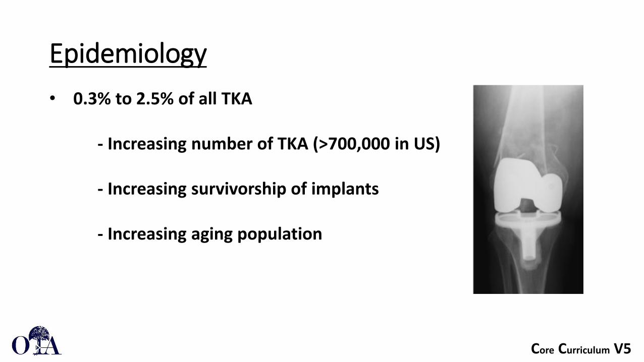
Epidemiology• 0.3% to 2.5% of all TKA
- Increasing number of TKA (>700,000 in US)
- Increasing survivorship of implants
- Increasing aging population

Core Curriculum V5
Factors that increase Prosthetic Fractures
• Mismatch between bone density and implant
• Aging and Osteoporosis
• Fragility fractures

Core Curriculum V5
Bone Stock after TKA
• TKA leads to decrease in periprosthetic bone mineral density (BMD) for up to 7 years postoperatively, with the greatest decline 3 months after surgery due to following factors:
• Stress shielding
• Osteolysis as result abrasion with periprosthetic bone
• Loosening of the implant
• Osteonecrosis

Core Curriculum V5
Preoperative Planning
• History and Physical Exam- Exclude other injuries - Preinjury pain- Complete neurovascular exam
• Preoperative assessment - Index procedure- Type of implant- Comorbidities- Pre-operative ambulatory status

Core Curriculum V5
Radiographic Work Up
• Radiographs- Proper AP and Lateral of Knee- Full length femur and tibia films - Previous x-rays if possible
• CT Scan- Fracture and component stability- Implant dimensions (eg size of implant)- Open box: opening in femoral component of TKA that allows for
possible passage of femoral nail- Artifact sparing cuts

Core Curriculum V5
Is the implant loose?
• Pain • Radiographic changes from index procedure • Lab values (ESR, CRP, CBC for WBC, ect) • Pre-injury mobility• Instability • Fracture
Core Curriculum V5
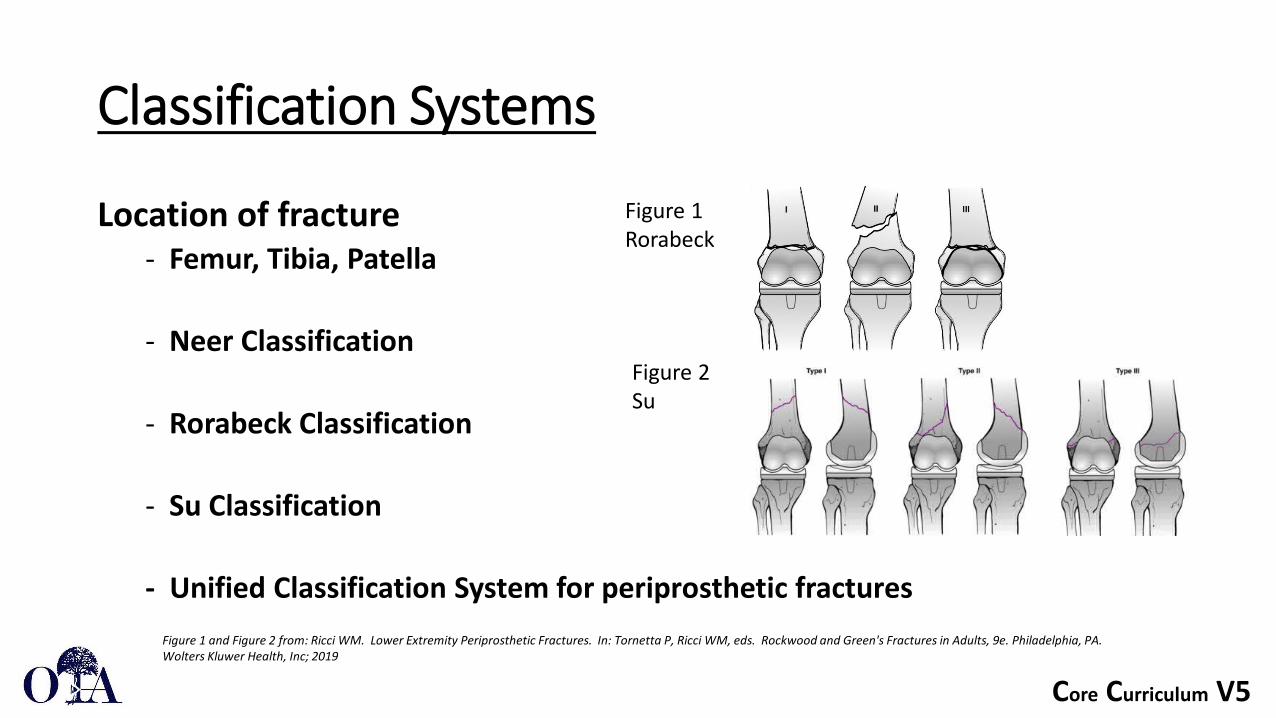
Classification Systems
Location of fracture- Femur, Tibia, Patella
- Neer Classification
- Rorabeck Classification
- Su Classification
- Unified Classification System for periprosthetic fractures
Figure 1 Rorabeck
Figure 2Su
Figure 1 and Figure 2 from: Ricci WM. Lower Extremity Periprosthetic Fractures. In: Tornetta P, Ricci WM, eds. Rockwood and Green's Fractures in Adults, 9e. Philadelphia, PA. Wolters Kluwer Health, Inc; 2019

Core Curriculum V5
Unified Classification System
• The Unified Classification System allows for simplified classification and treatment algorithm for any bone and joint that is involved. The core principles include:
• The location of the fracture
• The fixation of the component
• The adequacy of bone stock around the implant

Core Curriculum V5
Unified Classification System
Location
• I: Shoulder• II: Elbow • III: Wrist • IV: Hip • V: Knee • VI: Ankle
Types• A: Apophyseal• B: Bed of Implant• C: Clear of implant • D: Dividing the bone between two
implant • E: Each of two bone supporting one
arthroplasty • F: Facing and articulating with
hemiarthroplasty
UCS is based on Location and Type

Core Curriculum V5
Unified Classification System
Field testing the Unified Classification System for periprosthetic fractures of the femur, tibia and patella in association with knee replacement: an international collaboration. Van der Merwe JM, Haddad FS, Duncan CP. • 10 fellowship trained orthopedic surgeons (experts) and 10 residents of
orthopedic surgery in last two years of training (pre-experts)• 15 radiographs for evaluation to measure inter and intra-observer
reliability• Kappa value for inter-observer reliability for experts and pre-experts is
0.741 and 0.765 and intra-observer for experts and pre-experts is 0.898 and 0.878.
Core Curriculum V5
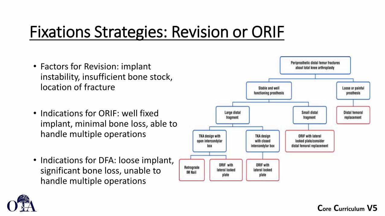
Fixations Strategies: Revision or ORIF
• Factors for Revision: implant instability, insufficient bone stock, location of fracture
• Indications for ORIF: well fixed implant, minimal bone loss, able to handle multiple operations
• Indications for DFA: loose implant, significant bone loss, unable to handle multiple operations
Core Curriculum V5
Fixations Strategies: Revision or ORIF
Primary Versus Secondary Distal Femoral Arthroplasty for Treatment of Total Knee Arthroplasty Periprosthetic Femur Fractures. Antonia F. Chen et al. - Retrospective Study, 48 patients, 35 with primary DFA, 13 with
secondary DFA- Increased postoperative complications (infections, dislocation, and
effusions) for secondary DFA patients - Increased number of surgeries for secondary DFA patients

Core Curriculum V5
ORIF Principles
• Fracture location helps determine LP vs IMN (or Nail Plate Combination)
• TKA implant must be stable • Restore mechanical and joint line axis • Achieve stable fixation to allow
immediate weight bearing if able • Span the femur, plating or nailing

Core Curriculum V5
Locked Plating vs IMN
• Equivalent union rates between intramedullary nail and locked plate fixation for distal femur periprosthetic fractures - a systematic review. Jay K Shah, Patrick Szukics, Arianna L Gianakos, Frank A Liporace, Richard S Yoon
- Meta analysis: 38 studies with 1,188 patients - No difference in IMN, and LCP when analysis union rate or time to union - LP significant lower complication and reoperation rate - IMN with higher percentage and quicker time to full weightbearing (100% and
7.6 weeks) when compared to plating (94% and 15.8 weeks)- IMN with higher percentage to preinjury activity when compared to those
treated with plating (70.8% vs. 61.6%)

Core Curriculum V5
Locked and Hybrid-locked Plating
• Indications: Stable implant, closed box TKA, low, distal fracture
• Approach: Midline or lateral parapatellar - Midline allows TKA assessment, easy to
transition to DFR if needed
• Important Techniques: - Span the femur - Utilize hybrid fixation (locking and non
locking screws)- Ensure proper length, alignment, and
rotation before leaving OR

Core Curriculum V5
Lateral Approach for Plating Periprosthetic Fracture • https://otaonline.org/video-library/45036/procedures-and-
techniques/multimedia/16731389/lateral-distal-femur-plate-for-periprosthetic
• *Note locking and nonlocking hybrid fixation for lateral plate

Core Curriculum V5
Intramedullary Nail• Indications: Stable implant, proximal enough for distally
locked screw- Confirm open box implant and size; important to have
the operative report, implant and nail mismatch possible
- Periprosthetic Supracondylar Femoral Fractures Above A Total Knee Replacement: Compatibility Guide for Fixation With Retrograde Intramedullary Nail. Thompson et al. Arthroplasty 2014.
• Antegrade vs Retrograde: Depends on proximal or distal fracture
• Important Techniques: - Utilize plate for hybrid fixation if needed - Remove/replace polyethylene- Ensure proper length, alignment, and rotation before
leaving OR
Core Curriculum V5
Technique Pearls for Nail placement with TKA
• Know preexisting implants (secure previous operative reports if possible)
• Implant measurement specifics from TKA and IMN can be obtained• Starting point is key to prevent valgus and recruvatum deformities • Service et al, JOT 2015: Much higher change of having a starting point
posterior to Blumensaat’s line on TKA implants (when compared to native knees) and CR implants (when compared to PS implants)
• Start point too posterior may cause extension deformity and injury to PCL

Core Curriculum V5
Retrograde Intramedullary Nail through a Total Knee Arthroplasty • OTA Technique Video for Retrograde IMN for Distal Femur Fracture
through a Total Knee Arthroplasty • https://otaonline.org/video-library/45036/procedures-and-
techniques/multimedia/18826480/retrograde-intramedullary-nail-for-distal-femur

Core Curriculum V5
Box and Implant sizes • Box implants, sizes and compatibility with supracondylar nails • Currall et al; Retrograde nailing for supracondylar fracture around
total knee replacement: A compatibility study using the Trigensupracondylar nail, 2007
• Thompson et al.; Periprosthetic Supracondylar Femoral Fractures Above a Total Knee Replacement: Compatibility Guide for Fixation With a Retrograde Intramedullary Nail, 2014
• Jones et al; Retrograde femoral nailing of periprosthetic fractures around total knee replacements, 2016
Core Curriculum V5
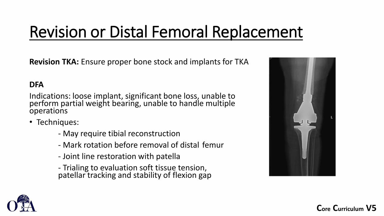
Revision or Distal Femoral Replacement
Revision TKA: Ensure proper bone stock and implants for TKA
DFAIndications: loose implant, significant bone loss, unable to perform partial weight bearing, unable to handle multiple operations • Techniques:
- May require tibial reconstruction- Mark rotation before removal of distal femur - Joint line restoration with patella- Trialing to evaluation soft tissue tension, patellar tracking and stability of flexion gap

Core Curriculum V5
DFR Outcomes
• “Distal Femoral Replacements for Acute Comminuted PeriprostheticKnee Fractures: Satisfactory Clinical Outcomes at Medium-Term Follow-Up.” Matar, Hosam E, et al. Arthroplasty Today, vol. 7. February 2021.
• Retrospective study of 31 patients measuring clinical outcomes of DFRs from 2010 – 2018
• 81 average age of patients, all Rorabeck type II/III fractures, 7.4% complication rate with 1 reoperation (polyethylene insert), avg length of hospital stay 17.8 days, 3 passed away due to multiple commodities. No cases of infection
• DFRs allow for early mobilization and rehabilitation to restore function in a challenging group of patients
Core Curriculum V5
DFR Outcomes
• Long-Term Results of Total Knee Arthroplasty with Contemporary Distal Femoral Replacement. Wyles et al. The Journal of Bone and Joint Surgery: January 2, 2020.
• Retrospective study of 144 patients who underwent TKA with DFR from 2000 to 2015 with greater than 2 years of follow up
• 10 year cumulative complication rates: aseptic loosening (17%), all-cause revision (27.5%), and any reoperation (46.3%), increased risk of re-operation for patients who underwent index DFR for aseptic loosening compared to peripostheitic or native femoral fractures. KSS (Knee society score) increased from 45 pre-op to 75 post op; 7 AKA at time of final followup
• Great clinical improvement indicated by KSS, but high changes of revision and reoperation for end stage revision procedure
Core Curriculum V5
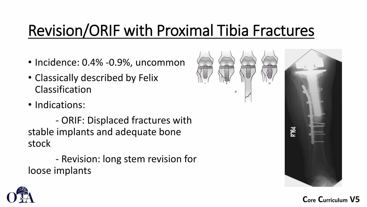
Revision/ORIF with Proximal Tibia Fractures
• Incidence: 0.4% -0.9%, uncommon• Classically described by Felix
Classification • Indications:
- ORIF: Displaced fractures with stable implants and adequate bone stock
- Revision: long stem revision for loose implants
Core Curriculum V5
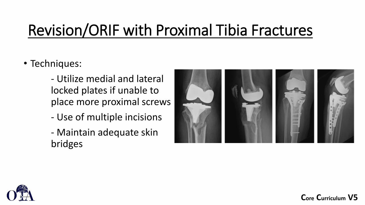
Revision/ORIF with Proximal Tibia Fractures
• Techniques:- Utilize medial and lateral locked plates if unable to place more proximal screws - Use of multiple incisions - Maintain adequate skin bridges
Core Curriculum V5
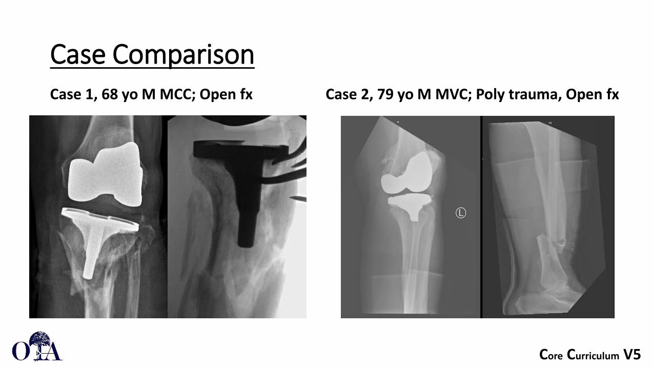
Case ComparisonCase 1, 68 yo M MCC; Open fx Case 2, 79 yo M MVC; Poly trauma, Open fx
Core Curriculum V5
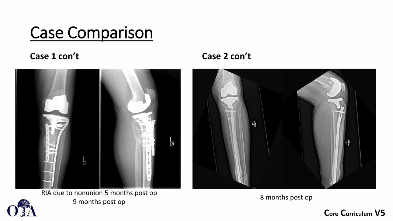
Case ComparisonCase 1 con’t Case 2 con’t
RIA due to nonunion 5 months post op 9 months post op 8 months post op
Core Curriculum V5
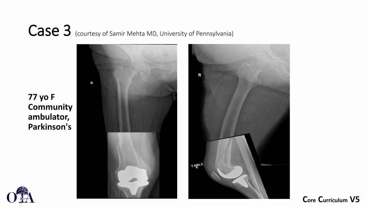
Case 3 (courtesy of Samir Mehta MD, University of Pennsylvania)
77 yo F Community ambulator, Parkinson's

Core Curriculum V5
Case 3
Nail-Plate combination for added strength and immediate weight-bearing
Core Curriculum V5
Nail-Plate Combination Outcomes• Nail Plate Combination Technique for Native and Periprosthetic
Distal Femur Fractures. Liporace FA, Yoon RS. J Orthop Trauma. 2019• Review of 15 patients (9 periprosthetic and 6 native), mean age 74.8• All patients made WBAT immediately after surgery • Mean follow up time 19.2 weeks, 1 deceased patient due to unrelated
comorbidity • No reports of nonunion, hardware failure, deep infections, or subsequent OR
returns; 1 superficial SSI• 14 patients remain ambulatory with assistive device • Limited study for periprosthetic fractures, but NPC allows for reproducible
technique with immediate weight-bearing opportunities in the elderly patient population
Core Curriculum V5
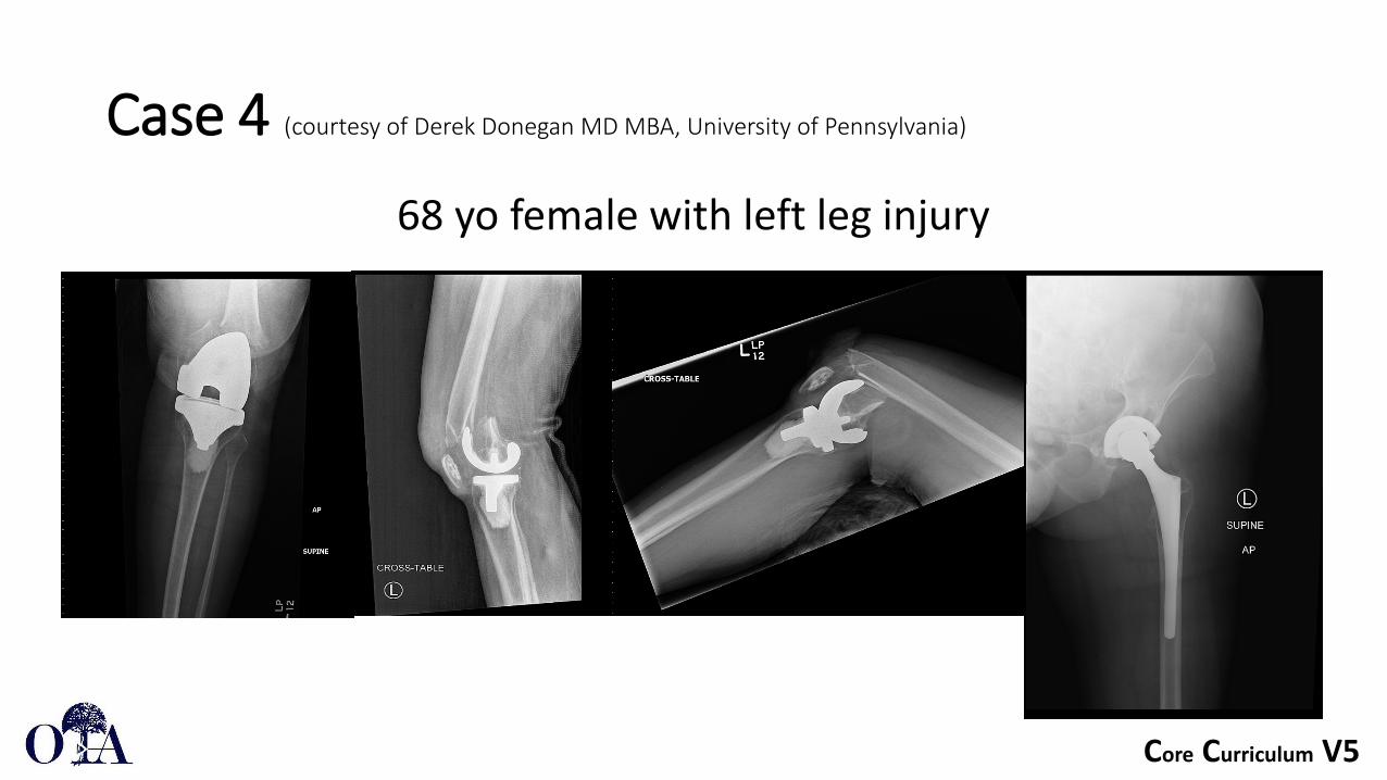
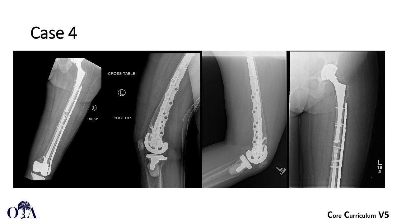
Case 4 (courtesy of Derek Donegan MD MBA, University of Pennsylvania)
68 yo female with left leg injury

Core Curriculum V5
Case 4

Core Curriculum V5
Case 4
Core Curriculum V5
Case 4
Core Curriculum V5
Postoperative Management
• Goal: Immediate/Rapid Mobilization and progression to full weight bearing
- Advantage of DFR, TKA revision, and IMN- WB with plate fixation: Generally restricted for 6-12 weeks postoperatively
• DVT Prophylaxis is essential during early post-operative day course - Duration and type of VTE treatment dependent on patient and
injury factors• Standard 1st gen IV Abx for 24 hours
Core Curriculum V5
Summary
• TKA Periprosthetic fracture incidence will continue to grow• Preoperative history, physical exam and radiographic assessment is essential
especially to…• …determine quality of bone, stability of implant that guide operative management • Plate if lower segment fracture above TKA or ‘closed box’ • IMN if metaphyseal fracture above TKA with ‘open box’• DFA allows for immediate weightbearing and should be considered if ORIF is not
possible• Plate-IMN combinations confer greater stability and strength (and confidence in WB)
but with increased physiologic / surgical and implant burdens

Core Curriculum V5
References1. National Center for Health Statistics: National Hospital Discharge Survey, 2010. Public-use data file (ftp://ftp.cdc.gov/pub/
Health_Statistics/NCHS/Datasets/NHDS/ nhds10/) and documentation (ftp://ftp.cdc. gov/pub/Health_Statistics/NCHS/ Dataset_Documentation/NHDS/ NHDS_2010_Documentation.pdf). Hyattsville, MD, US Centers for Disease Control and Prevention, National Center for Health Statistics, 2012. Accessed May 17, 2017.
2. Kuzyk, Paul, MD, MASc, Watts, Evan, MD, MSc, Backstein, David, MD, MEd. Revision Total Knee Arthroplasty for the Management of Periprosthetic Fractures. J Am Acad Orthop Surg. 2017;25(9):624-633. doi:10.5435/JAAOS-D-15-00680.
3. Duncan CP, Haddad FS. The Unified Classification System (UCS): improving our understanding of periprosthetic fractures. Bone Joint J. 2014 Jun;96-B(6):713-6. doi: 10.1302/0301-620X.96B6.34040. PMID: 24891568.
4. Van der Merwe JM, Haddad FS, Duncan CP. Field testing the Unified Classification System for periprosthetic fractures of the femur, tibia and patella in association with knee replacement: an international collaboration. Bone Joint J. 2014 Dec;96-B(12):1669-73. doi: 10.1302/0301-620X.96B12.34103. PMID: 25452371.
5. Antonia F. Chen, Lisa E. Choi, Matthew W. Colman, Mark A. Goodman, Lawrence S. Crossett, Ivan S. Tarkin, Richard L. McGough. Primary Versus Secondary Distal Femoral Arthroplasty for Treatment of Total Knee Arthroplasty Periprosthetic Femur Fractures,The Journal of Arthroplasty, Volume 28, Issue 9, 2013.
6. Equivalent union rates between intramedullary nail and locked plate fixation for distal femur periprosthetic fractures – a systematic review. Shah, Jay K. et al. Injury, Volume 51, Issue 4, 1062 – 1068
7. Thompson SM, Lindisfarne EA, Bradley N, Solan M. Periprosthetic supracondylar femoral fractures above a total knee replacement: compatibility guide for fixation with a retrograde intramedullary nail. J Arthroplasty. 2014 Aug;29(8):1639-41. doi: 10.1016/j.arth.2013.07.027. Epub 2014 Jun 11. PMID: 24929282.

Core Curriculum V5
References1. Hake ME, Davis ME, Perdue AM, Goulet JA. Modern Implant Options for the Treatment of Distal Femur Fractures. J Am Acad Orthop Surg. 2019 Oct
1;27(19):e867-e875. doi: 10.5435/JAAOS-D-17-00706. PMID: 30939565.
2. Wallace SS, Bechtold D, Sassoon A. Periprosthetic fractures of the distal femur after total knee arthroplasty : Plate versus nail fixation. Orthop Traumatol SurgRes. 2017 Apr;103(2):257-262. doi: 10.1016/j.otsr.2016.11.018. Epub 2017 Jan 13. PMID: 28089667.
3. Service BC, Kang W, Turnbull N, Langford J, Haidukewych G, Koval KJ. . Influence of Femoral Component Design on Retrograde Femoral Nail Starting Point. Journal of Orthopaedic Trauma. 2015;29(10):e380–e384. doi: 10.1097/BOT.0000000000000350.
4. Jones MD, Carpenter C, Mitchell SR, Whitehouse M, Mehendale S. Retrograde femoral nailing of periprosthetic fractures around total knee replacements. Injury. 2016 Feb;47(2):460-4. doi: 10.1016/j.injury.2015.10.030. Epub 2015 Oct 28. PMID: 26582217.
5. Currall VA, Kulkarni M, Harries WJ. Retrograde nailing for supracondylar fracture around total knee replacement: a compatibility study using the Trigensupracondylar nail. Knee. 2007 Jun;14(3):208-11. doi: 10.1016/j.knee.2006.12.001. Epub 2007 Feb 7. PMID: 17289394.
6. Rockwood, C. A., Green, D. P., Heckman, J. D., & Bucholz, R. W. (2001). Rockwood and Green's fractures in adults. Philadelphia: Lippincott Williams & Wilkins.
7. Liporace FA, Yoon RS. Nail Plate Combination Technique for Native and Periprosthetic Distal Femur Fractures. J Orthop Trauma. 2019 Feb;33(2):e64-e68. doi: 10.1097/BOT.0000000000001332. PMID: 30277982.