peripheral smear
TRANSCRIPT

Peripheral smearPeripheral smear

Types Types
1.1. Wedge Wedge 2.2. Spun – most uniform distribution of Spun – most uniform distribution of
blood cellsblood cells3.3. ThickThick4.4. Buffy layerBuffy layer5.5. Wet Wet

Wedge filmWedge film
Spreading slide 30 – 45 degreeSpreading slide 30 – 45 degree Severe anemia - > 60 degreeSevere anemia - > 60 degree Polycythemia -< 15 degreePolycythemia -< 15 degree

Thick filmThick film
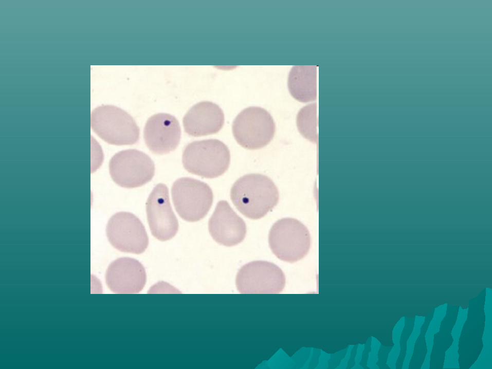
ParasitesParasites View large volumeView large volume 4 drops4 drops Join them over an area of 1 cm2Join them over an area of 1 cm2

Buffy layer filmBuffy layer film
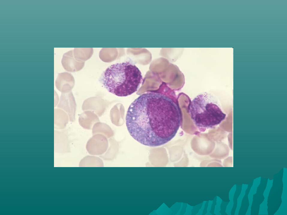
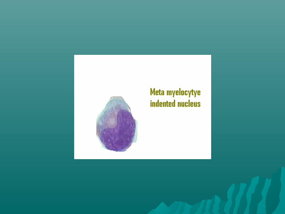
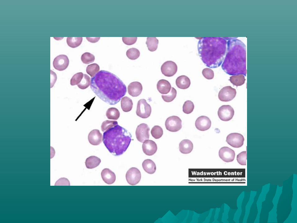
1.1. Pernicious anemia – demonstrate Pernicious anemia – demonstrate megaloblastsmegaloblasts
2.2. AIHA – erythrophagocytosisAIHA – erythrophagocytosis3.3. Tumor cells in aleukemic leukemiaTumor cells in aleukemic leukemia4.4. LE cellsLE cells

Wet filmWet film
Examination of blood cells suspende Examination of blood cells suspende in plasma +- supravital stainin plasma +- supravital stain
1.1. Demonstrate parasitesDemonstrate parasites2.2. Induce red cell sicklingInduce red cell sickling3.3. Study the red cells by Nomarski Study the red cells by Nomarski
interference microscopy after interference microscopy after fixation in Gluteraldehydefixation in Gluteraldehyde

Blood smear preparation Blood smear preparation EDTA EDTA Ideal smearIdeal smear1.1. Length of the smear shud be 2/3 rd of Length of the smear shud be 2/3 rd of
the slidethe slide2.2. Uniformly thickenedUniformly thickened3.3. Thin – printed matter can be readThin – printed matter can be read4.4. Margin shud be free and centrally placedMargin shud be free and centrally placed5.5. Tongue shapedTongue shaped6.6. Shud have adequate thin area –free from Shud have adequate thin area –free from
vacoule ,serration etcvacoule ,serration etc


Biological causes of poor filmBiological causes of poor film
1.1. Cold agglutininCold agglutinin2.2. LipemiaLipemia3.3. RouleauxRouleaux

Fixatives Fixatives
1.1. Methyl alcohol – Fixative Of ChoiceMethyl alcohol – Fixative Of Choice2.2. Ethyl alcoholEthyl alcohol

Staining of peripheral smearStaining of peripheral smear
Romanowsky type dyesRomanowsky type dyes1.1. LeishmanLeishman2.2. WrightWright3.3. May Grunwald GiemsaMay Grunwald Giemsa4.4. JennerJenner Compound dyesCompound dyes Neutral dyesNeutral dyes Field stain rapid stain for parasitesField stain rapid stain for parasites

EosinEosin
Cytoplasm , collagen ,muscle fibersCytoplasm , collagen ,muscle fibers Acidic Acidic H & E staining – H & E staining – Cytoplasm pink –orange Cytoplasm pink –orange Nuclei – blue / purpleNuclei – blue / purple Eosin stains red blood cells intensly Eosin stains red blood cells intensly
redred Haemetoxylin – nuclear stainHaemetoxylin – nuclear stain
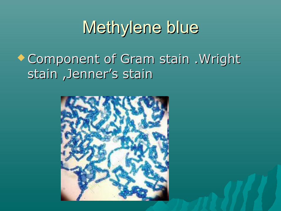
Methylene blueMethylene blue
Component of Gram stain .Wright Component of Gram stain .Wright stain ,Jenner’s stainstain ,Jenner’s stain

May Grunwald Giemsa stainMay Grunwald Giemsa stain
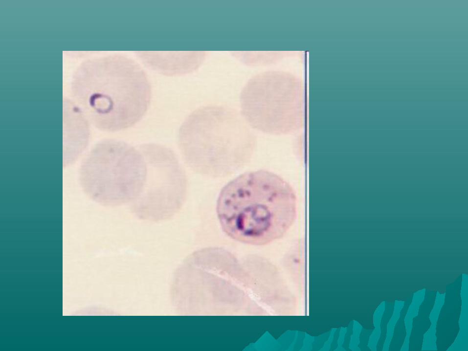
Histopathological diagnosis for Histopathological diagnosis for Malaria and other parasitesMalaria and other parasites
Fixative – acetone free methyl Fixative – acetone free methyl alcoholalcohol

Jenner’s stainJenner’s stain
Methylene blue eosinateMethylene blue eosinate

Wright stainWright stain
Eosin + MB Eosin + MB

Romanowsky stainRomanowsky stain
Basic dye forBasic dye for1.1. Nucleic acidNucleic acid2.2. NucleoproteinNucleoprotein3.3. Basophilic granulesBasophilic granules4.4. Weakly to neutrophil granulesWeakly to neutrophil granules Acidic dyes forAcidic dyes for1.1. HgHg2.2. Eosinophil granulesEosinophil granules

Leishman’s stainLeishman’s stain
William Boog Leishman –British William Boog Leishman –British PathologistPathologist
In India he studies Enteric Fever and In India he studies Enteric Fever and Kala AzarKala Azar
Buffered water PH 6.8Buffered water PH 6.8 ComponentsComponents1.1. Methylene Blue – Nuclear stainMethylene Blue – Nuclear stain2.2. Eosin – cytoplasmic stainEosin – cytoplasmic stain3.3. Acetone free Methyl Alcohol – solvent Acetone free Methyl Alcohol – solvent
and fixativeand fixative
Procedure Procedure
1.1. Enough stain to cover entire smear- Enough stain to cover entire smear- keep for 2 min – fixation by keep for 2 min – fixation by methanolmethanol
2.2. Add distilled water - double the Add distilled water - double the amount—10 min –actual staining amount—10 min –actual staining
3.3. Keep for 7 – 10 minKeep for 7 – 10 min4.4. WashWash5.5. Dry Dry

Good staining Good staining
Pinkish in colourPinkish in colour RBC – coppery red RBC – coppery red Eosinophil – orange red – with Eosinophil – orange red – with
yellowish tinged granulesyellowish tinged granules No depositNo deposit No cell shrinking or swellingNo cell shrinking or swelling If under atained add stain + water If under atained add stain + water
for another 5minfor another 5min If over stained –add stain only If over stained –add stain only

Special stainingSpecial staining
1.1. Peroxidase stainPeroxidase stain2.2. Sudan Black B stainSudan Black B stain3.3. PAS stainPAS stain4.4. Chloro Acetate esterase stainChloro Acetate esterase stain5.5. Non Specific EsterasesNon Specific Esterases6.6. Combined NSE & CAE stainingCombined NSE & CAE staining7.7. Iron stain Iron stain 8.8. Reticulin stainReticulin stain

Peroxidase stainPeroxidase stain Enzyme found in Primary and Enzyme found in Primary and
Secondary granules in WBCSecondary granules in WBC MPO – localised in the Azurophilic MPO – localised in the Azurophilic
granules of Neutrophils & Monocytesgranules of Neutrophils & Monocytes It can be demonstrated in It can be demonstrated in
Eosinophils and Basophils alsoEosinophils and Basophils also

MPO occurs onlu in the Primary Granules MPO occurs onlu in the Primary Granules of Myeloid cellsof Myeloid cells
It can be detected from the It can be detected from the Promyelocyte upto the granular Promyelocyte upto the granular stagestage
Lymphoid series are strongly MPO Lymphoid series are strongly MPO negativenegative
Reddish brown appearanceReddish brown appearance All myelocytic cell line except Myeloblasts All myelocytic cell line except Myeloblasts
shows Positiveshows Positive The more mature the cells become , the The more mature the cells become , the
weaker the reaction becomesweaker the reaction becomes Auer rods are strongly PositiveAuer rods are strongly Positive

Indications for peroxidase stainingIndications for peroxidase staining
Recognition of Acute Leukemias of Recognition of Acute Leukemias of myeloid origin – negative makes myeloid origin – negative makes lymphocyte origin – but does not lymphocyte origin – but does not exclude a myeloid originexclude a myeloid origin
Positive Positive sub division of Myeloid sub division of Myeloid Leukemias by FABLeukemias by FAB

Result Result Cells of Myeloid series positive with Cells of Myeloid series positive with
increasing intensity and no. of granules as increasing intensity and no. of granules as the cells maturethe cells mature
Early blasts negativeEarly blasts negative Lymphoid cells are negativeLymphoid cells are negative Erythroid cells and RBC may be Positive – Erythroid cells and RBC may be Positive –
due to Non enzymatic Reaction between due to Non enzymatic Reaction between Hb and stainHb and stain
Imp to distinguish AML from ALLImp to distinguish AML from ALL Neutrophils show decreased staining in Neutrophils show decreased staining in
congenital MPO deficiency and congenital MPO deficiency and Myelodysplastic syndromesMyelodysplastic syndromes


Sudan Black B stainSudan Black B stain
Fat soluble substanceFat soluble substance Stains Stains Fat cells ,Macrophages and Fat cells ,Macrophages and
GranulocytesGranulocytes Positive is dark brown to black in the Positive is dark brown to black in the
cytoplasm of the cell containing lipidcytoplasm of the cell containing lipid Granulocytes +ve inc with maturityGranulocytes +ve inc with maturity Cells of Burkitts’ Lymphoma +ve Cells of Burkitts’ Lymphoma +ve Large vacoulated ALL –L 3 cells +veLarge vacoulated ALL –L 3 cells +ve

Myeloblasts –veMyeloblasts –ve Lumphoblasts and Lymphocytes are Lumphoblasts and Lymphocytes are
–ve–ve Some cases of AML and CML –ve or Some cases of AML and CML –ve or
reduced sudanophiliareduced sudanophilia


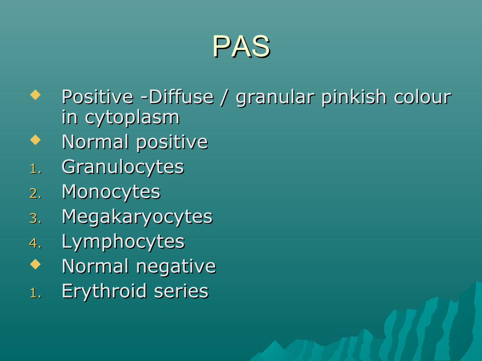
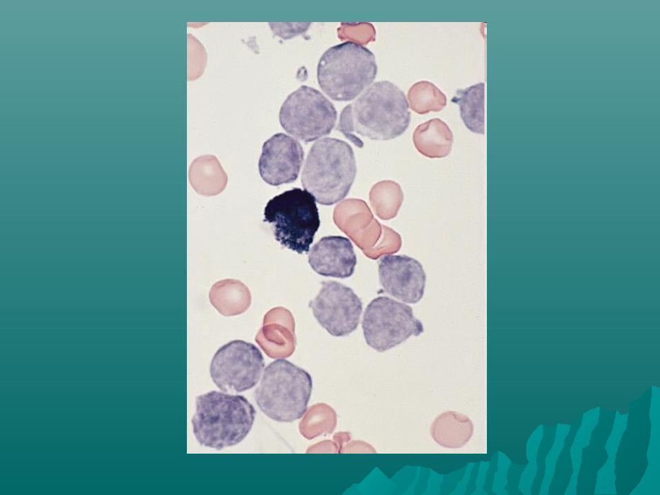
PASPAS
Positive -Diffuse / granular pinkish colour Positive -Diffuse / granular pinkish colour in cytoplasmin cytoplasm
Normal positiveNormal positive1.1. Granulocytes Granulocytes 2.2. Monocytes Monocytes 3.3. MegakaryocytesMegakaryocytes4.4. LymphocytesLymphocytes Normal negativeNormal negative1.1. Erythroid seriesErythroid series

Abnormal PASAbnormal PAS1.1. Lymphocytes in IMN +veLymphocytes in IMN +ve2.2. Late Erythroblasts in Thalssemia +veLate Erythroblasts in Thalssemia +ve3.3. Lymphoblasts in all ALL –Large Positive Lymphoblasts in all ALL –Large Positive
GranulesGranules4.4. 10 – 15% Myeloblasts in AML –diffuse 10 – 15% Myeloblasts in AML –diffuse
+ve+ve5.5. Erythroleukemia – AML –M6 –early Erythroleukemia – AML –M6 –early
normoblasts – shows coarse granular normoblasts – shows coarse granular positivity positivity late diffuse late diffuse
6.6. Hairy cells +veHairy cells +ve Myeloblasts inAML -veMyeloblasts inAML -ve


Non specific EsteraseNon specific Esterase
Positive for Monocytic SeriesPositive for Monocytic Series Diagnosis of AML M5Diagnosis of AML M5

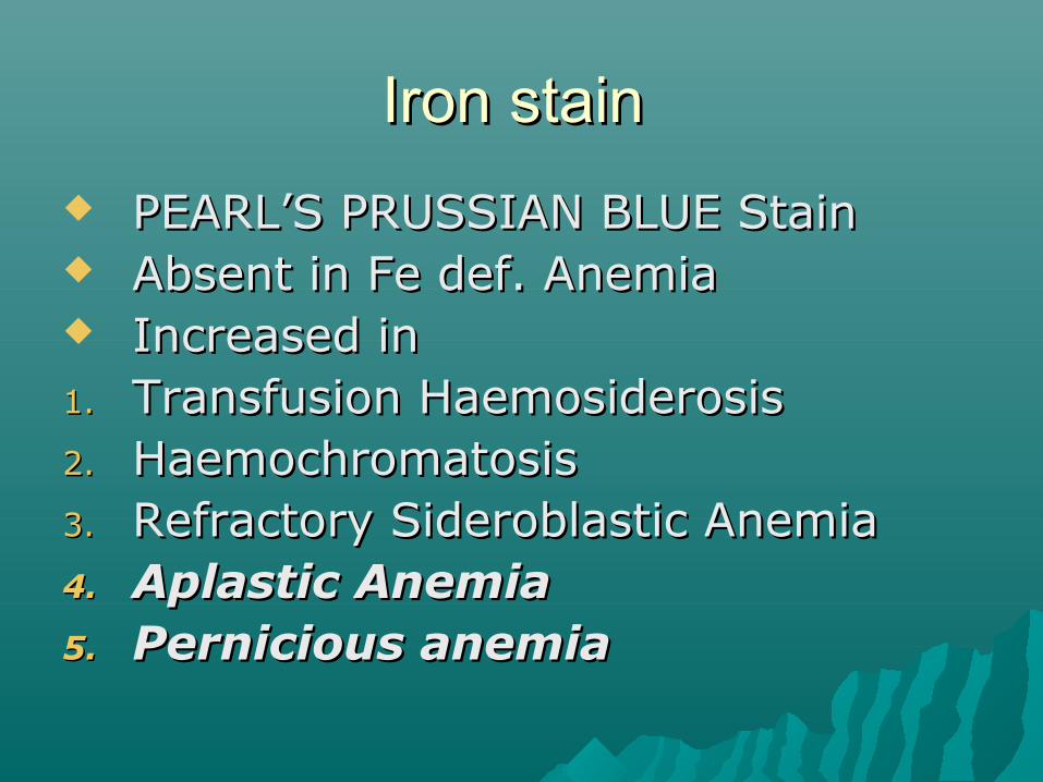
Iron stain Iron stain
PEARL’S PRUSSIAN BLUE StainPEARL’S PRUSSIAN BLUE Stain Absent in Fe def. AnemiaAbsent in Fe def. Anemia Increased inIncreased in1.1. Transfusion HaemosiderosisTransfusion Haemosiderosis2.2. HaemochromatosisHaemochromatosis3.3. Refractory Sideroblastic AnemiaRefractory Sideroblastic Anemia4.4. Aplastic AnemiaAplastic Anemia5.5. Pernicious anemiaPernicious anemia


Nucleus and Cytoplasm pinkNucleus and Cytoplasm pink Hemosiderin – bright blue – green Hemosiderin – bright blue – green
granulesgranules

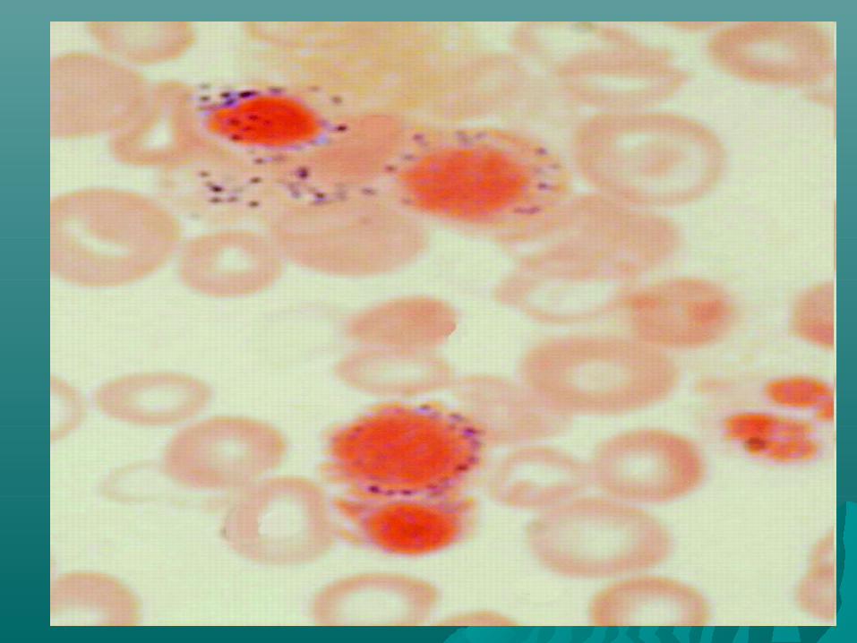


PS reportingPS reporting
Size of the RBC is compared to small Size of the RBC is compared to small lymphocyteslymphocytes
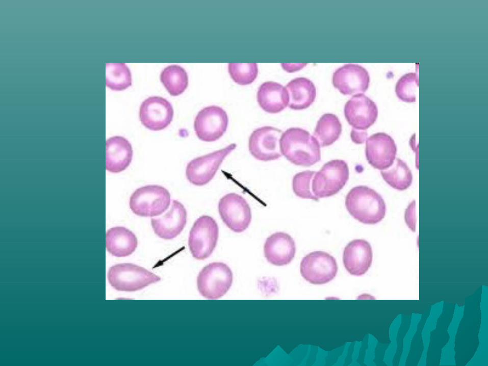
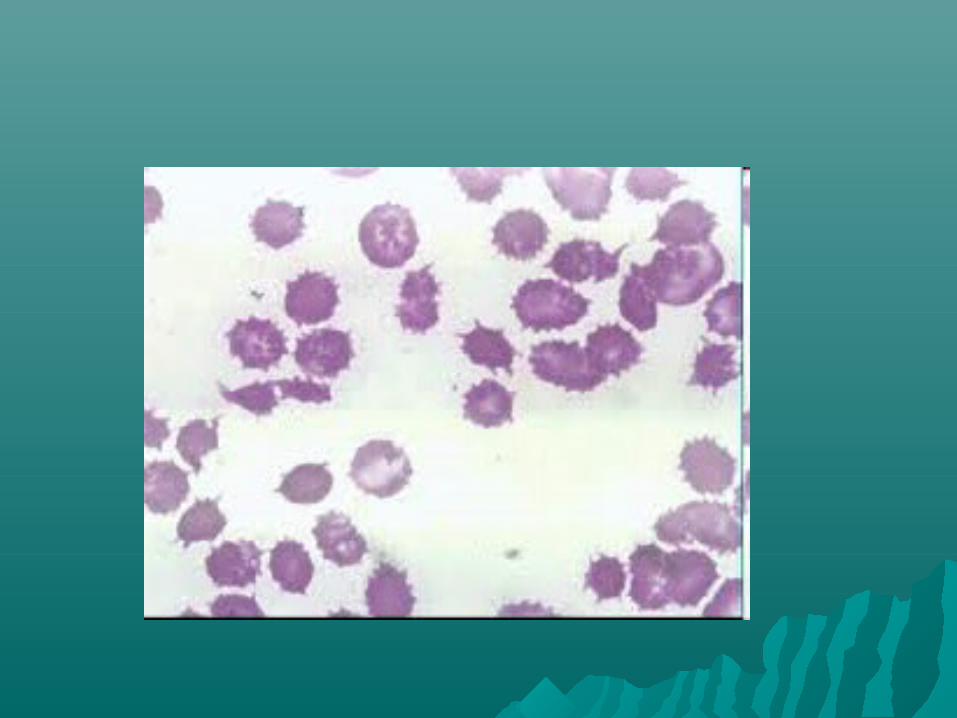
Microcytes –MCV<80 flMicrocytes –MCV<80 fl Macrocytes – MCV> 96 flMacrocytes – MCV> 96 fl AnisocytosisAnisocytosis PoikilocytosisPoikilocytosis

SpherocytesSpherocytes
Dark stained central pallor region – Dark stained central pallor region – hyperchromasiahyperchromasia
1.1. Heriditary SpherocytosisHeriditary Spherocytosis2.2. AIHAAIHA3.3. Chemical & Bacterial ToxinsChemical & Bacterial Toxins