periodontal ligament stress analysis during periodontal ... · pdf...
TRANSCRIPT
Abstract—Conservation of the PDL(periodontal ligament)
height during the orthodontic treatment phase is an important
aim during the treatment of periodontal disease. It was
hypothesized that due to periodontal resorption,, the same
amount of force determines an increase of maximum stress in
the PDL, which might affect its dimensions. The objective was
to quantify stress produced in the PDL at 10 different bone
levels under transverse and vertical orthodontic loadings, for
the special case of a two roots second mandibular premolar.
The alveolar bone and PDL has been reduced in height by 10%,
from 0% to 90%, and subjected to 6 constant loadings of 1-10
N/mm2. The von Mises stress values for the PDL were
calculated. For 0% bone loss, a 1 N/mm2 intrusive load
produced a maximum stress of 0.08 N/mm2. For 50%
resorption the stress was 0.41N/mm2 while for the 90% level
the stress was 2.96 N/mm2. For the 0-80% resorption the
mesio-distal load produced the highest stress values. The stress
gradually increased along with the PDL and bone height loss,
but declined steadily from the cervical to the apical level.
Approximately 5.7mm (30%) resorption is assumed to be the
starting point for dramatic changes in stress level. Results lead
to the conclusion that application of higher forces will
determine higher stresses in the cervical level which might
affect the height of the PDL during the orthodontic treatment
phase, and could worsen the periodontal problems.
Index Terms—Periodontal ligament biomechanics, finite
element analysis, stress, bone loss, bone biology
I. INTRODUCTION
NE of the major problems in dentistry is considered
to be the treatment of the periodontal disease [1]. The
periodontal resorption determines a reduction of the
periodontal capability to efficiently support current
functional loadings and influence its biomechanical behavior
under stress [1]. Conservation of the tooth-periodontal
ligament(PDL)-alveolar bone complex at various levels of
bone height, among other factors [2], is heavily dependent
on knowing the PDL's stress distribution [3]-[5].
With adequate orthodontic-periodontal coordination, it is
possible to reestablish a healthy and functional dentition [6].
Force systems in most orthodontic treatments are considered
indeterminate and the magnitude of forces and moments are,
Manuscript received February 29, 2016. This work was supported
from author's funds.
R. A. Moga is Assistant Professor with the Department of Odontology,
Faculty of Dental Medicine, University of Medicine and Pharmacy Iuliu
Hatieganu Cluj-Napoca, No.33 Motilor street, 400001 Cluj, Romania
(corresponding author's phone: 0040740000767; email:
C. G. Chiorean is Professor and head of Department of Structural
Mechanics, Technical University Cluj-Napoca, Cluj, Romania (e-mail:
in practice, largely unknown [7]-[9]. Hence, it is necessary
to limit the orthodontic force to prevent any damage [9]. The
physiological mechanism primarily responsible for tooth
movement in response to force is the PDL. Short-term tooth
movement is regarded as primarily governed by PDL
deformation [8]-[9].
The PDL's resorption process was widely investigated in
the last decades [10]-[19]. Published results showed a
significant increase in stress concentration at the apex with
bone loss [16] and that height reduction potentially causes
mechanical damage to the PDL [20]. It was found that 2.5
mm of alveolar bone loss can be considered as a limit
beyond which stress alterations were accelerated [17]. PDL's
accurate modeling affects the deformation and thus strains
magnitudes not only of the alveolar bone around the tooth
subjected to load, but also those of the whole mandible [21].
A better understanding of the PDL's biomechanical behavior
under physiological and traumatic loading conditions might
enhance the understanding of PDL's biological reaction in
health and disease [2]. Much still remains to be learned
about its 3D responses to load and the factors that control
them [22].
It was pointed out that when analyzing the bone resorption
process it is important to simulate various levels of bone
height in order to understand how axial loadings are
absorbed by PDL. Previous studies used models having
various levels of bone height [15]-[17], a PDL's thickness of
0.14-0.40 mm [15], [23], [24] and constantly loaded force
varying from 0.3 N up to 1500 N(46 MPa) [2]-[26].
However, they neither closely investigate the linear
periodontal resorption process nor used a highly accurate
anatomical model. The boundary conditions and input data
affect the accuracy of the results [3]-[4], [15], [27]-[32].
Therefore sensitivity analysis is needed for assess the
robusticity of the results [27]. Previous studies did not
mention anything regarding their sensitivity tests on their
FEA models.
Previous studies used models of human upper incisives
[11], [15]-[17], [19], upper [12] and lower [13], [14] canine
and upper molars [18]. However, no one used the second
lower premolar. The lower premolar, has a very complex
geometry(e.g. the one used in this study has two root canals
and two fused roots) but still similar with other lower and
upper premolars. It's location on the mandible(a mobile
bone) might influence the stress distribution in the
periodontal tissues due oral kinematics [21].
Using a new model created based on the information
provided by CT slices, being anatomical accurate and
simulating a bone loss of 10%-90%, this protocol is
Periodontal Ligament Stress Analysis During
Periodontal Resorption
R.A. Moga, C.G. Chiorean
O
Proceedings of the World Congress on Engineering 2016 Vol I WCE 2016, June 29 - July 1, 2016, London, U.K.
ISBN: 978-988-19253-0-5 ISSN: 2078-0958 (Print); ISSN: 2078-0966 (Online)
WCE 2016

expected to provide accurate new accurate information
regarding the behavior of PDL under different loadings. This
new data should complete the knowledge regarding the
human teeth investigated during periodontal resorption. It
should also fill the gaps regarding the way the stress
distribution occurs in the PDL of a premolar during the
gradual linear resorption process [22] and has potential
clinical applicability.
II. MATERIAL AND METHODS
A. Model creation
A CBCT (PaX-Reve3D,Vatech America), FOV 5×5 cm
and voxel size 0.08 mm, generating detailed 2D images was
used in this study. The investigated area contained the
second lower right premolar of a 34-year-old healthy male.
More than 2200 2D slices(80 µm) were taken. 3D models of
each components were generated, through image manual
segmentation and then were assembled into a 3D complete
model having the following dimensions 320×320×320 mm.
Tooth's support surrounding tissues had a height of 19.2
mm, bone beneath had 6 mm, and the second lower right
premolar had 26 mm of height, all measured on the CT
slices.
Then this model was replicated, thus obtaining ten second
human lower right premolar models having the same
configuration except for the alveolar bone height and PDL.
A homogenous horizontal reduction all around the tooth was
assumed. The alveolar bone and PDL has been reduced in
height by 0%, 10%, 20%, 30%, 40%, 50%, 60%, 70%, 80%
and 90% simulating the progressive resorption. Each 10%
height loss simulated a 1.92 mm (24 CT slices) of
periodontal loss [26], [32].
AMIRA, version 5.4.0.(AMIRA, version 5.4.0, Visage
Imaging Inc.), software was used for used for segmenting
and generating the 3D models. The image segmentation
process was done manually for each slice based on the
different gray scale values and on Houndsfield units. A clear
separation between dentin and cement was impossible, so
the entire cement layer, in the roots area, was considered to
have the same properties as dentin. This simplification
should be acceptable due to the similar properties of the
dentin and cement. The PDL was attached to the
intraalveolar surface and to the root. The PDL's thickness
varied between 2-3 voxel (0.16-0.24 mm), depending on the
anatomical topography and dimensions identified during the
manual image segmentation process. The cortical bone layer
had a minimum length and thickness of 2 mm, depending on
the anatomy of the segmented region. The rest of the bone
was filled with cancellous bone, according to the regions
designated by the CT slices.
The 3D models were calculated and described using
triangles in AMIRA. The surface models were strongly
simplified but maintaining the shapes of the original
surfaces. The meshes were obtained by filling the volumes
restricted by the surfaces with tetrahedrons. The final mesh
contained 14395 nodes and 73913 tetrahedrons
B. Finite element analysis
The FEA is a numerical form of analysis allowing stress
and displacements to be identified. It is based on the
discretization of continuum into a number of elements. The
final mesh was exported and analyzed in ABAQUS, version
6.1.1.1sofware (ABAQUS, version 6.1.1, Dassault
Systemes). Boundary conditions, material properties (elastic
constants) and loading conditions were then assigned to each
voxel, defining the way the model would deform under
stress. The model structures were solved as a series of nodal
displacements and the resulting von Mises stresses were
calculated and displayed. Were investigated the overall
stress value of the PDL, such as von Mises stress values in
N/mm2(MPa) at the cervical level and at the apical level
under each of the 6 orthodontic loadings [26]. The von
Mises stresses were preferred as they are usually used to
define the yielding criteria for isotropic materials under
different loading conditions and represents the overall stress
intensity in contrast with individual stress components such
as principal stresses. The FEA and results visualization were
conducted in ABAQUS software using a color-coded
projection onto the model geometry.
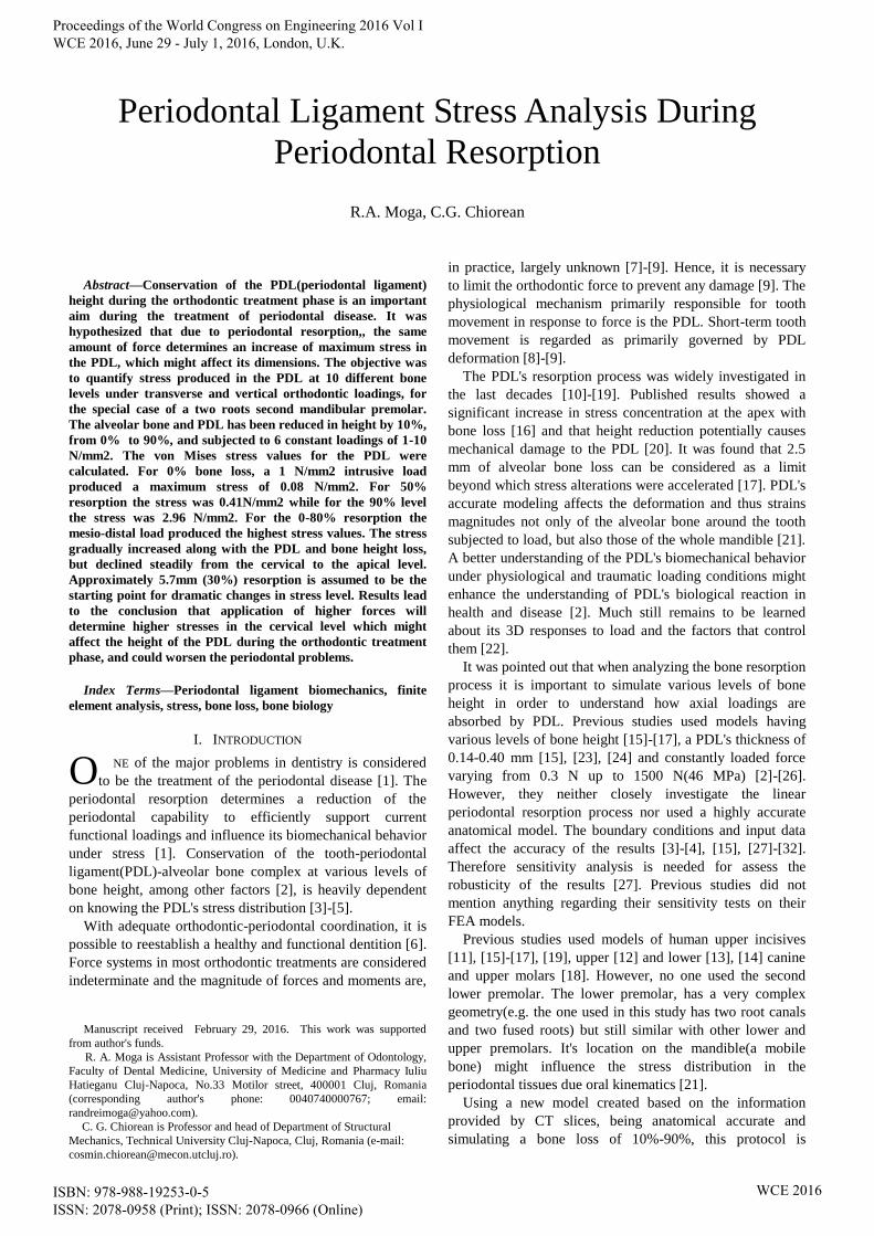
C. Material models and modeling assumptions
The components of the model, Fig. 1, were assumed to be
homogeneous and isotropic, to possess linear elasticity,
small deformations and displacements. However, the living
tissues exhibit inhomogeneous, anisotropic and nonlinear
behavior and normally they should be modeled as a elasto-
plastic porous materials with a complex microstructure [3].
Such approach involves complex finite element models
enhanced with advanced nonlinear constitutive equations
experimentally calibrated, but such approach is scarce in the
scientific literature. The main benefits of a FEA like the
present one is the simplicity and yet the accuracy of the
results. All the modeling decision from the present study
(e.g. material properties and inclusion of the PDL) complied
with the sensitivity test criteria presented in the literature.
Elastic constants of all used materials are given in Table I.
Young’s modulus describes the tendency of a material to
deform elastically under axial loading conditions, indicating
its stiffness while Poisson's ratio is related to Young’s
modulus and shows how much a material expands when
compressed, or contracts when stretched. Boundary
conditions consist of prescription of displacements in several
parts of the model. For restraining all forms of translational
movements the base of the model was considered to have
zero displacement, while all other parts were treated as free
of boundary conditions. This manner of restraining prevents
the models from any rigid body motion while the loadings
are acting. Because of localized nature of the problem
studied here, it must be emphasize that the lateral(mesial and
distal) edges of the developed model have been chosen at
some reasonable distance from the region of main interest(a
volume of bone equivalent to the volume of the neighboring
teeth roots) in order to obtain the solution without
perturbation due to the dimensions of the mandible.
Numerical studies performed for the model confirmed the
localized nature of the phenomenon, and that there were
almost no displacements at the mesial and distal edges. It has
been assumed that the interfaces between all components
were perfectly bonded interfaces all the components interact
Proceedings of the World Congress on Engineering 2016 Vol I WCE 2016, June 29 - July 1, 2016, London, U.K.
ISBN: 978-988-19253-0-5 ISSN: 2078-0958 (Print); ISSN: 2078-0966 (Online)
WCE 2016
with each other.
Coronal-apical(intrusive) loads of 1 N/mm2 and 10
N/mm2, and 1 N/mm
2 and 3 N/mm
2 vestibular-
palatinal(transversal) and mesio-distal(transversal) loads
were applied, one at the time (in different loading
sequences), at the enamel surface, centrally in the occlusal
face of the crown, in order to estimate the loads' effect over
the PDL. The other surfaces were treated as free surfaces
with zero loads.
Table I. Elastic properties of materials used in the study
Material Constitutive
equation
Young's
modulus,
E (GPa)
Poisson
ratio, ʋ
Refs.
Enamel
Isotropic,
homogeneous
and linear elastic
80 0.33 [22], [26]-
[32]
Dentin Isotropic,
homogeneous
and linear elastic
18.6 0.31 [22], [26]-
[32]
Pulp Isotropic,
homogeneous
and linear elastic
0.0021 0.45 [22], [26]-
[32]
PDL Isotropic,
homogeneous
and linear elastic
0.0689 0.45 [12],[ 22],
[26]-[32]
Cortical bone Isotropic,
homogeneous
and linear elastic
14.5 0.323 [1], [18],
[26]-[32]
Cancellous
bone
Isotropic,
homogeneous
and linear elastic
1.37 0.30 [1], [18],
[22], [26]-
[32]
III. RESULTS
Coronal apical load. 0% bone loss. For the coronal apical
1 N/mm2 directed force, the increased level of stress was
located at the PDL's lingual cervical margin (Fig. 2A and
Table II), decreasing to the apical region. The lowest stress
value was approximately 0.04% of the cervical stress (Table
III).
Alveolar bone loss. The highest stress values were found
at the lingual cervical level (Fig. 3A, 4A and Table II),
gradually increasing with the increase in bone resorption.
The stress increased 5.1 times when resorption reached 50%
of bone height and 36.6 times for the 90% bone loss (1
N/mm2 load). For the 10 N/mm2 directed force, the stress
level distribution maintains in the same locations, while the
values increase by 10 times.
Vestibulo-lingual load. 0% bone loss. For the 1 N/mm2
and 3 N/mm2 directed forces, the highest levels of stress
were located at the PDL's vestibular and distal cervical level
(Fig. 2B and Table II). Higher stress also appeared at the
apical level. The highest apical stress was located on the
vestibular and distal sides and was 17% of the cervical stress
while the lowest apical stress was approximately 0.13%
(Table III).
Alveolar bone loss. The highest stress values were found
at the cervical level, on the vestibular and distal sides,
shifting gradually versus the lingual and mesial sides (Fig.
3B, 4B and Table II) due to the resorption's direction and
tooth load. The maximum stress gradually increased
reaching 12.9 times for 50% bone resorption and 143 times
for 90% bone loss.
Mesio-distal load. 0% bone loss. For the 1 N/mm2 and 3
N/mm2 directed forces, the highest stress was located at the
PDL's vestibular, distal and lingual cervical level (Fig. 2C
and Table II). Higher stress level also appeared in the
vestibular and distal apical region. The highest apical stress
value was 26% of the cervical stress while the lowest was
0.25% (Table III).
Alveolar bone loss. The highest stress values were found
at the cervical level, on the vestibular, distal and lingual
sides (Fig. 3C, 4C and Table II), gradually shifting versus
the lingual and mesial sides according the resorption level
due to the resorption's direction and tooth load. The
maximum stress gradually increased reaching 13 times for
the 50% bone loss and 129 times for 90% level. A decrease
in stress value was manifest at 90% level (Table III),
explained by the local conditions due to massive bone height
loss.
During the resorption process (0-80%) the mesio-distal
load (1 N/mm2 load) produced the highest von Mises stress
values. At 90% bone loss the vestibulo-lingual load (1
N/mm2 load) determined the highest stress in the PDL (Fig.
5a). For the 10 N/mm2 load, a gradual increase of the stress
values appeared (Fig. 5b), with the highest values at 80%
and 90% bone loss. For the 3 N/mm2 loads, a gradual
increase of the mesio-distal load appeared(0-80% bone loss)
while for the 90% bone loss the vestibulo-lingual one
produced the maximum equivalent stress value.
Fig. 1(a, b, c) The FE Mesh used in Abaqus for 0% resorption level with
boundary conditions: a- the occlusal face of the crown were the loads were
applied; b-vestibular view; c-lingual view
IV. DISCUSSIONS
The tooth-PDL-alveolar bone complex is subjected to
high occlusal forces during mastication but their effect on
the PDL with different levels of resorption needs further
studies [1], [17]. Furthermore, the stress values that produce
biological changes in dental structures are not entirely
known and understood[3]-[5]. Based on this fact the presents
study tried to assess the PDL loss and its consequences.
The stress in the living PDL is difficult to be measured
experimentally so in this case the FEA is the proper method
to investigate the von Mises equivalent stress.
The FEA is an accurate method to study and describe the
biomechanical behavior of different living structures but
relays directly on the input data [15]. Therefore, the results
are directly influenced by the work precision. The models
used in this study were highly anatomical accurate, having
14395 nodes and 73913 tetrahedrons (created through image
manual segmentation) while some of the previous studies
[2]-[19] used models having 726 - 37884 nodes(created after
Ash's dental dental anatomy).
Furthermore, the discrepancies within the accepted values
Proceedings of the World Congress on Engineering 2016 Vol I WCE 2016, June 29 - July 1, 2016, London, U.K.
ISBN: 978-988-19253-0-5 ISSN: 2078-0958 (Print); ISSN: 2078-0966 (Online)
WCE 2016
of the physical properties of the 3D models can produce
results alteration [15]. Therefore for a proper comparison all
the boundary conditions used in this study were provided by
previous studies.
It was pointed out that when analyzing periodontal
resorption it is important to simulate various levels of bone
height [1]. However, little information regarding the stress
distribution during complete linear periodontal resorption of
0-90% is available, being hypothesized that a reduction in
alveolar support causes an increase in maximum stress in the
periodontal structures.
For a robust FEA's results sensitivity testes are advisable
[27]. Despite all these, previous studies failed to mention
anything about their sensitivity testes. This study complied
with the sensitivity tests criteria regarding the modeling
decisions [27] and conducted a geometric morphometric
analysis for confirming the deformation results.
Horizontal bone loss is the more common type of alveolar
bone resorption [6]. Previous studies used models having
various levels of bone height ranging from 25% to 75% bone
height resorption [15]-[17], however, none described a
periodontal resorption of 0% up to 90%.
For simplifying the FEA, all the interfaces were
considered perfectly bonded interfaces [3], [4]. This
assumption does not necessarily simulate clinical situations
and is considered to be an inherent limitation of this process.
However, the goal of this study was a qualitative analysis
and these assumptions do not alter the accuracy of the
results.
Previous studies [15] shown PDL stress values of 0.0012-
0.132 N/mm2 at the cervical margin for forces of 1 N applied
to the center of the tooth and mesio-distally, while the stress
values at the apex were 0.002-0.05 N/mm2. In this study the
results of the structural response of the system have been
obtained in terms of the von Mises cervical stress for the 1
N/mm2 load force (Table II) and were similar to those
provided by previous studies [4], [13], [15]-[17] for the
same value of the force. For the 3 and 10 N/mm2 load forces
the obtained results are higher than those previous cited. The
results for the apical stress for the 3-10 N/mm2 loads up to
70% bone height loss were much below the value [14] of
0.05 N/mm2, while the apical stress results for the 1 N/mm2
loads are in agreement with Cobo's [14]. The apical stress
value [14] at 8 mm of bone height loss was of 0.364 N/mm2.
The maximum apical stress level (Table III), for similar
bone height loss conditions, was of 0.023 N/mm2 for the 1
N/mm2 load and 0.238 N/mm
2 for the 10N/mm
2 load at 40%
height reduction. This behavior and the numerical results
were within the limits stated both by Geramy [14], [17] and
Cobo [14]. The difference occur because of differently
applied load directions and the 3D model design, in
agreement with Poiate, et al.[23]. This study found the
highest stress values at the cervical level which are in
agreement with Wilson et al.[29] and Wiejun et al. [30] but
differ from those reported by Geramy [15].
Alveolar bone height loss causes an increase in the PDL's
von Mises stress values at all levels [10]-[15]. This study is
in agreement with this and provides an accurate description
of the phenomenon. A certain predictability regarding the
rise of stress values and their tendency to increase can be
noted confirming the tendency shown in previous studies
[15], [17].
A previous study stated 19% bone loss as a limit beyond
which the stress increase accelerates [27]. The obtained
results are in agreement with all this. Each layer of
periodontal resorption(almost 2mm) determined an increase
of the stress values which is in agreement with previous
studies [15], [17]. This FEA's results showed a gradual
increase in stress values (0-20% bone loss) and a massive
increase after 30% bone loss which is in agreement with
Shang's results [1]. It also shown that 5.7 mm of alveolar
bone loss (30%) is assumed to be the starting point for
dramatic changes in stress level which contradicts Geramy's
[17] results.
The model used is the second lower right premolar, with
two root canals, while most of the studies used single-rooted
and single canal teeth [1], [15], [17] and this aspect might
also influence the biomechanical behavior.
The novelty in this approach consists in: the high
anatomical accuracy of the 3D models, the gradual
periodontal resorption that simulated a complete process (0-
90%), the FEA conducted according to sensitivity tests
criteria, totally new numerical stress values for the 70-90%
resorption levels and new values for 0-70% resorption
levels.
This qualitative analysis explains why after a long period
of slow resorption process of the tooth surrounding support
tissues, showing no warning signs, as soon as a certain
resorption degree is passed, the tooth is rapidly lost. In the
open literature there is no data regarding the maximum
tolerable stress for living tissues [17], however, the stress
increase during periodontal resorption is so high that it can
be considered to be out of the range of tolerable stress,
similar with Geramy's conclusions [17]. Further studies are
needed for finding the maximum tolerable stress and
allowing a more accurate discussion regarding this study's
results.
V. CONCLUSIONS
It was observed that during the linear periodontal
resorption simulation the von Mises stress gradually
increased along with the PDL and bone height loss, but
declined steadily from the cervical to the apical level, thus
validating the research hypothesis. Each 2 mm layer of PDL
protects from stress the PDL beneath. After 30% periodontal
resorption the stress significantly increase. Approximately
5.7 mm of periodontal loss (30%) is assumed to be the
starting point for dramatic changes in stress level. The
maximum stress values were found when the periodontal
loss reached 90%. The vestibulo-lingual loadings produced
the highest von Misses stress values for the 90% bone loss.
The results show that due to periodontal resorption the same
amount of force determines an increase of maximum stress
in the PDL cervical level, which ultimately might affect its
height and could worsen the periodontal problems. All these
should be taken into consideration by the orthodontist when
applies the orthodontic forces at patients having
periodontitis. This analysis provides an integrated approach
to the solutions of biological and biomedical problems.
Proceedings of the World Congress on Engineering 2016 Vol I WCE 2016, June 29 - July 1, 2016, London, U.K.
ISBN: 978-988-19253-0-5 ISSN: 2078-0958 (Print); ISSN: 2078-0966 (Online)
WCE 2016
Fig. 2(A, B, C) Von Mises stress distribution for 1N/mm2 for 0%
resorption level: A-Coronal apical load; B-Vestibulo-lingual load; C-
Mesio-distal load
Fig. 3(A, B, C) Von Mises stress distribution for 1N/mm2 for 50%
resorption level: A-Coronal apical load; B-Vestibulo-lingual load; C-
Mesio-distal load
Fig. 4(A, B, C) Von Mises stress distribution for 1N/mm2 for 90%
resorption level.: A-Coronal apical load; B-Vestibulo-lingual load; C-
Mesio-distal load
CA
VL
MD
0
2
4
6
8
10
12
0% 10% 20% 30% 40% 50% 60% 70% 80% 90%
(CA) Coronal aplical load 1 N/mm2(MPa)
(VL) Vestibulo-lingual load 1 N/mm2(MPa)
(MD) Mesio-distal load 1 N/mm2(MPa)
a
CAVL
MD
0
5
10
15
20
25
30
35
0% 10% 20% 30% 40% 50% 60% 70% 80% 90%
(CA) Coronal aplical load 10 N/mm2(MPa)
(VL) Vestibulo-lingual load 3 N/mm2(MPa)
(MD) Mesio-distal load 3 N/mm2(MPa)
b
Fig. 5(a,b): a-Maximum von Mises stresses at the cervical level for 1 N/mm2(MPa) loading forces for the 10 levels of bone height loss; b-Maximum von
Mises stresses at the cervical level for 3-10 N/mm2(MPa) loading forces for the 10 levels of bone height loss
Table II. PDL's von Mises stress values in N/mm2(MPa) at the cervical level, under the 6 loadings, during bone height resorption simulation
alveolar bone
reduced in
height
alveolar bone
and PDL
height
A.
Coronal apical
load
B.
Vestibular-
Lingual load
C.
Mesial-Distal
load
% mm 1 N/mm2 (MPa) 10 N/mm2
(MPa)
1 N/mm2 (MPa) 3 N/mm2
(MPa)
1 N/mm2 (MPa) 3 N/mm2
(MPa)
0% 19.2 0.08 0.81 0.06 0.20 0.05 0.16
10% 17.28 0.10 1.07 0.07 0.23 0.10 0.30
20% 15.36 0.11 1.14 0.20 0.61 0.26 0.80
30% 13.44 0.24 2.41 0.56 1.71 0.60 1.81
40% 11.52 0.27 2.77 0.60 1.81 0.60 1.81
50% 9.6 0.41 4.15 0.86 2.59 0.74 2.22
60% 7.68 0.48 4.81 1.27 3.82 1.20 3.61
70% 5.76 1.27 12.79 1.89 5.67 2.69 8.06
80% 3.84 2.59 25.94 5.77 17.32 6.58 19.77
90% 1.92 2.96 29.68 9.55 28.68 6.92 20.78
Table III. PDL's von Mises stress values in N/mm2(MPa) at the apical level, under the 6 loadings, during bone height resorption simulation
alveolar
bone
reduced in
height
alveolar
bone and
PDL height
A.
Coronal apical
load
B.
Vestibulo-
lingual load
C.
Mesio-distal load
% mm 1 N/mm2(MPa) 10 N/mm2(MPa) 1 N/mm2(MPa) 3 N/mm2(MPa) 1 N/mm2(MPa) 3 N/mm2(MPa)
0% 19.2 3.5E-05 35E-05 8.9E-05 26E-05 13.5E-05 40E-05
10% 17.28 7.4E-05 74E-05 12.5 E-05 37E-05 16.9E-05 50E-05
20% 15.36 22.2E-05 222E-05 35.5E-05 106E-05 33.5E-05 100E-05
30% 13.44 37.5E-05 375E-05 39.7E-05 119E-05 51.1E-05 153E-05
40% 11.52 84.1E-05 841E-05 165.8E-05 497E-05 109.3E-05 327E-05
50% 9.6 47.6E-05 476E-05 119.4E-05 358E-05 68.7E-05 206E-05
60% 7.68 157.1E-05 1571E-05 191.8E-05 575E-05 299.2E-05 897E-05
70% 5.76 118.2E-05 1182E-05 706.8E-05 2120E-05 230.5E-05 2098E-05
80% 3.84 1887E-05 18870E-05 4082E-05 12250E-05 3560E-05 10680E-05
90% 1.92 535E-05 5350E-05 2201E-05 6600 E-05 1155E-05 3460E-05
Proceedings of the World Congress on Engineering 2016 Vol I WCE 2016, June 29 - July 1, 2016, London, U.K.
ISBN: 978-988-19253-0-5 ISSN: 2078-0958 (Print); ISSN: 2078-0966 (Online)
WCE 2016
REFERENCES
[1] Shang S, Li C, Qian Q, Liang L. Three-dimensional finite element
analysis of the stress of mandibular incisor with different level of
alveolar bone. Sheng Wu Yi Xue Gong Cheng Xue Za Zhi.
2005;22:725-729
[2] Poiate IA, de Vasconcellos AB, de Santana RB, Poiate E. Three-
dimensional stress distribution in the human periodontal ligament in
masticatory, parafunctional, and trauma loads: finite element
analysis. J Periodontol 2009;80:1859-1867.
[3] Achour T, Merdji A, Bachir Bouiadjra B, Serier B, Djebbaral N.
Stress distribution in dental implant with elastomeric stress barrier.
Mater Design 2011;32:282–290.
[4] Merdji A, Bouiadjra B, Serier B, Djebbar N. Stress analysis in dental
prosthesis. Comp Mater Sci 2010;49:126–133. [5] Djebbar N, Serier B, Bachir Bouiadjra B, Benbarek S, Drai A.
Analysis of the effect of load direction on the stress distribution in
dental implant. Mater Design 2010;31:2097–2101
[6] Cao T, Xu L, Shi J, Zhou Y. Combined orthodontic-periodontal
treatment in periodontal patients with anteriorly displaced incisors.
Am J Orthod Dentofacial Orthop. 2015 Nov;148(5):805-13.
[7] Fill TS, Toogood RW, Major PW, Carey JP. Analytically determined
mechanical properties of, and models for the periodontal ligament:
critical review of literature. J Biomech. 2012 Jan 3;45(1):9-16.
[8] Fill TS, Carey JP, Toogood RW, Major PW. Experimentally
determined mechanical properties of, and models for, the periodontal
ligament: critical review of current literature. J Dent Biomech.
2011;2011:312980.
[9] Vikram NR, Senthil Kumar KS, Nagachandran KS, Hashir YM.
Apical stress distribution on maxillary central incisor during various
orthodontic tooth movements by varying cemental and two different
periodontal ligament thicknesses: a FEM study. Indian J Dent Res.
2012 Mar-Apr;23(2):213-20
[10] Tanne K, Sakuda M. Initial stress induced in the periodontal tissue at
the time of the application of various types of orthodontic force:
three-dimensional analysis by means of the finite element method. J
Osaka Dent Univ 1983;23:143-171.
[11] Tanne K, Nagataki T, Inoue Y, Sakuda M, Burstone CJ. Patterns of
initial tooth displacements associated with various root lengths and
alveolar bone heights. Am J Orthod Dentofacial Orthop
1991;100:66-71.
[12] McGuinness N, Wilson AN, Jones M, Middleton J, Robertson NR.
Stresses induced by edgewise appliances in the periodontal ligament--
a finite element study. Angle Orthod 1992;62:15-22.
[13] Puente MI, Galbán L, Cobo JM. Initial stress differences between
tipping and torque movements. A three-dimensional finite element
analysis. Eur J Orthod 1996;18:329-339.
[14] Cobo J, Sicilia A, Argüelles J, Suárez D, Vijande M. Initial stress
induced in periodontal tissue with diverse degrees of bone loss by an
orthodontic force: tridimensional analysis by means of the finite
element method. Am J Orthod Dentofacial Orthop 1993;104:448-
454.
[15] Geramy A. Initial stress produced in the periodontal membrane by
orthodontic loads in the presence of varying loss of alveolar bone: a
three-dimensional finite element analysis. Eur J Orthod 2002;24:21-
33.
[16] Reddy MK, Vandana KL. Three-dimensional finite element analysis
of stress in the periodontium. J Int Acad Periodontol 2005;7:102-
107.
[17] Geramy A, Faghihi S. Secondary trauma from occlusion: three-
dimensional analysis using the finite element method. Quintessence
Int 2004;35:835-843.
[18] Jeon PD, Turley PK, Moon HB, Ting K. Analysis of stress in the
periodontium of the maxillary first molar with a three-dimensional
finite element model. Am J Orthod Dentofacial Orthop
1999;115:267-274.
[19] Chen WP, Lee BS, Chiang YC, Lan WH, Lin CP. Effects of various
periodontal ligament elastic moduli on the stress distribution of a
central incisor and surrounding alveolar bone. J Formos Med Assoc
2005;104:830-838.
[20] Ona M, Wakabayashi N. Influence of alveolar support on stress in
periodontal structures. J Dent Res 2006;85:1087-1091.
[21] Groning F, Fagan MJ, O’Higgins P. The effects of the periodontal
ligament on mandibular stiffness: a study combining finite element
analysis and geometric morphometrics. J Biomech 2011;44:1304-
1312.
[22] Naveh GR, Lev-Tov Chattah N, Zaslansky P, Shahar R, Weiner S.
Tooth-PDL-bone complex: response to compressive loads
encountered during mastication - a review. Arch Oral Biol
2012;57:1575-1584
[23] Consolaro A, Consolaro MF M-O. ERM functions, EGF and
orthodontic movement or Why doesn't orthodontic movement cause
alveolodental ankylosis?. Dental Press J Orthod 2010;2:24-32.
[24] Coolidge ED. The thickness of human periodontal membrane. J Am
Dent Assoc 1937;24:1260-1270.
[25] Montegi E, Nomura M, Tachiki C, Miyazaki H, Takeuchi F, Takaku
S et al. Occlusal force in people in their sixties attending college for
elderly. Bull Tokyo Dent Coll 2009;50:135-140.
[26] Moga RA, Chiorean CG. Strain Analysis of a Human Tooth with
Support Tissues Resorption. World Congress on Engineering 2013,
pp.1374-1379.
[27] Gröning F, Fagan M, O'Higgins P. Modeling the human mandible
under masticatory loads: which input variables are important? Anat
Rec (Hoboken). 2012;295:853-863
[28] Li J, Li H, Shi L et al. A mathematical model for simulating the bone
remodeling process under mechanical stimulus. Dent Mater
2007;23:1073-1078.
[29] Wilson AN, Middleton J, Jones ML, McGuinness NJ. The finite
element analysis of stress in the periodontal liagment when subject to
vertical orthodontic forces. Br J Orthod 1994;21:161-167.
[30] Weijun Yan, Xiaohui Jiao, Ping Shao, Wei Cai. Stress distribution in
the mandibular central incisor and periodontal ligament while
opening the bite: A finite element analysis Biomed Res-India
2012;23:343-348.
[31] Piotr Kowalczyk., 2009. Influence of the shape of the layers in photo-
cured dental restorations on the shrinkage stress peaks—FEM study.
Dent Mater 25, e83–e91.
[32] Moga RA, Chiorean CG. Strain Analysis of a Human Tooth with
Support Tissues Resorption. World Congress on Engineering 2013,
pp.1374-1379.
Proceedings of the World Congress on Engineering 2016 Vol I WCE 2016, June 29 - July 1, 2016, London, U.K.
ISBN: 978-988-19253-0-5 ISSN: 2078-0958 (Print); ISSN: 2078-0966 (Online)
WCE 2016