peri-prosthetic trans-patellar fractures after total knee
TRANSCRIPT

REVIEW Open Access
Peri-prosthetic trans-patellar fractures afterTotal knee Arthroplasty: a case series andreview of literatureGaurav Govil1,2* , Lavindra Tomar1,3 and Pawan Dhawan1,4
Abstract: Peri-prosthetic patella fracture is the second most common peri-prosthetic fracture after total kneearthroplasty. This report presented the treatment results in 6 patients with peri-prosthetic patella fractures. FromJanuary 2015 to February 2019, six patients with peri-prosthetic patella fractures were treated. The mean age at surgerywas 64 years (range, 48–72 years). Four patients with displaced fractures were treated surgically, and two patients withnon-displaced fractures were treated non-surgically. Outcomes were assessed in terms of motion, functional kneescore, and Knee Society score. The mean follow-up period lasted 16months (range: 12–20months). The average arc ofmotion was 110° (range: 80°–130°). The mean functional knee score was 77 (range: 70–87). The mean Knee Societyscore was 84 (range: 75–89). The non-surgical treatment may be a good choice for non-displaced peri-prostheticpatella fractures. For displaced fractures, surgical treatments yielded good functional outcomes.
Level of evidence: IVa
Keywords: Peri-prosthetic patella fracture, Patella, Osteosynthesis, Complication, Trans-patellar fracture
IntroductionPeri-prosthetic patella fracture (PPPF) represents thesecond most common peri-prosthetic fracture after totalknee arthroplasty (TKA). The reported prevalence ratesstood somewhere between 0.2–21% in resurfaced patellaeand was about 0.5% in un-resurfaced patellae [1–5]. MostPPPFs occur within 2 years after arthroplasty. Treatmentsinclude non-surgical and surgical methods, depending onthe features of fractures [2, 4].Currently, there is no universally-accepted validated
classification system for PPPFs. The Ortiguera and Berryclassification is most commonly used. It takes into accountboth stability of patellar implant and the extensor mechan-ism [6]. Goldberg et al [7] also developed a classifica-tion on the basis of extensor apparatus continuity andstability of patella resurfacing. However, this classificationdoes not consider variability of fracture configuration and
involvement of quadriceps tendon. Trans-patellar fractureswith stable implant and intact extensor mechanism can betreated non-surgically [8]. On the other hand, displacedtrans-patellar fractures require open reduction and internalfixation [5]. Patellar fractures with unstable implant requirea revision arthroplasty.This report introduced the treatments of PPPFs, with
a review of the literature conducted.
Patients and methodsInformed consent was obtained from each patient beforetreatments. From January 2015 to February 2019, six pa-tients with PPPFs were treated in our hospital, includingtwo males and four females. The mean age at surgery was64 years (range, 48–72 years). The trans-patellar fracturesoccurred, on average, 14months (range: 2 to 24months)after TKA. Against the Ortiguera and Berry classification, 3were of type I and 3 type II. Combined rheumatoid arthritiswas found in one patient, and combined osteoarthritis wasfound in the other five patients. Post-traumatic PPPFsoccurred in four knees, and atraumatic PPPFs were foundin two knees. Four patients had pain. The PPPFs occurred
© The Author(s). 2020 Open Access This article is licensed under a Creative Commons Attribution 4.0 International License,which permits use, sharing, adaptation, distribution and reproduction in any medium or format, as long as you giveappropriate credit to the original author(s) and the source, provide a link to the Creative Commons licence, and indicate ifchanges were made. The images or other third party material in this article are included in the article's Creative Commonslicence, unless indicated otherwise in a credit line to the material. If material is not included in the article's Creative Commonslicence and your intended use is not permitted by statutory regulation or exceeds the permitted use, you will need to obtainpermission directly from the copyright holder. To view a copy of this licence, visit http://creativecommons.org/licenses/by/4.0/.
* Correspondence: [email protected] of orthopaedics, Max Super Speciality Hospital, 108 A, I.P.Extension, Patparganj, Delhi 110092, India2D-101, Sunshine Helios, Sector 78, Noida, Uttar Pradesh 201305, IndiaFull list of author information is available at the end of the article
ArthroplastyGovil et al. Arthroplasty (2020) 2:35 https://doi.org/10.1186/s42836-020-00050-8

on the left knee (n = 4) and right knee (n = 2) (Table 1).The index TKA was performed through the medial parapa-tellar incision. The prostheses included cemented posteriorstabilized press-fit condylar implants (Sigma, DePuy Ortho-paedics Inc., IN, USA) (n = 4) and implants with a tibialstem extender and a high-flexion rotating platform (Sigma,DePuy Orthopaedics Inc., IN, USA) (n = 2). There were tworesurfaced patellae and four non-resurfaced patellae. Weused a three-peg oval polyethylene patella for resurfacing.All treatments were performed by the same senior surgeon(LT). Two type I PPPFs were treated non-surgically, andthe remaining four PPPFs (one type I and three type II)were treated surgically. After surgery, early mobilizationwith brace or plaster support was used whenever possible.The operated knees were immobilized for 4 weeks, andnon-operated knees for 6 weeks. Range of motion exercisewas started thereafter. Progressive knee flexion was advisedbased on the radiological evidence of fracture healing andclinical assessment of quadriceps muscle strength. Threemonths after treatment, patients were allowed to walk with-out walking aids.Outcome assessments covered anterior knee pain,
extensor lag, arc of motion, and functional ability (Excel-lent: arc of motion > 110°, extension lag < 5°, no pain;Good: arc of motion = 80° –110°, extension lag = 5–10°,mild pain; Fair and Poor: arc of motion < 80°, extension
lag > 10°, severe pain) [5]. The knees were also assessedin terms of Knee Society score.
ResultsThere was no reoperation. None of the patients devel-oped infection, deep vein thrombosis, or pulmonaryembolism. The mean follow-up period lasted 16months(range: 12–20months) (Table 1). The average arc ofmotion was 110° (range: 80°–130°). The functional kneescore was 77 (range: 70–87). The mean Knee Societyscore was 84 (range: 75–89) (Table 2). The findings ofpre- and postoperative X-ray examination of implantsare presented in Figs. 1, 2, 3, 4.
DiscussionEpidemiologyPPPFs usually occur postoperatively and intraoperatively[9]. We conducted a comprehensive systematic reviewtill 2006, and found that the majority of PPPFs was oftype III (55%), followed by type I (25%) and type II(20%) [10]. In our series, traumatic events accounted for2/3 of PPPFs, and two PPPFs were asymptomatic. Thefemale-to-male ratio was reverse probably because in-creasing TKA was performed in elderly female patientswho had a combined osteoporosis.
Table 1 Details of 6 patients with peri-prosthetic patella fractures
Case Age(year)
Sex Side Implant Cause Association Surface TFITT (month) aType Treatment
1 68 F L Rotating platformPS knee
Fall OA Re 2 II ORIF- CW + SE
2 65 F L PFC Sigma Fall OA NRe 23 II ORIF- SE + bracing
3 72 F R PFC Sigma +tibial stem
RRC OA NRe 8 I ORIF – TBW + SE
4 64 F L PFC Sigma RTA OA NRe 24 I Cast
5 48 M L PFC Sigma No trauma RA Re 15 I Cast
6 69 M R PFC Sigma No trauma OA NRe 12 II ORIF – TBW + SE
Mean 64 14
RA Rheumatoid arthritis, OA Osteoarthritits, Re Resurfaced patella, NRe Non-resurfaced patella, RRC Rising from a chair, TFITT Time from injuries to treatments;aOrtiguera and Berry classification; ORIF Open reduction and internal fixation, CW Cerclage wiring, TBW Tension band wiring, SE Suturing with Ethibond
Table 2 Outcomes after one year
Case Follow-up(month)
AOM(°)
Extensor lag(°)
Outcome KSS Functional score
1 18 110 10 Good 84 75
2 15 100 10 Fair 83 70
3 18 80 20 Poor 75 70
4 20 130 0 Excellent 89 87
5 12 120 5 Excellent 89 86
6 15 110 10 Good 86 76
Mean 16 108 9 84 77
AOM Arc of motion, KSS Knee society score
Govil et al. Arthroplasty (2020) 2:35 Page 2 of 6

Risk factorsThe risk factors of PPPFs include advanced age, osteo-porosis, over-clamping of the patella during resurfacing,over-reaming of the patella, slippage of the reamer,aggressive patella resection with remaining bone stockless than 10 to 15mm, thermal injury, bone necrosis dueto polymethylmethacrylat cement, and revision of thepatellar component particularly in patients with lessbone stock [2–4]. Resurfaced patellae are more prone tofracture than their non-resurfaced counterparts [3].Either under-correction or over-correction should beavoided [11]. A thicker patella may cause the loss offlexion and lateral subluxation, whereas a thinner patellamay result in patellar stress fracture and anteroposteriorinstability of the knee [4, 5, 10]. During lateral release,preserving the lateral vessels, superior lateral genicularartery, and the fat pad can decrease patellar devasculari-sation [2–4, 10]. Associated medical comorbidities, in-cluding rheumatoid arthritis, diabetes mellitus, chronicrenal failure, obesity, and hyperthyroidism, may be asso-ciated with poor outcomes [1, 4–7].During the primary TKA, it is important to select a
fitting patellar component, correctly position the compo-nents, and achieve proper patellar match and tracking.Larger femoral components and mal-positioning of the
femoral components in flexed position increase the reac-tion force of patellofemoral joint, resulting in an elevatedrisk for PPPFs [9]. Other issues that deserve our atten-tion include: (1) a posterior-stabilized total knee pros-thesis had increased contact stress across the femoralcomponent, thereby increasing the patellofemoral con-tact stresses and the risk of patellar fracture [9]; (2)using a single large central peg component may disruptthe intraosseous vascular supply [4]; (3) revision TKAmay be an independent risk factor, which increasespossibility of immediate post-operative fractures [1].
Clinical manifestationsMost PPPFs are asymptomatic, and often have underlyingpre-existing factors causing aseptic loosening, infection,arthrofibrosis, and patellofemoral complications [1–5, 9,10, 12]. Patients with missed injuries may suffer frominstability of the knee joint, followed by the failure of TKA[8]. In patients highly suspected of the injuries, extensormechanism ruptures and fractures should be ruled out.
Imaging studyX-ray examination usually suffices to identify PPPFs. Askyline view may be helpful sometimes. However, X-raysmay not provide definitive evidence of component stability
Fig. 1 A 68-year-old female patient who suffered a peri-prosthetic patella fracture (PPPF) in her left knee (case 1 in Table 1). a Preoperative lateralX-ray shows a PPPF with extensor mechanism rupture and rotating hinge knee system (Depuy) with stable patellar implant. b Postoperativeanteroposterior view shows the fracture is reduced and fixed with cerclage wiring. c Lateral X-ray shows good approximation
Govil et al. Arthroplasty (2020) 2:35 Page 3 of 6
[11]. CT scan shows a better fracture geometry [1]. Tech-netium TC99m medronate bone scan may be useful in thedifferentiation between old and new fractures [9].
Treatment selectionStable and undisplaced trans-patellar fractures are usu-ally treated conservatively. Unstable and displaced trans-patellar fractures may require an internal fixation. If theimplant is unstable, a revision surgery is needed [1, 3, 4,13]. After reviewing 19 studies regarding PPPFs, Chalidiset al [10] found that 67% of PPPFs were treated non-surgically. They emphasized that surgical revision is re-quired only if there is an injury to the extensor appar-atus or patellar implant loosening [1].We conducted a review of articles published between
2006 and 2020. We found high failure rates (approxi-mately 92%) of surgeries for PPPF. Therefore, simple openreduction and internal fixation were not routinely recom-mended [2, 10]. Surgical procedures should include restor-ing continuity of the extensor mechanism by excisingsmall poor-quality osseous fragments, and repairing theremaining extensor tendon [1, 2, 11]. Internal fixation canbe achieved using anchor sutures [14], tension wire [15],lag screws with neutralizing plate [16], and locked mesh
plates [17]. However, tension-band wiring is desirable forthis purpose. Excising the displaced distal pole fragmentfollowed by patellar tendon repair and even patellectomyis indicated if all other treatments fail [12]. Currently, nooptimal technique is available for open reduction andinternal fixation due to the fact that only limited cases ofPPPFs were reported. Usually, superior or inferior polefractures are treated with tension band technique. If thepassive flexion is less than 75° with tension, augmentationof the extensor mechanism using a semi-tendinosus oriliotibial band tendon autograft, allograft, or xenograft isindicated [8, 9].
Expected outcomesGood functional outcomes are often achieved in patientswithout extension lag and with sufficient bone stock [2, 4].Poor outcomes may be attributed to the coexisting osteo-porosis, especially in elderly female patients with combinedrheumatoid arthritis. In order to prevent PPPFs, it is im-portant to follow the basic principles of TKA, i.e., achievingthe proper extensor mechanism alignment, balancing thesoft tissues, and obtaining accurate bone cuts [4, 5, 12, 18].Type II PPPFs are associated with high rates of com-plications (50%) and recurrent surgeries (42%) after
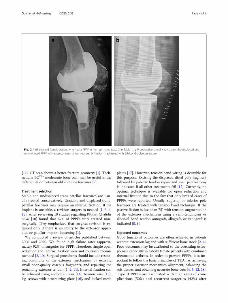
Fig. 2 A 65-year-old female patient who had a PPPF in her right knee (case 2 in Table 1). a Preoperative lateral X-ray shows the displaced andcomminuted PPPF with extensor mechanism rupture. b Fixation is achieved with Ethibond polyester suture
Govil et al. Arthroplasty (2020) 2:35 Page 4 of 6
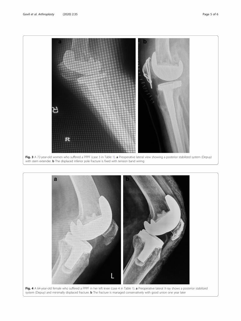
Fig. 4 A 64-year-old female who suffered a PPPF in her left knee (case 4 in Table 1). a Preoperative lateral X-ray shows a posterior stabilizedsystem (Depuy) and minimally displaced fracture. b The fracture is managed conservatively with good union one year later
Fig. 3 A 72-year-old women who suffered a PPPF (case 3 in Table 1). a Preoperative lateral view showing a posterior stabilized system (Depuy)with stem extender. b The displaced inferior pole fracture is fixed with tension band wiring
Govil et al. Arthroplasty (2020) 2:35 Page 5 of 6

osteosynthesis. Rehabilitation plays an important rolein regaining the pre-fracture level of activity. Whenremoving the patella component, leaving an osseousshell may contribute to the development of anteriorknee discomfort and crepitus [9]. Generally, extensor mech-anism repair often produces poor results.
LimitationsOur study has some limitations. The study lacks a con-trol group. The sample size of the series was too smalland cannot be used to identify the factors for predictingoutcomes. The study cannot serve as a fixation guidelinefor PPPFs. Future studies should be done in larger co-horts, with control groups set.
ConclusionGenerally, undisplaced PPPFs should be treated non-operatively whenever possible. For displaced fractures,open reduction and internal fixation and repairing thecombined extensor mechanism injury can yield goodfunctional outcomes.
AbbreviationsPPPF: Periprosthetic patella fracture; TKA: Total knee arthroplasty
AcknowledgementsNot applicable.
Authors’ contributionsLT, GG and PD conceptualised the study design and collected data for thestudy. GG prepared the manuscript. The authors read and approved the finalmanuscript.
FundingNo funding was received for this study.
Availability of data and materialsThe datasets used and/or analysed during the current study are availablefrom the corresponding author on request.
Ethics approval and consent to participateNot required as it was a retrospective case series.
Consent for publicationInformed consent that data will be used for publication was taken from allpatients.
Competing interestsNone of the authors have competing interests to declare.
Author details1Department of orthopaedics, Max Super Speciality Hospital, 108 A, I.P.Extension, Patparganj, Delhi 110092, India. 2D-101, Sunshine Helios, Sector 78,Noida, Uttar Pradesh 201305, India. 3Address: A-702 Vardhman apartment,Mayur Vihar phase1extension, Delhi 110091, India. 4Address: House no 37,Sukh Vihar, Delhi 110051, India.
Received: 26 May 2020 Accepted: 4 October 2020
References1. Benkovich V, Klassov Y, Mazilis B, Bloom S. Periprosthetic fractures of the
knee: a comprehensive review. Eur J Orthop Surg Traumatol. 2020;30:387–99. https://doi.org/10.1007/s00590-019-02582-5.
2. Canton G, Ratti C, Fattori R, Hoxhaj B, Murena L. Periprosthetic kneefractures. A review of epidemiology, risk factors, diagnosis, managementand outcome. Acta Biomed. 2017;88:118–28. https://doi.org/10.23750/abm.v88i2-S.6522.
3. Chun KA, Ohashi K, Bennett DL, El-Khoury GY. Patellar fractures after totalknee replacement. Am J Roentgenol. 2005;185:655–60. https://doi.org/10.2214/ajr.185.3.01850655.
4. Yoo JD, Kim NK. Periprosthetic fractures following total knee arthroplasty.Knee Surg Relat Res. 2015;27:1–9. https://doi.org/10.5792/ksrr.2015.27.1.1.
5. Parvizi J, Kim KI, Oliashirazi A, Ong A, Sharkey PF. Periprosthetic patellarfractures. Clin Orthop Relat Res. 2006;446:161–6. https://doi.org/10.1097/01.blo.0000218722.83601.18.
6. Ortiguera CJ, Berry DJ. Patellar fracture after total knee arthroplasty. J BoneJoint Surg Am. 2002;84:532–40. https://doi.org/10.2106/00004623-200204000-00004.
7. Goldberg VM, Figgie HE III, Inglis AE. Patellar fracture type and prognosis incondylar total knee arthroplasty. Clin Orthop Relat Res. 1998;236:115–22.
8. Vaishya R, Agarwal A, Vijay V. Extensor mechanism disruption after totalknee arthroplasty: a case series and review of literature. Cureus. 2016;8:479doi:10.7759.
9. Sheth NP, Pedowitz DI, Lonner JH. Current concepts review-periprostheticpatellar fractures. J Bone Joint Surg Am. 2007;89:2285–96. https://doi.org/10.2106/JBJS.G.00132.
10. Chalidis BE, Tsiridis E, Tragas AA, Stavrou Z, Giannoudis PV. Management ofperiprosthetic patellar fractures: a systematic review of literature. Injury.2007;38:714–24.
11. Putman S, Boureau F, Girard J, Migaud H, Pasquier G. Patellar complicationsafter total knee arthroplasty. Orthop Traumatol Surg Res. 2019;105:S43–51.https://doi.org/10.1016/j.otsr.2018.04.028.
12. Sayeed SA, Johnson A, Delanois RE. The treatment of patellar fractures aftertotal knee arthroplasty. Semin Arthroplasty. 2018;21:139–41.
13. Rayan F, Konan S, Haddad FS. A review of periprosthetic fractures aroundtotal knee arthroplasties. Curr Orthop. 2008;22:52–61.
14. Maniar RN, Nayak RM, Vatchha S, Singhi T. Periprosthetic patellar fracturefixation using suture anchors. Orthopedics. 2013;36(11). https://doi.org/10.3928/01477447-20131021-36.
15. Masmoudi K, Grissa Y, Benzarti S, Cheikhrouhou H, Mensi Z. Openperiprosthetic patellar fracture after total knee replacement. J OrthopaedicCase Rep. 2016;6:89–91. https://doi.org/10.13107/jocr.2250-0685.452.
16. Lindemeier SC, Brazier BG, Ruhland ED, Cochran JM. Treatment ofperiprosthetic patella fractures using a lag screw and neutralization plateconstruct: a novel surgical technique. Tech Orthop. 2019;00:000.
17. Siljander M, Gandhi S, Koueiter DM, Wiater PJ. Osteosynthesis of aperiprosthetic patella fracture with a locked mesh plate. Int J Orthod. 2017;28:811–3. https://doi.org/10.17554/j.issn.2311-5106.2017.04.229.
18. Agarwal S, Sharma RK, Jain JK. Periprosthetic fractures after total kneearthroplasty. J Orthop Surg. 2014;22:24–9.
Publisher’s NoteSpringer Nature remains neutral with regard to jurisdictional claims inpublished maps and institutional affiliations.
Govil et al. Arthroplasty (2020) 2:35 Page 6 of 6