performance study of and optoelectronic localization system for functional proton radiosurgery
TRANSCRIPT
S712 I. J. Radiation Oncology d Biology d Physics Volume 69, Number 3, Supplement, 2007
characteristics, accuracy, and system components have previously been reported (1,2). Calibration data were conducted with 6MVphotons and 6–16 MeV electron energies in a phantom and showed approximately a 1:1 correlation with slight dependency onelectron energy. Data was obtained on 24 patients (part of FDA approved pilot and pivotal studies on DVS) receiving electron boostbreast treatments. Each patient (pt) had one dosimeter implanted in the tumor bed and one in normal tissue in the opposite quadrantof the breast. Radiation dose was calculated and prescribed to a point at depth in the tumor bed. Daily dose measurements from theDVS for the electron boost treatments were recorded and compared with the predicted dose values at the MOSFET position for eachdosimeter location.
Results: Dosimeters implanted in the tumor bed were close to the 180 cGy electron dose prescription. The frequency of measuredto expected dose variability of .±7% was found in 13 out of the 24 patients (6 of the 13 pts had dose variability in the range.±15%). Dose measurements were obtained from the dosimeter implanted in normal tissue for all patients and measurabledata was obtained in 6 pts. For dosimeters in or at the edge of the field (4 pts), the measured dose was in the range of 68–184cGy vs. an expected dose .180 cGy. Surprisingly, dosimeters located outside the field recorded doses in the range of 64 cGy com-pared to a expected dose of 0.0 cGy. This large variability is indicative of possible setup errors or target movement during treatmentas daily dose measurements varied widely in individual patients.
Conclusions: The DVS was able to measure electron dose for breast boost patients. A majority of patients had .7% variabilitybetween predicted and measured dose. The dosimeter has the potential to help evaluate the daily dose delivered, quantify the ac-curacy of electron dose calculations, quantify the effects of daily intra- and inter- fractional motion and verify small field marginaccuracy (such as partial breast irradiation). The variance noted with electron boost doses suggests an important role for DVS toimprove consistency and accuracy of electron dose delivery for breast patients.
1. Int J Rad Onc Bio Phys, 62 (2), pp 606–613, 2005.2. Phys Med Biol, 50, pp 141–149, 2005.
Author Disclosure: C.W. Scarantino, Shareholder, E. Ownership Interest; Medical Director, F. Consultant/Advisory Board; G.P.Beyer, Medical Physicist, A. Employment; L. McCumber, Clinical Application Specialist, A. Employment; J. Pursley, Mastersresearch project, G. Other.
2912 Performance Study of and Optoelectronic Localization System for Functional Proton Radiosurgery
R. W. Schulte1, F. Shihadeh2, K. E. Schubert2
1Loma Linda University & Medical Center, Loma Linda, CA, 2Californial State University, San Bernardino, CA
Purpose/Objective(s): To study the performance of an optoelectronic localization system (OLS) with three high-resolutioncameras and passive retroreflective markers for potential application in positioning and alignment control in functional protonradiosurgery.
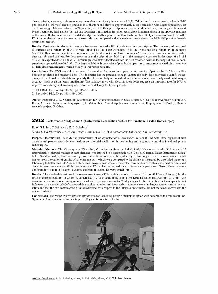
Materials/Methods: The Vicon system (Vicon 260, Vicon Motion Systems, Ltd, Oxford, UK) was used as the OLS. A set of 15retroreflective spherical markers (6 mm diameter) was attached to a stereotactic halo (Leksell G frame, Elekta Instruments, Stock-holm, Sweden) and captured repeatedly. We tested the accuracy of the system by performing distance measurements of eachmarker from the center of gravity of all other markers, which were compared to the distances measured by a certified metrologylaboratory to better than 0.025 mm. Before each measurement session. the system was calibrated with a static marker frame anddynamic wand movements. Within each session 17–18 data individual data captures were performed. Two different cameraconfigurations and four different dynamic calibration techniques were tested (Fig.).
Results: The standard deviation of the measurement error (95% confidence interval) were 0.16 mm (0.12 mm, 0.26 mm) for thefirst camera configuration for which the camera axes met at an acute angle of about 50 deg at isocenter, and 0.24 mm (0.19 mm, 0.38mm) for the second camera configuration for which the camera axes met at 90-deg angles. Different calibration techniques did notinfluence the accuracy. ANOVA showed that marker variation and intersession variations were the largest components of the var-iation and that the two camera configurations differed with respect to the intersession variance but not the residual error and themarker variance.
Conclusions: The Vicon system appears appropriate for localizing passive markers in space with better than 0.4 mm resolution.System performance can be further improved by careful marker selection.
Author Disclosure: R.W. Schulte, None; F. Shihadeh, None; K.E. Schubert, None.