performance of a laser fluorescence device in detecting oclussal caries ... · performance of a...
TRANSCRIPT

48
IntroductionThe contemporary approach to clinical man-
agement of dental caries is based on the detectionof the presence of the disease in early stage, whenany hard tissue changes are minimal. Treatment isbased on non-invasive measures to arrest the dis-ease process and reverse the hard tissue damage.
Today there is an increasing concern regardingso – called “hidden” occlusal caries: carious lesionswhich develop beneath macroscopically intact fis-sures and are difficult to detect by conventionalvisual, tactile and radiographic techniques.
Since early caries detection is not simple, anew system has been developed in the monitoringof early caries lesions. This system based on laserfluorescence is the DIAGNOdent*. This system isdesigned to capture increase fluorescence in theocclusal or proximal areas of the tooth [6,7, 12].
Changes in the tooth substances associated withprogression of the carious process are reflected inan increased amount of fluorescent light [5]. Thecause of this increased level of fluorescence wasthe presence of chromofores associated with bacte-ria present in the infected tooth structure. A numer-ical value is assigned to the degree of fluorescenceas an indicator of the extent of caries [6,7].
ObjectiveThe aim of this study was to establish morpho-
clinical correlation between the results obtainedwith Diagnodent device and the histologicalaspects of carious lesion in polarized lightmicroscopy and stereomicroscopy.
Material and methodFourty-three extracted third molars and premo-
Performance of a laser fluorescence device in detecting oclussal caries in vitro
Roxana Oancea1, Ruxandra Sava-Roºianu1, Atena Gãluºcan2, Daniela Jumanca3,Ramona Amina Popovici1, Angela Codruþa Podariu4
Timisoara, Romania
AbstractThe aim of this study was to compare the validity of the measurements of the laser fluorescence device, KaVoDIAGNOdent, with the result of polarized light microscopy in the detection of occlusal fissure caries in extract-ed third molars and premolars. Fourty-three extracted third molars and premolars which had macroscopicallyintact occlusal surface were selected. The DIAGNOdent measurements of the occlusal test site were recorded bytwo observers at intervals of 2 days. The teeth were then sectioned at the specified test sites for histological exam-inations.Prepared specimens were evaluated under the polarized light microscopy and all images were scored with thecaries classification of D1 (sound and fissure lesion in the half of the outer enamel), D2 (enamel decay) and D3(dentin decay) level (gold standard). Value of specificity for the detection of enamel caries at D1 level was 0,72 andsensitivity values at D2 and D3 levels were 0,66 and 1, respectively. The present study indicates that theDIAGNOdent provides not only almost perfect agreement but also sufficient repeatability at D1, D2, D3 levelsand better specificity at D1 level as well as lower sensitivity at D2 level and excellent sensitivity at D3 level.
Key words: caries detection, laser fluorescence, occlusal caries, polarized light microscopy.
1 Assistent Professor, DMD, Faculty of Dentistry, Preventive, Community and Oral Health Department, University of Medicine andPharmacy „Victor Babeº” Timiºoara2 Lecturer, DMD, Faculty of Dentistry, Preventive, Community and Oral Health Department, University of Medicine and Pharmacy „VictorBabeº” Timiºoara3 Associate Professor, Faculty of Dentistry, Preventive, Community and Oral Health Department, University of Medicine and Pharmacy„Victor Babeº” Timiºoara4 Professor, DMD, PhD, Faculty of Dentistry, Preventive, Community and Oral Health Department, University of Medicine and Pharmacy„Victor Babeº” Timiºoara
lars which had macroscopically intact occlusal sur-face (10 impacted third molars with completed rootformation, 20 premolars extracted in orthodonticpurposes and 13 premolars with different grades ofmobility) were surgically removed without anydamage. The teeth were stored in 10% buffered for-malin immediately following extraction and hadnone of the following: occlusal restorations; fissuresealants; developmental defects; and frank cavita-tion, i.e. cavitation visible on initial examination.
All teeth had been pooled within an interval ofless than 1 month and stored in physiological salinesolution. They were then thoroughly rinsed withwater, cleaned with a tooth brush and pumice anddried with paper tissues in order to obtain optimalconditions for laser detection.
The measurements of the occlusal test sitewere made by using laser fluorescence deviceDIAGNOdent. At the specific wavelength (655-nm) that the DIAGNOdent laser operates, cleanhealthy tooth structure exhibits little or no fluores-cence, resulting in very low scale readings (D1>0–13) on the display. The presence and extend ofcarious lesions were classified as follows: soundfissures and enamel fissure lesions (D1), enamelcaries (D2) and dentinal caries (D3). According tothe caries level, carious tooth structure exhibits flu-orescence in elevated scale readings (D2 >14–19)and (D3 > 20) [8].
A tapered fibre optic probe (Tip A) has been spe-cially designed for the detection of fissure caries. Thelaser probe scans over the fissure in question and hasto be inclined to the right and to the left over the mainfissures to ensure that the tip picks up the fluores-cence from the slopes of the fissure walls, where thecarious process often begins. The maximum value isshown by the appliance as a ‘peak’ value.
The observers were asked to recordDIAGNOdent peak value on the charts above thedrawings on the same day. Two days later, eachmeasurement was repeated on the same test site ofdifferently enumerated teeth to ensure that theobservers did not recall the previous result.
Teeth were then sectioned for histologicalexamination. Teeth were hemi-sectioned in amesial–distal direction through the fissure patternwith a high-speed drill and fine diamond bur.
The specimens were submerged in water andexamined using a polarized light microscope.Stereomicroscopy allows the study of tridimens-sional images. This qualities are based on largefields of interpretation and the large distances
between 92 mm -286 mm, with a magnificationfrom 1,95 to 225 x.
For the otical study in stereomicroscopy wasused Olympus microscope SZ x 7 and Olympuscamera with 2,5 x digital zoom and 3 x optical zoom.
Specificity was defined at D1 level and sensi-tivity was calculated at D2 and D3 level.
ResultsResults obtained with Diagnodent device
revealed: out of 43 apparently sound teeth: 18 hadvalues between 2 and 12 (D1), 13 values between14 and 20 (D2), 12 values over 20 (D3).
Four of the specimens were damaged duringthe histological preparations so they were not eval-uated. After histological examination, 25 teethwere sound, 10 teeth had enamel caries and 8 teethwere shown to have dentinal caries.
From the 10 surfaces with enamel cariouslesions histologicaly distinguised in stereomi-croscopy, 6 presented at the examination withDiagno-dent device values between 14-20 (D2)(fig.1, 2) and 4 values between 20-32 (D3) (fig.3, 4).
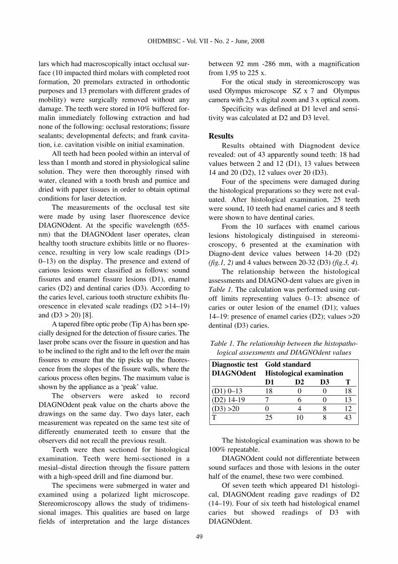
The relationship between the histologicalassessments and DIAGNO-dent values are given inTable 1. The calculation was performed using cut-off limits representing values 0–13: absence ofcaries or outer lesion of the enamel (D1); values14–19: presence of enamel caries (D2); values >20dentinal (D3) caries.
Table 1. The relationship between the histopatho-logical assessments and DIAGNOdent values
The histological examination was shown to be100% repeatable.
DIAGNOdent could not differentiate betweensound surfaces and those with lesions in the outerhalf of the enamel, these two were combined.
Of seven teeth which appeared D1 histologi-cal, DIAGNOdent reading gave readings of D2(14–19). Four of six teeth had histological enamelcaries but showed readings of D3 withDIAGNOdent.
Diagnostic test Gold standardDIAGNOdent Histological examination
D1 D2 D3 T(D1) 0–13 18 0 0 18(D2) 14-19 7 6 0 13(D3) >20 0 4 8 12T 25 10 8 43
49
OHDMBSC - Vol. VII - No. 2 - June, 2008

Histological dentin caries did not differ fromthe results of DIAGNOdent.
This study predicted the probability that whenhistological caries was confined to enamel, the cut-off limits were between 19 and 26 (D2) and indentin they were more than 27 (D3). Values ofspecificity for the detection of caries level was 0,72at D1 level and sensitivities at D2 and D3 levelwere 0,60 and 1, respectively (Table 2).
Table 2. Specificity and sensitivity forDIAGNOdent
Figure 1. Oclussal surface with deep fissures andplaque deposits, stereomicroscopy, x 32.
Figura 2. Morfometry of occlusal surface withdeep fissured and plaque deposits, stereomi-
croscopy, x 32.
Figure 3. Stereomicroscopy of a carious lesiondiffused under amelo-dentinal junction, politopic
lisys of enamel and dentin nearby carious lesion, x32.
Figure 4. Oclussal surface with multiple defects oftisular compounds, reactionary hipermineraliza-
tion adiacent to the carious lesion, stereomi-croscopy, x 8.
DisscusionThe modern clinician’s aim should be to diag-
nose caries before caries cavitation occurs. Earlydiagnosis of occlusal caries and the initiation ofmore effective treatment presents a considerablechallenge [9,13,15]. Much attention has been paidto the possible use of lasers for early caries detec-tion. [9]. This study was conducted on third molarswhich were obtained in less than 1 month time andstored in physiological saline solution to preventthe effect of storing solution and time [4].
Contrary to present study, the material used inother in vitro DIAGNOdent studies consisted ofextracted molar and/or premolar teeth of unknownhistory.
Results PercentageSpecificity D1 72The value of sensitivity for D2 60The value of sensitivity for D2 100
50
OHDMBSC - Vol. VII - No. 2 - June, 2008

Therefore, the number of teeth selected in thisstudy was limited (n= 43). Ten of the selected teethwere impacted third molars. Since the impactedthird molars had comparatively poorly maturedenamel, the measurements of high DIAGNOdentmight have been influenced by incomplete erup-tion, but this cut-off limit was not obviously asso-ciated with histological caries.
So that impacted third molars could be exam-ined to establish ‘gold standards’ for sound fis-sures.
Ten impacted teeth had a minimumDIAGNOdent score of 4 and maximum 14 butnever had 0. This finding had been in agreementwith the result of longitidunal diagnosis of justerupted first molars [10].
Erupted premolars (25) without macroscopicbreakdown exhibited also minimum reading of 4.
Based on the result of this study, numericalscale readings of device were defined as follows:3–18, no caries, or histological enamel caries limit-ed to the outer half of enamel thickness; 19–26, his-tological caries extending beyond the outer half,but confined to the enamel; 27–99, histologicaldentinal caries.
It is clear that the cut-off limits obtained in thisstudy are quite different from those of the earlier invitro studies [7, 12], but almost in agreement withrecent clinical studies [8, 11]. An explanation may beprovided by the in vivo mimetic characteristic of thestorage solution and time used in the present study.
It is obvious from Table 2 that the use ofDIAGNOdent to detect caries at D1 thresholdshowed substantial specificity (0,72). The resultswere roughly in accordance with the literature. Inan in vitro study of natural decay of occlusal fissureenamel, the specificities of DIAGNOdent were(0,72–0,78) [7]. In another in vitro study (Shi et al.,2000) with micro radiography as the gold standard,the laser device had a specificity of 0,95 [12].
In this study, DIAGNOdent showed perfectsensitivity value (1,00) at D3 level supported byother recent studies. One of these evaluated theDIAGNOdent system in vitro and diagnosis was
confirmed by micro radiography and showed high-er diagnostic accuracy in the detection of dentinalcaries than in enamel [12].
Attrill and Ashley (2001) found that the sensi-tivity values of the DIAGNOdent system for thedetection of occlusal caries extending into dentinein extracted primary molars was 0,77–0,80 [2].
DIAGNOdent showed a high sensitivity (0,95) for detection of caries when the cut-off limitwas between sound fissures and caries in enameland dentin. For the diagnosis of enamel decay (D2)on occlusal surfaces a previous study had comparedDIAGNOdent device with electronic caries moni-tor (ECM) in an in vitro study with histologicalmeasures as the gold standard. It found higher sen-sitivities (0,76 – 0,84) for DIAGNOdent than thevalue found in the present study (0,66) (with a cut-off 14–19) [3].
High sensitivity for dentin caries (at D3 thresh-old) with good specificity for sound fissures andlesions in the outer half of the enamel (at D1 thresh-old) indicates that this diagnostic method can accu-rately detect both dentin caries and sound fissures.
Although this experiment was carried out invitro, and therefore some care must be taken intoaccount to interpret these results for in vivo condi-tion, the ability of the device to detect dentinallesions before substantial tissue loss would allowconsiderable changes in the treatment decisionstrategy.
Good specificity for enamel caries detection(recognizing sound surfaces) would have signifi-cant influence on the overall use of preventiontechniques.
ConclusionThis in vitro study revealed that the
DIAGNOdent device has high diagnostic validityfor the detection of sound surfaces and the dentinalcaries. DIAGNOdent provides not only almost per-fect agreement but also sufficient repeatability atall levels and good specificity at D1 level, andlower sensitivity at D2 level than D3 level inextracted third molars.
51
OHDMBSC - Vol. VII - No. 2 - June, 2008
References1. Ástvaldsdottir A, Holbrook P, Tranæus S. Consistency
of DIAGNOdent instruments for clinical assessment of fissurecaries. Acta Odontol Scand 2004; 62:193–8.
2. Attrill DC, Ashley PF. Occlusal caries detection in pri-mary teeth: a comparation of DIAGNOdent with conventionalvisual examination. Caries Res 2001; 190: 440-3.
3. Bamzahim M, Shi X-Q, Angmar-Mänsson B. Occlusalcaries detection and quantification by DIAGNOdent and elec-

tronic caries monitor: in vitro comparison. Acta Odontol Scand2002; 60:360–4.
4. Francescut, P. & Lussi, A. Impact on DIAGNOdentvalues of formalin and chloramin storage solutions. CariesResearch 2000, 34: 325.
5. Hibst R, Gall R. Development of a diode laser basedfluorescence caries detector. Caries Res 1998; 32:294.
6. Longbottom C, Pitts NB, Lussi A, Reich E. In vitrovalidity of a new laser-based caries detection device. J DentRes 1998; 77:766-9.
7. Lussi, A, Hibst, R. Methods for occlusal caries detec-tion used in daily practice. In: Early Detection of Dental CariesII.Proceedings of the 4th Annual Indiana Conference, (ed.G.W.Stookey), Indianapolis, Indiana, 1999; pp. 171.
8. Lussi A, Megert B, Longbottom C, Reich E,Francescut P. Clinical performance of a laser fluorescencedevice for detection of occlusal caries lesions. Eur J Oral Sci2001; 109:14–9.
9. Mount, G.J. & Ngo, H. Minimal intervention: a newconcept for operative dentistry. Quintessence International2000a; 31:527-531.
10. Sheehy, E.C., Brailsford, S.R., Zoitopoulos, L., Kidd,E.A., Beighton D. Longitudinal assessment of occlusal caries inchildren using a laser fluorescence system (DIAGNOdent).Caries Research 2001a; 35: 269.
11. Sheehy EC, Brailsford SR, Kidd EAM, Beighton D,Zoitopoulos L. Comparison between visual examination and alaser fluorescence system for in vivo diagnosis of occlusalcaries. Caries Res 2001b; 35:421–6.
12. Shi X-Q, Welander U, Angmar-Mannsson B.Occlusal caries detection with KaVo DIAGNOdent and radi-ographic examination: an in vitro comparison. CariesRes2000; 34:151–8.
13. Tinanoff N, Douglass JM. Clinical decision makingfor caries management in children. Pediatr Dent 2002;24(5):386–92.
14. Tranæus S, Shi X-Q, Lindgren L, Trollsans K,Angmar-Mannsson B. In vivo repeatability and reproducibilityof quantitative light-induced fluorescence. Caries Res 2002;36:3–9.
15. Verdonschot, E.H., Angmar-Mansson, B., ten Bosch,J.J., Deery,C.H., Huysmans, M.C.D.N.S.M., Pitts, N.B.,Waller, E. Development in caries diagnosis and their relation-ship to treatment decisions and quality of care. ORCASaturdayAfternoon Symposium 1997. Caries Research 1999;33: 32.
***
Corespondance to: Prof.Dr. Angela Codruta Podariu, Faculty of Dentistry Preventive,Community and Oral Health Department, University of Medicine and Pharmacy „Victor Babes"
Timisoara, P-ta Eftimie Murgu nr. 2, 300041, Timisoara
OHDMBSC - Vol. VII - No. 2 - June, 2008
52