percutaneous recanalization of total occlusions of the
TRANSCRIPT
Percutaneous recanalization of total occlusions ofthe iliac veinSeshadri Raju, MD,a and Peter Neglén, MD, PhD,b Jackson and Flowood, Miss
Background: Venovenous bypass has been the standard in relieving chronic total occlusions of iliac veins. The technicalfeasibility of percutaneous recanalization was previously reported. Routine applicability of this technique in a widespectrum of lesions and patients, stent patency, and clinical outcome forms the basis of this presentation.Methods: During a 9-year period, 167 limbs in 159 unselected patients in a consecutive series with post-thromboticchronic total occlusions of the iliac and adjacent vein segments underwent percutaneous attempts at recanalization.Patients were not selected based on venographic appearance or extent of the lesion, or excluded because of a preemptivechoice of open venovenous bypass surgery.Results: Percutaneous recanalization was successful in 139 of 167 limbs (83%), including patients with bilateral occlusionsand 14 patients with inferior vena cava filters incorporated in the treated occlusion. Median age was 53 years (range,18-84 years). Thrombophilia was identified in 44 patients. Venous dermatitis/ulcer was found in 46% of the treatedlimbs. Recanalization involved three or more totally occluded vein segments in 42% of the limbs. The cumulativesecondary stent patency rate at 4 years was 66%. The cumulative marked relief of pain and swelling at 3 years was 79% and66%, respectively. Cumulative healing of venous ulcer at 33 months was 56%. Quality of life metrics improvedsignificantly.Conclusions: Most femoroiliocaval chronic total occlusions lesions can be successfully recanalized percutaneously with verylittle morbidity, minimal downtime, sustained long-term stent patency, and substantial clinical improvement. Theprocedure has wide applicability in a broad spectrum of symptomatic patients, including those with extensive lesions, and
can be considered for routine use. ( J Vasc Surg 2009;50:360-8.)Venovenous bypasses using the saphenous vein1 or aprosthesis2 have been the standard in the treatment ofsymptomatic iliac vein occlusions. Early results of percuta-neous endovenous recanalization in 38 limbs were de-scribed in 2002.3 Since then, procedural success has in-creased with experience, reducing the need for openvenovenous bypass procedures. This study analyzed thesuccess rate, stent patency rate, and clinical outcomes inpatients after stent recanalization of chronic total occlu-sions (CTO) of femoroiliocaval vein segments.
METHODS
Patients. From 1999 through 2007, obstructive le-sions of the femoroiliocaval veins segments were treated bystent placement in 1402 limbs; of these, 603 were post-thrombotic which included CTO limbs. A consecutiveseries of 167 limbs in 159 patients with femoroiliocavalCTO underwent attempts at percutaneous recanalizationduring that time. No limbs were excluded because of theextent or severity of CTO or because of a preemptive choiceof venovenous bypass. In seven patients a previous veno-venous bypass (3 prosthetic, 4 Palma) had been performed,and five had occluded. Two Palma bypasses had remainedpatent; however, the patients continued to be symptom-
From the University of Mississippi Medical Center, Jackson,a and River OaksHospital, Flowood.b
Competition of interest: none.Reprint requests: Seshadri Raju, MD, 1020 River Oaks Dr, Ste 420,
Flowood, MS 39232 (e-mail: [email protected]).0741-5214/$36.00Copyright © 2009 Published by Elsevier Inc. on behalf of the Society for
Vascular Surgery.
doi:10.1016/j.jvs.2009.01.061360
atic. Limbs with acute or acute/chronic thromboses re-quiring thrombolysis before stenting were excluded be-cause these subsets were considered different from CTOlimbs. The procedure was successful in 139 limbs, repre-senting 23% of all post-thrombotic limbs treated withstents.
Preoperative investigations, indications, technique, andfollow-up protocol have been described in detail else-where4-6 and are presented here in abbreviated form.
Indications. Patients with significant limb symptoms,including pain, swelling, venous dermatitis and ulcer, re-current cellulitis, or a combination of these symptoms, whohad failed conservative therapy were considered for inter-vention. Severity of symptom presentation, and not thevenographic finding of CTO, was the determinant forintervention; some patients with extensive venographic oc-clusive lesions may be asymptomatic or only mildly symp-tomatic.
A preoperative thrombophilia workup, consisting ofantithrombin 3, protein C and S, anticardiolipin antibody,lupus anticoagulant, prothrombin and Leiden gene muta-tions, and homocystinemia �15 mg, was routinely per-formed. Venous investigations included arm/foot venouspressure differential, ambulatory venous pressure measure-ment, air plethysmography, duplex ultrasound (DUS) ex-amination, and ascending (pedal contrast injection) andtransfemoral antegrade venography with exercise femoralpressure measurement.
After recanalization, patients were monitored by clini-cal examination at 6 weeks, 3 months, 6 months, and atyearly intervals thereafter. The cumulative rate of ulcer
healing (100% epithelialization) was calculated. Pain was
JOURNAL OF VASCULAR SURGERYVolume 50, Number 2 Raju and Neglén 361
evaluated using a visual analog scale (VAS)7 from 0 to 10,wherein 10 is the most severe pain. Swelling was assessedas grade 0 (absent), grade 1 (pitting, not obvious), grade2 (visible ankle edema), and grade 3 (gross, encompassingthe entire leg) according to reporting standards. TheChronic Venous Insufficiency Questionnaire (CIVIQ) qualityof life method8 validated for chronic venous disease wasused, and the latest patient response was used in outcomeanalysis. Contrast venography was routinely performed at 6to 12 weeks, at 6 months after the procedure, and thenyearly. DUS examination, as recently described by Labro-poulos et al,9 has been increasingly used to assess thepatency of the stented iliac vein. Preliminary experienceindicates the method is comparable to venography in as-sessing gross stent patency.
Technique of recanalization. The procedure is per-formed under general anesthesia for safer cardiopulmonarycontrol and lack of adequate pain control under localanesthesia. A midthigh femoral vein access under US guid-ance is used. The femoral vein is found posterolateral to theartery at midthigh level. Progressive dilatation of the accessneedle track with over-the-wire dilators may be required inpost-thrombotic veins before the introduction of thesheath. With routine use of a sealing device (VasoSeal,Datascope Corp, Montvale, NJ), access site complicationsare rare. With attention to depth markings on the device,inadvertent intravenous placement of the sealant is avoided(1 suspected in �1500 uses), even when large sheaths areused.
The midthigh access allows the use of shorter-lengthinstrumentation with superior pushability compared withpopliteal or internal jugular access. Sufficient space remainscephalad to the sheath (size 10-13, 13-cm length) belowthe inguinal ligament to deploy a stent into the commonfemoral vein if needed. When the femoral vein is occluded,as is frequently found, access is often possible through theupper 3 to 5 cm of the vein, which tends to remain open, orthrough parallel collaterals in the vicinity. The profundafemoris vein is usually open if the femoral vein is occludedand can be accessed 2 to 5 cm below the lesser trochanter.When access is possible only in the upper thigh, the sheathtip has to be withdrawn below the common femoral veinfor stent deployment if needed; stent extension into theprofunda femoris vein is allowed when the femoral vein isoccluded.
An antegrade venogram is performed to define existingvenous anatomy. A 0.035-inch soft or stiff Glidewire(Terumo Inc, Ann Arbor, Mich) is inserted up to theocclusion. Further progress into the occlusion is madewith the tip of the Glidewire with straight or angledcatheter support. Unlike in arterial occlusions, a leadingloop is not helpful at this stage. Cutting balloons andtunneling devices (Frontrunner CTO, LuMend Inc, Red-wood City, Calif) may be helpful to gain initial entry intothe occluded segment. After gaining access into the oc-cluded vein, further progress into the occlusion is madewith the Glidewire tip, with or without catheter (or long
sheath) support. A leading loop may be advantageous togain passage through the external and common iliac veins ifthe trabeculae are not dense.
A low-profile catheter with a sharp edge (Quick-Crosscatheter; Spectranetics, Colorado Springs, Colo) appears tofacilitate progress, but other hydrophilic catheters, either4F or 5F size with standard or angled tips, are helpful aswell. If forward progress stalls, other Glidewires of differentstiffness, caliber, or tip angle should be tried. Usually, somecombination of Glidewire and supporting catheter will befound to advance progress. Passage of the Glidewirethrough the occluded segment meets with variable diffi-culty. With some experience, most occluded iliofemoralveins can be recanalized in 30 to 40 minutes and evenextensive inferior vena cava (IVC) occlusions in 60 to 75minutes. The progress is sometimes tedious and patience isnecessary.
The Glidewire probably advances through small vascu-lar channels present in the organizing fibrous tissue withinthe vein. Because the occluded vein segment remains invis-ible on venography, Glidewire passage must be guided byintuitive sense of the normal anatomic course of the vein. Inthe frontal projection, the left femoral vein overlies themedial third of the femoral head, coursing up to the lowerpelvic brim over the pelvic tubercle, and from there bowsslightly outward, moving across the sacroiliac joint joiningthe IVC variably between the fourth and fifth lumbarvertebral bodies at their right margin. The right femoraland iliac veins course in a straighter line (frontal projection)to their junction with the IVC. The Glidewire should trackaccordingly. It is useful to view progress of the recanaliza-tion intermittently by 45° or 60° oblique projections toensure that the Glidewire initially follows the curve of thesacrum and then turns anteriorly towards the promonto-rium and anterior to the spine.
Progress of Glidewire passage through the occludedvein may be impeded to a variable degree by vascular andligamentous structures compressing the vein. The post-thrombotic fibrous tissue in and around the affected vein isparticularly dense at these anatomic “chokepoints,” wherecompression occurs and post-thrombotic recanalization isknown to be poor.10-12 These sites differ between the rightand left sides (Fig 1). Intermediate balloon dilatations tofacilitate Glidewire passage are often counterproductivebecause the wire loses the support of surrounding tissueand coils in the dilated space, making further progress evenmore difficult. The Glidewire may be diverted into collat-erals and tributaries, particularly adjacent to these choke-points. This is easily recognized if normal anatomy is kept inmind.
An angled-tip catheter is useful in redirecting theGlidewire in the proper direction when it seems to veer offcourse. In a few instances, the Glidewire has passed throughthe vertebral veins into the vertebral canal where it ispositioned in the middle of the vertebral column ratherthan at the right lateral edge where the IVC is situated. Thisis easily recognized by oblique fluoroscopic projections.The Glidewire is withdrawn and redirected with no com-
plications.
JOURNAL OF VASCULAR SURGERYAugust 2009362 Raju and Neglén
Passage of the Glidewire away from the expected courseof the vein with sudden ease denotes perforation. TheGlidewire can be withdrawn and usually manipulated in theproper direction without sequelae. Withdrawal of bloodthrough the catheter may or may not be possible during therecanalization process and has no particular significance(procedure may not be aborted) as long as the Glidewiretracks approximate to normal anatomy.
Approaching the IVC, the Glidewire can track parallelto the vena cava, possibly within the vasa vasorum in itswall, for 3 to 5 cm before entering the proper lumen of theIVC. This is not harmful. Interval contrast injections intothe track are usually not helpful, and pooling of the dye intolakes can be misleading and should not suggest that the
Fig 1. The Glidewire passage during recanalization procedurecommonly meets resistance at several anatomic chokepoints wherethe iliac or the hypogastric artery crosses the vein; the inguinalligament and the diaphragmatic hiatus may also prevent passage.Relevant arterial crossover levels differ slightly between the rightand left side, as shown. The Glidewire can veer off course from themain vein through tributaries and collaterals (shown numbered)when meeting resistance. On the right side, the Glidewire mayenter medial or lateral collaterals because the iliac vein runs astraighter course to the inferior vena cava. The left iliac vein has aconvex curvature, and the Glidewire tends to enter lateral tributar-ies, usually the ascending lumbar vein. When the suprarenal venacava is occluded, the azygos and hemiazygos collaterals are en-larged and the Glidewire enters them frequently. 1, Phrenic vein; 2,azygos/hemiazygos complex; 3, prevertebral plexus; 4, hypogas-tric vein; 5, ascending lumbar vein; 6, tributaries from the iliopsoasmuscles; 7, femoral vein tributaries; 8, lumbar veins.
procedure be aborted.
Entry into the IVC is verified by further easy passage ofthe wire or catheter along the normal course of the vesselinto the right atrium. This should be confirmed by fluoros-copy, contrast injection through a catheter placed in theIVC, and intravascular US (IVUS). From this stage of therecanalization procedure, the use of IVUS13 becomes cru-cial to assure integrity of the recanalized channel, to selectoptimal proximal and distal landing sites preferably free ofpost-thrombotic disease, to ensure proper deployment andexpansion of the stents, and to minimize radiation expo-sure. Venography alone is distinctly inferior in these re-spects, and exclusive dependence on it may result in aninferior outcome with poor patency.
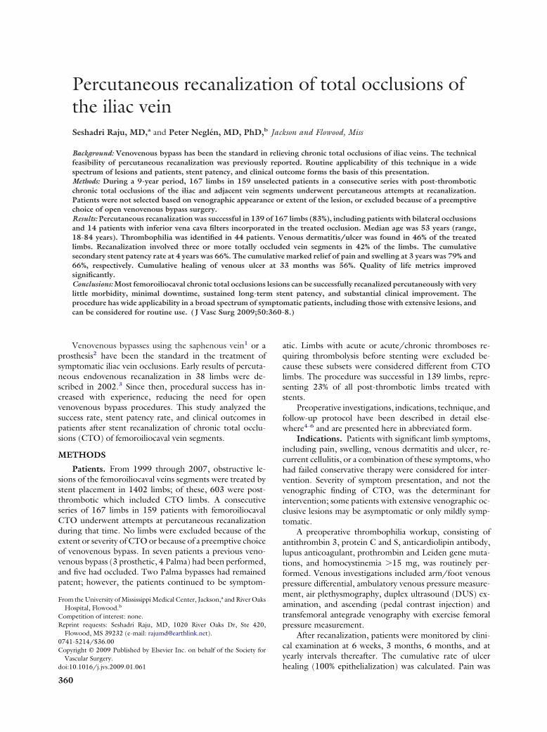
The recanalized channel may be dilated in a single stepto the ultimate desired size after IVUS examination. Predi-latation up to 10 mm may be necessary, starting withsmaller-sized (3-mm) balloons and matched guidewires insome cases to gain easy passage of a 6F IVUS catheter andlarge-caliber balloons. By IVUS, the Glidewire is invariablyfound to lie within the confines of the recanalized occludedvein (Fig 2).
Repeated balloon dilatation at the anatomic choke-points may be necessary. It is useful to use high-pressureballoons (16 to 18 atm) and keep the balloon expanded atmaximum level for at least a minute until the balloonpressure stabilizes at this level. Overdilatation and oversiz-ing of the stent diameter by 2 to 4 mm for the anatomiclocation is recommended to compensate for the variablerecoil of the recanalized channel. Oversizing the vena cavalstent is important to facilitate later contralateral stenting ifrequired. Optimal stent diameters after recoil are 20 mmfor the IVC, 16 to 18 mm for the common iliac vein, 14 to16 mm for the external iliac vein, and 12 to 14 mm for thecommon femoral vein in normally sized adults.
Self-expanding woven braided stainless stents (Wall-stent, Boston Scientific, Nantucket, Mass) in series with 3-to 5-cm overlap to minimize shelving along the complexcourse of the iliac vein were used in most patients (Fig 3). Asmooth transition at overlaps is facilitated by using stentswith the same diameter (used for the largest segment)through all of the recanalized segments, but limiting post-deployment balloon dilatation to the size appropriate foreach segment. This results in oversizing of the stents, whichis an advantage during later interventional corrections, ifneeded.
Because multiple stents are typically required, the max-imum manufactured length for the various diameter sizes(typically 7- to 9-cm length) should be used, restrictingshorter lengths for use at either end of the stack to tailorlength. Unless the recoil is severe (�40%), dilatation afterdeployment should be delayed until all of the stents are inplace. This speeds up restoration of flow through the stentassembly and helps to reduce the chances of thrombusnidus formation. A completion IVUS examination andvenogram terminate the procedure after noted defects arecorrected by repeat ballooning. Patients can be discharged
after an overnight stay.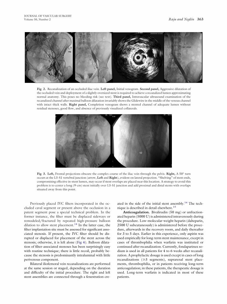
isual
JOURNAL OF VASCULAR SURGERYVolume 50, Number 2 Raju and Neglén 363
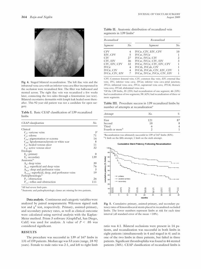
Previously placed IVC filters incorporated in the oc-cluded caval segment or present above the occlusion in apatent segment pose a special technical problem. In theformer instance, the filter must be displaced sideways orremodeled/fractured by repeated high-pressure balloondilation to allow stent placement.14 In the latter case, thefilter implantation site must be assessed for significant asso-ciated stenosis. If present, the IVC filter should be dis-rupted or displaced for placement of the stent across thestenosis; otherwise, it is left alone (Fig 4). Balloon dilata-tion of filter-associated stenoses has been surprisingly easywith routine technique; there is little recoil, probably be-cause the stenosis is predominantly intraluminal with littleperivenous component.
Bilateral iliofemoral vein recanalizations are performedat the same session or staged, depending on the durationand difficulty of the initial procedure. The right and left
Fig 2. Recanalization of an occluded iliac vein. Left pathe occluded vein and deployment of a slightly oversizednormal anatomy. This poses no bleeding risk (see text)recanalized channel after maximal balloon dilatation invarwith intact thick walls. Right panel, Completion venoresidual stenoses, good flow, and absence of previously v
Fig 3. Left, Frontal projections obscure the complex coccurs at the L5-S1 vertebral junction (arrow, Left and Rcompromising effective in-stent lumen, may occur if stenproblem is to center a long (9-cm) stent initially over L5situated away from this point.
stent assemblies are connected through a fenestration cre-
ated in the side of the initial stent assembly.14 The tech-nique is described in detail elsewhere.15
Anticoagulation. Bivalirudin (50 mg) or unfraction-ated heparin (5000 U) is administered intravenously duringthe procedure. Low-molecular-weight heparin (dalteparin,2500 U subcutaneously) is administered before the proce-dure, afterwards in the recovery room, and daily thereafterfor 3 to 5 days. Earlier in this experience, only aspirin wasused empirically for long-term stent maintenance, except incases of thrombophilia when warfarin was instituted orcontinued after recanalization. Currently, fondaparinux so-dium is used in all patients for 4 to 6 weeks after recanali-zation. A prophylactic dosage is used except in cases of longrecanalizations (�3 segments), suprarenal stent place-ments, thrombophilia, or in patients receiving long-termanticoagulation; in these patients, the therapeutic dosage isused. Long-term warfarin is indicated in most of these
nitial venogram. Second panel, Aggressive dilatation ofis required to achieve a recanalized lumen approximatingird panel, Intravascular ultrasound examination of theshows the Glidewire in the middle of the venous channelshows a stented channel of adequate lumen without
ized collaterals.
of the iliac vein through the pelvis. Right, A 50° turn), evident on lateral projection. “Shelving” of stent ends,laps are placed near this location. A strategy to avoid thisnction and add proximal and distal stents with overlaps
nel, Istent. Thiablygram
ourseight
t over-S1 ju
patients.
JOURNAL OF VASCULAR SURGERYAugust 2009364 Raju and Neglén
Data analysis. Continuous and categoric variables wereanalyzed by paired nonparametric Wilcoxon signed ranktest and �2 test, respectively. Primary, assisted-primary,and secondary patency rates, as well as clinical outcomewere calculated using survival analysis with the Kaplan-Meier method. Prism 3 software (GraphPad, San Diego,Calif) was used for analysis. A value of P � .05 wasconsidered significant.
RESULTS
The procedure was successful in 139 of 167 limbs in131 of 159 patients. Median age was 53 years (range, 18-92
Fig 4. Staged bilateral recanalization. The left iliac vein and theinfrarenal vena cava with an inferior vena cava filter incorporated inthe occlusion were recanalized first. The filter was ballooned andstented across. The right iliac vein was recanalized a few weekslater, connecting the two sides through a fenestration (see text).Bilateral excoriative dermatitis with lymph leak healed soon there-after. This 92-year-old patient was not a candidate for open sur-gery.
Table I. Basic CEAP classification of 139 recanalizedlimbs
CEAP classification No.
ClinicalC2: varicose veins 3a
C3: edema 71C4a: pigmentation or eczema 14C4b: lipodermatosclerosis or white scar 8C5: healed venous ulcer 11C6: active venous ulcer 32
EtiologicEp: primary 0Es: secondary 139
Anatomicb
Ad: deep veins 39As,d: superficial and deep veins 76Ad,p: deep and perforator veins 3As,d,p: superficial, deep, and perforator veins 19
Pathophysiologicb
Po: obstruction 26Pr,o: reflux and obstruction 111
aAll had severe limb pain.bAnatomic and pathophysiologic classes are missing for two patients.
years). Female to male ratio was 2:1, and left to right limb
ratio was 4:1. Bilateral occlusions were present in 14 pa-tients, and recanalization was successful in both limbs ineight patients (simultaneously in 4 and staged in 4) and inone of the two limbs in three patients, but failed in threepatients. Significant thrombophilia was found in 44 stented
Fig 5. Cumulative primary, assisted-primary, and secondary pa-tency rates of femoroiliocaval stents placed in recanalized occludedlimbs. The lower numbers represent limbs at risk for each timeinterval (all standard error of the mean �10%).
Table II. Anatomic distribution of recanalized veinsegments in 139 limbsa
Recanalized Recanalized
Segment No. Segment No.
CFV 4 IVCir, CIV, EIV, CFV 10EIV, CFV 5 IVCsr, IVCir 1CIV 27 IVCsr, IVCir, CIV 2CIV, EIV 36 IVCsr, IVCir, CIV, EIV 3CIV, EIV, CFV 32 IVCsr, IVCir, CIV, EIV, CFV 1IVCir 4 IVCth, IVCab, CIV 1IVCir, CIV 4 IVCth, IVCab, CIV, EIV, CFV 1IVCir, CIV, EIV 7 IVCat, IVCsr, IVCir, CIV, EIV 1
CFV, Common femoral vein; CIV, common iliac vein; EIV, external iliacvein; IVC, inferior vena cava; IVCat, inferior vena cava-atrial junction;IVCir, infrarenal vena cava; IVCsr, suprarenal vena cava; IVCth, thoracicvena cava; IVCab, abdominal vena cava.aOf the 139 limbs, 35 (25%) had recanalization of one segment; 46 (33%)had recanalization of two segments; 58 (42%) had recanalization of three ormore segments.
Table III. Procedure success in 139 recanalized limbs bynumber of attempts at recanalizationa
Attempt No. %
First 121 87Second 10 8Third 6 4Fourth or moreb 2 1
aRecanalization was ultimately successful in 139 of 167 limbs (83%).b1 limb on the fifth attempt; 1 limb on the sixth attempt.
patients (34%). CEAP classification of recanalized limbs is

n sele
JOURNAL OF VASCULAR SURGERYVolume 50, Number 2 Raju and Neglén 365
reported in Table I. Distribution of recanalized venoussegments of the limbs is summarized in Table II. A total of14 IVC filters of various types were remodeled and stentedthrough, without any apparent complications. No associ-ated stenosis was found in two other limbs, and the filterwas left undisturbed above the stent. No pararenal orsuprarenal filters were encountered in this experience.
The upper and lower landing sites of the stents com-monly extended one or more segments beyond the recana-lized section to cover adjoining nonocclusive stenoses. Thelower landing site of the stent was below the inguinalligament (common femoral vein) in 65 limbs (47%), in theexternal iliac vein in 42 (30%), and in the common iliac veinin 32 (23%). The upper landing site was in the IVC in allbut two limbs for previously described technical reasons.4
These two limbs had distal external iliac vein occlusion, andthe upper landing site was in the common iliac vein.
Procedural success. The overall recanalization successrate was 83% (139 of 167 limbs), and most procedures weresuccessful on the first attempt (Table III). Recanalizationfailed in two of five patents with occluded venovenousbypasses performed earlier because the prior venotomy sitecould not be traversed. Endophlebectomies16 of the fem-oral vein were performed in seven limbs that were impervi-ous to percutaneous entry into the occluded femoral veinsegment. Subsequent recanalization was attempted in fivelimbs, but succeeded only in two limbs, both of which lateroccluded.
Morbidity. There was no 30-day mortality. There wasno clinically apparent procedurally related bleeding. (Acontained pelvic hematoma requiring transfusions oc-curred in one patient in 2008 who was not included in thisseries.) No access complications occurred that requiredintervention. About 25% of the patients reported back pain,which was limited (commonly days, occasionally up to 2weeks) and easily controlled with ibuprofen. No patientrequired hospitalization for pain. One patient had a contrast-related transient rise in the serum creatinine concentration.Early thrombotic events (�30 days after stenting) occurred
Fig. 6. Removal of an occluded Wallstent through a listrands under fluoroscopy (arrow) will result in unravelistrands. The technique may allow repeat recanalization i
in 10 limbs, all associated with concurrent thrombosis of
the stent. Late thrombotic events (�30 days after stenting)occurred in 32 limbs (ipsilateral, involving the stent in 29;contralateral nonstented limbs in 3 patients).
Stent patency. Long-term primary, assisted-primary,and secondary patency rates are shown in Fig 5. During thefollow-up period, 39 stents (28%) occluded, including onepatient in whom the lower portion (common femoral in-flow) has occluded and only the upper part of the stentstack is open (hypogastric inflow through the stent weave).Stent thrombosis was unrelated to preexisting thrombo-philia (P � .68). Pharmacomechanical thrombectomy orcatheter-directed thrombolysis, or both, were attempted in23 limbs with occluded stents. This was successful in restor-ing stent patency in 17 limbs; 10 remained patent, butseven later occluded. One occluded stent spontaneouslylysed after having been occluded for 3 years, and bothoccurrences were confirmed by transfemoral venography.An adjunctive arteriovenous fistula to improve inflow afterthrombectomy was used in one patient, but the stentrethrombosed. In two limbs, the occluded iliac stent wasremoved (Fig 6) through a limited transverse venotomy.One limb was recanalized and restented later, but throm-bosed �6 weeks.
Other reinterventions. Precautionary reinterventionswere completed in 38 recanalized limbs (27%; Table IV).Indication for reintervention was the presence of significant
Table IV. Details of reintervention procedures in 38recanalized limbs
Reintervention procedure No. (n � 38)
Balloon dilation 22Plus add proximal stent 2Plus add distal stent 8Plus add proximal and distal stents 1Plus add stent separation stent 2
Add proximal stent 1Removal of Wallstent; restented 2
transverse venotomy. Left, Steady pull on one of thethe stent weave and (Right) serial removal of all of thected cases.
mitedng of
(�50%) in-stent restenosis or residual or recurrent symp-
JOURNAL OF VASCULAR SURGERYAugust 2009366 Raju and Neglén
toms, or both. Types and details of reintervention tech-niques performed in stented patients, including the recana-lized CTO subset, are described elsewhere.17
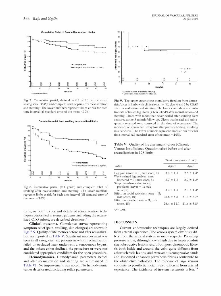
Clinical outcome. Cumulative curves representingsymptom relief (pain, swelling, skin changes) are shown inFigs 7-9. Quality of life metrics before and after recanaliza-tion are reported in Table V. Significant improvement wasseen in all categories. Six patients in whom recanalizationfailed or occluded later underwent a venovenous bypass,and the others either declined the procedure or were notconsidered appropriate candidates for the open procedure.
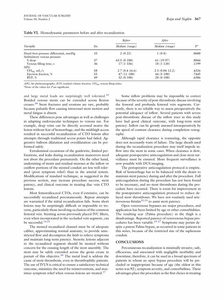
Hemodynamics. Hemodynamic parameters beforeand after recanalization and stenting are summarized inTable VI. No improvement was noted. No hemodynamic
Fig 7. Cumulative partial, defined as �3 of 10 on the visualanalog scale (VAS), and complete relief of pain after recanalizationand stenting. The lower numbers represent limbs at risk for eachtime interval (all standard error of the mean �10%).
Fig 8. Cumulative partial (�1 grade) and complete relief ofswelling after recanalization and stenting. The lower numbersrepresent limbs at risk for each time interval (all standard error ofthe mean �10%).
values deteriorated, including reflux parameters.
DISCUSSION
Current endovascular techniques are largely derivedfrom arterial experience. The venous system obviously dif-fers from the arterial system in many respects. Prevailingpressure is low, although flow is high due to larger conduitsize; obstructive lesions result from post-thrombotic fibro-sis both inside and around the vein, quite different fromatherosclerotic lesions; and extravenous compressive bandsand associated enhanced perivenous fibrosis contribute tothe obstructive pathology. The response of large venousconduits to prosthetic metal is also different from arterial
Fig 9. The upper curve shows cumulative freedom from derma-titis/ulcer in limbs with clinical severity (C) class 4 and 5 by CEAPafter recanalization and stenting. The lower curve shows cumula-tive rate of healed leg ulcers (C6 in CEAP) after recanalization andstenting. Limbs with ulcers that never healed after stenting werecensored at the 3-month follow-up. Ulcers that healed and subse-quently recurred were censored at the time of recurrence. Theincidence of recurrence is very low after primary healing, resultingin a flat curve. The lower numbers represent limbs at risk for eachtime interval (all standard error of the mean �10%).
Table V. Quality of life assessment values (ChronicVenous Insufficiency Questionnaire) before and afterrecanalization in 128 limbs
Value
Total score (mean � SD)
Before After
Leg pain (none � 1, max score, 5) 3.5 � 1.3 2.6 � 1.3a
Work-related leg problem (notbothered � 1; max score, 5) 3.7 � 1.3 2.9 � 1.2a
Sleep disturbance due to legproblems (never � 1; maxscore, 5) 3.2 � 1.3 2.5 � 1.3a
Effect on social activities (none � 8;max score, 40) 26.8 � 8.8 21.3 � 8.7a
Effect on morale (none � 9; maxscore, 45) 26.4 � 11.1 21.6 � 8.8a
aP � .001.
experience. The incidence of in-stent restenosis is less,18
JOURNAL OF VASCULAR SURGERYVolume 50, Number 2 Raju and Neglén 367
and large metal loads are surprisingly well tolerated.19
Braided venous stents can be extended across flexioncreases.20 Stent fractures and erosions are rare, probablybecause pulsatile flow causing intravessel stent motion andmetal fatigue is absent.
These differences pose advantages as well as challengesin adapting endovascular techniques to venous use. Forexample, deep veins can be directly accessed nearer thelesion without fear of hemorrhage, and the midthigh accessresulted in successful recanalization of CTO lesions afterattempts through traditional access points had failed. Ag-gressive balloon dilatation and overdilatation can be per-formed safely.
Extraluminal excursions of the guidewire, limited per-forations, and leaks during recanalization maneuvers neednot abort the procedure prematurely. On the other hand,undersizing of stents and residual stenoses at the inflow oroutflow portions of the stented conduit are less well toler-ated (poor symptom relief) than in the arterial system.Modifications of standard technique, as suggested in theprevious section, may increase procedure success, stentpatency, and clinical outcome in treating iliac vein CTOlesions.
Most femoroiliocaval CTOs, even if extensive, can besuccessfully recanalized percutaneously. Repeat attemptsare warranted if the initial recanalization fails. Some shortlesions may be surprisingly difficult or impossible to tra-verse, particularly those involving occlusion of the commonfemoral vein. Stenting across previously placed IVC filters,even when incorporated in the occluded vein segment, canbe successful.14,21
The stented recanalized channel must be of adequatecaliber, approximating normal anatomy, to provide unre-stricted flow and decompress the limb to relieve symptomsand maintain long-term patency. Stenotic lesions adjacentto the recanalized segment should be stented withoutconcern for the ensuing length of the stent assembly. Thestent may be safely extended across the groin crease inpursuit of this objective.20 The metal load is seldom thecause of stent thrombosis, even in thrombophilic patients.The use of IVUS is critical to ensure a satisfactory technicaloutcome, minimize the need for reinterventions, and max-
Table VI. Hemodynamic parameters before and after reca
Variable No.
Hand-foot pressure differential, mmHg 33Ambulatory venous pressure
% drop 37Venous filling time, s 36
APGVFI90, mL/s 58Ejection fraction, % 55RVF, % 49
APG, Air plethysmography; RVF, residual volume fraction; VFI90, venousaNone of the values for P are significant.
imize symptom relief when venous lesions are treated.17
Some inflow problems may be impossible to correctbecause of the severity of post-thrombotic disease involvingthe femoral and profunda femoral vein segments. Cur-rently, there is no reliable way to assess preoperatively thepotential adequacy of inflow. Several patients with severepost-thrombotic disease of the inflow tract in this studyhave had good clinical outcome, with long-term stentpatency. Inflow can be grossly assessed intraoperatively bythe speed of contrast clearance during completion venog-raphy.
Although rapid clearance is reassuring, the oppositedoes not necessarily warn of failure. The large sheath usedduring the recanalization procedure may itself impede in-flow into the stent in some cases. When clearance is slow,adequate postoperative anticoagulation and close stent sur-veillance must be ensured. More frequent surveillance isnow possible with DUS imaging.
The perioperative anticoagulation protocol is empiric.Risk of hemorrhage has to be balanced with the desire tomaintain stent patency during and after the procedure. Fullanticoagulation during the procedure has not been foundto be necessary, and no stent thromboses during the pro-cedure have occurred. There is room for improvement inthe postoperative anticoagulation protocol to reduce de-layed stent thromboses. We have not routinely used arte-riovenous fistulas22,23 to assist stent patency.
Open venovenous bypasses are major procedures, andapplication has been limited by age or other comorbidities.The resulting scar (Palma procedure) in the thigh is adisadvantage. Reported patency of venovenous bypass pro-cedures has been variable.23-27 Symptoms may persist de-spite a patent Palma bypass, as occurred in some patients inthis series, because of the restricted size of the saphenousconduit.
CONCLUSIONS
Percutaneous recanalization is minimally invasive, safe,efficacious, and associated with negligible morbidity anddowntime; therefore, it can be used in a broad spectrum ofpatients in whom an open bypass procedure will be pre-cluded or inappropriate because of age (the oldest in thisseries was 92), symptom severity, and comorbidities. These
ation
Before After
PaMedian (range) Median (range)
2 (0-22) 1 (0-8) .0688
63.5 (0-100) 61 (19-97) .804417 (1-336) 18 (1-120) .1199
2.7 (0.4-16.6) 2.3 (0.04-12.2) .432647 (11-100) 44 (5-100) .744532 (0-100) 28 (0-100) .6386
ndex.
naliz
filing i
advantages place the procedure as the first choice in treating
JOURNAL OF VASCULAR SURGERYAugust 2009368 Raju and Neglén
symptomatic femoroiliocaval vein occlusion. If stentingwere to fail, subsequent open surgery would not be pre-cluded. The open vein segment below the occluded stent orthe occluded femoral vein after endovenectomy, combinedwith partial resection of the occluded stent, can then beused for inflow to the bypass. Conversely, prior failed opensurgical interventions in the groin area (venovenous by-pass) and the resulting dense fibrosis may render the per-cutaneous recanalization technically impossible.
A current deficit is the absence of a reliable tool to assesshemodynamic severity.6,12 If available, such a metric wouldbe expected to be grossly abnormal in CTO lesions. Noneof the currently available techniques are useful in thisrespect. Selection of patients for intervention continues tobe based on symptom severity and morphologic assessmentof obstruction.
AUTHOR CONTRIBUTIONS
Conception and design: SR, PNAnalysis and interpretation: SR, PNData collection: SR, PNWriting the article: SR, PNCritical revision of the article: SR, PNFinal approval of the article: SR, PNStatistical analysis: SR, PNObtained funding: Not applicableOverall responsibility: SR
REFERENCES
1. Palma EC, Esperon R. [Treatment of the post-thrombophlebitic syn-drome by means of internal saphenous transplants.]. Bol Soc Cir Urug1959;30:115-25.
2. May R. [Femoral bypass in the post-phlebitic state]. Phlebologie 1974;27:469-72.
3. Raju S, McAllister S, Neglen P. Recanalization of totally occluded iliacand adjacent venous segments. J Vasc Surg 2002;36:903-11.
4. Neglen P, Berry MA, Raju S. Endovascular surgery in the treatment ofchronic primary and post- thrombotic iliac vein obstruction. Eur J VascEndovasc Surg 2000;20:560-71.
5. Neglen P, Hollis KC, Olivier J, Raju S. Stenting of the venous outflowin chronic venous disease: long-term stent-related outcome, clinical,and hemodynamic result. J Vasc Surg 2007;46:979-90.
6. Neglen P, Raju S. Proximal lower extremity chronic venous outflowobstruction: recognition and treatment. Semin Vasc Surg 2002;15:57-64.
7. Scott J, Huskisson EC. Graphic representation of pain. Pain 1976;2:
175-84.8. Launois R, Reboul-Marty J, Henry B. Construction and validation of aquality of life questionnaire in chronic lower limb venous insufficiency(CIVIQ). Qual Life Res 1996;5:539-54.
9. Labropoulos N, Borge M, Pierce K, Pappas PJ. Criteria for definingsignificant central vein stenosis with duplex ultrasound. J Vasc Surg2007;46:101-7.
10. Cockett FB, Thomas ML, Negus D. Iliac vein compression.–Its relationto iliofemoral thrombosis and the post-thrombotic syndrome. Br Med J1967;2:14-9.
11. Negus D, Fletcher EW, Cockett FB, Thomas ML. Compression andband formation at the mouth of the left common iliac vein. Br J Surg1968;55:369-74.
12. Raju S, Neglen P. High prevalence of nonthrombotic iliac vein lesionsin chronic venous disease: a permissive role in pathogenicity. J Vasc Surg2006;44:136-43; discussion 44.
13. Neglen P, Raju S. Intravascular ultrasound scan evaluation of theobstructed vein. J Vasc Surg 2002;35:694-700.
14. Raju S, Owen SJ, Neglen P. The clinical impact of iliac venous stents inthe management of chronic venous insufficiency. J Vasc Surg 2002;35:8-15.
15. Neglen P, Raju S. Bilateral venous stenting in chronic occlusive disease.2008, submitted.
16. Puggioni A, Kistner RL, Eklof B, Lurie F. Surgical disobliteration ofpostthrombotic deep veins–endophlebectomy–is feasible. J Vasc Surg2004;39:1048-52; discussion 52.
17. Raju S, Tackett P Jr, Neglen P. Reinterventions for non-occlusiveiliofemoral venous stent malfunctions. J Vasc Surg 2009;49:511-8.
18. Neglen P, Raju S. In-stent recurrent stenosis in stents placed in thelower extremity venous outflow tract. J Vasc Surg 2004;39:181-7.
19. Raju S, Hollis K, Neglen P. Obstructive lesions of the inferior vena cava:clinical features and endovenous treatment. J Vasc Surg 2006;44:820-7.
20. Neglen P, Tackett TP Jr, Raju S. Venous stenting across the inguinalligament. J Vasc Surg 2008;48:1255-61.
21. Vedantham S, Vesely TM, Parti N, Darcy MD, Pilgram TK, Sicard GA,et al. Endovascular recanalization of the thrombosed filter-bearinginferior vena cava. J Vasc Interv Radiol 2003;14:893-903.
22. Lalka SG, Cosentino C, Malone JM, Reinert RL, Bernhard VM. He-modynamics of revascularization for iliofemoral venous occlusion: ashort-term canine model. J Vasc Surg 1988;8:592-9.
23. Raju S. New approaches to the diagnosis and treatment of venousobstruction. J Vasc Surg 1986;4:42-54.
24. Gruss JD, Bartels D, Vargas-Montano H. [Reconstruction of unilateraliliac vein occlusion]. Langenbecks Arch Chir 1988;Suppl 2:181-6.
25. Harris JP, Kidd J, Burnett A, Halliday P, May J. Patency of femoro-femoral venous crossover grafts assessed by duplex scanning and phle-bography. J Vasc Surg 1988;8:679-82.
26. Jost CJ, Gloviczki P, Cherry KJ Jr, McKusick MA, Harmsen WS,Jenkins GD, et al. Surgical reconstruction of iliofemoral veins and theinferior vena cava for nonmalignant occlusive disease. J Vasc Surg2001;33:320-7; discussion 7-8.
27. Meissner MH, Eklof B, Smith PC, Dalsing MC, DePalma RG, Glovic-zki P, et al. Secondary chronic venous disorders. J Vasc Surg 2007;46(suppl S):68S-83S.
Submitted Dec 3, 2008; accepted Jan 25, 2009.