perception of social interaction compresses subjective
TRANSCRIPT

*For correspondence:
[email protected] (YJ);
[email protected] (WZ)
†These authors contributed
equally to this work
Competing interests: The
authors declare that no
competing interests exist.
Funding: See page 12
Received: 18 September 2017
Accepted: 28 March 2018
Published: 22 May 2018
Reviewing editor: Kevin
Ochsner, Columbia University,
USA
Copyright Liu et al. This article
is distributed under the terms of
the Creative Commons
Attribution License, which
permits unrestricted use and
redistribution provided that the
original author and source are
credited.
Perception of social interactioncompresses subjective duration in anoxytocin-dependent mannerRui Liu1,2,3,4†, Xiangyong Yuan2,3,5†, Kepu Chen1,2,3†, Yi Jiang2,3,5*, Wen Zhou1,2,3*
1CAS Key Laboratory of Behavioral Science, Institute of Psychology, ChineseAcademy of Sciences, Beijing, China; 2Department of Psychology, University ofChinese Academy of Sciences, Beijing, China; 3CAS Center for Excellence in BrainScience and Intelligence Technology, Shanghai, China; 4Donders Institute for Brain,Cognition and Behaviour, Radboud University, Nijmegen, Netherlands; 5State KeyLaboratory of Brain and Cognitive Science, Institute of Psychology, ChineseAcademy of Sciences, Beijing, China
Abstract Communication through body gestures permeates our daily life. Efficient perception of
the message therein reflects one’s social cognitive competency. Here we report that such
competency is manifested temporally as shortened subjective duration of social interactions:
motion sequences showing agents acting communicatively are perceived to be significantly shorter
in duration as compared with those acting noncommunicatively. The strength of this effect is
negatively correlated with one’s autistic-like tendency. Critically, intranasal oxytocin administration
restores the temporal compression effect in socially less proficient individuals, whereas the
administration of atosiban, a competitive antagonist of oxytocin, diminishes the effect in socially
proficient individuals. These findings indicate that perceived time, rather than being a faithful
representation of physical time, is highly idiosyncratic and ingrained with one’s personality trait.
Moreover, they suggest that oxytocin is involved in mediating time perception of social interaction,
further supporting the role of oxytocin in human social cognition.
DOI: https://doi.org/10.7554/eLife.32100.001
IntroductionAs a joke, Albert Einstein once gave this picture to explain his theory of relativity: “Put your hand on
a hot stove for a minute, and it seems like an hour. Sit with a pretty girl for an hour, and it seems like
a minute. That’s relativity.” This seemingly intuitive picture has no bearing on the structure of space-
time, yet nicely illustrates the now established finding that mental time deviates, sometimes signifi-
cantly, from physical time (Eagleman, 2008).
To date, the deviation between our experienced time and the physical time has mostly been
attributed to sensory properties of the external stimuli (Eagleman, 2008) and their context
(Shi et al., 2013). It has been proposed that subjective time is ‘warped’ by the neural energy
involved in representing sensory inputs (Eagleman and Pariyadath, 2009; Zhou et al., 2018). For
instance, intense and/or moving stimuli are generally experienced as longer in duration
(Fraisse, 1984) as they evoke stronger perceptual responses in cortical neurons (Mayo and
Sommer, 2013). Little is known as to what role, if any, we the experiencers play in the time we expe-
rience. Considering our gregarious nature and the ubiquity of social interactions in daily life, we set
out to probe this issue by examining time perception of social interactions and the inter-individual
differences therein.
Liu et al. eLife 2018;7:e32100. DOI: https://doi.org/10.7554/eLife.32100 1 of 16
RESEARCH ARTICLE

An important medium of social interaction is body gestures, from which most humans efficiently
extract others’ attitudes and intentions even when the gestures are portrayed by only a handful of
point lights attached to the head and major joints (Johansson, 1973). Such efficacy is considered
evolutionarily endowed –– Human newborns and infants exhibit a predisposition to attend to the
motions of biological entities (i.e. biological motion)(Fox and McDaniel, 1982; Simion et al., 2008);
and the perception of biological motion engages a specific network of distributed neural areas, par-
ticularly the superior temporal sulcus (STS) (Grossman et al., 2005; Grossman and Blake, 2002;
Vaina et al., 2001) that plays an important role in social perception in both monkeys and humans
(Allison et al., 2000). Meanwhile, inter-individual variation is noteworthy. People with autism charac-
terized by impaired social interaction and communication show both a deficit of biological motion
perception (Blake et al., 2003; Klin et al., 2009) and abnormalities in the STS (Zilbovicius et al.,
2006). Social proficiency varies widely even among neurotypical individuals, and is manifested
behaviorally as a stable personality trait (Digman, 1990). This inter-individual variance has been
associated with plasma concentrations of oxytocin, a well-documented prosocial neuropeptide, as
well as polymorphisms of its receptor gene OXTR (Andari et al., 2014; Donaldson and Young,
2008; Modahl et al., 1998; Parker et al., 2014; Skuse et al., 2014; Tost et al., 2010). Likely
through interactions with endogenous oxytocin, intranasally administered oxytocin (Dal Monte
et al., 2014; Freeman et al., 2016; Lee and Weerts, 2016; Striepens et al., 2013) is found to alter
the processing of social stimuli including biological motion in manners that depend on one’s social
proficiency (Bartz et al., 2011; Keri and Benedek, 2009) as well as blood oxytocin concentration
(Parker et al., 2017).
There has been limited research on the perceptual processing of social interaction between bio-
logical entities, particularly as depicted in biological motion displays. Nonetheless, social interaction
is far beyond the movements of individuals. In its simplest form, it involves two agents acting in a
meaningful manner: one agent executes a gesture, the other recognizes it and acts accordingly.
eLife digest Einstein once joked: “Put your hand on a hot stove for a minute, and it seems like
an hour. Sit with a pretty girl for an hour, and it seems like a minute. That’s relativity.” While it may
not have helped explain the space-time continuum, his joke neatly captures how time can appear to
pass at different rates. This perception depends in part on the sensory properties of the stimuli we
are experiencing. Intense stimuli, such as bright and fast-moving objects, trigger stronger responses
in the brain than less intense stimuli, and so we perceive them as longer lasting.
But what role do we, as the experiencers, play in how we perceive time? To find out, Liu, Yuan,
Chen et al. showed volunteers pairs of movie clips, each featuring two human figures outlined by
dots. In one clip, the two figures interacted socially, for example by passing an object between
them. In the other, the two figures moved independently of each other. The volunteers had to
decide which clip lasted longer.
The volunteers generally judged clips containing social interactions to be shorter than those
without such interactions, even when this was not the case. Moreover, volunteers with better social
skills tended to underestimate the length of the social interaction clips to a greater extent.
Previous studies have shown that people who are more social tend to have higher levels of a
hormone called oxytocin in their blood. Oxytocin is sometimes referred to as the ‘love hormone’
because it promotes social behavior and bonding. Applying an oxytocin nasal spray to the
volunteers who were less socially proficient caused them to perceive the social interaction clips as
shorter than before. By contrast, socially proficient volunteers who used a nasal spray that blocks the
effects of oxytocin perceived these clips as longer than they had done previously (although they still
judged the clips to be shorter than videos that did not show people interacting).
The perception of time thus varies between people and may depend at least in part on
personality. These results open up a new avenue for studying and manipulating how we process
social situations. This could eventually benefit people who struggle with social interactions, such as
those with autism spectrum disorders.
DOI: https://doi.org/10.7554/eLife.32100.002
Liu et al. eLife 2018;7:e32100. DOI: https://doi.org/10.7554/eLife.32100 2 of 16
Research Article Neuroscience

Temporally adjacent actions generally tend to be inferred as a causal sequence and hence communi-
cative (Lagnado and Sloman, 2006). But ultimately that social ‘meaning’ is derived from the observ-
er’s interpretations of the agents’ actions and the relationship in between, and is, to a certain
degree, subjective in nature.
The present study aimed to address the effect of perceived social interaction on subjective time
and its relationship with one’s intrinsic social proficiency. In a series of experiments, we carefully
manipulated point-light displays of acting agents to dissociate the perception of social interaction
and that of biological motion. We assessed temporal perception of such displays in individuals vary-
ing in social proficiency, and critically examined the role of oxytocin in this process. Given the afore-
mentioned relationships among social proficiency, oxytocin and neural processing of social stimuli,
our hypothesis was that both social proficiency, which reflects endogenous oxytocin level, and exog-
enous manipulations of oxytocin level would influence the neural efficacy in processing social interac-
tions, and thereby modulate the subjective time of perceived social interactions.
Results
Perception of social interactions portrayed by point-light displaysshortens subjective duration in a manner dependent on the observer’ssocial proficiencyAs an initial step to qualify the influence of perceived social interaction on subjective duration, we
selected 10 point-light displays of motion sequences from the Communicative Interaction Database
(Manera et al., 2010), each portraying two agents engaging in a communicative interaction that
usually involved an object (triadic interaction, see Materials and methods), and made from them an
essentially physically matched set of 10 noncommunicative motion sequences by cross-pairing the
agents from different interactions (see Supplementary file 1). Observers were shown two motion
sequences in each trial –– one communicative, the other noncommunicative, one after the other in
random order –– and were asked to report via button press which (the first or the second) appeared
longer in duration (Figure 1). We kept the duration of one motion sequence fixed at 1000 ms (com-
municative or noncommunicative, each in 50% of trials in random order), and varied the duration of
the other one from trial to trial (from 400 to 1600 ms). In different blocks, the two motion sequences
were either both shown upright, or both inverted. By assessing which motion sequence observers
perceived as being longer in duration, we obtained psychometric curves that depicted the probabil-
ity of choosing the communicative over the noncommunicative as a function of their physical dura-
tion difference (communicative – noncommunicative). The duration difference corresponding to a
probability of 50% marks the point of subjective equality (PSE), which would be around 0 if there is
no influence of social interaction on time perception. Half the interquartile range of the fitted psy-
chometric function marks difference limen, an index of one’s sensitivity in temporal perception.
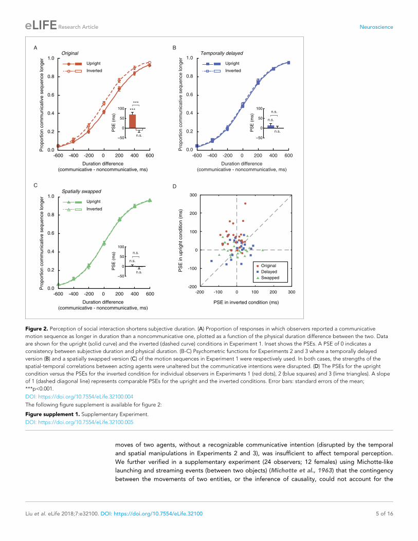
Twenty-four observers (12 females) performed the duration judgment task in Experiment 1. In the
upright condition, the mean PSE was 69.3 ms, significantly above 0 (t23 = 5.60, p<0.001, Cohen’s
d = 1.14, Figure 2A). In other words, an upright communicative motion sequence compressed sub-
jective duration such that it needed to be 69.3 ms longer to be perceived as equal in duration to an
upright noncommunicative motion sequence. In the inverted condition, by contrast, the mean PSE
was significantly smaller (t23 = �5.82, p<0.001, Cohen’s d = �1.19) and not different from 0
(t23 = �1.51, p=0.15) (Figure 2A). Inversion is known to impair the perception of biological motion
and thereby social interactions mediated by biological motion, yet does not affect low-level visual
features (Troje and Westhoff, 2006). This result hence verified that the temporal compression effect
of perceived social interactions could not be due to low-level non-biological visual features. The
data from individual observers conformed with the averaged patterns. They are summarized in
Figure 2D, in which each observer’s PSEs from the upright (y-axis) and the inverted (x-axis) condi-
tions are plotted against each other and shown as a red dot. Most red dots fell above the dashed
line of slope 1, although several of them were close to the dashed line; that is, the observers were
largely biased towards perceiving the communicative motion sequences as shorter in duration than
the noncommunicative ones when the agents were shown upright as opposed to upside down,
despite that the task did not require explicit social processing. Their temporal sensitivities remained
Liu et al. eLife 2018;7:e32100. DOI: https://doi.org/10.7554/eLife.32100 3 of 16
Research Article Neuroscience

unchanged between the two conditions, as indicated by a comparison of the difference limens
(t23 = 0.70, p=0.49).
The point-light trajectories of two agents acting communicatively (coordinately) could be spatially
and temporally more correlated than those of two agents acting noncommunicatively (indepen-
dently) (Bassili, 1976; Chartrand and Bargh, 1999). Extraction of such a spatial-temporal relation-
ship between two upright agents, rather than the social meaning per se, could have caused the
observed temporal compression effect. To address this possibility, we conducted Experiments 2 and
3, where we eliminated the social aspect of the original communicative displays while keeping the
spatial-temporal pattern differences between the communicative and the noncommunicative motion
sequences unchanged. In Experiment 2, this was done by inserting a temporal lag of 700 ms in
between every two acting agents (upright and inverted, communicative and noncommunicative)
(Manera et al., 2013). In Experiment 3, we spatially swapped the two agents in each display such
that they faced in opposite directions instead of facing each other. The two experiments were other-
wise identical to Experiment 1. Analyses of the results from 48 observers (24 in each of Experiments
2 and 3; 27 females) indicated that neither the temporally delayed ‘communicative’ motion sequen-
ces nor the spatially swapped ones altered temporal perception relative to their ‘noncommunicative’
counterparts (Figure 2B and C). The PSEs were not significantly different from 0 regardless of
whether the motion sequences were presented upright (t23s = 1.39 and 0.34, ps = 0.18 and 0.74, for
Experiments 2 and 3, respectively) or upside down (t23s = 0.01 and �0.14, ps = 0.99 and 0.89,
respectively). Between the upright and the inverted conditions, there was no significant difference in
PSE (t23s = �1.02 and �0.28, ps = 0.32 and 0.78, for Experiments 2 and 3, respectively) or difference
limen (t23s = �0.07 and 0.84, ps = 0.95 and 0.41, respectively). We also examined individual data
and plotted each observer’s PSEs from the upright and the inverted conditions against each other.
As shown in Figure 2D, the values fell on both sides of the dashed line of slope 1 and were centered
around the origin for both Experiments 2 (blue squares) and 3 (lime triangles). Moreover, an omnibus
ANOVA of the pooled PSEs across Experiments 1 to 3 confirmed a significant interaction between
the vertical orientation of the agents (upright or inverted) and experiment (Experiment 1, 2, or 3) (F2,
69 = 9.98, p<0.001, Cohen’s f = 0.54). No such interaction was found with the difference limens (F2,
69 = 0.23, p=0.80). We hence concluded that the temporal compression effect observed in Experi-
ment 1 was absent in Experiments 2 and 3. The mere spatial-temporal correlation between the
Time
Fixation
(1000 ms)
... ...
Communicative motion sequence
(400~1600 ms)
... ...
... ...... ...
Fixation
(400~600 ms) Noncommunicative motion sequence
(1000 ms)
Which longer
in duration?
Figure 1. Schematic illustration of an exemplar trial in the duration judgment task.
DOI: https://doi.org/10.7554/eLife.32100.003
Liu et al. eLife 2018;7:e32100. DOI: https://doi.org/10.7554/eLife.32100 4 of 16
Research Article Neuroscience
moves of two agents, without a recognizable communicative intention (disrupted by the temporal
and spatial manipulations in Experiments 2 and 3), was insufficient to affect temporal perception.
We further verified in a supplementary experiment (24 observers; 12 females) using Michotte-like
launching and streaming events (between two objects) (Michotte et al., 1963) that the contingency
between the movements of two entities, or the inference of causality, could not account for the
C
BA
D
PSE in inverted condition (ms)
-200 -100 0 100 200 300
PS
E in u
pright
conditio
n (
ms)
-200
-100
0
100
200
300
Original
Swapped
Delayed
Duration difference
(communicative - noncommunicative, ms)
-600 -400 -200 0 200 400 600
Pro
po
rtio
n c
om
mu
nic
ative
se
qu
en
ce
lo
ng
er
0.0
0.2
0.4
0.6
0.8
1.0Original
Upright
Inverted
PS
E (
ms)
***
n.s.
***
50
0
50
100
Duration difference
(communicative - noncommunicative, ms)
-600 -400 -200 0 200 400 600
Pro
po
rtio
n c
om
mu
nic
ative
se
qu
en
ce
lo
ng
er
0.0
0.2
0.4
0.6
0.8
1.0Temporally delayed
Upright
Inverted
n.s.
n.s.
n.s.PS
E (
ms)
50
0
50
100
Duration difference
(communicative - noncommunicative, ms)
-600 -400 -200 0 200 400 6000.0
0.2
0.4
0.6
0.8
1.0
Pro
po
rtio
n c
om
mu
nic
ative
se
qu
en
ce
lo
ng
er
Spatially swapped
Upright
Inverted
PS
E (
ms)
n.s.
n.s.
n.s.50
0
50
100
Figure 2. Perception of social interaction shortens subjective duration. (A) Proportion of responses in which observers reported a communicative
motion sequence as longer in duration than a noncommunicative one, plotted as a function of the physical duration difference between the two. Data
are shown for the upright (solid curve) and the inverted (dashed curve) conditions in Experiment 1. Inset shows the PSEs. A PSE of 0 indicates a
consistency between subjective duration and physical duration. (B-C) Psychometric functions for Experiments 2 and 3 where a temporally delayed
version (B) and a spatially swapped version (C) of the motion sequences in Experiment 1 were respectively used. In both cases, the strengths of the
spatial-temporal correlations between acting agents were unaltered but the communicative intentions were disrupted. (D) The PSEs for the upright
condition versus the PSEs for the inverted condition for individual observers in Experiments 1 (red dots), 2 (blue squares) and 3 (lime triangles). A slope
of 1 (dashed diagonal line) represents comparable PSEs for the upright and the inverted conditions. Error bars: standard errors of the mean;
***p<0.001.
DOI: https://doi.org/10.7554/eLife.32100.004
The following figure supplement is available for figure 2:
Figure supplement 1. Supplementary Experiment.
DOI: https://doi.org/10.7554/eLife.32100.005
Liu et al. eLife 2018;7:e32100. DOI: https://doi.org/10.7554/eLife.32100 5 of 16
Research Article Neuroscience

temporal compression effect associated with the perception of social interactions (Figure 2—figure
supplement 1).
On the other hand, the recognition of communicative intention is unlikely to spontaneously occur
in all observers. Social communicative ability has been shown to be a continuum that extends from
patients with autism into the neurotypical population (Baron-Cohen, 1995; Frith, 1991;
Nummenmaa et al., 2012; von dem Hagen et al., 2011). The degree of autistic traits (or lack of
social proficiency), as measured by the Autism Spectrum Quotient (AQ), varies substantially even
among healthy young adults. Such variance is particularly pronounced in males, who generally score
higher than females on the AQ (Baron-Cohen et al., 2001). We wondered if the extent of temporal
compression induced by the perception of social interactions (see Figure 2D for inter-individual dif-
ferences) was a manifestation of one’s social proficiency. To this end, we recruited a larger sample of
90 male observers and carried out Experiment 4, which employed the same task as Experiment 1
except that all motion sequences were presented upright (i.e., the inverted condition that served as
a control in Experiment 1 was not included). Each observer’s autistic-like tendency was also assessed
with the AQ. Overall, Experiment 4 replicated the temporal compression effect observed in Experi-
ment 1. The mean PSE was 38.5 ms, comparable to that of the male observers in Experiment 1 (45.8
ms) and significantly above 0 (t89 = 6.07, p<0.001, Cohen’s d = 0.64, Figure 3A). Critically, inspec-
tion of the individual data revealed a significant negative correlation between PSE and AQ score:
those with higher AQ scores, namely stronger autistic-like tendencies and lower social proficiencies,
were less biased in making duration judgments of the communicative and the noncommunicative
motion sequences, and had PSEs closer to 0 (r90 = �0.40, p<0.001, Figure 3B). The median AQ
score of this sample was 19, roughly corresponding to a cut-off between low and intermediate levels
of autistic traits (Baron-Cohen et al., 2001). A median split of the observers by AQ score showed
that the social interaction-induced temporal compression effect was evident in the low AQ group
(AQ scores < 20, mean PSE = 58.9 ms, significantly above 0; t45 = 7.06, p<0.001, Cohen’s d = 1.04),
yet barely reached statistical significance in the high AQ group (AQ scores � 20, mean PSE = 17.1
ms, t43 = 2.00, p=0.052), with a significant group difference in PSE (t88 = 3.51, p=0.001, Cohen’s
d = 0.74), but not difference limen (t88 = 0.53, p=0.60) (Figure 3C). These results, while reaffirming
the influence of perceived social interactions on subjective duration, highlighted the idiosyncrasy of
subjective time for social interactions, and tied it to a stable personality trait –– social proficiency.
A B C
PS
E (
ms)
-50
0
50
100
Low AQ High AQ
†
***
***
Duration difference
(communicative - noncommunicative, ms)
-600 -400 -200 0 200 400 600
Pro
po
rtio
n c
om
mu
nic
ative
se
qu
en
ce
lo
ng
er
0.0
0.2
0.4
0.6
0.8
1.0
***
PS
E (
ms)
50
0
50
100
AQ score
0 105 15 20 25 3530
PS
E (
ms)
-100
-50
0
50
100
150
200
250
r = -0.40***
High AQ
Low AQ
Figure 3. The degree of temporal compression induced by the perception of social interactions reflects one’s social proficiency. (A) Psychometric
function for Experiment 4 which contained only the upright condition. Inset shows the overall PSE. (B) Across the observers, one’s PSE negatively
correlated with his score on the Autism Spectrum Quotient (AQ). (C) Comparison of the PSEs for low AQ (<20) versus high AQ (�20) observers. Error
bars: standard errors of the mean; †: marginally significant, ***p�0.001.
DOI: https://doi.org/10.7554/eLife.32100.006
Liu et al. eLife 2018;7:e32100. DOI: https://doi.org/10.7554/eLife.32100 6 of 16
Research Article Neuroscience

Oxytocin mediates temporal perception of social interactionsAutistic traits have been widely associated with reduced levels of oxytocin (Clark et al., 2013;
Green et al., 2001; Modahl et al., 1998; Parker et al., 2014), and can be ameliorated with intrana-
sal oxytocin administration (Anagnostou et al., 2012; Gordon et al., 2013; Guastella et al., 2010;
Watanabe et al., 2015; Yatawara et al., 2016). The link between autistic-like tendency and subjec-
tive duration of social interactions thus raises the intriguing question of whether oxytocin plays a
role therein. Experiment 5 probed this question by testing if the application of oxytocin would pro-
mote the social interaction-induced temporal compression effect in socially less proficient individu-
als. The same task as in Experiment 4 was employed. Eighty males with AQ scores larger than or
equal to 20 (range: 20–36, same cutoff value as in Experiment 4) completed the duration judgment
task twice, once before and once 40 min after the nasal administration of either oxytocin (for 40
observers) or atosiban (for the other 40 observers). Atosiban is a desamino-oxytocin analogue and a
competitive oxytocin receptor antagonist (Sanu and Lamont, 2010), and has been shown to be cen-
trally available when administered intranasally (Lamont and Kam, 2008; Lundin et al., 1986). We
used it as a comparison treatment and hypothesized that its influence on subjective duration of
social interactions, if any, would be in the opposite direction of oxytocin. The results were consistent
with our hypotheses. In those treated with oxytocin, the mean PSE significantly increased by 36.9 ms
(t39 = 3.68, p=0.001, Cohen’s d = 0.58), from 13.3 ms at baseline, which was not significantly differ-
ent from 0 (t39 = 1.34, p=0.19), to 50.1 ms after oxytocin administration (significantly above 0,
t39 = 4.22, p<0.001, Cohen’s d = 0.66) (Figure 4A). By contrast, in those treated with atosiban, the
mean PSE was not significantly different from 0 both at baseline (9.7 ms, t39 = 1.14, p=0.26) and
after atosiban administration (�9.4 ms, t39 = �1.11, p=0.28), yet numerically showed a significant
reduction (t39 = �2.24, p=0.031, Cohen’s d = �0.35) (Figure 4B). Between the two drug groups,
there was a marked difference in the changes in PSEs pre- and post- drug administration (t78 = 4.25,
p<0.001, Cohen’s d = 0.95). These effects could not be due to changes of the observers’ temporal
sensitivity, as their difference limens remained unaltered before and after drug administration
(t39s = 0.35 and �0.66, ps = 0.73 and 0.51, for oxytocin and atosiban, respectively). Besides, their
transient mood states, as reflected by ratings on the Profile of Mood States (POMS) scale
(McNair et al., 1971), were also unaffected by drug condition (drug condition �testing session; total
mood disturbance: F1, 78 = 0.18, p=0.68; all subscales: ps > 0.1).
Nonetheless, there is a reason to suspect that the influence of atosiban on socially less proficient
individuals, as observed in Experiment 5, was unreliable –– they were not significantly biased by the
social aspect of interactions in making duration judgments to begin with. To further verify if antago-
nizing the effect of oxytocin would diminish the temporal compression effect of perceived social
interactions, we turned to socially proficient individuals. In Experiment 6, 80 males with AQ scores
less than 20 (range: 10–19, same cutoff value as in Experiment 4) completed the duration judgment
task both before and 40 min after the nasal administration of either atosiban (for 40 observers) or
saline (for the other 40 observers), following the same procedure as in Experiment 5. Saline served
as a placebo control here to address potential confounds including practice and fatigue. At baseline,
the observers in both drug groups were significantly biased towards perceiving the communicative
motion sequences as shorter in duration than the noncommunicative ones (mean PSEs = 48.6 ms
and 54.5 ms, t39s = 4.32 and 5.94, ps < 0.001, Cohen’s ds = 0.68 and 0.94, for atosiban and saline,
respectively), with no difference in between (t78 = 0.41, p=0.68) (Figure 4C–D). After drug treat-
ments, however, a significant group difference emerged (mean PSEs = 17.5 ms and 46.8 ms,
t78 = 2.64, p=0.010, Cohen’s d = 0.59). In those treated with atosiban, the mean PSE dropped signif-
icantly by 31.1 ms (t39 = �3.90, p<0.001, Cohen’s d = �0.62), albeit still significantly above 0
(t39 = 2.04, p=0.048, Cohen’s d = 0.32) (Figure 4C). By contrast, in those treated with saline, the
PSEs were unaffected (t39 = �1.13, p=0.26; Figure 4D). There was a significant difference between
the two drug groups in the changes in PSEs pre- and post- drug administration (t78 = 2.22, p=0.029,
Cohen’s d = 0.50). Meanwhile, the difference limens in both groups remained unchanged
(t39s = �1.07 and �1.11, ps = 0.29 and 0.27, for atosiban and saline, respectively), and the POMS
ratings were unaffected by drug condition (drug condition �testing session; total mood disturbance:
F1, 78 = 0.47, p=0.50; all subscales: ps > 0.2).
We plotted in Figure 4E the distributions of PSEs for individual observers in Experiments 5 and 6
before (x-axis) and after (y-axis) drug treatment. Their central tendencies are respectively highlighted
Liu et al. eLife 2018;7:e32100. DOI: https://doi.org/10.7554/eLife.32100 7 of 16
Research Article Neuroscience

A B
DC
High AQ individuals
Duration difference
(communicative - noncommunicative, ms)
-600 -400 -200 0 200 400 600
Pro
po
rtio
n c
om
mu
nic
ative
se
qu
en
ce
lo
ng
er
0.0
0.2
0.4
0.6
0.8
1.0No drug
Oxytocin
PS
E (
ms) ***
n.s.
***
50
0
50
100
High AQ individuals
Duration difference
(communicative - noncommunicative, ms)
-600 -400 -200 0 200 400 600
Pro
po
rtio
n c
om
mu
nic
ative
se
qu
en
ce
lo
ng
er
0.0
0.2
0.4
0.6
0.8
1.0No drug
Atosiban
n.s.
*
n.s.
PS
E (
ms)
50
0
50
100
Duration difference
(communicative - noncommunicative, ms)
-600 -400 -200 0 200 400 6000.0
0.2
0.4
0.6
0.8
1.0Low AQ individuals
Pro
po
rtio
n c
om
mu
nic
ative
se
qu
en
ce
lo
ng
er
No drug
Saline
PS
E (
ms) ***
n.s.
50
0
50
100
***
Duration difference
(communicative - noncommunicative, ms)
-600 -400 -200 0 200 400 6000.0
0.2
0.4
0.6
0.8
1.0Low AQ individuals
Pro
po
rtio
n c
om
mu
nic
ative
se
qu
en
ce
lo
ng
er
No drug
Atosiban
PS
E (
ms) ***
***
50
0
50
100
*
E F
PSE before drug adminstration (ms)
-200 -100 0 100 200 300
PS
E a
fter
dru
g a
dm
inis
tration (
ms)
-200
-100
0
100
200
300
High AQ w/ oxytocin
High AQ w/ atosiban
Low AQ w/ atosiban
Low AQ w/ saline
PSE before drug adminstration (ms)
-
PS
E a
fter
dru
g a
dm
inis
tration (
ms)
-80
-40
0
40
80
120
-80 -40 0 40 80 120
Figure 4. Oxytocin mediates temporal perception of social interactions. (A-D) Psychometric functions for Experiments 5 and 6 where high AQ observers
(A-B) and low AQ observers (C-D) completed the duration judgment task of upright motion sequences both before (light gray curves) and after the
nasal administration of oxytocin (red curve in A), atosiban (blue curves in B and C) or saline (dark gray curve in D). Insets show the PSEs. (E) The PSEs
after drug administration versus those before drug administration for high AQ individuals treated with oxytocin (light brown dots with red circles), high
Figure 4 continued on next page
Liu et al. eLife 2018;7:e32100. DOI: https://doi.org/10.7554/eLife.32100 8 of 16
Research Article Neuroscience
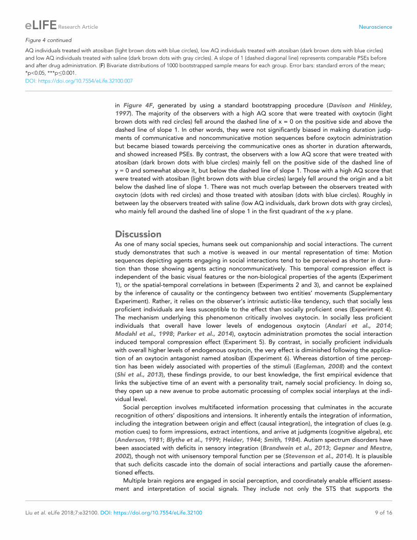
in Figure 4F, generated by using a standard bootstrapping procedure (Davison and Hinkley,
1997). The majority of the observers with a high AQ score that were treated with oxytocin (light
brown dots with red circles) fell around the dashed line of x = 0 on the positive side and above the
dashed line of slope 1. In other words, they were not significantly biased in making duration judg-
ments of communicative and noncommunicative motion sequences before oxytocin administration
but became biased towards perceiving the communicative ones as shorter in duration afterwards,
and showed increased PSEs. By contrast, the observers with a low AQ score that were treated with
atosiban (dark brown dots with blue circles) mainly fell on the positive side of the dashed line of
y = 0 and somewhat above it, but below the dashed line of slope 1. Those with a high AQ score that
were treated with atosiban (light brown dots with blue circles) largely fell around the origin and a bit
below the dashed line of slope 1. There was not much overlap between the observers treated with
oxytocin (dots with red circles) and those treated with atosiban (dots with blue circles). Roughly in
between lay the observers treated with saline (low AQ individuals, dark brown dots with gray circles),
who mainly fell around the dashed line of slope 1 in the first quadrant of the x-y plane.
DiscussionAs one of many social species, humans seek out companionship and social interactions. The current
study demonstrates that such a motive is weaved in our mental representation of time: Motion
sequences depicting agents engaging in social interactions tend to be perceived as shorter in dura-
tion than those showing agents acting noncommunicatively. This temporal compression effect is
independent of the basic visual features or the non-biological properties of the agents (Experiment
1), or the spatial-temporal correlations in between (Experiments 2 and 3), and cannot be explained
by the inference of causality or the contingency between two entities’ movements (Supplementary
Experiment). Rather, it relies on the observer’s intrinsic autistic-like tendency, such that socially less
proficient individuals are less susceptible to the effect than socially proficient ones (Experiment 4).
The mechanism underlying this phenomenon critically involves oxytocin. In socially less proficient
individuals that overall have lower levels of endogenous oxytocin (Andari et al., 2014;
Modahl et al., 1998; Parker et al., 2014), oxytocin administration promotes the social interaction
induced temporal compression effect (Experiment 5). By contrast, in socially proficient individuals
with overall higher levels of endogenous oxytocin, the very effect is diminished following the applica-
tion of an oxytocin antagonist named atosiban (Experiment 6). Whereas distortion of time percep-
tion has been widely associated with properties of the stimuli (Eagleman, 2008) and the context
(Shi et al., 2013), these findings provide, to our best knowledge, the first empirical evidence that
links the subjective time of an event with a personality trait, namely social proficiency. In doing so,
they open up a new avenue to probe automatic processing of complex social interplays at the indi-
vidual level.
Social perception involves multifaceted information processing that culminates in the accurate
recognition of others’ dispositions and intensions. It inherently entails the integration of information,
including the integration between origin and effect (causal integration), the integration of clues (e.g.
motion cues) to form impressions, extract intentions, and arrive at judgments (cognitive algebra), etc
(Anderson, 1981; Blythe et al., 1999; Heider, 1944; Smith, 1984). Autism spectrum disorders have
been associated with deficits in sensory integration (Brandwein et al., 2013; Gepner and Mestre,
2002), though not with unisensory temporal function per se (Stevenson et al., 2014). It is plausible
that such deficits cascade into the domain of social interactions and partially cause the aforemen-
tioned effects.
Multiple brain regions are engaged in social perception, and coordinately enable efficient assess-
ment and interpretation of social signals. They include not only the STS that supports the
Figure 4 continued
AQ individuals treated with atosiban (light brown dots with blue circles), low AQ individuals treated with atosiban (dark brown dots with blue circles)
and low AQ individuals treated with saline (dark brown dots with gray circles). A slope of 1 (dashed diagonal line) represents comparable PSEs before
and after drug administration. (F) Bivariate distributions of 1000 bootstrapped sample means for each group. Error bars: standard errors of the mean;
*p<0.05, ***p�0.001.
DOI: https://doi.org/10.7554/eLife.32100.007
Liu et al. eLife 2018;7:e32100. DOI: https://doi.org/10.7554/eLife.32100 9 of 16
Research Article Neuroscience

understanding of actions (Allison et al., 2000), but also higher cortical areas like the temporal-parie-
tal junction (TPJ) that represents mental states (Carter and Huettel, 2013) and the dorsal medial
prefrontal cortex (dmPFC) that is implicated in the uniquely human representation of triadic interac-
tions between two minds and an object (Saxe, 2006). The observed temporal compression effect,
being independent of the perception of biological motion, likely arises from these higher stages of
social processing (Cusack et al., 2015; von der Luhe et al., 2016). Subjective time has been pro-
posed to be a signature of the amount of energy expended in representing a stimulus
(Eagleman and Pariyadath, 2009). It is plausible that communicative motion sequences are proc-
essed with increased efficiency in TPJ and dmPFC relative to motion sequences without a recogniz-
able communicative intention, thus leading to lowered metabolic cost (Gutnisky and Dragoi, 2008;
Laughlin, 2001) and shortened subjective duration, particularly in socially proficient individuals. Of
note here is that this temporal compression effect can hardly be accounted for by the operation of a
dedicated neural module specialized for representing the temporal relationships between events
(Ivry and Schlerf, 2008), since it is specific to the perception of social interactions.
Oxytocin partially drives social motivation (Dolen et al., 2013) and has increasingly been identi-
fied as an important modulator of social behaviors (Carter, 2014; Donaldson and Young, 2008)
ranging from social recognition (Oettl et al., 2016) to consolation (Burkett et al., 2016) and ethno-
centrism (De Dreu et al., 2011). It has also been linked to repetitive behaviors –– a core feature of
autism (Hollander et al., 2003; Insel et al., 1999). Results from human brain imaging studies indi-
cate that the application of oxytocin modulates responses in the STS, TPJ and the prefrontal cortex,
among others, in various social cognitive tasks (Zink and Meyer-Lindenberg, 2012). Such alterations
of neural activities presumably facilitate social processing in socially less proficient individuals, where
exogenous oxytocin has been shown to exert a more definitive prosocial effect (Bartz et al., 2010;
Bartz et al., 2011), thereby giving rise to their shortened subjective duration of social interactions
following oxytocin administration. In socially proficient individuals, the influence of exogenous oxyto-
cin is more complicated, even hard to predict and interpret (Bartz, 2016; Bartz et al., 2010;
Bartz et al., 2011; Olff et al., 2013). Nevertheless, we were able to show that antagonizing the
effect of endogenous oxytocin with atosiban in this group of observers reduced the temporal com-
pression effect as compared to a placebo control. The comparison between oxytocin and atosiban
spoke directly to the role of oxytocin in mediating time perception of social interactions. This finding
also adds to and extends the extant pharmacological research on time perception, which has primar-
ily focused on dopamine, serotonin and acetylcholine (Allman and Meck, 2012; Meck, 1998).
We conclude that the perceived duration of social interactions is a product of complex neuroen-
docrine and neural processes, the exact mechanism awaiting further investigation, and is ingrained
with one’s social traits. Subjective time is not only nonuniform, like Einstein alluded to in his analogy,
but also idiosyncratic.
Materials and methods
ParticipantsSeventy-two young adults (39 females; mean age ± SD = 22.8 ± 2.5 years) participated in Experi-
ments 1 to 3, 24 in each experiment. Sample sizes were determined by G*Power to be sufficient to
detect a large temporal distortion effect in time perception (d � 0.8), at a power larger than 95%.
The effect size was estimated based on an earlier study that employed biological motion stimuli and
similar psychophysical testing procedures to those in the current study (Wang and Jiang, 2012). An
independent group of 90 males (22.5 ± 2.7 years) took part in Experiment 4. In addition, 80 male
non-smokers (22.1 ± 2.6 years) who scored above or equal to 20 on the Autism Spectrum Quotient
(AQ) (mean AQ score ±SD = 24.9±4.3) participated in Experiment 5. Another 80 male non-smokers
(22.8 ± 2.3 years) with AQ scores below 20 (15.5 ± 2.7) participated in Experiment 6. Only males
were recruited in Experiments 4–6 for the following reasons: In Experiment 4, this was done to take
advantage of men’s pronounced variance in social proficiency (Baron-Cohen et al., 2001). In Experi-
ments 5 and 6, this was due to pragmatic reasons (like most intranasal oxytocin studies), as oxytocin
and atosiban respectively cause and antagonize contractions of the uterus. All participants reported
to have normal or corrected-to-normal vision. Those in Experiments 5 and 6 also reported to have
no respiratory allergy or upper respiratory infection at the time of testing. They gave informed
Liu et al. eLife 2018;7:e32100. DOI: https://doi.org/10.7554/eLife.32100 10 of 16
Research Article Neuroscience

consent to participate in procedures approved by the Institutional Review Board at Institute of Psy-
chology, Chinese Academy of Sciences.
Visual stimuliTen communicative point-light motion sequences (C1-10 in Supplementary file 1), each portraying
two agents of the same gender engaging in either a face-to-face (dyadic, two motion sequences) or
a person-object-person (triadic, eight motion sequences) interaction, were chosen from the Commu-
nicative Interaction Database (Manera et al., 2010). By cross-pairing the agents of the same gender
from different interactions, we produced an essentially physically matched set of 10 noncommunica-
tive motion sequences (NC1-10 in Supplementary file 1). We verified in an independent group of
24 observers (half male, 27.5 ± 3.3 years) that the noncommunicative motion sequences were per-
ceived as significantly less communicative than the communicative ones (normalized communicative-
ness rating: 0.37 vs. 0.71, t23 = �8.11, p<0.0001, Cohen’s d = �1.66). These twenty motion
sequences and their inverted (upside-down) counterparts were used in Experiment 1, shown at 30
frames per second with a visual angle of approximately 6˚�9˚ (each agent was approximately 2˚�9˚).In Experiments 2 and 3, a temporally delayed version and a spatially swapped version of the stimuli
in Experiment 1 were respectively adopted. Specifically, we introduced a temporal lag of 700 ms (21
frames) in between every two acting agents (communicative and noncommunicative, upright and
inverted) in Experiment 2 (Manera et al., 2013), and spatially swapped the two agents in each dis-
play in Experiment 3 such that they faced in opposite directions instead of facing each other. The
original upright communicative and noncommunicative motion sequences were employed in Experi-
ments 4–6.
Behavioral proceduresEach trial of the duration judgment task in Experiment 1 began with a fixation on a central cross
(1˚�1˚) for 1000 ms, followed by two sequentially presented motion sequences –– one communica-
tive, the other noncommunicative, in random order with a blank screen of 400–600 ms in between
(Figure 1). One of the motion sequences (communicative or noncommunicative, each in half of the
trials in random order) was presented for 1000 ms, the other for 400, 600, 800, 1000, 1200, 1400 or
1600 ms with equal possibility. That is, the duration difference between the communicative and the
noncommunicative motion sequences ranged from �600 ms to 600 ms in steps of 200 ms. Observ-
ers were asked to press one of two buttons to indicate which motion sequence (the first or the sec-
ond) was longer in duration, a task that did not require recognitions of the nature of the motion
sequences. The next trial began immediately after a response was made. There were 35 trials in
each block and a total of 8 blocks. In half of the blocks, the motion sequences were presented
upright; in the other half, they were presented upside down. The order of the ‘upright’ and the
‘inverted’ blocks was balanced across observers. There was a short break of 1 to 2 min in between
the blocks.
Experiments 2 to 6 followed the same procedures as in Experiment 1 except for the followings: In
Experiments 2 and 3, a temporally delayed version and a spatially swapped version of the visual
stimuli were respectively used (see Visual stimuli above). As a result of the temporal lag inserted
between the acting agents, each motion sequence in Experiment 2 was presented for 700 ms longer
than in Experiment 1 on average. In Experiment 4, observers only completed 4 ‘upright’ blocks. In
Experiments 5 and 6, observers completed 4 ‘upright’ blocks at baseline and another 4 ‘upright’
blocks 40 min after drug treatment (see Drug Application below). They also provided ratings on the
Profile of Mood States scale (McNair et al., 1971) following the duration judgment task both at
baseline and after drug treatment.
Drug applicationObservers in Experiments 5 and 6 self-administered a single intranasal dose of 24 IU of oxytocin
(ProSpec,>99.0% as determined by RP-HPLC, dissolved in saline; three puffs per nostril, each with 4
IU of oxytocin; for half of the observers in Experiment 5), 60 mg of atosiban (ProSpec,>99.0% as
determined by RP-HPLC, dissolved in saline; three puffs per nostril, each with 10 mg of atosiban; for
half of the observers in Experiments 5 and 6) or saline (three puffs per nostril, for half of the observ-
ers in Experiment 6), in a randomized between-subjects manner, after they completed the baseline
Liu et al. eLife 2018;7:e32100. DOI: https://doi.org/10.7554/eLife.32100 11 of 16
Research Article Neuroscience

blocks of the duration judgment task and the Profile of Mood States scale. Neither the participants
nor the experimenters were aware of the identity of the drug used. Atosiban is a desamino-oxytocin
analogue and a competitive oxytocin receptor antagonist (Sanu and Lamont, 2010). Both oxytocin
(Dal Monte et al., 2014; Freeman et al., 2016; Lee and Weerts, 2016; Striepens et al., 2013) and
atosiban (Lamont and Kam, 2008; Lundin et al., 1986) have been shown to be bioavailable when
administered intranasally.
Fresh oxytocin and atosiban solutions were made every 3 days during the period of data collec-
tion, such that for each participant in the drug administration experiments, the solution he received
was prepared in less than 3 days before. The prepared solutions were stored in 10 ml sterilized nasal
spray bottles at 4˚C until usage.
AnalysisFor each observer under each condition, we calculated the proportions that a communicative motion
sequence was judged as longer in duration than a noncommunicative one, and fitted them with a
Boltzmann sigmoid function f xð Þ ¼ 1= 1þ expð x� x0ð Þ=!ð ÞÞ, where x corresponds to the physical dura-
tion difference between a communicative motion sequence and a noncommunicative one (-600 ms, -
400 ms, -200 ms, 0 ms, 200 ms, 400 ms, or 600 ms), x0 corresponds to the point of subjective equal-
ity (PSE), at which the observer perceived a communicative motion sequence as equal in duration to
a noncommunicative one; and half the interquartile range of the fitted function corresponds to dif-
ference limen, an index of temporal discrimination sensitivity. PSE and difference limen served as the
dependent variables and were subsequently compared between conditions and groups.
AcknowledgementsWe thank Yuting Ye for assistance, and Zhi Yang and Qian Xu for comments and suggestions. This
work was supported by the National Natural Science Foundation of China (31422023 and
31525011), the Strategic Priority Research Program (XDB02010003 and XDB02030006) and the Key
Research Program of Frontier Sciences (QYZDB-SSW-SMC030 and QYZDB-SSW-SMC055) of the Chi-
nese Academy of Sciences.
Additional information
Author contributions
Rui Liu, Xiangyong Yuan, Kepu Chen, Data curation, Software, Formal analysis, Validation, Investiga-
tion; Yi Jiang, Conceptualization, Supervision, Funding acquisition, Methodology, Writing—review
and editing; Wen Zhou, Conceptualization, Supervision, Funding acquisition, Methodology, Writ-
ing—original draft, Writing—review and editing
Author ORCIDs
Yi Jiang https://orcid.org/0000-0002-5746-7301
Wen Zhou https://orcid.org/0000-0001-6730-2116
Funding
Funder Grant reference number Author
National Natural ScienceFoundation of China
31525011 Yi Jiang
Chinese Academy of Sciences XDB02010003 Yi Jiang
Chinese Academy of Sciences QYZDB-SSW-SMC030 Yi Jiang
National Natural ScienceFoundation of China
31422023 Wen Zhou
Chinese Academy of Sciences XDB02030006 Wen Zhou
Chinese Academy of Sciences QYZDB-SSW-SMC055 Wen Zhou
Liu et al. eLife 2018;7:e32100. DOI: https://doi.org/10.7554/eLife.32100 12 of 16
Research Article Neuroscience

The funders had no role in study design, data collection and interpretation, or the
decision to submit the work for publication.
Decision letter and Author response
Decision letter https://doi.org/10.7554/eLife.32100.012
Author response https://doi.org/10.7554/eLife.32100.013
Additional filesSupplementary files. Source data 1. Raw data. PSEs and difference limens (DLs) for individual participants in Experiments
1-6.
DOI: https://doi.org/10.7554/eLife.32100.008
. Supplementary file 1. Description of the point-light motion sequences used in the duration judg-
ment task. C1-10 were chosen from the Communicative Interaction Database (Manera et al., 2010).
NC1-10 were produced by cross-pairing the agents of the same gender from C1-10.
DOI: https://doi.org/10.7554/eLife.32100.009
. Transparent reporting form
DOI: https://doi.org/10.7554/eLife.32100.010
ReferencesAllison T, Puce A, McCarthy G. 2000. Social perception from visual cues: role of the STS region. Trends inCognitive Sciences 4:267–278. DOI: https://doi.org/10.1016/S1364-6613(00)01501-1, PMID: 10859571
Allman MJ, Meck WH. 2012. Pathophysiological distortions in time perception and timed performance. Brain135:656–677. DOI: https://doi.org/10.1093/brain/awr210, PMID: 21921020
Anagnostou E, Soorya L, Chaplin W, Bartz J, Halpern D, Wasserman S, Wang AT, Pepa L, Tanel N, Kushki A,Hollander E. 2012. Intranasal oxytocin versus placebo in the treatment of adults with autism spectrumdisorders: a randomized controlled trial. Molecular Autism 3:16. DOI: https://doi.org/10.1186/2040-2392-3-16,PMID: 23216716
Andari E, Schneider FC, Mottolese R, Vindras P, Sirigu A. 2014. Oxytocin’s fingerprint in personality traits andregional brain volume. Cerebral Cortex 24:479–486. DOI: https://doi.org/10.1093/cercor/bhs328, PMID: 23118193
Anderson NH. 1981. Foundations of Information Integration Theory. Academic Press.Baron-Cohen S, Wheelwright S, Skinner R, Martin J, Clubley E. 2001. The autism-spectrum quotient (AQ):evidence from Asperger syndrome/high-functioning autism, males and females, scientists and mathematicians.Journal of Autism and Developmental Disorders 31:5–17. DOI: https://doi.org/10.1023/A:1005653411471,PMID: 11439754
Baron-Cohen S. 1995. Mindblindness: An Essay on Autism and Theory of Mind. Boston: MIT Press.Bartz JA, Zaki J, Bolger N, Hollander E, Ludwig NN, Kolevzon A, Ochsner KN. 2010. Oxytocin selectivelyimproves empathic accuracy. Psychological Science 21:1426–1428. DOI: https://doi.org/10.1177/0956797610383439, PMID: 20855907
Bartz JA, Zaki J, Bolger N, Ochsner KN. 2011. Social effects of oxytocin in humans: context and person matter.Trends in Cognitive Sciences 15:301–309. DOI: https://doi.org/10.1016/j.tics.2011.05.002, PMID: 21696997
Bartz JA. 2016. Oxytocin and the pharmacological dissection of affiliation. Current Directions in PsychologicalScience 25:104–110. DOI: https://doi.org/10.1177/0963721415626678
Bassili JN. 1976. Temporal and spatial contingencies in the perception of social events. Journal of Personalityand Social Psychology 33:680–685. DOI: https://doi.org/10.1037/0022-3514.33.6.680
Blake R, Turner LM, Smoski MJ, Pozdol SL, Stone WL. 2003. Visual recognition of biological motion is impaired inchildren with autism. Psychological Science 14:151–157. DOI: https://doi.org/10.1111/1467-9280.01434,PMID: 12661677
Blythe PW, Todd PM, Miller GF. 1999. How motion reveals intention: Categorizing social interactions. In:Gigerenzer G, Todd P. M, Group A. R (Eds). Simple Heuristics That Make Us Smart. New York: OxfordUniversity Press.
Brandwein AB, Foxe JJ, Butler JS, Russo NN, Altschuler TS, Gomes H, Molholm S. 2013. The development ofmultisensory integration in high-functioning autism: high-density electrical mapping and psychophysicalmeasures reveal impairments in the processing of audiovisual inputs. Cerebral Cortex 23:1329–1341.DOI: https://doi.org/10.1093/cercor/bhs109, PMID: 22628458
Burkett JP, Andari E, Johnson ZV, Curry DC, de Waal FB, Young LJ. 2016. Oxytocin-dependent consolationbehavior in rodents. Science 351:375–378. DOI: https://doi.org/10.1126/science.aac4785, PMID: 26798013
Liu et al. eLife 2018;7:e32100. DOI: https://doi.org/10.7554/eLife.32100 13 of 16
Research Article Neuroscience
Carter CS. 2014. Oxytocin pathways and the evolution of human behavior. Annual Review of Psychology 65:17–39. DOI: https://doi.org/10.1146/annurev-psych-010213-115110, PMID: 24050183
Carter RM, Huettel SA. 2013. A nexus model of the temporal-parietal junction. Trends in Cognitive Sciences 17:328–336. DOI: https://doi.org/10.1016/j.tics.2013.05.007, PMID: 23790322
Chartrand TL, Bargh JA. 1999. The chameleon effect: the perception-behavior link and social interaction. Journalof Personality and Social Psychology 76:893–910. DOI: https://doi.org/10.1037/0022-3514.76.6.893,PMID: 10402679
Clark CL, St John N, Pasca AM, Hyde SA, Hornbeak K, Abramova M, Feldman H, Parker KJ, Penn AA. 2013.Neonatal CSF oxytocin levels are associated with parent report of infant soothability and sociability.Psychoneuroendocrinology 38:1208–1212. DOI: https://doi.org/10.1016/j.psyneuen.2012.10.017,PMID: 23507187
Cusack JP, Williams JH, Neri P. 2015. Action perception is intact in autism spectrum disorder. Journal ofNeuroscience 35:1849–1857. DOI: https://doi.org/10.1523/JNEUROSCI.4133-13.2015, PMID: 25653346
Dal Monte O, Noble PL, Turchi J, Cummins A, Averbeck BB. 2014. CSF and blood oxytocin concentrationchanges following intranasal delivery in macaque. PLoS One 9:e103677. DOI: https://doi.org/10.1371/journal.pone.0103677, PMID: 25133536
Davison AC, Hinkley D. 1997. Bootstrap Methods and Their Application. Cambridge: Cambridge UniversityPress. DOI: https://doi.org/10.1017/CBO9780511802843
De Dreu CK, Greer LL, Van Kleef GA, Shalvi S, Handgraaf MJ. 2011. Oxytocin promotes human ethnocentrism.PNAS 108:1262–1266. DOI: https://doi.org/10.1073/pnas.1015316108, PMID: 21220339
Digman JM. 1990. Personality structure: emergence of the five-factor model. Annual Review of Psychology 41:417–440. DOI: https://doi.org/10.1146/annurev.ps.41.020190.002221
Donaldson ZR, Young LJ. 2008. Oxytocin, vasopressin, and the neurogenetics of sociality. Science 322:900–904.DOI: https://doi.org/10.1126/science.1158668, PMID: 18988842
Dolen G, Darvishzadeh A, Huang KW, Malenka RC. 2013. Social reward requires coordinated activity of nucleusaccumbens oxytocin and serotonin. Nature 501:179–184. DOI: https://doi.org/10.1038/nature12518,PMID: 24025838
Eagleman DM, Pariyadath V. 2009. Is subjective duration a signature of coding efficiency? PhilosophicalTransactions of the Royal Society B: Biological Sciences 364:1841–1851. DOI: https://doi.org/10.1098/rstb.2009.0026, PMID: 19487187
Eagleman DM. 2008. Human time perception and its illusions. Current Opinion in Neurobiology 18:131–136.DOI: https://doi.org/10.1016/j.conb.2008.06.002, PMID: 18639634
Fox R, McDaniel C. 1982. The perception of biological motion by human infants. Science 218:486–487.DOI: https://doi.org/10.1126/science.7123249, PMID: 7123249
Fraisse P. 1984. Perception and estimation of time. Annual Review of Psychology 35:1–37. DOI: https://doi.org/10.1146/annurev.ps.35.020184.000245, PMID: 6367623
Freeman SM, Samineni S, Allen PC, Stockinger D, Bales KL, Hwa GG, Roberts JA. 2016. Plasma and CSFoxytocin levels after intranasal and intravenous oxytocin in awake macaques. Psychoneuroendocrinology 66:185–194. DOI: https://doi.org/10.1016/j.psyneuen.2016.01.014, PMID: 26826355
Frith U. 1991. Autism and Asperger’s Syndrome. Cambridge: Cambridge University Press. DOI: https://doi.org/10.1017/CBO9780511526770
Gepner B, Mestre D. 2002. Rapid visual-motion integration deficit in autism. Trends in Cognitive Sciences 6:455.DOI: https://doi.org/10.1016/S1364-6613(02)02004-1, PMID: 12457894
Gordon I, Vander Wyk BC, Bennett RH, Cordeaux C, Lucas MV, Eilbott JA, Zagoory-Sharon O, Leckman JF,Feldman R, Pelphrey KA. 2013. Oxytocin enhances brain function in children with autism. PNAS 110:20953–20958. DOI: https://doi.org/10.1073/pnas.1312857110, PMID: 24297883
Green L, Fein D, Modahl C, Feinstein C, Waterhouse L, Morris M. 2001. Oxytocin and autistic disorder:alterations in peptide forms. Biological Psychiatry 50:609–613. DOI: https://doi.org/10.1016/S0006-3223(01)01139-8, PMID: 11690596
Grossman ED, Battelli L, Pascual-Leone A. 2005. Repetitive TMS over posterior STS disrupts perception ofbiological motion. Vision Research 45:2847–2853. DOI: https://doi.org/10.1016/j.visres.2005.05.027,PMID: 16039692
Grossman ED, Blake R. 2002. Brain areas active during visual perception of biological motion. Neuron 35:1167–1175. DOI: https://doi.org/10.1016/S0896-6273(02)00897-8, PMID: 12354405
Guastella AJ, Einfeld SL, Gray KM, Rinehart NJ, Tonge BJ, Lambert TJ, Hickie IB. 2010. Intranasal oxytocinimproves emotion recognition for youth with autism spectrum disorders. Biological Psychiatry 67:692–694.DOI: https://doi.org/10.1016/j.biopsych.2009.09.020, PMID: 19897177
Gutnisky DA, Dragoi V. 2008. Adaptive coding of visual information in neural populations. Nature 452:220–224.DOI: https://doi.org/10.1038/nature06563, PMID: 18337822
Heider F. 1944. Social perception and phenomenal causality. Psychological Review 51:358–374. DOI: https://doi.org/10.1037/h0055425
Hollander E, Novotny S, Hanratty M, Yaffe R, DeCaria CM, Aronowitz BR, Mosovich S. 2003. Oxytocin infusionreduces repetitive behaviors in adults with autistic and Asperger’s disorders. Neuropsychopharmacology 28:193–198. DOI: https://doi.org/10.1038/sj.npp.1300021, PMID: 12496956
Insel TR, O’Brien DJ, Leckman JF. 1999. Oxytocin, vasopressin, and autism: is there a connection? BiologicalPsychiatry 45:145–157. DOI: https://doi.org/10.1016/S0006-3223(98)00142-5, PMID: 9951561
Liu et al. eLife 2018;7:e32100. DOI: https://doi.org/10.7554/eLife.32100 14 of 16
Research Article Neuroscience

Ivry RB, Schlerf JE. 2008. Dedicated and intrinsic models of time perception. Trends in Cognitive Sciences 12:273–280. DOI: https://doi.org/10.1016/j.tics.2008.04.002, PMID: 18539519
Johansson G. 1973. Visual perception of biological motion and a model for its analysis. Perception &Psychophysics 14:201–211. DOI: https://doi.org/10.3758/BF03212378
Keri S, Benedek G. 2009. Oxytocin enhances the perception of biological motion in humans. Cognitive, Affective,& Behavioral Neuroscience 9:237–241. DOI: https://doi.org/10.3758/CABN.9.3.237, PMID: 19679759
Klin A, Lin DJ, Gorrindo P, Ramsay G, Jones W. 2009. Two-year-olds with autism orient to non-socialcontingencies rather than biological motion. Nature 459:257–261. DOI: https://doi.org/10.1038/nature07868,PMID: 19329996
Lagnado DA, Sloman SA. 2006. Time as a guide to cause. Journal of Experimental Psychology: Learning,Memory, and Cognition 32:451–460. DOI: https://doi.org/10.1037/0278-7393.32.3.451, PMID: 16719658
Lamont RF, Kam KYR. 2008. Atosiban as a tocolytic for the treatment of spontaneous preterm labor. ExpertReview of Obstetrics & Gynecology 3:163–174. DOI: https://doi.org/10.1586/17474108.3.2.163
Laughlin SB. 2001. Energy as a constraint on the coding and processing of sensory information. Current Opinionin Neurobiology 11:475–480. DOI: https://doi.org/10.1016/S0959-4388(00)00237-3, PMID: 11502395
Lee MR, Weerts EM. 2016. Oxytocin for the treatment of drug and alcohol use disorders. BehaviouralPharmacology 27:640–648. DOI: https://doi.org/10.1097/FBP.0000000000000258, PMID: 27603752
Lundin S, Akerlund M, Fagerstrom PO, Hauksson A, Melin P. 1986. Pharmacokinetics in the human of a newsynthetic vasopressin and oxytocin uterine antagonist. European Journal of Endocrinology 112:465–472.DOI: https://doi.org/10.1530/acta.0.1120465, PMID: 3751461
Manera V, Schouten B, Becchio C, Bara BG, Verfaillie K. 2010. Inferring intentions from biological motion: astimulus set of point-light communicative interactions. Behavior Research Methods 42:168–178. DOI: https://doi.org/10.3758/BRM.42.1.168, PMID: 20160297
Manera V, Schouten B, Verfaillie K, Becchio C. 2013. Time will show: real time predictions during interpersonalaction perception. PLoS One 8:e54949. DOI: https://doi.org/10.1371/journal.pone.0054949, PMID: 23349992
Mayo JP, Sommer MA. 2013. Neuronal correlates of visual time perception at brief timescales. PNAS 110:1506–1511. DOI: https://doi.org/10.1073/pnas.1217177110, PMID: 23297217
McNair DM, Lorr M, Droppleman LF. 1971. Profile of Mood States. San Francisco: Educational and IndustrialTesting Service.
Meck WH. 1998. Neuropharmacology of timing and time perception. Cognitive Brain Research 6:233.DOI: https://doi.org/10.1016/S0926-6410(97)00031-1, PMID: 9479075
Michotte A. 1963.Miles T, Miles E (Eds). The Perception of Causality. New York: Basic Books.Modahl C, Green L, Fein D, Morris M, Waterhouse L, Feinstein C, Levin H. 1998. Plasma oxytocin levels in autisticchildren. Biological Psychiatry 43:270–277. DOI: https://doi.org/10.1016/S0006-3223(97)00439-3, PMID:9513736
Nummenmaa L, Engell AD, von dem Hagen E, Henson RN, Calder AJ. 2012. Autism spectrum traits predict theneural response to eye gaze in typical individuals. NeuroImage 59:3356–3363. DOI: https://doi.org/10.1016/j.neuroimage.2011.10.075, PMID: 22062191
Oettl LL, Ravi N, Schneider M, Scheller MF, Schneider P, Mitre M, da Silva Gouveia M, Froemke RC, Chao MV,Young WS, Meyer-Lindenberg A, Grinevich V, Shusterman R, Kelsch W. 2016. Oxytocin enhances socialrecognition by modulating cortical control of early olfactory processing. Neuron 90:609–621. DOI: https://doi.org/10.1016/j.neuron.2016.03.033, PMID: 27112498
Olff M, Frijling JL, Kubzansky LD, Bradley B, Ellenbogen MA, Cardoso C, Bartz JA, Yee JR, van Zuiden M. 2013.The role of oxytocin in social bonding, stress regulation and mental health: an update on the moderatingeffects of context and interindividual differences. Psychoneuroendocrinology 38:1883–1894. DOI: https://doi.org/10.1016/j.psyneuen.2013.06.019, PMID: 23856187
Parker KJ, Garner JP, Libove RA, Hyde SA, Hornbeak KB, Carson DS, Liao CP, Phillips JM, Hallmayer JF, HardanAY. 2014. Plasma oxytocin concentrations and OXTR polymorphisms predict social impairments in children withand without autism spectrum disorder. PNAS 111:12258–12263. DOI: https://doi.org/10.1073/pnas.1402236111, PMID: 25092315
Parker KJ, Oztan O, Libove RA, Sumiyoshi RD, Jackson LP, Karhson DS, Summers JE, Hinman KE, Motonaga KS,Phillips JM, Carson DS, Garner JP, Hardan AY. 2017. Intranasal oxytocin treatment for social deficits andbiomarkers of response in children with autism. PNAS 114:8119–8124. DOI: https://doi.org/10.1073/pnas.1705521114, PMID: 28696286
Sanu O, Lamont RF. 2010. Critical appraisal and clinical utility of atosiban in the management of preterm labor.Therapeutics and Clinical Risk Management 6:191–199. DOI: https://doi.org/10.2147/TCRM.S9378,PMID: 20463780
Saxe R. 2006. Uniquely human social cognition. Current Opinion in Neurobiology 16:235–239. DOI: https://doi.org/10.1016/j.conb.2006.03.001, PMID: 16546372
Shi Z, Church RM, Meck WH. 2013. Bayesian optimization of time perception. Trends in Cognitive Sciences 17:556–564. DOI: https://doi.org/10.1016/j.tics.2013.09.009, PMID: 24139486
Simion F, Regolin L, Bulf H. 2008. A predisposition for biological motion in the newborn baby. PNAS 105:809–813. DOI: https://doi.org/10.1073/pnas.0707021105, PMID: 18174333
Skuse DH, Lori A, Cubells JF, Lee I, Conneely KN, Puura K, Lehtimaki T, Binder EB, Young LJ. 2014. Commonpolymorphism in the oxytocin receptor gene (OXTR) is associated with human social recognition skills. PNAS111:1987–1992. DOI: https://doi.org/10.1073/pnas.1302985111, PMID: 24367110
Liu et al. eLife 2018;7:e32100. DOI: https://doi.org/10.7554/eLife.32100 15 of 16
Research Article Neuroscience

Smith ER. 1984. Model of social inference processes. Psychological Review 91:392–413. DOI: https://doi.org/10.1037/0033-295X.91.3.392
Stevenson RA, Siemann JK, Schneider BC, Eberly HE, Woynaroski TG, Camarata SM, Wallace MT. 2014.Multisensory temporal integration in autism spectrum disorders. The Journal of Neuroscience 34:691–697.DOI: https://doi.org/10.1523/JNEUROSCI.3615-13.2014, PMID: 24431427
Striepens N, Kendrick KM, Hanking V, Landgraf R, Wullner U, Maier W, Hurlemann R. 2013. Elevatedcerebrospinal fluid and blood concentrations of oxytocin following its intranasal administration in humans.Scientific Reports 3:3440. DOI: https://doi.org/10.1038/srep03440, PMID: 24310737
Tost H, Kolachana B, Hakimi S, Lemaitre H, Verchinski BA, Mattay VS, Weinberger DR, Meyer-Lindenberg A.2010. A common allele in the oxytocin receptor gene (OXTR) impacts prosocial temperament and humanhypothalamic-limbic structure and function. PNAS 107:13936–13941. DOI: https://doi.org/10.1073/pnas.1003296107, PMID: 20647384
Troje NF, Westhoff C. 2006. The inversion effect in biological motion perception: evidence for a "life detector"?Current Biology 16:821–824. DOI: https://doi.org/10.1016/j.cub.2006.03.022, PMID: 16631591
Vaina LM, Solomon J, Chowdhury S, Sinha P, Belliveau JW. 2001. Functional neuroanatomy of biological motionperception in humans. PNAS 98:11656–11661. DOI: https://doi.org/10.1073/pnas.191374198, PMID: 11553776
von dem Hagen EA, Nummenmaa L, Yu R, Engell AD, Ewbank MP, Calder AJ. 2011. Autism spectrum traits inthe typical population predict structure and function in the posterior superior temporal sulcus. Cerebral Cortex21:493–500. DOI: https://doi.org/10.1093/cercor/bhq062, PMID: 20439317
von der Luhe T, Manera V, Barisic I, Becchio C, Vogeley K, Schilbach L. 2016. Interpersonal predictive coding,not action perception, is impaired in autism. Philosophical Transactions of the Royal Society B: BiologicalSciences 371:20150373. DOI: https://doi.org/10.1098/rstb.2015.0373
Wang L, Jiang Y. 2012. Life motion signals lengthen perceived temporal duration. PNAS 109:E673–E677.DOI: https://doi.org/10.1073/pnas.1115515109, PMID: 22215595
Watanabe T, Kuroda M, Kuwabara H, Aoki Y, Iwashiro N, Tatsunobu N, Takao H, Nippashi Y, Kawakubo Y,Kunimatsu A, Kasai K, Yamasue H. 2015. Clinical and neural effects of six-week administration of oxytocin on coresymptoms of autism. Brain 138:3400–3412. DOI: https://doi.org/10.1093/brain/awv249, PMID: 26336909
Yatawara CJ, Einfeld SL, Hickie IB, Davenport TA, Guastella AJ. 2016. The effect of oxytocin nasal spray onsocial interaction deficits observed in young children with autism: a randomized clinical crossover trial.Molecular Psychiatry 21:1225–1231. DOI: https://doi.org/10.1038/mp.2015.162, PMID: 26503762
Zhou B, Feng G, Chen W, Zhou W. 2018. Olfaction warps visual time perception. Cerebral Cortex 28:1718–1728.DOI: https://doi.org/10.1093/cercor/bhx068, PMID: 28334302
Zilbovicius M, Meresse I, Chabane N, Brunelle F, Samson Y, Boddaert N. 2006. Autism, the superior temporalsulcus and social perception. Trends in Neurosciences 29:359–366. DOI: https://doi.org/10.1016/j.tins.2006.06.004, PMID: 16806505
Zink CF, Meyer-Lindenberg A. 2012. Human neuroimaging of oxytocin and vasopressin in social cognition.Hormones and Behavior 61:400–409. DOI: https://doi.org/10.1016/j.yhbeh.2012.01.016, PMID: 22326707
Liu et al. eLife 2018;7:e32100. DOI: https://doi.org/10.7554/eLife.32100 16 of 16
Research Article Neuroscience