peperomin e reactivates silenced tumor suppressor genes in lung
TRANSCRIPT
Peperomin E reactivates silenced tumor suppressorgenes in lung cancer cells by inhibition of DNAmethyltransferaseXin-zhi Wang,1 Ying Cheng,1 Kui-long Wang,1 Rui Liu,1 Xiao-lin Yang,1 Hong-mei Wen,1 Chuan Chai,1
Jing-yu Liang2 and Hao Wu1
1College of Pharmacy, Nanjing University of Chinese Medicine, Nanjing; 2Department of Natural Medicinal Chemistry, China Pharmaceutical University,Nanjing, China
Key words
Anticancer activity, DNA (cytosine-5-)-methyltransferase 1,non-small-cell lung cancer, peperomin E, tumorsuppressor genes
Correspondence
Hao Wu, College of Pharmacy, Nanjing University ofChinese Medicine, Xianlin Avenue No. 138, 210023Nanjing, China.Tel: +86-25-858-11839; Fax: +86-025-858-11839;E-mail: [email protected]
Funding InformationNatural Science Foundation of China (81402812); NaturalScience Foundation of Jiangsu Province (BK20130954).
Received April 21, 2016; Revised July 26, 2016; AcceptedAugust 1, 2016
Cancer Sci 107 (2016) 1506–1519
doi: 10.1111/cas.13029
Advanced lung cancer has poor prognosis owing to its low sensitivity to current
chemotherapy agents. Therefore, discovery of new therapeutic agents is urgently
needed. In this study, we investigated the antitumor effects of peperomin E, a
secolignan isolated from Peperomia dindygulensis, a frequently used Chinese folk
medicine for lung cancer treatment. The results indicate that peperomin E has
antiproliferative effects, promoting apoptosis and cell cycle arrest in non-small-
cell lung cancer (NSCLC) cell lines in a dose-dependent manner, while showing
lower toxicity against normal human lung epidermal cells. Peperomin E inhibited
tumor growth in A549 xenograft BALB/c nude mice without significant secondary
adverse effects, indicating that it may be safely used to treat NSCLC. Further-
more, the mechanisms underlying the anticancer effects of peperomin E have
been investigated. Using an in silico target fishing method, we observed that
peperomin E directly interacts with the active domain of DNA methyltransferase
1 (DNMT1), potentially affecting its genome methylation activity. Subsequent
experiments verified that peperomin E decreased DNMT1 activity and expression,
thereby decreasing global methylation and reactivating the epigenetically
silenced tumor suppressor genes including RASSF1A, APC, RUNX3, and p16INK4,
which in turn activates their mediated pro-apoptotic and cell cycle regulatory sig-
naling pathways in lung cancer cells. The observations herein report for the first
time that peperomin E is a potential chemotherapeutic agent for NSCLC. The anti-
cancer effects of peperomin E may be partly attributable to its ability to
demethylate and reactivate methylation-silenced tumor suppressor genes
through direct inhibition of the activity and expression of DNMT1.
L ung cancer is the leading cause of cancer-related deathworldwide.(1) Owing to a large smoking population and
serious air pollution, the incidence of lung cancer has grownrapidly in China, resulting in a large social and economic bur-den.(2–4) Diagnosis of lung cancer, especially non-small-celllung cancer (NSCLC), often occurs so late that approximatelytwo-thirds of patients have lost the opportunity for radical sur-gery.(4) Four chemotherapeutic regimens – cisplatin and gemc-itabine, cisplatin and docetaxel, carboplatin and paclitaxel, andcisplatin and paclitaxel – have been used for clinical treatmentof NSCLC.(5) However, the aforementioned chemotherapy isnot very efficient and causes severe side-effects, including neu-rotoxicity, renal damage, and bone marrow inhibition.(6–8)
Patients usually die within 1–2 years of diagnosis. Therefore,identifying new drug targets and novel therapeutic agents toreduce mortality and improve quality of life for patients withadvanced lung cancer remains a major goal for researchers anddrug developers.Peperomia dindygulensis (Piperaceae) is commonly used in
southern China as a folk medicine to treat various cancers.(9)
Chemical analysis of this plant has indicated that it contains
secolignans,(10–13) tetrahydrofuran lignans,(14) flavonoids,(15)
and polyketides.(16–18) Secolignans are characteristic of thePeperomia species and have been shown to possess variousbioactivities that make them effective treatments againsttumors,(10–13,19) inflammation,(20) and HIV infection.(21)
Among the secolignans that have been isolated, peperomin E(PepE; Fig. 1a), which is characterized by an a-methylene-c-butyrolactone moiety, has shown the strongest inhibition ofcancer cell growth in lung,(11) breast,(19) leukemia,(19)
liver,(11) and cervical cancer cells.(19) However, antitumoractivity and safety of PepE have not been investigatedin vivo, and the mechanism of its anticancer activity has notbeen elucidated.In this study, we evaluated the safety and efficacy of PepE
for treatment of NSCLC. We undertook an in silico target fish-ing study of PepE, which showed that PepE possessed thehighest affinity to the active pocket of the DNA methyltrans-ferase 1 (DNMT1) enzyme. Subsequent experiments investi-gated PepE activity against DNMT1 activity and expressionand evaluated the effect of PepE on the expression of the epi-genetically silenced tumor suppressors (i.e., RASSF1A and
Cancer Sci | October 2016 | vol. 107 | no. 10 | 1506–1519 © 2016 The Authors. Cancer Science published by John Wiley & Sons Australia, Ltdon behalf of Japanese Cancer Association.This is an open access article under the terms of the Creative CommonsAttribution-NonCommercial License, which permits use, distribution andreproduction in any medium, provided the original work is properly cited and isnot used for commercial purposes.

APC genes in A549 cells, p16INK4 gene in H1299 cells, andRUNX3 gene in NCI-H460 cells), which may further activatepro-apoptotic and cell cycle regulatory signaling pathways inthese NSCLC cell lines.
Materials and Methods
Plant material. Peperomia dindygulensis (whole plant) wascollected from Yunnan Province, China, in February 2014 andidentified by Professor She-ban Pu from the China Pharmaceu-tical University (Nanjing, China). Voucher specimens (PDg2014-2) were deposited at the College of Pharmacy, NanjingUniversity of Chinese Medicine, (Nanjing, China).
Chemicals and antibodies. Peperomin E was previously iso-lated from P. dindygulensis in our laboratory by a series ofchromatographic procedures. Its structure (Fig. 1) wasunequivocally elucidated by spectroscopic methods (i.e., massspectrometry, proton (1H) nuclear magnetic resonance (NMR),and carbon-13 (13C) NMR; Fig. S1). Purity of PepE was veri-fied by HPLC peak area normalization and peak purity analy-sis. Results showed that the purity was >98% and the peakpurity angle/peak purity threshold was <1 (Fig. S2).Peperomin E powder was dissolved in DMSO (Sigma-
Aldrich, St. Louis, MO, USA) to produce a 10�2 M solution,which was stored at �20°C. 5-Aza-2-deoxycytidine (5-Aza-dC, CAS No. 2353-33-5) was purchased from Sigma-Aldrichand dissolved in water immediately before use.Monoclonal rabbit antibodies against DNMT1, Ras associa-
tion domain family member 1 (RASSF1A), macrophage stimu-lating (MST)1, MST2, Bax, Bcl-2, cleaved caspase 3, cleavedcaspase 9, poly(ADP-ribose) polymerase (PARP), and GAPDHwere all purchased from Abcam (San Francisco, CA, USA).Cyclin D1, runt related transcription factor 3 (RUNX3),p16INK4, adenomatous polyposis coli (APC), modulator ofapoptosis 1 (MOAP1), Connector enhancer of kinase suppres-sor of ras 1 (CNK1), b-catenin, and HRP-conjugated secondaryantibodies (goat anti-rabbit) were all purchased from SantaCruz Biotechnology (Dallas, TX, USA). Gibco FBS and high-glucose DMEM media were obtained from Thermo Fisher Sci-entific (Rockford, IL, USA).
Cell culture. BEAS-2B, A549, H1229, and NCI-H460 cellswere obtained directly from ATCC (Manassas, VA, USA) andpassaged in our laboratory for <6 months after resuscitation.Cells were cultured and maintained in high-glucose DMEMsupplemented with 10% (v/v) FBS, 100 U/mL penicillin, and100 lg/mL streptomycin and grown at 37°C in a 5% CO2
humidified incubator.In vitro cytotoxicity assay. The effects of PepE on lung cancer
cell viability were investigated using MTT assays. The tumorcells (4 9 104 cells/well in 100 lL medium) were seeded into
96-well plates for 24 h before drug treatment. We used 5-Aza-dC, which exhibits activity against lung cancer cells, as a posi-tive control.(22) After treatment with various concentrations ofPepE (0–100 lM) and 5-Aza-dC for 48 h, the cell plates weretreated with MTT solution (20 lL; 5 mg/mL in PBS) for anadditional 4 h at 37°C. The formazan crystals in viable cellswere solubilized with DMSO (150 lL) and the absorbance wasmeasured on a microplate reader (ELX 800; BioTek Instru-ments, Winooski, VT, USA) at a wavelength of 490 nm. Cellviability was calculated as the inhibition ratio (I%) using thefollowing equation (optical density at 490 nm [OD490]):
I% ¼ OD490(control)� OD490(treated)
OD490(control)� 100%:
In vivo tumorigenicity assay. Six-week-old male BALB/cathymic nude mice were purchased from Cavens animal cen-ter (Changzhou, China; No. SCXK 2011-0003). The micewere kept under specific pathogen-free conditions, providedwith sterilized food and water, and housed in positive pres-sure isolators with 12:12-h light:dark cycles. A549 cells(5.0 9 106; 0.2 mL) were injected s.c. into the right armpit.Tumor growth and bodyweight were monitored every otherday. When tumor diameter reached 5 mm, mice were ran-domly divided into treatment and control groups (n = 6/group). Peperomin E was dissolved in soybean oil and givenby i.v. injection at a dose of 25 or 50 mg/kg every otherday. As a positive control, 5-Aza-dC was given by i.v. injec-tion at a dose of 5 mg/kg twice a week. The control groupreceived soybean oil only. Mice were treated for 3 weeks.Tumor volume was calculated using the formula V = 1/2(A 9 B2), where A indicates the length and B indicates thewidth of the tumor as measured by calipers. At the end ofthe treatment period, blood samples were collected from theretro-orbital plexus. Serum was tested for alanine aminotrans-ferase and creatinine levels. Plasma was tested for blood pla-telet and neutrophilic granulocyte cell levels. Analyses werecarried out using a DXC800 Synchron spectrophotometer(Beckman Coulter, Brea, CA, USA). Mice were then killedand the tumor tissue, lung, liver, and kidney were surgicallyremoved from the body, fixed in 10% neutral formalin, andembedded in paraffin for analysis. Experiments were carriedout in accordance with the Guidelines for Animal Experimen-tation and approved by the Animal Ethics Committee at Nan-jing University of Chinese Medicine.
Apoptosis and cell cycle analysis. Cell apoptosis was deter-mined using an annexin V–FITC apoptosis detection kit (Key-Gen Biotechnology, Nanjing, China) according to themanufacturer’s instructions. Briefly, A549 cells were treatedwith PepE (0.5, 2.0, or 4.0 lM) or 5-Aza-dC (2.0 lM) for
Fig. 1. In vitro antitumor activity of peperomin E(PepE) against non-small-cell lung cancer cells. (a)Chemical structure of peperomin E (PepE); and (b)growth inhibition rates of normal cell line BEAS-2Band lung cancer cell lines treated with PepE at theindicated concentrations for 48h. Data shown aremeans�SD (n = 3).
Cancer Sci | October 2016 | vol. 107 | no. 10 | 1507 © 2016 The Authors. Cancer Science published by John Wiley & Sons Australia, Ltdon behalf of Japanese Cancer Association.
Original Articlewww.wileyonlinelibrary.com/journal/cas Wang et al.

48 h. Cells were harvested by trypsinization, washed twicewith pre-chilled PBS, and centrifuged at 671 9 g for 5 min.The cell pellet was resuspended in binding buffer and stainedwith annexin V–FITC/propidium iodide. After incubation at15°C for 15 min in the dark, the samples were quantified byBecton Dickinson FACSCalibur flow cytometry (FranklinLakes, NJ, USA), and data were analyzed using CellQuestsoftware (Becton Dickinson).Cell cycle analysis was undertaken using a cell cycle detec-
tion kit (KeyGen Biotechnology) according to the manufac-turer’s instructions. Briefly, A549 cells were treated with PepE(2.0 lM) or 5-Aza-dC (2.0 lM) for 48 h. The cells were har-vested and fixed with 70% alcohol at 4°C for 12 h, DNA wasstained with propidium iodide in the presence of 1% DNase-free RNase A at 37°C for 30 min before flow cytometry analy-sis (FACSCalibur; Becton Dickinson). The distribution of cellsin distinct cell cycle phases was analyzed using CellQuestsoftware (Becton Dickinson).
In silico target fishing studies. Target fishing and moleculardocking studies were carried out with Discovery Studioversion 4.0 (Accelrys, San Diego, CA, USA) using the DSTarget Fishing program (based on the LibDock and PipelinePilot module designed by Neotrident Corp., Beijing, China).To identify potential binding proteins of PepE, the AnnotatedDatabase of Druggable Binding Sites from the Protein Data-Bank (sc-PDB), (http://bioinfo-pharma.u-strasbg.fr/scPDB/),which contains structural information (e.g., active sites) ofmore than 3678 known or potential protein drug targets, wasused. A protein preparation protocol was used to perform taskssuch as inserting missing atoms in incomplete residues, delet-ing alternate conformations, removing water, and protonatingtitratable residues. For energy minimization of PepE, an MerckMolecular Force Field (MMFF) was used in the parameteriza-tion step. Molecular docking studies were carried out to gener-ate bioactive binding poses of PepE in the active site (orbinding pocket) of druggable targets using the LibDock andCDOCKER programs. Peperomin E was docked to each drug-gable target and ranked by LibDock score with the target fish-ing program. More details on the target fishing procedures aregiven in Figure S3.
Determination of genomic DNA methylation levels. A549,NCI-H460, and H1299 cells were treated with PepE (0.5,2.0, or 4.0 lM) or 5-Aza-dC (2.0 lM) for 48 h. GenomicDNA was isolated and purified from the above cells andtumor tissues extracted from nude mice using the DNeasytissue kit (Qiagen, Valencia, CA, USA) following the manu-facturer’s instructions and stored at �80°C. Global methyla-tion levels were determined by the liquid chromatography–electrospray ionization tandem mass spectrometry (MS/MS)method as described previously.(23) Briefly, 500 ng genomicDNA was enzymatically hydrolyzed to single nucleotidesusing the Epiquik one-step DNA hydrolysis kit (Cat. No. P-1023; Epigentek, Farmingdale, NY, USA) following themanufacturer’s instructions. Digested DNA was separated ona Prominence ultra-fast liquid chromatography system (Shi-madzu, Kyoto, Japan) using an Acquity ultra performanceliquid chromatography high strength silica T3 column(100 mm 9 2.1 mm, i.d. 1.7 lm; Waters, Dublin, Ireland)with a guard cartridge at 35°C. The mobile phase was com-posed of acetonitrile and 0.1% formic acid aqueous solution(elution gradient, 0–10 min, 2–15% acetonitrile; flow rate,0.3 mL/min; injection volume, 2 lL). Detection was carriedout on a triple quadrupole linear ion trap mass spectrometerequipped with a TurboIonSpray ion source (5500 Q-Trap;
Applied Biosystems, Foster City, CA, USA). Quantificationwas undertaken in multiple reaction monitoring mode bymonitoring transition pairs of m/z 242.0/126.1 for 5-methyl-deoxy-cytidine (5mdC) and 267.9/152.0 for deoxyguanosine(dG). Genomic DNA methylation was expressed as [5mdC]/[dG]%, which was quantified by using a calibration curvebetween the peak area ratio of 5mdC to dG (A5mdC/AdG)versus [5mdC]/[dG]%.
In vitro DNA methyltransferase inhibition assay. Nuclearextracts were prepared from A549 cells using the EpiQuiknuclear extraction kit (Cat. No. OP-0002-1; Epigentek) follow-ing the manufacturer’s instructions and stored at �80°C.Nuclear extract quantity was determined using Quick StartBradford protein assay kit (Bio-Rad, Hercules, CA, USA). TheDNMT activity was determined using an Epiquik DNMTactivity/inhibition assay kit (Cat. No. P-3001; Epigentek) fol-lowing the manufacturer’s instructions. Briefly, 10 lg A549nuclear extract was added to sample wells coated with cyto-sine-rich DNA substrate. Then PepE or 5-Aza-dC was addedto the inhibition wells; control wells received DMSO only.The substrate reaction was read spectrophotometrically at450 nm using the 96-well Multiskan GO microplate reader(Thermo Scientific). Inhibition of DNMT was calculated usingthe following formula; IC50 was determined from the dose–in-hibition curves.
DNMT inhibition%¼ 1� Inhibitor sample OD - Blank OD
No inhibitor sample OD - Blank OD
� �
�100%:
Bisulfite modification and methylation-specific PCR. DNA wasextracted from PepE-treated and untreated A549, NCI-H460,and H1229 cells using the DNeasy tissue kit (Qiagen) accord-ing to the manufacturer’s instructions. Extracted DNA wasmodified with sodium bisulfite and analyzed according to amodification of the method described by Herman et al.(24)
Briefly, 1 lg genomic DNA was modified with bisulfite usinga CpGenome DNA modification kit (Chemicon, Temecula,CA, USA). The modified DNA was amplified with primers forthe methylated and unmethylated gene sequences designedaccording previously published reports (Table S1).(25–28) Thereactions were carried out using the Veriti 96-well thermalcycler (Applied Biosystems). Polymerase chain reaction condi-tions are listed in Table S2. The PCR products were thenloaded onto 3.5% low-range agarose gels (Thermo Fisher Sci-entific), stained with ethidium bromide, and observed underUV illumination using Gel Doc XR (Bio-Rad). CpGenomeUniversal methylated DNA and unmethylated DNA (Chemi-con) were used as positive controls for DNA methylation andunmethylation.
Quantitative real-time PCR assays. RNA was extracted fromNSCLC cells (including A549, NCI-H460, and H1299 cells)using the RNeasy RNA isolation kit (Qiagen), and on-columnDNA digestion was done using the RNase-Free DNase Set (Qia-gen). cDNA was synthesized from 2 lg RNA with the iScriptcDNA synthesis kit (Bio-Rad) according to the manufacturer’sinstructions, and gene expression levels of DNMT1, RASSF1A,and APC in A549 cells, RUNX3 in NCI-H460 cells, andp16INK4 in H1299 cells were quantitatively measured in tripli-cate with a real-time quantitative PCR system (Tianlong TL988-IV; Tianlong Scientific Technology Corp., Xi’an, China), usingSYBR Green I (Applied Biosystems) and corresponding primers(Table S3, designed by the software Primer premier 5.0 and
© 2016 The Authors. Cancer Science published by John Wiley & Sons Australia, Ltdon behalf of Japanese Cancer Association.
Cancer Sci | October 2016 | vol. 107 | no. 10 | 1508
Original ArticlePeperomin E reactivates TSGs in lung cancer www.wileyonlinelibrary.com/journal/cas

synthesized by Genewiz Corp., Suzhou, China). The PCR condi-tions are listed in Table S4. The mRNA levels of all tested geneswere normalized against GAPDH, results were reported as fold-change (2�DDCt ) relative to non-treated samples. To visuallyevaluate the mRNA expression of RASSF1A, APC, RUNX3, andp16INK4 genes, the amplified products derived from the Veriti96-well thermal cycler (Applied Biosystems) were loaded onto1.8% agarose gels containing ethidium and observed using theGel Doc XR system (Bio-Rad).
Western blot analysis. Protein levels were analyzed by Wes-tern blotting. Briefly, A549, NCI-H460, or H1299 cells were cul-tured at 2 9 106 cells/mL in a 100 9 20-mm cell culture dishand treated with different doses of PepE or 5-Aza-dC (2.0 lM)for 48 h. Subsequently, the cells were collected and incubated inRIPA buffer (Santa Cruz Biotechnology). Cell lysates were cen-trifuged and used as protein extracts. The concentration of eachextract was determined by Bradford protein assay (Bio-Rad).Proteins (approximately 25–50 lg) were subjected to 10% SDS-PAGE (Thermo Fisher Scientific). Proteins were then transferredonto a nitrocellulose transfer membrane (Abcam) and blockedfor 1 h using 5% non-fat dried milk at room temperature. Afterincubating in TBS/0.1% Tween-20, primary antibodies againstDNMT1, RASSF1A, MST1/2, Bax, Bcl-2, cleaved caspase 3,cleaved caspase 9, cleaved PARP, GAPDH (Abcam), and cyclinD1, RUNX3, p16INK4, APC, MOAP1, CNK1, and b-catenin(Santa Cruz Biotechnology) were added (1:1000 v/v). Afterovernight incubation, the primary antibodies were washed awayand the appropriate secondary antibodies (diluted 1:5000) wereadded. Then, protein bands were visualized by chemilumines-cence using an ECL Plus chemiluminescence kit (ApplygenTechnologies, Beijing, China) on X-ray film. Gray levels wereanalyzed using Gel-Pro 32 software (Media Cybernetics, Rock-ville, MD, USA) with normalization to GAPDH or b-actin.Experiments were repeated three times.
Pathologic and immunohistochemical observation and TUNEL
analysis. The tumor, lung, liver, and kidney were excised fromthe mice, fixed in 10% neutral formalin, embedded in paraffin,and cut into 4-lm-thick sections. At least 10 randomly selectedtissue sections from each group were stained with HE forhistopathological analysis. The tumor tissue sections wereincubated in 3% hydrogen peroxide for 15 min and then innormal goat serum for 20 min to block endogenous peroxidaseactivity and unspecific binding sites, respectively. Sectionswere then incubated for 2 h with primary rabbit antibodiesagainst DNMT1, RASSF1A, APC, MST1/2, MOAP1, CNK1,cyclin D1, b-catenin, and TUNEL (1:100 dilution) (KeyGenBiotechnology). Sections were then treated with biotinylatedsecondary antibodies (1:200 dilution), and antibody-bindingsites were visualized by 3,30-diaminobenzidine staining (Dako,Carpinteria, CA, USA). Sections were observed under aninverted phase contrast microscope (IX51; Olympus, Tokyo,Japan). Photographs were taken and analyzed using Image-ProPlus software (Media Cybernetics, Inc, Rockville, MD, USA).
Statistical analysis. Statistical analyses were undertaken usingGraphPad Prism 6.0 software (Graphpad software Inc., LaJolla, CA, USA). One-way ANOVA followed by Student’s t-testwas used for multiple comparisons. P < 0.05 was consideredsignificant and P < 0.01 was considered more significant.
Results
Peperomin E inhibits cell viability, promotes apoptosis, and
induces G1/S phase cell cycle arrest of lung cancer cells in vitro
and in vivo. Human lung cancer cell lines A549, NCI-H460,
and H1229 were treated with PepE at various concentrations(0–100 lM) for 48 h. The inhibitory effect of PepE on thesecell lines was dose-dependent (Fig. 1b). The PepE IC50 valueswere as follows: A549, 2.177 lM; H1229, 6.862 lM; andNCI-H460, 6.485 lM. Selectivity for tumor versus normalcells was determined by comparing the efficacy of PepE onthe above tumor cells to its efficacy on the normal human lungepithelial cell line BEAS-2B. Inhibition of PepE on BEAS-2Bcells was far less potent than on the gastric tumor cell lines(Fig. 1b), indicating that PepE is specific for cancer cells.Peperomin E given at 25 or 50 mg/kg every other day slo-
wed the growth of A549 tumors in BALB/c mice (Fig. 2a).Mice treated with PepE showed significantly lower tumor vol-ume on day 21 compared to negative control mice (P < 0.01,Fig. 2a). As indicated in Figure 2(a), the proliferation inhibi-tion efficacy of PepE at 50 mg/kg every day was lower thanthat of 5-Aza-dC (positive control) at 10 mg/kg twice a week(57.35% and 67.11% inhibition, respectively). However, lowertoxicity was observed in PepE- than in 5-Aza-dC-treated micebased on parallel monitoring of body weight. As shown in Fig-ure 2(b), 5-Aza-dC treatment led to significant weight loss inmice with xenograft tumors compared with PepE treatment. Asshown in Figure 2(c), no significant alterations of blood bio-chemical parameters were observed after PepE treatment.Additionally, no significant histological or pathologic alter-ations were found in the lung, liver, or kidney after PepE treat-ment (Fig. S4). However, 5-Aza-dC treatment led to asignificant decrease in platelet and neutrophilic granulocytecell levels in xenograft mice compared with negative controlmice (Fig. 2c). These results indicated that PepE can effec-tively protect against tumor growth in vivo with fewer adverseeffects than 5-Aza-dC.Typical characteristics of apoptosis, such as cell shrinkage
and pyknosis, were observed in PepE-treated lung cancer cells,indicating that PepE may cause lung cancer cell death byinducing cell apoptosis. Therefore, flow cytometry usingFITC–annexin V/propidium iodide staining coupling were usedto detect cancer cell apoptosis and Western blot analysis wasused to detect apoptosis-related proteins after PepE treatment.Following 48 h of treatment, the proportion of apoptotic cellswas significantly higher in cells exposed to 0.5 lM PepE com-pared with vehicle (P < 0.01; Fig. 3a). A dose-dependentincrease was also observed in the percentage of apoptotic cells(17.5%, 24.9%, and 39.2% following treatment with 0.5, 2.0,and 4.0 lM PepE, respectively) (Fig. 3a). Meanwhile, a con-centration-dependent increase of the pro-death protein Bax wasobserved following PepE treatment, whereas Bcl-2 levelsdecreased with increasing PepE concentration (Fig. 3b).Decrease in the Bcl-2/Bax ratio led to the cleavage of caspase9 and caspase 3 (Fig. 3b), and cleaved PARP, a hallmark ofapoptosis, significantly increased after PepE treatment(Fig. 3b). The above results indicated that PepE may causelung cancer cell death by inducing cell apoptosis. The extentof apoptosis in tumor tissues was further examined using theTUNEL assay. The PepE (50 mg/kg) and 5-Aza-dC groupsshowed more TUNEL-positive cells than the control group(Fig. 3c), indicating that PepE can also induce lung cancer cellapoptosis in vivo.The cell cycle progression of A549 cells was also evaluated.
As shown in Figure 4(a), the percentage of cells in G1 wassignificantly increased after treatment with 2 lM PepE and 5-Aza-dC for 48 h than with the negative control (PepE, 77.3%vs 57.4%; 5-Aza-dC, 84.7% vs 57.4%; P < 0.01), suggestingthat 2 lM PepE arrested A549 cells in the G1 phase. The
Cancer Sci | October 2016 | vol. 107 | no. 10 | 1509 © 2016 The Authors. Cancer Science published by John Wiley & Sons Australia, Ltdon behalf of Japanese Cancer Association.
Original Articlewww.wileyonlinelibrary.com/journal/cas Wang et al.

proportion of sub-G1 phase cells was significantly increased inA549 cells exposed to 2 lM PepE and 5-Aza-dC comparedwith negative control (PepE, 23.4% vs 2.3%; 5-Aza-dC, 37.1%vs 2.3%; P < 0.01), suggesting that 2 lM PepE led to a nota-ble level of cell apoptosis (Fig. 4a). The expression of cellcycle-related protein cyclin D1 in A549 cells was then evalu-ated following PepE treatment. A concentration-dependentdecrease of cyclin D1, which can lead to G1/S-phase cell cyclearrest in cancer cells, was observed in vitro after PepEtreatment (Fig. 4b).The above results indicate that the antiproliferation effect of
PepE was mediated primarily by promoting apoptosis and sup-pressing the cell cycle in vitro and in vivo.
Identification of potential PepE targets by computational tar-
get fishing. The top 10 tumor-related targets, ranked by Lib-Dock score in descending order, are shown in Table 1. HumanDNMT1 (PDB ID: 3SWR) and mouse DNMT1 (PDB ID:3AV6) ranked first and second, respectively. Peperomin E dock-ing on DNMT1 (PDB ID: 3SWR) active sites (Fig. 5a) showedthree hydrogen bond formations with glycine (GLY1147), glu-tamic acid (GLU1168), and phenylalanine (PHE1145). Simi-larly, molecular binding of 5-Aza-dC, a standard inhibitor ofDNMT1, showed five hydrogen bonds with glycine (GLY1147),
glutamic acid (GLU1168, GLU1266), asparagine (ASN1578),and phenylalanine (PHE1145). Moreover, as shown in Fig-ure 5(b) and Table 2, superimposition of co-crystallized sine-fungin and docked compounds PepE and 5-Aza-dC on DNMT1showed similar binding affinity. Peperomin E bound well withinthe DNMT1 active pocket (Fig. 5b), but showed slightly lowerbinding affinity than 5-Aza-dC and sinefungin. The moleculardocking 2-D diagram for PepE, sinefugin, 5-Aza-dC, and S-ade-nosylmethionine (Fig. 5c–f) shows interaction of DNMT1hydrophobic amino acid residues with bond ligands.DNA methyltransferase 1 is responsible for duplicating the
pattern of DNA methylation during replication and is essentialfor mammalian development. Targeting DNMT1 can stronglyinhibit the formation of tumors by reverting the hypermethyla-tion of tumor suppressor genes and suppressing cancer-specificcellular phenotypes.(29) Typical DNMT1 inhibitor such as5-Aza-dC can inhibit DNMT1 activity by covalent binding tothe active site of the enzyme then blocking the entry of keynucleotide cytosine into such site, thus, preventing DNAmethylation of cancer cells.(30–33) The in silico modelingresults suggested that PepE might have the same inhibitoryeffect on DNMT1 by showing similar binding affinity to theactive site of this enzyme.
Fig. 2. In vivo antitumor efficacy and safety evaluation of peperomin E against non-small-cell lung cancer. (a) Illustration shows the tumorexcised, the mean tumor weight, and the tumor growth curve for mice with subcutaneous tumors when treated with peperomin E and 5-Aza-dC; (b) The change in body weight during the treatment; and (c) Measurement of serum or plasma parameters of the xenograft mice. Datarepresents the mean � SD (n = 6). *P < 0.05; **P < 0.01. 5-Aza-dC, 5-Aza-2-deoxycytidine.
© 2016 The Authors. Cancer Science published by John Wiley & Sons Australia, Ltdon behalf of Japanese Cancer Association.
Cancer Sci | October 2016 | vol. 107 | no. 10 | 1510
Original ArticlePeperomin E reactivates TSGs in lung cancer www.wileyonlinelibrary.com/journal/cas

Fig. 3. Effects of peperomin E (PepE) on the changes of apoptotic rate and apoptosis-related proteins of A549 non-small-cell lung cancer cells.(a) The Annexin V/PI double-staining assay of A549 cells, cell populations in the lower right (Annexin+/PI�) represents early apoptotic cells, upperright (Annexin+/PI+) represents late apoptotic cells; (b) Expression of Bcl-2, Bax, cleaved caspase 9, cleaved caspase 3 and PARP proteins in A549cells, the intensity of the bands was quantified by optical density (OD) and normalized to the OD of b-actin; and (c) Representative TUNEL stain-ing of tumors harvested 21 days after subcutaneous inoculation is shown for control group, 50 mg/kg PepE and 5-Aza-dC treatment group. Dataare presented as means � SD (n = 3 for a and b; n=6 for c). **P < 0.01. 5-Aza-dC, 5-Aza-2-deoxycytidine; PI, propidium iodide.
Cancer Sci | October 2016 | vol. 107 | no. 10 | 1511 © 2016 The Authors. Cancer Science published by John Wiley & Sons Australia, Ltdon behalf of Japanese Cancer Association.
Original Articlewww.wileyonlinelibrary.com/journal/cas Wang et al.

Peperomin E inhibits DNA methyltransferase enzyme activity
and expression, thereby decreasing global DNA methylation in
NSCLC cells. To validate the results of in silico virtual screen-ing, we determined DNMT activity in the nuclear extract ofA549 cells after treatment with various concentrations of PepE(10�4–106 nM) or 5-Aza-dC (positive control). We found thatboth PepE and 5-Aza-dC inhibited DNMT activity in thenuclear extracts in a dose-dependent manner (Fig. 6a), withIC50 values of 0.205 and 2.15 lM, respectively.To examine whether PepE induces degradation of DNMT1,
we carried out Western blot analysis with extracts from A549
cells treated with PepE at different concentrations (1.0, 4.0,and 8.0 lM) and found that PepE treatment was associatedwith a statistically significant dose-dependent depletion ofDNMT1 in 4.0 and 8.0 lM doses (P < 0.01, Fig. 6b). Next,we measured DNMT1 mRNA levels by quantitative RT-PCR(RT-qPCR) following treatment with PepE. As shown in Fig-ure 6(c), DNMT1 mRNA levels decreased in a dose-dependentmanner, indicating that, unlike 5-Aza-dC, which inducesdegradation of DNMT1 through the proteasomal pathway,(34)
PepE mediates depletion of DNMT1 by altering its mRNAexpression. Previous studies indicated that the Sp1/nuclearfactor-jB (NF-jB) pathway positively modulates the geneexpression of DNMT1.(35,36) Therefore, the changing of Sp1and NF-jB (p65) proteins in A549 cells after PepE treatmentwas further investigated. As shown in Figure 6(d), proteinlevels of Sp1 and NF-jB (p65) were both significantly down-regulated in a dose-dependent manner after PepE treatment(P < 0.01). This result indicated that the transcriptional repres-sion activity of PepE against DNMT1 might be owing to itsability to reduce the expression of NF-jB (p65) and Sp1,which correlated with a reduction in binding of these transcrip-tion factors to the DNMT1 promoter. This conjecture requiresfurther experimental proof in following studies.Because DNMT catalyzes the process of DNA methylation,
we assessed whether PepE alters genomic DNA methylationlevels in NSCLC cells or xenograft tumor tissues. Typical ultra-fast liquid chromatography tandem mass spectrometry multiplereaction monitoring ion chromatograms for dG and 5-mdC areshown in Figure S5(A,B). Calibration curves of 5-mdC and dG(0–10%) are shown in Figure S5(C) (R2 = 0.9995). DNA methy-lation levels in NSCLC cells treated with 2.0 and 4.0 lM PepE(i.e., 2.86% and 2.61% methylation in A549 cells, 3.61% and3.30% methylation in NCI-H460 cells, and 3.47% and 3.08%methylation in H1299 cells, respectively) were significantlylower than those treated with the negative control (i.e., 3.84%,4.77%, and 4.20% methylation in untreated A549, NCI-H460,and H1299 cells, respectively) (Fig. 7a). Similarly, Figure 7(b)shows that genomic methylation levels were significantlyreduced in xenograft tumor tissues collected from mice treated
Fig. 4. Induction of G1 cell cycle arrest and cyclinD1 downregulation in non-small-cell lungcancer cells in response to peperomin E (PepE)treatment. (a) The cell cycle distributions (includingG1, S, G2/M and Sub-G1 phases) of A549 cells aftertreatment with PepE and 5-Aza-dC for 48h arepresented as a histogram graph. (b) Effects of PepEand 5-Aza-dC on changes of Cyclin D1 protein for48h.All data are presented as means � SD (n = 3).*P < 0.05; **P < 0.01. 5-Aza-dC, 5-Aza-2-deoxycytidine.
Table 1. Top 10 potential tumor-related targets of peperomin E
predicted by LibDock of Discovery Studio 4.0
RankPDB
ID.
Libdock
scoreTargets
1 3SWR 155.7 DNA (cytosine-5)-methyltransferase 1 (human)
2 3AV6 139.5 DNA (cytosine-5)-methyltransferase 1 (mouse)
3 3TLM 136.9 Sarcoplasmic/endoplasmic reticulum calcium
ATPase 1 (cattle)
4 4EKL 134.8 RAC-alpha serine/threonine-protein kinase
(human)
5 1T5T 132.1 Sarcoplasmic/endoplasmic reticulum calcium
ATPase 1 (rabbit)
6 4FJY 130.4 Phosphatidylinositol 4,5-bisphosphate
3-kinase catalytic subunit gamma isoform
(human)
7 3QKK 130.1 RAC-alpha serine/threonine-protein kinase
(human)
8 3ZK6 129.1 Bcl-2 like protein 1 (human)
9 3ML8 124.0 Phosphatidylinositol-4,5-bisphosphate
3-kinase catalytic subunit gamma isoform
(human)
10 3OW4 123.8 RAC-alpha serine/threonine-protein kinase
(human)
Annotated Database of Druggable Binding Sites from the ProteinDataBank (PDB) was used to identify potential binding proteins ofpeperomin E.
© 2016 The Authors. Cancer Science published by John Wiley & Sons Australia, Ltdon behalf of Japanese Cancer Association.
Cancer Sci | October 2016 | vol. 107 | no. 10 | 1512
Original ArticlePeperomin E reactivates TSGs in lung cancer www.wileyonlinelibrary.com/journal/cas

Fig. 5. Molecular interactions between peperomin E and DNA methyltransferase 1. (a) Structure model of DNMT1 (PDB: 3SWR) with co-crystal-lized ligand Sinefugin binding site (yellow color). (b) Superimposition of the best conformation of 5-Aza-dC (in red), Sinefugin (in green), S-ade-nosylmethionine (SAM, in blue) and PepE (in yellow) docked in the active site pocket of DNMT1 by Discovery Studio 4.0 Libdock protocol. 2-Ddiagrams illustrating protein-ligand interactions of (c) SAM, (d) Sinefugin, (e) 5-Aza-dC and (f) PepE.
Cancer Sci | October 2016 | vol. 107 | no. 10 | 1513 © 2016 The Authors. Cancer Science published by John Wiley & Sons Australia, Ltdon behalf of Japanese Cancer Association.
Original Articlewww.wileyonlinelibrary.com/journal/cas Wang et al.

with 25 and 50 mg/kg PepE (i.e., 3.76% and 3.12% methylation,respectively) compared to the negative control (4.33% methyla-tion). Treatment of A549 cells and xenograft tumors with 5-Aza-dC also significantly decreased global DNA methylation(Fig. 7).The above results indicated that PepE can act as both chemi-
cal inhibitor and transcriptional modulator of DNMT1. Suchactivities might be attributable to its ability to block the activesite and downregulate the positive transcriptional mediators(Sp1/NF-jB pathway) of the DNMT1 enzyme.
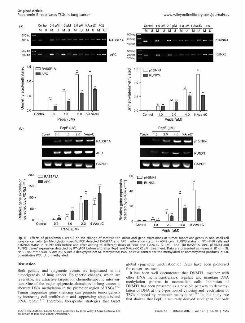
Peperomin E demethylates tumor suppressor genes and reacti-
vates their expressions in NSCLC cells. One of the primary bio-logical outcomes of DNA demethylation in cancer cells istranscriptional reactivation of silenced tumor suppressor genes(TSGs).(37–39) The RASSF1A, APC, p16INK4 (in malepatients), and RUNX3 genes were discovered more frequentlymethylated in lung tissue from NSCLC patients than non-can-cerous tissue.(40–45) RASSF1A and APC genes were found fullyhypermethylated in A549 cells,(46) the RUNX3 gene was foundsilenced in NCI-H460 cells by promoter methylation,(47) andthe promoter of p16INK4 gene was found completely methy-lated in H1299 cells.(40) Demethylation of these TSGs andrestoration of their expression in cancer cell lines impairstumorigenicity. Thus, factors that restore the expression ofthese TSGs have immense potential in inhibiting tumorgrowth. As PepE inhibits DNMT1 activity and expression, wespeculated that PepE might be involved in demethylation andre-expression of TSGs in NSCLC cells. We therefore investi-gated methylation status of RASSF1A and APC genes in A549cells, the p16INK4 gene in H1299 cells, and the RUNX3 genein NCI-H460 cells using methylation-specific PCR before andafter PepE treatment. As expected, RASSF1A and APC geneswere fully methylated in untreated A549 cells, confirming thatthey are complete silenced in A549 cells (Fig. 8a). Following0.5 lM PepE treatment, there was a noticeable increase in thelevels of unmethylated RASSF1A or APC compared to the con-trol; and after 1.0 or 2.0 lM treatment, this effect was evenmore significant (P < 0.01, Fig. 8a). Quantitative real-timePCR analysis revealed that PepE and 5-Aza-dC each reacti-vated silenced RASSF1A and APC mRNA expression in a con-centration-dependent manner in A549 cells (i.e., ≥74-fold forRASSF1A and ≥19 fold for APC at 2 lM dose of PepE,Fig. 8b). The findings are much the same concerning p16INK4and RUNX3 genes. As shown in Figure 8(a), these two geneswere entirely methylated in NCI-H460 and H1299 cells. Aftertreatment with PepE, the levels of unmethylated p16INK4 andRUNX3 genes were significantly increased, whereas the methy-lated levels of these two genes were significantly reduced(P < 0.01). The RT-qPCR results proved that the mRNAexpression of p16INK4 and RUNX3 were significantly restoredin a dose-dependent manner following PepE treatment (i.e.≥30-fold for p16INK4 and ≥87-fold for RUNX3 at 4 lM doseof PepE; Fig. 8b).In vitro protein reactivation levels were determined using
Western blot analysis. As shown in Figure 9(a), PepE and 5-Aza-dC each significantly reactivated silenced RASSF1A andAPC protein levels in A549 cells in a concentration-dependentmanner (P < 0.01). The protein levels of p16INK4 andRUNX3 were also significantly restored in NCI-H460 cells ina dose-dependent manner (P < 0.01). Immunohistochemicalanalysis showed that there was a significant increase inRASSF1A- and APC-positive cells in the tumor tissues of the50 mg/kg PepE-treated group when compared to the controlgroup (Fig. 9b).T
able
2.
Details
of
LibDock
score,
CDOCKER
energy,
CDOCKER
interaction
energy,
active
site
residues,
and
hydrogen
bonds
revealed
through
molecu
lar
docking
of
S-adenosylm
ethionine(SAM),5-aza
-2-deoxycytidine(5-A
za-dC),peperomin
E(PepE),andsinefugin
onDNA
methyltransferase
1(D
NMT1)(Protein
DataBankID:3SWR)ofHomosapiens
Target
Ligand
LibDock
score
CDOCKER
energy
CDOCKER
interaction
energy
Residuesofsinefugin
binding
Residuesinvo
lvedin
H-bond
No.of
H-bonds
Human
DNMT1
Sinefugin
160.33
27.25
55.87
GLY
1147,GLY
1149,GLY
1150,LE
U1151,ILE1167,
LEU1247,PHE1145,MET1169,ASP
1190,
VAL1
580,ALA
1579,GLU
1266,GLU
1168,PRO1225,ASN
1578,CYS1
148
GLY
1149,GLY
1150,LE
U1151,VAL1
580,
GLU
1168,GLU
1266
6
SAM
161.44
22.05
50.26
ALA
1579,MET1169,GLU
1168,PHE1145,PRO1225,GLY
1147,GLY
1149,LE
U1151,
GLY
1150,VAL1
580,CYS1
148,ALA
1579,ASN
1578,GLU
1266
GLY
1147,GLY
1149,GLY
1150,GLU
1168,
LEU1151,VAL1
580,PRO1225
7
5-A
za-dC
160.36
4.92
38.2
VAL1
580,LE
U1151,GLY
1149,GLY
1147,PHE1145,
GLU
1168,GLU
1266,ALA
1579,ASN
1578
GLY
1147,GLU
1266,GLU
1168,
ASN
1578,PHE1145
5
PepE
155.7
–14.81
53.19
PRO1225,GLY
1147,GLY
1150,LE
U1151,PHE1145,GLU
1266,VAL1
580,
GLU
1168,ASN
1578,MET1169,CYS1
148
GLY
1147,PHE1145,GLU
1168
3
TheLibDock
andCDOCKERprogramsare
providedbyDisco
very
studio
version4.0
software
(Accelrys,Sa
nDiego,CA,USA
).
© 2016 The Authors. Cancer Science published by John Wiley & Sons Australia, Ltdon behalf of Japanese Cancer Association.
Cancer Sci | October 2016 | vol. 107 | no. 10 | 1514
Original ArticlePeperomin E reactivates TSGs in lung cancer www.wileyonlinelibrary.com/journal/cas

According to previous studies, RASSF1A, APC, RUNX3, andp16INK4 tumor suppressors can scaffold multiple pro-apoptoticpathways and regulate cell cycle progressions in lung cancer.RASSF1A directly activates pro-apoptotic and cell cycle regula-tion factors in lung cancer cells including MST1/2 (pro-apopto-tic serine/threonine kinases), CNK1 (a pro-apoptotic adapterprotein), MOAP-1 (a Bax binding protein), and cyclin D1.(48,49)
APC can directly destroy b-catenin (a crucial regulating proteinof Wnt signaling pathway), thus promoting apoptosis and cellcycle arrest of NSCLC cells.(50,51) RUNX3 can induce NSCLCcell apoptosis by blocking the transforming growth factor-bsuperfamily signaling pathway.(52,53) p16INK4 mediates apopto-sis and cell cycles in NSCLC cells by direct downregulating Rband indirectly downregulating the anti-apoptotic protein
Bcl-2.(54,55) In this study, the protein levels of some of theaforementioned pro-apoptotic and cell cycle regulation factors(i.e., RASSF1A-mediated MST1/2, CNK1, and MOAP-1, andAPC-mediated b-catenin) were also investigated to further eluci-date the mechanisms involved in PepE-induced apoptosis andcell cycle arrest in A549 cells. As shown in Figure S6, afterPepE treatment, the protein levels of MST1, MST2, CNK1, andMOAP-1 were significantly increased, whereas b-catenin wassignificantly decreased in a concentration-dependent manner(P < 0.01). Immunohistochemical analysis showed that therewas a significant increase in MST1-, MST2-, CNK1-, andMOAP-1-positive cells (P < 0.01) but a decrease in b-catenin-positive cells (P < 0.01) in the tumor tissues of the PepE-treated(50 mg/kg) group (Fig. S7).
Fig. 6. Effects of peperomin E (PepE) on changesof activity and expression of DNA methyltransferase1 (DNMT1). (a) Dose-response plots of PepE and 5-Aza-dC against DNMT. The IC50 concentrationswere determined by biochemical DNMT assaysunder identical conditions; (b) Western-blot bandsfor DNMT1 protein expression in A549 cells beforeand after PepE treatment. The intensity of thebands was quantified by optical density (OD) andnormalized to the OD of GAPDH; (c) Representativeimmunostaining for DNMT1 in the paraffin section(original magnification 9200). 3 slides per mousewere reviewed. The positive rate was quantified byIOD values and normalized to the IOD values ofcontrol group; and (d) Western-blot bands for Sp1and NF-kB (p65) proteins expression in A549 cellsbefore and after PepE treatment. The intensity ofthe bands was quantified by optical density (OD)and normalized to the OD of b-actin. All data arepresented as means � SD (n = 3 for a, b, d and e;n = 6 for c). **P < 0.01. NF-jB, nuclear factor jB;qPCR, quantitative PCR.
Fig. 7. Effect of peperomin E (PepE) on globalDNA methylation in non-small-cell lung cancer cellsin vitro and in vivo. (a) Effects of PepE and 5-Aza-dC on global DNA methylation levels in NSCLC cells.DMSO served as control; and (b) Effects of PepEand 5-Aza-dC on the global DNA methylation levelsin A549 tumor tissues extracted from nude mice.Data are presented as means � SD (n = 3 for a;n = 6 for b). *P < 0.05; **P < 0.01. 5-Aza-dC, 5-Aza-2-deoxycytidine; dG, deoxyguanosine; 5mdC, 5-methyl-deoxy-cytidine.
Cancer Sci | October 2016 | vol. 107 | no. 10 | 1515 © 2016 The Authors. Cancer Science published by John Wiley & Sons Australia, Ltdon behalf of Japanese Cancer Association.
Original Articlewww.wileyonlinelibrary.com/journal/cas Wang et al.
Discussion
Both genetic and epigenetic events are implicated in thetumorigenesis of lung cancer. Epigenetic changes, which arereversible, are attractive targets for chemotherapeutic interven-tion. One of the major epigenetic alterations in lung cancer isaberrant DNA methylation in the promoter region of TSGs.(42)
Tumor suppressor gene silencing can promote tumorigenesisby increasing cell proliferation and suppressing apoptosis andDNA repair.(37) Therefore, therapeutic strategies that target
global epigenetic inactivation of TSGs have been pioneeredfor cancer treatment.It has been well documented that DNMT1, together with
other DNA methyltransferases, regulate and maintain DNAmethylation patterns in mammalian cells. Inhibition ofDNMT1 has been presented as a possible pathway to demethy-lation of DNA at the 5-position of cytosine and reactivation ofTSGs silenced by promoter methylation.(29) In this study, wefirst showed that PepE, a naturally derived secolignan, not only
Fig. 8. Effects of peperomin E (PepE) on the change of methylation status and gene expressions of tumor suppressor genes in non-small-celllung cancer cells. (a) Methylation-specific PCR detected RASSF1A and APC methylation status in A549 cells, RUNX3 status in NCI-H460 cells andp16INK4 status in H1299 cells before and after adding to different doses of PepE and 5-Aza-dC (2 lM); and (b) RASSF1A, APC, p16INK4 andRUNX3 genes’ expression detected by RT-qPCR before and after PepE and 5-Aza-dC (2 lM) treatment. Data are presented as means � SD (n = 3).*P < 0.05; **P < 0.01. 5-Aza-dC, 5-Aza-2-deoxycytidine; M, methylated; POS, positive control for the methylated or unmethylated products; qPCR,quantitative PCR; U, unmethylated.
© 2016 The Authors. Cancer Science published by John Wiley & Sons Australia, Ltdon behalf of Japanese Cancer Association.
Cancer Sci | October 2016 | vol. 107 | no. 10 | 1516
Original ArticlePeperomin E reactivates TSGs in lung cancer www.wileyonlinelibrary.com/journal/cas

inhibits DNMT1 enzymatic activity, but also decreases its tran-scription levels by blocking the active site and inhibiting thepositive transcription regulators (Sp1/NF-jB pathway) of thisenzyme. That is, PepE may serve dual functions: acting asboth chemical inhibitor and transcriptional modulator ofDNMT1. Both of these activities drive promoter demethyla-tion, resulting in re-expression of certain silenced TSGs inNSCLC cells. It was also found that, in response to PepE treat-ment, NSCLC cells underwent apoptosis and became arrestedat the G1 phase of the cell cycle. All the aforementionedevents occur together and subsequently contribute to PepE’santicancer effects against NSCLC, as evidenced by our in vitroand in vivo studies.Up to now, the extensively studied DNMT inhibitors were
nucleoside analogs including 5-azacytidine and5-Aza-dC,which have been approved by the US FDA for the treatmentof myelodysplastic syndrome and cutaneous T-cell lym-phoma.(33) Although no DNMT inhibitors are currentlyapproved for treatment of lung cancer, a recently completedclinical trial combining 5-azacytidine and entinostat (a histonedeacetylase inhibitor) for the treatment of advanced NSCLCdemonstrated promising results.(56) However, there are a num-ber of additional limitations for the use of these nucleosideanalogs. In particular, these drugs can cause severe side-effects, including hematopoietic toxicity and neutropenia. Fur-thermore, these drugs are unstable, and can be degraded byhydrolytic cleavage and deamination by cytidine
deaminase.(33,57) In contrast, PepE, derived from nature, maybe devoid of several of these limitations. According to ourresearch data, although the demethylating activity is lower thanthat of the currently used agent 5-Aza-dC, PepE appears to beconsiderably safer in animals. Therefore, this agent might fur-ther serve as a complementary therapy, especially when usedin conjunction with nucleoside analogs or in the setting ofnucleoside resistance, during treatment of cancer.In summary, we identified a new DNMT1 inhibitor, PepE,
that induced DNA demethylation and reversed the silencing ofcertain TSGs in NSCLC cells and induced NSCLC cell apop-tosis and cell cycle arrest. These findings are of importance forunderstanding the mechanisms of PepE against lung cancerand provides a promising epigenetically targeting agent forcancer treatment. Further mechanism of action and structuralmodification studies to increase the efficacy of this natural pro-duct are ongoing.
Acknowledgments
This work was supported by grants from the Natural ScienceFoundation of China (Grant No. 81402812) and the Natural ScienceFoundation of Jiangsu Province (Grant No. BK20130954).
Disclosure Statement
The authors have no conflict of interest.
Fig. 9. Effect of peperomin E (PepE) on the change of protein expression of epigenetically silenced tumor suppressor in non-small-cell lung can-cer cells in vitro and in vivo. (a) Western-blot bands for RASSF1A and APC proteins in A549 cells, p16INK4 and RUNX3 proteins in NCI-H460 cells.The intensity of the bands was quantified by optical density (OD) and normalized to the OD of b-actin; and (b) Representative immunostainingfor RASSF1A and APC in the paraffin section (original magnification 9200). 3 slides per mouse were reviewed. The positive rate was quantifiedby IOD values and normalized to the IOD values of control group. Data are presented as means � SD (n = 3 for a; n = 6 for b). **P < 0.01. 5-Aza-dC, 5-Aza-2-deoxycytidine.
Cancer Sci | October 2016 | vol. 107 | no. 10 | 1517 © 2016 The Authors. Cancer Science published by John Wiley & Sons Australia, Ltdon behalf of Japanese Cancer Association.
Original Articlewww.wileyonlinelibrary.com/journal/cas Wang et al.

References
1 Youlden DR, Cramb SM, Baade PD. The international epidemiology of lungcancer: geographical distribution and secular trends. J Thorac Oncol 2008;3: 819–31.
2 She J, Yang P, Hong Q, Bai C. Lung cancer in China: challenges and inter-ventions. Chest 2013; 143: 1117–26.
3 Chen W, Zheng R, Zeng H et al. Epidemiology of lung cancer in China.Thorac Cancer 2015; 6: 209–15.
4 Hong QY, Wu GM, Qian GS et al. Prevention and management of lung can-cer in China. Cancer 2015; 121: 3080–8.
5 Schiller JH, Harrington D, Belani CP et al. Comparison of four chemother-apy regimens for advanced non-small-cell lung cancer. N Engl J Med 2002;346: 92–8.
6 Florea AM, Busselberg D. Cisplatin as an anti-tumor drug: cellular mecha-nisms of activity, drug resistance and induced side effects. Cancers 2011; 3:1351–71.
7 Rajeswaran A, Trojan A, Burnand B et al. Efficacy and side effects of cis-platin- and carboplatin-based doublet chemotherapeutic regimens versus non-platinum-based doublet chemotherapeutic regimens as first line treatment ofmetastatic non-small cell lung carcinoma: a systematic review of randomizedcontrolled trials. Lung Cancer 2008; 59: 1–11.
8 Rowinsky EK, Eisenhauer EA, Chaudhry V et al. Clinical toxicities encoun-tered with paclitaxel (Taxol). Semin Oncol 1993; 20: 1–15.
9 Nanjing New Medical School. Dictionary of Chinese Herbal Drugs. Shang-hai: Shanghai Science and Technology Press, 1978; 622.
10 Govindachari TR, Krishna Kumari GN, Partho PD. Two secolignans fromPeperomia dindygulensis. Phytochemistry 1998; 49: 2129–31.
11 Wu JL, Li N, Hasegawa T et al. Bioactive secolignans from Peperomiadindygulensis. J Nat Prod 2006; 69: 790–4.
12 Chen L, Yu Y, Dong JX. Chemical constituents of Peperomia dindygulensis.Chin Tradit Herb Drugs 2007; 38: 491–3.
13 Lin MG, Yu DH, Wang QW et al. Secolignans with antiangiogenic activitiesfrom Peperomia dindygulensis. Chem Biodivers 2011; 8: 862–71.
14 Wu JL, Li N, Hasegawa T et al. Bioactive tetrahydrofuran lignans fromPeperomia dindygulensis. J Nat Prod 2005; 68: 1656–60.
15 Chen L, Zhou Y, Dong JX. Three new flavonoid glycosides from Peperomiadindygulensis. Acta Pharm Sin 2007; 42: 183–6.
16 Wang QW, Du DH, Lin MG et al. Antiangiogenic polyketides from Pepero-mia dindygulensis Miq. Molecules 2012; 17: 4474–83.
17 Wang XZ, Qu W, Liang JY. New long-chain aliphatic compounds fromPeperomia dindygulensis. Nat Prod Res 2013; 27: 796–803.
18 Wang XZ, Qu W, Liang JY. Two N-containing polyketide derivatives fromPeperomia dindygulensis. Chem Nat Comp 2013; 48: 1027–30.
19 Xu S, Li N, Ning MM et al. Bioactive compounds from Peperomia pellu-cida. J Nat Prod 2006; 69: 247–50.
20 Tsutsui C, Yamada Y, Ando M et al. Peperomins as anti-inflammatoryagents that inhibit the NF-kappa B signaling pathway. Bioorg Med ChemLett 2009; 19: 4084–7.
21 Zhang XJ, Yang GY, Wang RR et al. 7,8-Secolignans from Schisandrawilsoniana and their anti-HIV-1 activities. Chem Biodivers 2011; 7: 2962–71.
22 Momparle RL, Ayoub J. Potential of 5-aza-20-deoxycytidine (Decitabine) apotent inhibitor of DNA methylation for therapy of advanced non-small celllung cancer. Lung Cancer 2001; 34: 111–5.
23 Song LG, James SR, Kazim L et al. Specific method for the determinationof genomic DNA methylation by liquid chromatography-electrospray ioniza-tion tandem mass spectrometry. Anal Chem 2005; 77: 504–10.
24 Herman JG, Graff JR, Myohanen S et al. Methyation-specific PCR: a novelPCR assay for methylation status of CpG islands. Proc Natl Acad Sci USA1996; 93: 9821–6.
25 Kawaguchi K, Oda Y, Saito T et al. DNA hypermethylation status of multi-ple genes in soft tissue sarcomas. Mod Pathol 2006; 19: 106–14.
26 Scarpa M, Scarpa M, Castagliuolo I et al. Aberrant gene methylation in non-neoplastic mucosa as a predictive marker of ulcerative colitis-associatedCRC. Oncotarget 2016; 7: 10322–31.
27 Chim CS, Pang R, Fung TK et al. Epigenetic dysregulation of Wnt signalingpathway in multiple myeloma. Leukemia 2007; 21: 2527–36.
28 Sushma PS, Jamil K, Kumar PU et al. PTEN and p16 genes as epigeneticbiomarkers in oral squamous cell carcinoma (OSCC): a study on southIndian population. Tumour Biol 2016; 37: 7625–32.
29 Robert RF, Morin S, Beaulieu N et al. DNMT1 is required to maintain CpGmethylation and aberrant gene silencing in human cancer cells. Nat Genet2002; 33: 61–5.
30 Erdmann A, Halby L, Fahy J et al. Targeting DNA methylation with smallmolecules: what’s next? J Med Chem 2015; 58: 2569–83.
31 Fahy J, Jeltsch A, Arimondo PB. DNA methyltransferase inhibitors in can-cer: a chemical and therapeutic patent overview and selected clinical studies.Expert Opin Ther Pat 2012; 22: 1427–42.
32 Stresemann C, Brueckner B, Musch T et al. Functional diversity of DNAmethyltransferase inhibitors in human cancer cell lines. Cancer Res 2006;66: 2794–800.
33 Christman JK. 5-Azacytidine and 5-aza-20-deoxycytidine as inhibitors ofDNA methylation: mechanistic studies and their implications for cancer ther-apy. Oncogene 2002; 21: 5483–95.
34 Ghoshal K, Datta J, Majumder S et al. 5-Aza-deoxycytidine induces selec-tive degradation of DNA methyltransferase 1 by a proteasomal pathway thatrequires the KEN box, bromo-adjacent homology domain and nuclear local-ization signal. Mol Cell Biol 2005; 25: 4727–41.
35 Yu J, Peng Y, Wu LC et al. Curcumin down-regulates DNA methyltrans-ferase 1 and plays an anti-leukemic role in acute myeloid leukemia. PLoSOne 2013; 8: e55934.
36 Liu SJ, Liu ZF, Xie ZJ et al. Bortezomib induces DNA hypomethylationand silenced gene transcription by interfering with Sp1/NF-jB-dependentDNA methyltransferase activity in acute myeloid leukemia. Blood 2008;111: 2364–73.
37 Baylin SB. DNA methylation and gene silencing in cancer. Nat Clin PractOncol 2005; 2: 4–11.
38 Jones PA, Takai D. The role of DNA methylation in mammalian epigenetics.Science 2001; 293: 1068–70.
39 Fang MA, Wang YM, Ai N et al. Tea polyphenol (�)-epigallocatechin-3-gallate inhibits DNA methyltransferase and reactivates methylation-silencedgenes in cancer cell lines. Cancer Res 2003; 63: 7653.
40 Shames DS, Girard L, Gao BN et al. A genome-wide screen for promotermethylation in lung cancer identifies novel methylation markers for multiplemalignancies. PLoS Med 2006; 3: e486.
41 Drilon A, Sugita H, Sima CS et al. A prospective study of tumor suppressorgene methylation as a prognostic biomarker in surgically resected stage I toIIIA non-small-cell lung cancers. J Thorac Oncol 2014; 9: 1272–7.
42 Langevin SM, Kratzke RA, Kelsey KT et al. Epigenetics of lung cancer.Trans Res 2015; 165: 74–90.
43 Yanagawa N, Tamura G, Oizumi H et al. Promoter hypermethylation ofRASSF1A and Runx3 genes as an independent prognostic prediction marker insurgically resected non-small cell lung cancers. Lung Cancer 2007; 58: 131–8.
44 Zhang YW, Wang R, Song HZ et al. Methylation of multiple genes as a candi-date biomarker in non-small cell lung cancer. Cancer Lett 2011; 303: 21–8.
45 Yanagawa N, Tamura G, Oizumi H et al. Promoter hypermethylation oftumor suppressor and tumor-related genes in non-small cell lung cancers.Cancer Sci 2003; 94: 589–92.
46 Lin Q, Geng JF, Ma KL et al. RASSF1A, APC, ESR1, ABCB1 andHOXC9, but not p16INK4A, DAPK1, PTEN and MT1G genes were fre-quently methylated in the stage I non-small cell lung cancer in China. JCancer Res Clin Oncol 2009; 135: 1675–84.
47 Li QL, Kim HR, Kim WJ et al. Transcriptional silencing of the Runx3 geneby CpG hypermethylation is associated with lung cancer. Biochem BiophsRes Commun 2004; 314: 223–8.
48 Richter AM, Pfeifer GP, Dammann RH. The RASSF proteins in cancer;from epigenetic silencing to functional characterization. Biochim BiophysActa 2009; 1796: 114–28.
49 Donninger H, Vos DM, Clark GJ. The RASSF1A tumor suppressor. J CellSci 2007; 120: 3163–72.
50 Su LK, Vogelstein B, Kinzler KW et al. Association of the APC tumor sup-pressor protein with catenins. Science 1993; 262: 1734.
51 Stewart DJ. Wnt signaling pathway in non–small cell lung cancer. J NatlCancer Inst 2014; 106: 1–11.
52 Bae SC, Choi JK. Tumor suppressor activity of Runx3. Oncogene 2004; 23:4336–40.
53 Ito Y, Miyazono K. Runx transcription factors as key targets of TGF-bsuperfamily signaling. Curr Opin Genet Dev 2003; 13: 43–7.
54 Shapiro GI, Edwards CD, Kobzik L et al. Reciprocal Rb inactivation andp16INK4 expression in primary lung cancers and cell lines. Cancer Res1995; 55: 505–9.
55 Kataoka M, Wiehle S, Schumacher F et al. Down-regulation of bcl-2 isassociated with p16INK4-mediated apoptosis in non-small cell lung cancercells. Oncogene 2000; 12: 1589–95.
56 Juergens RA, Wrangler J, Vendetti FP et al. Combination epigenetic therapyhas efficacy in patients with refractory advanced non–small cell lung cancer.Cancer Discov 2011; 1: 598.
57 Jean-Pierre I. Decitabine. Curr Opin Oncol 2003; 15: 446–51.
© 2016 The Authors. Cancer Science published by John Wiley & Sons Australia, Ltdon behalf of Japanese Cancer Association.
Cancer Sci | October 2016 | vol. 107 | no. 10 | 1518
Original ArticlePeperomin E reactivates TSGs in lung cancer www.wileyonlinelibrary.com/journal/cas

Supporting Information
Additional Supporting Information may be found online in the supporting information tab for this article:
Fig. S1. Proton nuclear magnetic resonance (a) and carbon-13 nuclear magnetic resonance (b) spectrum of peperomin E.
Fig. S2. High-performance liquid chromatography peak purity analysis of peperomin E.
Fig. S3. In silico target fishing protocol using the LibDock module in Discovery Studio 4.0.
Fig. S4. Histopathological analysis of liver, kidney, and lung tissue structures in xenograft mice.
Fig. S5. Chromatogram using ultra-fast liquid chromatography tandem mass spectrometry of 5-methyl-deoxy-cytidine (5mdC) and deoxyguanosine(dG) in DNA of hydrolyzed A549 cells.
Fig. S6. Effects of peperomin E on changes of Ras association domain family member 1 (RASSF1A)-modulated pro-apoptotic and cell cyclearrest proteins.
Fig. S7. Effects of peperomin E on changes of adenomatous polyposis coli (APC)-modulated pro-apoptotic protein b-catenin.
Table S1. Primer list for methylation-specific PCR.
Table S2. Polymerase chain reaction amplification protocol in methylation-specific PCR test.
Table S3. Primer list for quantitative RT-PCR.
Table S4. Polymerase chain reaction amplification protocol in quantitative RT-PCR test.
Cancer Sci | October 2016 | vol. 107 | no. 10 | 1519 © 2016 The Authors. Cancer Science published by John Wiley & Sons Australia, Ltdon behalf of Japanese Cancer Association.
Original Articlewww.wileyonlinelibrary.com/journal/cas Wang et al.