peer-reviewed article bioresources€¦ · observed with a light microscope (olympus bx61, japan)...
TRANSCRIPT

PEER-REVIEWED ARTICLE bioresources.com
Zhang et al. (2015). “DNA conc. distrib. xylem cells,” BioResources 10(1), 1304-1317. 1304
Concentration and Distribution of Nuclei and Plastids in Xylem Cells in Cunninghamia lanceolata and Aquilaria sinensis
Rong Zhang,a,b Kuiwu Xu,a and Kelin Ye b,*
After programmed cell death (PCD), heartwood formation, storage, and processing, wood DNA degradation occurs to varying degrees. The concentration and distribution of nuclei and plastids in xylem cells of Cunninghamia lanceolata and Aquilaria sinensis, treated under different conditions of processing and storing, were studied by analyzing the distribution frequency, area, and signal intensity, in specimens that had been stained with aceto-carmine, DAPI, and I2-KI. Most of the nuclei and plastids were present in the ray cells, and a small quantity of nuclei and plastids were present in the axial parenchyma cells. There was an indication that the concentration of the remaining nuclei and plastids in the xylem cells was mainly affected by the xylem heartwood formation, storage time, and temperature. The nuclei and plastids content of the sapwood was greater than that of the heartwood. However, the nuclei and plastids content of the fresh wood was greater than that of the processed and stored wood. An estimation of the quantity of nuclei and plastids using staining methods could provide a direct basis for the appropriate selection of a procedure for DNA extraction.
Keywords: Nuclei; Plastid; Xylem; Aceto-carmine; DAPI; I2-KI
Contact information: a: College of Materials Science and Engineering, Nanjing Forestry University,
No.159 Longpan Road, Nanjing, China; b: Research Institute of Wood Industry, Chinese Academy of
Forestry, No.1 Dongxiaofu, Beijing, China; * Corresponding author: [email protected]
INTRODUCTION
Nuclear ribosomal DNA and plastid DNA play an important role in the
identification of timber species from the general to the individual level. In eukaryotic
organisms, the nuclear ribosomal DNA (rDNA) has two internal transcribed spacers
(ITS), i.e., ITS1 and ITS2. The length and sites of ITS sequences are different, which can
be used as a basis for the identification of wood species. The popularity of the rDNA-ITS
region for molecular systematic analyses of closely related species can be attributed to
both rapid evolution of the ITS spacers and PCR-amplification with conserved primers
(Gonzalez et al. 2009; China Plant BOL Group 2011; Hanssen et al. 2011). Plastids are
specific organelles of plants, and their forms include chloroplast, chromoplast, leucoplast,
proplastids, etioplasts, gerontoplasts, and more specialized leucoplasts. These plastids can
be transformed into each other. Compared with unique nuclei, there are a number of
copies of plastid genomes in each cell (Deguilloux et al. 2002). The conserved gene order
of chloroplast genome, the widespread availability of primers, and a general lack of
heteroplasmy and recombination have made the chloroplast genome an attractive tool for
phylogenetic studies of plants (Olmstead and Palmer 1994). DNA barcoding has been

PEER-REVIEWED ARTICLE bioresources.com
Zhang et al. (2015). “DNA conc. distrib. xylem cells,” BioResources 10(1), 1304-1317. 1305
researched and applied in wood identification, using genes such as rpoC1, psbK–psbI,
trnH–psbA, rbcL, matK (CBOL Plant Working Group 2009; Kress and Erickson 2007).
Wood is arborous, containing secondary xylem and many kinds of cells
differentiated from cambium, such as vessel, ray, xylem fiber, and tracheid. DNA that
remains in cells has provided a new approach to wood identification, viz. a DNA barcode
and a DNA fingerprint. Secondary growth includes several consecutive processes, such
as vascular tissue differentiation, secondary cell wall deposition, lignification,
programmed cell death (PCD), and heartwood formation (Tian et al. 2007). In the process
of PCD, nuclei are decreased and draped. At the same time, DNA is cut into fragments
among the nucleosomes (Turner et al. 2005). Subsequently, DNA goes through further
degradation during storage and processing.
DNA extraction is the front line for wood identification. Such identification is
done by using an applied genetic method and is used for regulating the timber trade and
fighting illegal logging. However, DNA in wood cells gradually breaks down, which
makes DNA extraction and molecular identification very difficult, particularly from
heartwood. So far, an all-round procedure for DNA extraction is still unavailable for
application to different wood materials, although some successful cases have been
published (Asif and Cannon 2005; Jiao et al. 2012; Tnah et al. 2012; Jiao et al. 2014).
Furthermore, in some experiments, DNA cannot be amplified or sequenced even after
successful DNA extraction (Asif and Cannon 2005; Zhang et al. 2014). Therefore, in
order to choose the appropriate procedure, it is very important to estimate the
concentration and quality of DNA before extraction occurs.
Nuclei can be observed effectually by using aceto-carmine, 4’-6-diamidino-2-
phenylindole (DAPI), and aceto-carmine. Carmine is a natural dye extracted from a red
pigment called cochineal. Apart from carmine, the aqueous solution of aceto-carmine
contains acetic acid and ferrum. Acetic acid is a solvent for carmine, and it increases cell
permeability and helps the ions enter the cells (Belling 1926; Rattenbury 1952).
Chromosomes become crimson or red after an aceto-carmine reaction, and these appear
visibly different from the other cell organelle and tissues. Consequently, it has been used
in studies of the nuclei distribution of xylem (Islam and Begum 2011; Nakaba et al.
2012) and ray parenchyma cells in Pinus dendiflora, Pinus rigida (Nakaba et al. 2008), in
Populus sieboldii, Populis grandidentata (Nakaba et al. 2012), and in Tectona grandis
(Islam and Begum 2011). DAPI is a kind of DNA-specific probe, which forms a
fluorescent complex by becoming attached in the minor groove of A-T rich sequences of
DNA. In both sites, DAPI is bound with a long axis, approximately parallel to the
grooves of the DNA helix (Kubista et al. 1987; Kapuscinski 1995). DAPI can penetrate a
cell membrane and is applied in DNA locating and the quantitative analysis of clones
(Nguyen et al. 1995). On the other hand, amyloplast, a kind of plastid, will show navy
blue after being stained with Lugol’s solution (I2-KI). It can then be effectively
distinguished from nuclei, cell walls, and so on (Abe et al. 2011).
The aim of this study was to understand the degree of degradation of wood nuclei
and plastids under different sapwood, heartwood, storage, and processing conditions. The
distribution frequency of nuclei and plastids, the area of nuclei, the signal intensity of
nuclei in the ray cells, and the percentage of plastid area in rays of Cunninghamia
lanceolata (Cuppressaceae) and Aquilaria sinensis (Thymelaeaceae) were investigated
after being stained with aceto-carmine, DAPI, and I2-KI in order to assist in choosing the
appropriate DNA extraction procedure.
PEER-REVIEWED ARTICLE bioresources.com
Zhang et al. (2015). “DNA conc. distrib. xylem cells,” BioResources 10(1), 1304-1317. 1306
EXPERIMENTAL
Materials C. lanceolata is widely distributed in China and is an important conifer species
used in architecture and industry. Wood samples were collected from a 36 year old
standing tree in the Chenshan Forestry Centre in Ji’an, in the Jiang Xi Province of China.
Twelve wood discs (10 mm × 10 mm × 5 mm) were cut at a level of 2 m from the
ground, with a diameter of 24.2 cm at breast height. Six wood samples were immediately
placed into 2.5% glutaraldehyde in order to keep fresh. Other six wood samples were
keeping in air temperature. On arrival at the laboratory, they were treated according to
Table 1.
Table 1. Sample Information of C. lanceolata
Sample type Location Sample number
Storage
temperature(℃) Storage
time(years) Radial position
(distance to pith)(cm)
Fresh wood sapwood 3 4 2 18
heartwood 3 4 2 3
Air-dried wood
sapwood 3 Room
temperature 2 18
heartwood 3 Room
temperature 2 3
A. sinensis is a precious and unique resource for the production of “agarwood” in
China and an endangered flora that has been listed since 2004 in Appendix II of the
Convention on International Trade in Endangered Species of Wild Fauna and Flora
(CITES) (http://www.cites.org/eng/app/appendices ). Wood samples were collected from
a standing tree of A. sinensis in the city of Guang Zhou in the Guang Dong Province of
China.
The tree height was 2.3 m, the diameter at breast height was 7.48 cm, and the
average diameter of the heartwood was 5.77 cm. The wood samples were immediately
placed into an ice storage box after being collected from the stumpage. They were then
immediately transported and stored in a refrigerator at -20 C on return to the laboratory.
Eighteen sticks (10 mm × 10 mm × 5 mm) were randomly chosen from the sapwood and
heartwood respectively. They were treated according to Table 2.
Table 2. Sample Information for A. sinensis
Sample type
Location Sample number
Storage or treated
temperature C
Processing time(hours)
Radial position (distance to pith)
(cm)
Fresh wood Sapwood 3 4 24 6.5
Heartwood 3 4 24 2
Dried wood Sapwood 3 80 3 6.5
Heartwood 3 80 3 2
Dried wood Sapwood 3 120 240 6.5
Heartwood 3 120 240 2

PEER-REVIEWED ARTICLE bioresources.com
Zhang et al. (2015). “DNA conc. distrib. xylem cells,” BioResources 10(1), 1304-1317. 1307
Methods Section preparation
First, the surfaces of the wood sticks were washed with 70% ethanol and then
with purified water to reduce the influence of microbes on the DNA. Second, samples
were fixed in a 2.5% solution of glutaraldehyde (pH=7.4) at room temperature for one
day and thereafter washed four times with 0.2 mol/L phosphate buffer (PB) (0.2 mol/L
monosodium orthophosphate and disodium hydrogen phosphate) (pH=7.4). Radial
sections with a thickness of 10 μm were cut using a freeze sliding microtome (Leica
CM3050S, Germany).
Control sample
Sections washed with PB or PBS without any staining, as control samples, were
observed with a light microscope (Olympus BX61, Japan) and a Leica TCS SPE confocal
microscope (Leica Microsystems, Germany)
Aceto-carmine staining
The sections were washed twice with 0.2 mol/L PB (pH=7.4) and then immersed
in a 2% aqueous solution of aceto-carmine for 20 min. After washing twice with 0.2
mol/L PB (pH=7.4) to remove any unreacted aceto-carmine, the sections were placed
under a light microscope (Olympus BX61, Japan) for observation.
DAPI staining
The sections were washed three times with a phosphate buffer solution (PBS)
(pH=7.4) and then immersed in a 2.9 mM/L solution of DAPI for 20 min. After washing
with PBS twice, the sections were then placed on a glass slide with glycerol. A Leica
TCS SPE confocal microscope (Leica Microsystems, Germany) was adapted to observe
at a 405 nm wavelength (PMT Gain value=900; PMT Offest=0; Laser output
power=15%).
I2-KI staining
The sections were washed twice with 0.2 mol/L PB (pH=7.4) and then immersed
in an I2-KI solution for 20 min. After washing twice with 0.2 mol/L PB (pH=7.4) to
remove any unreacted I2-KI, the sections were placed under a light microscope (Olympus
BX61, Japan) for observation.
Statistical analysis
Three specimens were selected for each treatment and five fields of each
specimen were used for analysis. For each of the detected fields, the nuclei frequency (the
number of signals of DAPI and aceto-carmine of nuclei of rays (mm-2)), the nuclei area
(the single nuclei area of rays, μm-2), the signal intensity (0-255) of nuclei, and the
percentage of plastid area (the percentage of plastid area in rays) were recorded. A double
factor analysis of variance (ANOVA) was carried out using the SAS program, version 9.0,
to evaluate quantitative nuclei and plastids differences among the samples.
Nuclei and plastids in axial parenchyma cells were ignored, because they could
only be observed occasionally and the amount was so small compared with the nuclei and
plastids in ray cells. In the evaluation of nuclei and plastids area and signal intensity, 50
signals of nuclei were selected at random in every sapwood sample. Fifteen signals of
heartwood were measured because the number of signals of heartwood was small.

PEER-REVIEWED ARTICLE bioresources.com
Zhang et al. (2015). “DNA conc. distrib. xylem cells,” BioResources 10(1), 1304-1317. 1308
RESULTS AND DISCUSSION
Nuclei Distribution In the control samples, the nuclei could not be observed under the light
microscope (Fig. 1a, 1b, 1f, 2a, 2b, and 2f); however, they appeared black without auto
fluorescence (Fig. 3a, 3b, 4a, 4b, and 4e), as detected by the confocal microscope. The
nuclei stained with aceto-carmine appeared crimson in the ray cells and the axial
parenchyma cells. These were effectively separated from the lipids, amyloplast, and cell
walls (Fig. 1c, 1d, 1e, 1g, 2c, 2d, and 2g). Meanwhile, the nuclei stained with DAPI was
detected by fluorescence under a confocal microsystem (Fig. 3c, 3d, 3g, and 4d).
Fig. 1. Radial sections of C. lanceolata. a, b and h: Control samples. c, d, e, f and g: Samples stained with aceto-carmine. a: Rays of sapwood of fresh wood. b: Magnification of a. c: Rays of sapwood of fresh wood. d: Magnification of d. e: Axial parenchyma cells of fresh wood. f and g: Rays of heartwood of fresh wood. h: Rays of heart of air-dried wood. Parts indicated by arrows are nuclei in figure c, d, e, and g
Fig. 2. Radial sections of A. sinensis. a, b and f: Control samples. c, d, e and g: Samples stained with aceto-carmine. a: Rays of sapwood. b: Magnification of a.c: Rays of sapwood. d: Magnification of c. e: Rays of heartwood of air-dried wood. f and g: Rays of heartwood of fresh wood. Parts indicated by arrows are nuclear chromatin in figure c, d and g

PEER-REVIEWED ARTICLE bioresources.com
Zhang et al. (2015). “DNA conc. distrib. xylem cells,” BioResources 10(1), 1304-1317. 1309
Fig. 3. Radial sections of C. lanceolata. a, b and e: Control samples. c, d, f and g: Samples stained with DAPI. a: Rays of sapwood. b: Magnification of a. c: Rays of sapwood. d: Magnification of c. e and f: Rays of heartwood. g: Axial parenchyma cells of fresh wood. Parts in figure a, b c, d and g indicated by arrows are nuclei
Fig. 4. Radial sections of A. sinensis. a, b and e: Control samples. c, d and f: Samples stained with DAPI. a: Rays of sapwood. b: Magnification of a. c: Rays of sapwood. d: Magnification of c. e and f: Heartwood. Parts in figure a, b and d indicated by arrows are nuclear chromatin
Generally, the chromatins of C. lanceolata were spindly in the cell lumen of ray
parenchyma cells and axial parenchyma cells (Figs. 1 and 3). Most of the nuclear
chromatin was localized in the upper middle and lower lines of the ray cells. However,
when the nuclear chromatins of A. sinensis were attached to a cell wall, it was difficult to
separate the chromatin from the cell wall (Figs. 2 and 4). When the nuclei of heartwood
had been dispersed, there was not enough fluorescence emitted (Figs. 2g and 4f).
In the case of C. lanceolata sapwood samples, signals of DAPI and aceto-carmine
were equally distributed in rays of both early wood and late wood. However, in
heartwood samples, these signals were mainly distributed in rays of late wood near the
growth ring boundary. For A. sinensis, crimson signals of nuclei were distributed in
almost all ray parenchyma cells of fresh sapwood. Numbers of these signals in heartwood
and dried sapwood were less than fresh sapwood, but they too were evenly distributed. It
is possible that because A. sinensis trees grow in tropical areas, wood formation was not
influenced by seasons.
PEER-REVIEWED ARTICLE bioresources.com
Zhang et al. (2015). “DNA conc. distrib. xylem cells,” BioResources 10(1), 1304-1317. 1310
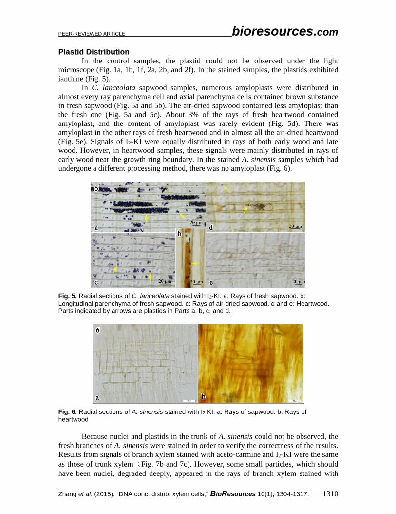
Plastid Distribution In the control samples, the plastid could not be observed under the light
microscope (Fig. 1a, 1b, 1f, 2a, 2b, and 2f). In the stained samples, the plastids exhibited
ianthine (Fig. 5).
In C. lanceolata sapwood samples, numerous amyloplasts were distributed in
almost every ray parenchyma cell and axial parenchyma cells contained brown substance
in fresh sapwood (Fig. 5a and 5b). The air-dried sapwood contained less amyloplast than
the fresh one (Fig. 5a and 5c). About 3% of the rays of fresh heartwood contained
amyloplast, and the content of amyloplast was rarely evident (Fig. 5d). There was
amyloplast in the other rays of fresh heartwood and in almost all the air-dried heartwood
(Fig. 5e). Signals of I2-KI were equally distributed in rays of both early wood and late
wood. However, in heartwood samples, these signals were mainly distributed in rays of
early wood near the growth ring boundary. In the stained A. sinensis samples which had
undergone a different processing method, there was no amyloplast (Fig. 6).
Fig. 5. Radial sections of C. lanceolata stained with I2-KI. a: Rays of fresh sapwood. b: Longitudinal parenchyma of fresh sapwood. c: Rays of air-dried sapwood. d and e: Heartwood. Parts indicated by arrows are plastids in Parts a, b, c, and d.
Fig. 6. Radial sections of A. sinensis stained with I2-KI. a: Rays of sapwood. b: Rays of heartwood
Because nuclei and plastids in the trunk of A. sinensis could not be observed, the
fresh branches of A. sinensis were stained in order to verify the correctness of the results.
Results from signals of branch xylem stained with aceto-carmine and I2-KI were the same
as those of trunk xylem(Fig. 7b and 7c). However, some small particles, which should
have been nuclei, degraded deeply, appeared in the rays of branch xylem stained with
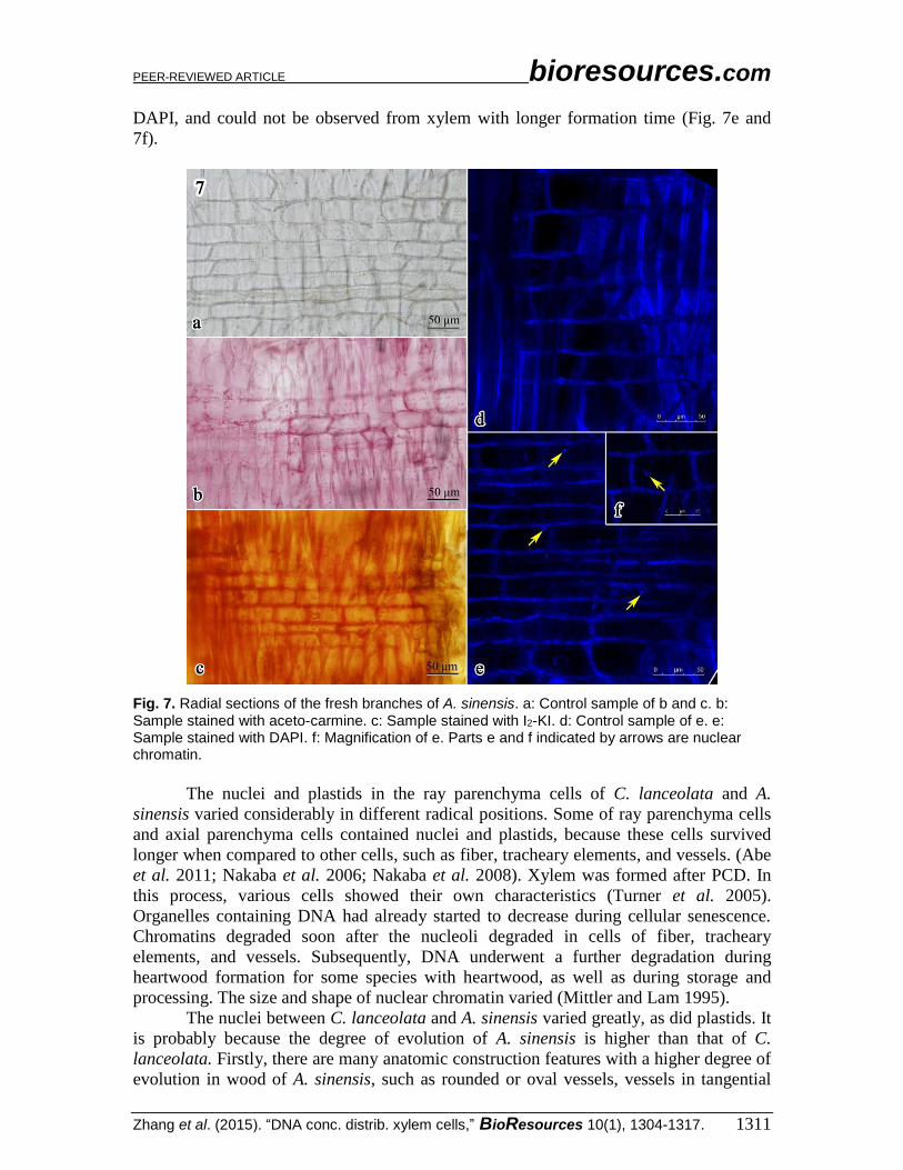
PEER-REVIEWED ARTICLE bioresources.com
Zhang et al. (2015). “DNA conc. distrib. xylem cells,” BioResources 10(1), 1304-1317. 1311
DAPI, and could not be observed from xylem with longer formation time (Fig. 7e and
7f).
Fig. 7. Radial sections of the fresh branches of A. sinensis. a: Control sample of b and c. b: Sample stained with aceto-carmine. c: Sample stained with I2-KI. d: Control sample of e. e: Sample stained with DAPI. f: Magnification of e. Parts e and f indicated by arrows are nuclear chromatin.
The nuclei and plastids in the ray parenchyma cells of C. lanceolata and A.
sinensis varied considerably in different radical positions. Some of ray parenchyma cells
and axial parenchyma cells contained nuclei and plastids, because these cells survived
longer when compared to other cells, such as fiber, tracheary elements, and vessels. (Abe
et al. 2011; Nakaba et al. 2006; Nakaba et al. 2008). Xylem was formed after PCD. In
this process, various cells showed their own characteristics (Turner et al. 2005).
Organelles containing DNA had already started to decrease during cellular senescence.
Chromatins degraded soon after the nucleoli degraded in cells of fiber, tracheary
elements, and vessels. Subsequently, DNA underwent a further degradation during
heartwood formation for some species with heartwood, as well as during storage and
processing. The size and shape of nuclear chromatin varied (Mittler and Lam 1995).
The nuclei between C. lanceolata and A. sinensis varied greatly, as did plastids. It
is probably because the degree of evolution of A. sinensis is higher than that of C.
lanceolata. Firstly, there are many anatomic construction features with a higher degree of
evolution in wood of A. sinensis, such as rounded or oval vessels, vessels in tangential

PEER-REVIEWED ARTICLE bioresources.com
Zhang et al. (2015). “DNA conc. distrib. xylem cells,” BioResources 10(1), 1304-1317. 1312
bands, vessel clusters common, simple perforation plates, and alternating intervessel pits
(Bailey and Tupper 1918; Bailey 1944; Frost 1930a, 1930b and 1931). Secondly, the
taxonomy of this Aquilaria genus is as follows—angiosperm: dicotyledoneae: rose
subclass: myrtles: Thymelaeaceae. On the one hand, the degree of evolution of
angiosperms is higher than that of gymnosperms. On the other hand, myrtle were located
at the highest evolution level in four evolutionary classification systems of angiosperms,
including A. Engler’s system, J. Hutchinson’s system, A. Takhtajan’s system, and A.
Cronquist’s system (Qiang 2006).
Nuclear Quantity Analysis Large quantities of nuclei remained in the fresh sapwood of C. lanceolata, while
the amount of nuclei that remained in the fresh heartwood, air-dried sapwood, and air-
dried heartwood was less obvious. Comparing the sapwood to the heartwood, the
variation was significant (F-probability <0.0001) and greater than the value of the
variation between the fresh wood and the air-dried wood (0.0006). Furthermore, there
was more nuclei in the sapwood that had been stored for two years than there was in the
fresh heartwood. Therefore, heartwood formation degraded the nuclei more than the
storage time did (Table 3).
Table 3. Distribution Frequency of Nuclei Remaining in the Ray Cells (mm-2) and the Quantity of DNA (ng/mg) Extracted from Freshwood using CTAB and Kit Methods of C. lanceolata
Fresh wood Air-dried wood
(2 years) F-probability
DNA quantity of freshwood *
CTAB Kit
Sapwood 212.20 A 79.09 B 69.95 107.02
(96.61) (52.91) (8.40) (7.63)
Heartwood 12.57C 2.96 C 12.70 13.83
(23.33) (5.49) (3.06) (4.43)
<0.0001 0.0006 0.0097
The values indicate the amount of nuclei remaining in wood per square mm2. The values in
parentheses represent standard deviations. A, B, C, and D: the same letter indicates that there was no significant difference among them. The significance level is 0.05. * This parameter was normalized according to the procedure of Jiao (2012). Its treatment conditions and sample source of wood were same to this paper.
For A. sinensis, the variation in the distribution frequency of the cells containing
nuclei from sapwood that were treated by keeping them fresh, dried at 80 C, and dried at
120 C were significantly different, which was the same result as with the C. lanceolata
samples. The variation was significant (F-probability<0.0001) between the sapwood and
heartwood. Additionally, the nuclei distribution frequency of the sapwood dried at 120
C was greater than that of all heartwood samples, i.e., the variation among the
heartwood treated by keeping it fresh, dried at 80 C, and dried at 120 C was
insignificant. Therefore, heartwood formation degraded the nuclei more than the high
temperature did (Table 4).

PEER-REVIEWED ARTICLE bioresources.com
Zhang et al. (2015). “DNA conc. distrib. xylem cells,” BioResources 10(1), 1304-1317. 1313
Table 4. Distribution Frequency of the Ray Cells Containing Nuclei Remaining (mm-2) and the Quantity of DNA (ng/mg) Extracted using Kit Method of A. sinensis
Distribution frequency DNA quantity *
Fresh wood
Dried at 80
C
Dried at
120 C F-probability
Fresh wood
Dried at
80 C
Dried at
120C
Sapwood 411.79 A 98.98B 38.27 B 8.01 4.67 4.44
(106.33) (89.37) (30.47) (0.80) (0.64) (0.66)
Heartwood 8.73 C 6.11 C 3.90 C 4.39 4.73 4.24
(12.5) (17.27) (11.04) (0.49) (0.53) (0.60)
<0.0001 <0.0001 <0.0001
The values indicate the amount of nuclei remaining in wood per mm2. The values in parentheses
represent standard deviations. A, B and C: the same letter indicates that there was no significant difference among them. The significance level is 0.05. * This parameter was normalized according to the procedure of Jiao (2014). Its treatment conditions and sample source were similar to this paper.
The area of nuclei that remained in the fresh sapwood of C. lanceolata was
significantly greater when compared to other fresh heartwood, dried sapwood, and dried
heartwood samples. However, among fresh heartwood, air-dried sapwood, and air-dried
heartwood, these values were not significant (Table 5). These results were similar to the
distribution frequency results for C. lanceolata. This means that the heartwood nuclei
degraded almost completely. The nuclei area of A. sinensis could not be computed,
because it was difficult to separate the nuclear chromatin from the cell walls.
Table 5. Area of Nuclei Remaining in the Ray Cells of C. lanceolata (μm2)
Fresh wood Air-dried wood (2 years) F-probability
Sapwood 173.14 A 81.47 B
(99.36) (68.79)
Heartwood 54.51 B 38.76 B
(24.62) (24.95)
<0.0001 <0.0001 <0.0001
The values indicate the area of nuclei remaining in wood per mm2. The values in parentheses
represent standard deviations. A and B: the same letter indicates that there was no significant difference among them. The significance level is 0.05.
Table 6. Signal Intensity of the Ray Cells in C. lanceolata
Fresh wood Air-dried wood F-probability
Sapwood 103.38A 41.49 B
(24.42) (9.41)
Heartwood 50.01 B 35.78B
(14.30) (10.33)
0.0321 <0.0001 0.0012
The values indicate the signal intensity (0-255) of nuclei remaining in the wood. The values in
parentheses represent standard deviations. A and B: the same letter indicates that there was no significant difference among them. The significance level is 0.05.
A greater signal intensity of nuclei means a greater concentration of DNA. The
value for fresh sapwood was significantly different compared to the values from fresh

PEER-REVIEWED ARTICLE bioresources.com
Zhang et al. (2015). “DNA conc. distrib. xylem cells,” BioResources 10(1), 1304-1317. 1314
heartwood, air-dried sapwood and air-dried heartwood (Table 6). This means that the best
DNA came from the fresh sapwood. However, the qualities of the nuclei that remained in
the fresh heartwood, air-dried sapwood, and air-dried heartwood were similar to each
other.
Plastid Quantity Analysis The percentage of plastid area in rays was determined according to the ianthine
signal of amyloplast in C. lanceolata stained with I2-KI. The amyloplast area of 14.56%
in rays of fresh sapwood was higher than 1.01% in air-dried sapwood, 0.05% in fresh
heartwood, and 0.0023% in air-dried heartwood. Comparing the sapwood to the
heartwood, the variation was significant (F-probability <0.0001), and the same as that
between fresh and air-dried wood (F-probability <0.0001). Furthermore, there was more
plastids in the sapwood that had been stored for two years than there was in the fresh
heartwood. Therefore, PCD degraded the plastids more than the storage time did (Table
7).
Table 7. Percentage of Plastid Area in Rays in C. lanceolata (%)
Fresh wood Air-dried wood
(2 years) F-probability
Sapwood 14.56A 1.01B
(3.60) (0.33)
Heartwood 0.05C 0.0023C
(0.108) (0.0063)
<0.0001 <0.0001 <0.0001
The values indicate the percentage of plastid area in rays. The values in parentheses represent standard deviations. A, B, and C. the same letter indicates that there was no significant difference among them. The significance level is 0.05.
To sum up, sapwood contained more plastids than heartwood, while fresh wood
contained more plastids than processed wood. The variation tendency of the plastids
quality was synchronous with the quantity (Rachmayanti et al. 2009; Tnah et al. 2012).
Taking all the factors into consideration, there was an indication that the concentration of
the remaining plastids in xylem cells was mainly affected by the xylem’s PCD,
heartwood formation, the storage time, and the drying temperature. The PCD made the
strongest impact on the plastids degradation, heartwood formation is next, when
compared to the effect of storage time and high temperature.
Content of Nuclei and Plastids and DNA Isolation It was reported that hexadecyl trimethyl ammonium bromide (CTAB), the
DNeasy Plant Mini Kit (Qiagen, Germany), and N-phenacylthiazolium bromide (PTB)
had been successfully used in wood DNA extraction after modification (Asif and Cannon
2005; Jiao et al. 2012; Jiao et al. 2014). However, two or three extraction methods were
used on the same wood sample in most of the existing research, because it is difficult to
select the right wood DNA extraction procedure for wood that has unknown degrees of
DNA degradation.
The differences among the distribution frequency of the nuclei and plastids
remaining in ray cells of A. sinensis were similar to the quantity of DNA extraction from
wood under the same treatment conditions. A DNeasy Plant Mini Kit (QIAGEN,

PEER-REVIEWED ARTICLE bioresources.com
Zhang et al. (2015). “DNA conc. distrib. xylem cells,” BioResources 10(1), 1304-1317. 1315
Germany) succeeded in extracting the DNA in fresh sapwood, fresh heartwood, and dried
heartwood of A. sinensis, but failed with heartwood dried at a high-temperature (Jiao et
al. 2014). The same was true for both the Kit and CTAB when it came to DNA extraction
from C. lanceolata (Jiao et al. 2012). This indicates that the extraction quantity varies
according to the content of nuclei and plastids. The quantity of DNA extracted from the
same samples using the DNeasy Plant Mini Kit was 1.5 to 2 times more than what was
extracted using CTAB (Jiao et al. 2012). However, the kit did not succeed with the
heartwood dried at a high-temperature (Jiao et al. 2014). CTAB and the kit yielded low-
qualities and low quantities of DNA; these samples could not be amplified even though
they were detectable. However, PTB succeeded in extracting DNA from the dried
heartwood of Ramin (Gonystylus bancanus) (Asif and Cannon 2005), due to having
greater DNA quantity and quality than what was obtained from CTAB and the kit. In
another report, CTAB and the kit responded well when used in extracting DNA from the
sapwood and heartwood of Penak (Neobalanocarpus heimii), but the result was not as
good as that which was optimized using PTB. In general, fresh wood that contains a mass
of DNA could be extracted using the CTAB or the kit without PTB. For aged wood that
contains less nuclei and plastids than fresh wood, the kit or PTB ought to be chosen.
When there is no sign of nuclei and plastids observable, a modified PTB procedure needs
to be developed. However, PTB does not always work for all wood with substantially
degraded nuclei and plastids.
In practice, the freshness and processing stage of wood is hard to evaluate
visually. However, staining methods can be used for a fast estimation of remaining nuclei
and plastids in wood materials. The optimized extraction procedures can be chosen
according to the observation of the nuclei and plastids amount and quality by staining.
CONCLUSIONS
1. Nuclei and plastids were degraded by PCD first in xylem formation. Fiber, tracheary
elements, and vessels lost nuclei and plastids. Subsequently, nuclei and plastids in ray
cells and axial parenchyma cells underwent a further degradation during heartwood
formation for some species with heartwood. After the tree was cut, nuclei and plastids
were degraded continually by storage and temperature.
2. Staining methods, such as aceto-carmine, DAPI, and I2-KI, can be adopted in the
evaluation of nuclei and plastids quantity. It is advised that the optimized extraction
procedures for wood DNA extracting be chosen.
ACKNOWLEDGMENTS
The authors would like to thank Professor Yin Yafang of the Chinese Research
Institute of Wood Industry, the Chinese Academy of Forestry for his valuable comments
on experiment and data analysis, Dr. Liu Bo for the guide of confocal microscope, and
Mr. Jiao Lichao and Mr. Song Kunlin for collecting the samples. This work was
financially supported by a project at the Chinese State Forestry Administration (No.
201304508) and the Priority Academic Program Development of Jiangsu Higher
Education Institutions (PAPD).

PEER-REVIEWED ARTICLE bioresources.com
Zhang et al. (2015). “DNA conc. distrib. xylem cells,” BioResources 10(1), 1304-1317. 1316
REFERENCES CITED
Abe, H., Watanabe, U., Yoshida, K., Kuroda, K., and Zhang, C. (2011). “Changes in
organelle and DNA quality, quantity, and distribution in the wood of Cryptomeria
Japonica over long-term storage,” IAWA J. 32(2), 263-272. DOI: 10.1163/22941932-
90000056
Asif, M. J., and Cannon, C. H. (2005). “DNA extraction from processed wood: A case
study for the identification of an endangered timber species (Gonystylus bancanus),”
Plant Mol. Biol. Rep. 23(2), 185-192. DOI: 10.1007/BF02772709
Bailey, I. W., and Tupper, W. W. (1918). “Size variation in tracheary cells I. A
comparison between the secondary xylems of vascular cryptogams, gymnosperms
and angiosperms,” Pro. Amer. Acad. Arts 54, 159-204. DOI: 10.2307/20025747
Bailey, I. W. (1944). “The development of vessels in angiosperms and its significance in
morphological research,” Amer. J Bot. 31, 421-428. DOI: 10.2307/2437302
Belling, J. (1926). “The iron-acetocarmine method of fixing and staining chromosomes,”
The Biological Bulletin Wood's Hole 50, 160-162. DOI: 10.2307/1536680
CBOL Plant Working Group. (2009). “A DNA barcode for land plants,” Proc. Natl. Acad.
Sci. USA 106, 12794-12797. DOI: 10.1073/pnas.0905845106
China Plant BOL Group. (2011). “Comparative analysis of a large dataset indicates that
internal transcribed spacer (ITS) should be incorporated into the core barcode for seed
plants,” Proc Natl. Acad. Sci. USA 108(49), 19641-19646. DOI:
10.1073/pnas.1104551108
Deguilloux, M. F., Pemonge, M. H., and Petit, R. J. (2002). “Novel perspectives in wood
certification and forensics: Dry wood as a source of DNA,” Proc. Biol. Sci. 269,
1039-1046. DOI: 10.1098/rspb.2002.1982
Frost, F. H. (1930a). “Specialization in secondary xylem of dicotyledons. I. Origin of
vessel,” Bot. Gaz. 89, 67-94. DOI: 10.1086/334026
Frost, F. H. (1930b). “Specialization in secondary xylem of dicotyledons. II. Evolution
of end wall of vessel segment,” Bot. Gaz. 90, 198-212. DOI: 10.2307/2471138
Frost, F. H. (1931). “Specialization in secondary xylem of dicotyledons. III.
Specialization of lateral wall of vessel segment,” Bot. Gaz. 91, 88-96. DOI:
10.1086/334128
Gonzalez, M. A., Baraloto, C., and Engel, J. (2009). “Identification of Amazonian trees
with DNA barcodes,” PLoS ONE 4(10):e7483. DOI:10.1371/journal.pone.0007483
Hanssen, F., Wischnewski, N., Moreth, U., and Magel, E., A. (2011). “Molecular
identification of Fitzroya cupressoides, Sequoia sempervirens, and Thuja plicata
wood using taxon-specific rDNA-ITS primers,” IAWA J 32(2), 273-284. DOI:
10.1163/22941932-90000057
Islam, M., A., and Begum, S. (2011). “Distribution of starch, lipid and nuclei in xylem
and phloem of tectona grandis Linn,” J. BioScience 19, 29-35. DOI:
10.3329/jbs.v19i0.12997
Jiao, L., Yin, Y., Xiao, F., Sun, Q., and Song, K. (2012). “Comparative analysis of two
DNA extraction protocols from fresh and dried wood of Cunninghamia lanceolata
(Taxodiaceae),” IAWA J 33(4), 441-456. DOI: 10.1163/22941932-90000106
Jiao, L., Yin, Y., Chen, Y., and Jiang, X. (2014). “DNA barcoding for identification of
the endangered species Aquilaria sinensis: Comparison of data from heated or aged
wood samples,” Holzforschung 68(4), 487-494. DOI: 10.1515/hf-2013-0129
Kapuscinski, J. (1995). “DAPI: A DNA-specific fluorescent probe,” Biotechnic and

PEER-REVIEWED ARTICLE bioresources.com
Zhang et al. (2015). “DNA conc. distrib. xylem cells,” BioResources 10(1), 1304-1317. 1317
Histochemistry 70(5), 220-233. DOI: 10.3109/10520299509108199
Kubista, M., Akerman, B., and Norden, B. (1987). “Characterization of interaction
between DNA and 4’,6-diamidino-2-phenylindole by optical spectroscopy,”
Biochemistry 26, 4545-4553. DOI: 10.1021/bi00388a057
Kress, W., J., and Erickson, D., L. (2007). “A two-locus global DNA barcode for land
plants: The coding rbcL gene complements the non-coding trnH-psbA spacer region,”
PLoS ONE 56, 295-299. DOI: 10.1371/journal.pone.0000508
Mittler, R., and Lam, E. (1995). “In situ detection of nDNA fragmentation during the
differentiation of tracheary elements on higher plants,” Plant Physiol. 108, 489-493.
DOI: 10.1104/pp.108.2.489
Nakaba, S., Begum, S., Yamagishi, Y., Jin, H., Kubo, T., and Funada, R. (2012).
“Differences in the timing of cell death, differentiation and function among three
different types of ray parenchyma cells in the hardwood Populus sieboldii ×P.
grandidentata,” Trees 26, 743-750. DOI: 10.1007/s00468-011-0640-0
Nakaba, S., Kubo, T., and Funada, R. (2008). “Differences in patterns of cell death
between ray parenchyma cells and ray tracheid in the conifers Pinus densiflora and
Pinus rigida,” Trees 22, 623-630. DOI: 10.1007/s00468-008-0220-0
Nakaba, S., Sano, Y., Kubo, T., and Funada, R. (2006). “The positional distribution of
cell death of ray parenchyma in a conifer, Abies sachalinensis,” Plant Cell Rep. 25,
1143-1148. DOI: 10.1007/s00299-006-0194-6
Nguyen, T., Erb, L., Weisman, G. A., Marchese, A., Heng, H. H., Garrad, R. C., George,
S. R., Turner, J. T., and O'Dowd, B. F. (1995). “Cloning, expression, and
chromosomal localization of the human uridine nucleotide receptor gene,” J. Biol.
Chem. 270, 30845-30848. DOI: 10.1074/jbc.270.52.30845
Olmstead, R. G., and Palmer, J. D. (1994). “Chloroplast DNA systematics: A review of
methods and data analysis,” Amer. J. Bot. 81, 1205-1224. DOI: 10.2307/2445483
Rattenbury, J. A. (1952). “Specific staining of nucleoli substance with aceto-carmine,”
Biotechnic and Histochemistry 27(2), 113-120. DOI: 10.3109/10520295209105069
Rachmayanti, Y., Leinemann, L., Gailing, O., and Finkeldey, R. (2009). “DNA from
processed and unprocessed wood: Factors influencing the isolation success,” Forensic
Sci. Int. Genet. 3,185-192. DOI: 10.1016/j.fsigen.2009.01.002
Qiang, S. (2006). “The origin and systems of plant,” in: Botany, Qiang, S. (ed.), Higher
Education Press, Beijing (in Chinese), ISBN: 9787040187632
Tian, M., Xia, Q., and Li, J. (2007). “The secondary growth in plant and its molecular
regulation,” Hereditas 29(11), 1324-1330. DOI: 10.1360/yc-007-1324
Tnah, L., H., Lee, S., L., Ng, K., S., Bhassu, S., and Othman, R.Y. (2012). “DNA
extraction from dry wood of Neobalanocarpus heimii (Dipterocarpaceae) for forensic
DNA profiling and timber tracking,” Wood Sci. Tect. 46(5), 813-825. DOI:
10.1007/s00226-011-0447-6
Turner, P. C., McLennan, A. G., Bates, A. D., and White, M. H. (2005). “DNA damage
repair and recombination,” in: Molecular Biology, P. C. Turner (ed.), BIOS Scientific
Publishing, London, ISBN: 9780415351676
Zhang, R., Yin, Y., Xu, K., and Ye, K. (2014). “Progress of wood identification based on
DNA methods,” J. Nanjing For. Uni. (Natural Sciences Edition) 38(1), 151-156. (in
Chinese) DOI: 10.3969/j.issn.1000-2006.2014.01.027
Article submitted: September 12, 2014; Peer review completed: October 15, 2014;
Revised version received and accepted: December 19, 2014; Published: January 8, 2015.