pediatric strabismus review
TRANSCRIPT

Pediatric Strabismus
Lea Ann Lope, D.O. Children’s Eye Center
Children’s Hospital of Pittsburghof UPMC

How big of a problem is strabismus?
Population Based Studies• Largely occur through school based
studies-
• may underestimate developmentally delayed children or those attending a private school
• Clinic based studies have a referral bias
• Preschool studies are relatively uncommon due to difficulty identifying schools

Population Based Studies of Preschool Age Children• Baltimore Pediatric Eye Disease Study
• Population based study of preschool aged White and African American in the Baltimore, MD area revealed that manifest strabismus affected 1 in 30 white (3.3%) , and 1 in 47 (2.1%) African American children in this age group
• The Multi-ethnic Pediatric Eye Disease Study
• Population based study of preschool aged Hispanic and African American children in the Los Angeles area reported the prevalence of strabismus of 2.5% in both groups

Population Based Studies
Conclusions from both studies
National population projections suggest approximately 677,000 cases of manifest strabismus in
children 6-71 months

Olmstead County, Minnesota, 1994
• Retrospective Chart Review of all patients younger than 19 years of age diagnosed with esotropia from 1985-1994
• Annual incidence identified as 111.0 cases of childhood esotropia per 100, 000 patients younger than 19 years of age
• approximately 2.0% prevalence of esotropia in patients 5 years old and younger in 10 year time period
Why is it important to identify and treat
strabismus? early?
• prevent the development of amblyopia
• restore binocularity - aid in the development of stereopsis
• improve treatment outcomes

What are the risks of uncorrected strabismus?• Amblyopia
• Increased rate of severe bilateral vision loss as patients with amblyopia are more likely to suffer injury to the healthy eye
• Decreased Stereovision• Disqualification for applicants of Class I Air
force Pilot and Naval Aviator• Qualifications for surgical subspeciality
training?• Psychosocial Discrimination

Risks of uncorrected strabismus

• Esotropia
• Infantile/Congenital
• Duane Syndrome
• Moebius Syndrome
• Accomodative
• Nystagmus Blockage Syndrome
• CNVI Palsy
• Exotropia
• Infantile
• Intermittent
• Duane Syndrome
• CNIII Palsy
• Hypertropia
• CNIV Palsy
• Hypotropia
• Entrapped IR
• Double Elevator Palsy
• Syndromes
• Congenital Fibrosis Syndrome
• CPEO
• Craniosynostosis
• Myasthenia Gravis

Esodeviationsnonfixating eye deviates in

Congenital Esotropia
• Present by 6 months of age
• Distance and near measurements equal
• Normative refractive error for age
• Large angle of deviation, often alternating
• Amblyopia uncommon
• Associated with DVD and IOOA - present by 3-4 years of age
• Family history common

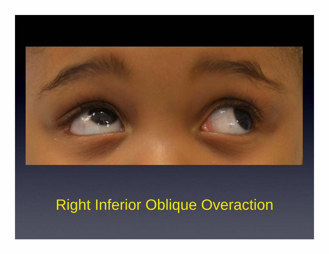
Right Inferior Oblique Overaction

DVD• Slow upward drifting of the nonfixating
eye
• Constant in all positions of gaze - no pattern
• Movement is upward, abduction and excyclotorsion
• Can be unilateral, bilateral, assymetric
• No compensatory hypodeviation in the contralateral eye (violates Herring’s Law)

Accomodative Esotropia• Develops between 6 months and 7 years
• Can appear acutely, or start out intermittent
• Associated with high hyperopic refractive error
• +3.00-+10.00 D (average +4.75 D)
• High incidence of amblyopia
• Distance near deviation similar

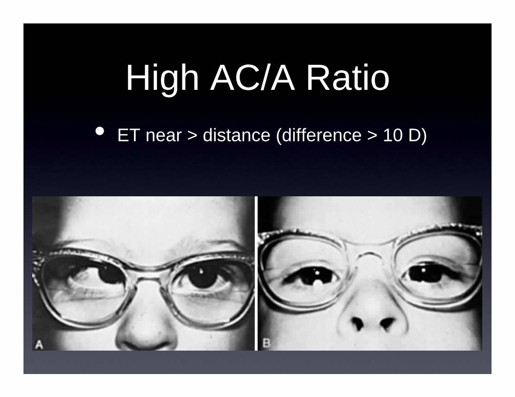
High AC/A Ratio• ET near > distance (difference > 10 D)

Mixed Mechanism and Nonaccomodative
Esotropia• Partially Accomodative
• esotropia only partially controlled by full hyperopic correction
• residual esotropia treated with eye muscle surgery
• Nonaccomodative• acquired, occurring in older children• unknown etiology, nml refractive error• most rule out associated neurologic disease

Duane’s Syndrome
Failure of formation of the abducens nucleus and sixth nerve, innervation of the lateral rectus by an anomalous
branch of the third nerve within the orbit

• Female > Male
• OS > OD > OU
• ET > Ortho > XT
• A-V patterns common
• Anisometropic Amblyopia
• Associations: Goldenhaar Syndrome(limbal dermoid/lipodermoid, preauricular skin tag, superior eyelid coloboma), deafness, crocodile tears (nerve fibers from mandibular and sublingual gland reinnervate the lacrimal gland

• Congenital• rare, well documented• prognosis for recovery good• focal damage to the peripheral nerve,
but nerve still intact• Acquired
• etiologies include: traumatic, increased ICP, meningitis
Congenital or Acquired Cranial Nerve VI Palsy
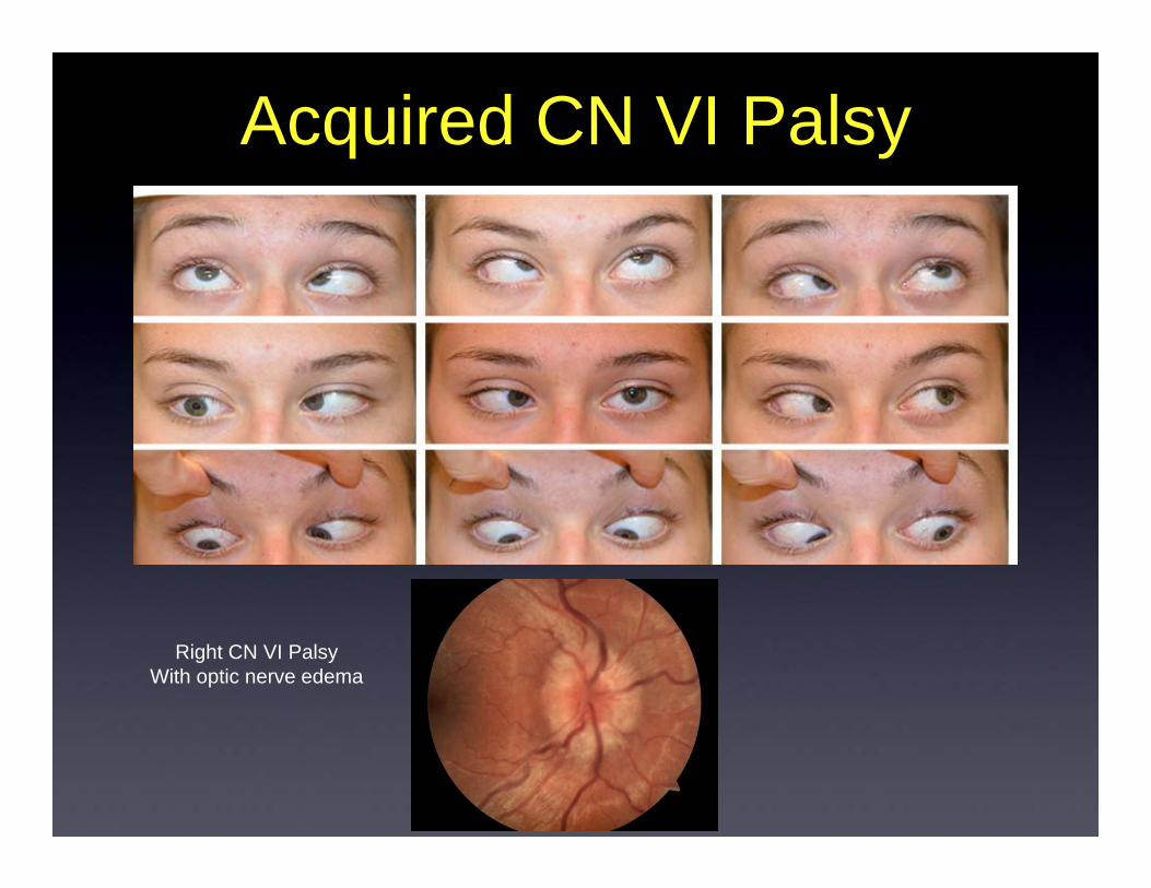
Acquired CN VI Palsy
Right CN VI PalsyWith optic nerve edema

Differentiating CN VI Palsy from Duane Syndrome
Right CN IV Palsy
Left Type 1 Duane Syndrome with Eyelid fissure narrowing in adduction

Less Common Esodeviations
• Nystagmus Blockage Syndrome
• convergence dampens nystagmus, pts fixate with adducted eye, causing head turn to side of deviating eye
• Mobius Syndrome
• CN 6, 7, 9 palsy
• “mask like facies”
• limb, chest and tongue defects
• ET or ortho

Exodeviationsnonfixating eye deviates out
Types of Exodeviations
• Congenital Exotropia• Occurs under the age of 1 year - rare
• Angle of deviation is large, average 35 prism diopters
• Amblyopia is not common
• Similar refractive error to general population
• Early surgical correction indicated
Types of Exodeviations• Sensory exotropia
• Secondary deviation due to poor vision in one eye • Uncorrected refractive error, amblyopia, media opacity,
organic lesion
• Intermittent Exotropia• Onset varies: infancy to 4 years of age• Most common form of exotropia
• Outward drifting of one eye, interspersed with periods of good alignment
• May be progressive - may start out as phoria, progress to intermittent exotropia, and then become manifest exotropia
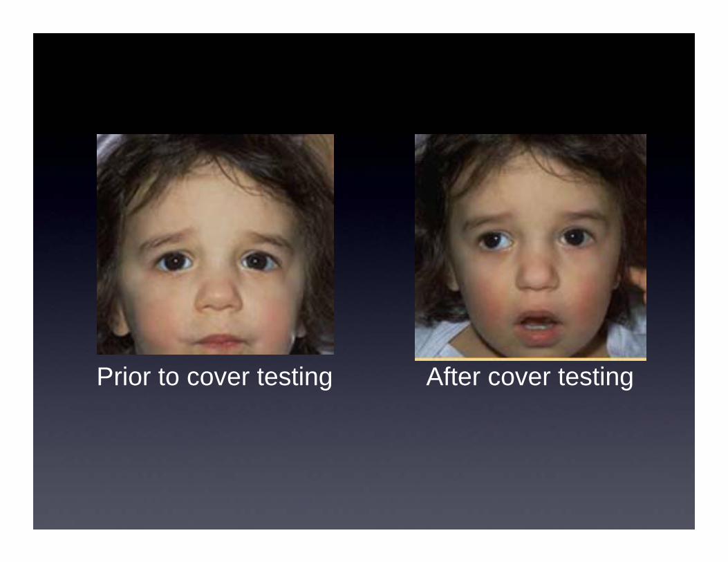
Prior to cover testing After cover testing


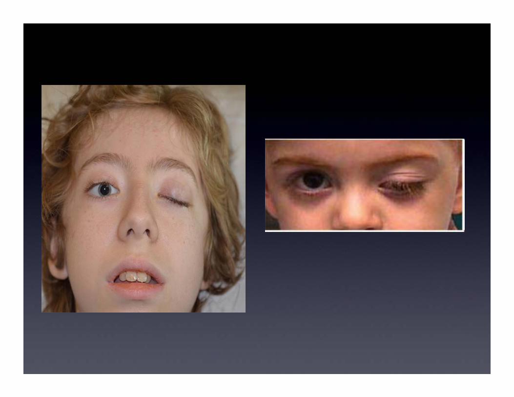
Cranial Nerve III Palsy -congenital and acquired
• Congenital• rare, most commonly unilateral• ptosis, exotropia, hypotropia• smaller pupil on affected eye• midbrain maldevelopment, intrauterine injury,
perinatal damage• 50% have associated neurologic signs
• Acquired• trauma, infection, tumor and vascular
etiologies• damage of nerve as it courses subarachnoid
space

Hypertropias(nonfixating eye elevates)

Congenital or Acquired Cranial Nerve IV Palsy• Most common congenital oculomotor
palsy• Half of all isolated CN IV palsies are
congenital• Most common presentation is with
abnormal head tilt• vertical deviations usually large (av. 18-
20 pd)• large vertical fusional amplitudes, up to
20 D• Hypodevelopment of contralateral face• May decompensate later in life

Congenital CN IV Palsy
• Etiology:• not associated with prenatal disorders
or birth trauma• possible underdevelopment of the
superior oblique tendon, or muscle
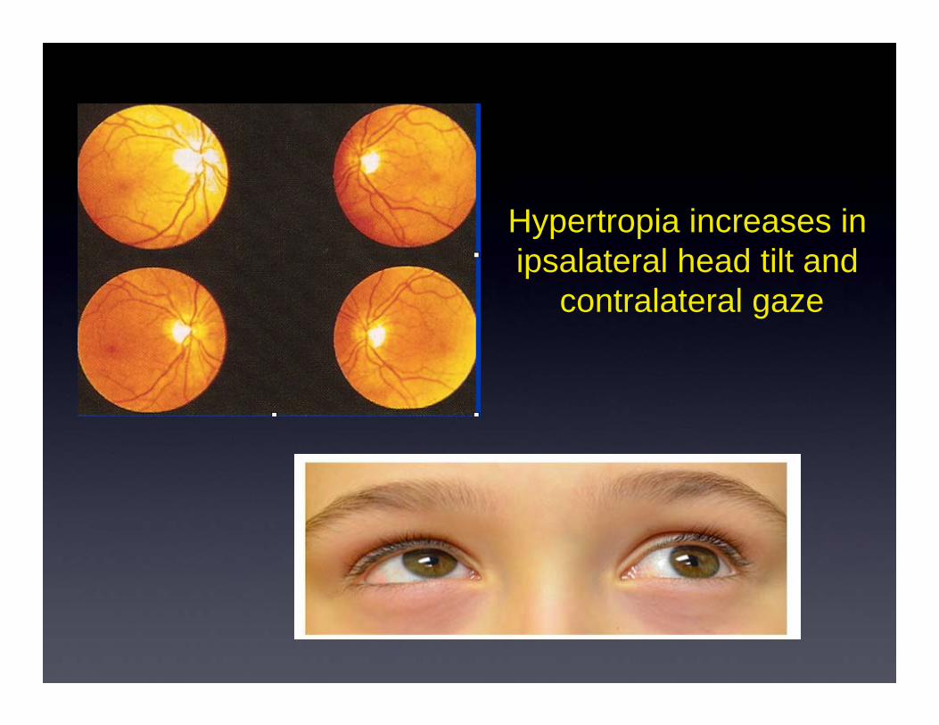
Hypertropia increases in ipsalateral head tilt and
contralateral gaze

Hypotropianonfixating eye deviates downward
Brown Syndrome• Limitation of elevation in adduction
• Relatively normal elevation in abduction
• V pattern exotropia in upgaze
• No overacting SO (distinguishes from IO palsy)
• Etiology: restriction of the SO tendon, or the troclea/tendon complex
• Congenital > Acquired• Acquired: SO tuck, valve or buckle near SO, inflammatory,
infectious, traumatic in the trochlear region

No elevation of the adducted right eye

Monocular Elevation Deficit(formerly known as Double Elevator Palsy)
Paresis of both inferior oblique and superior rectus
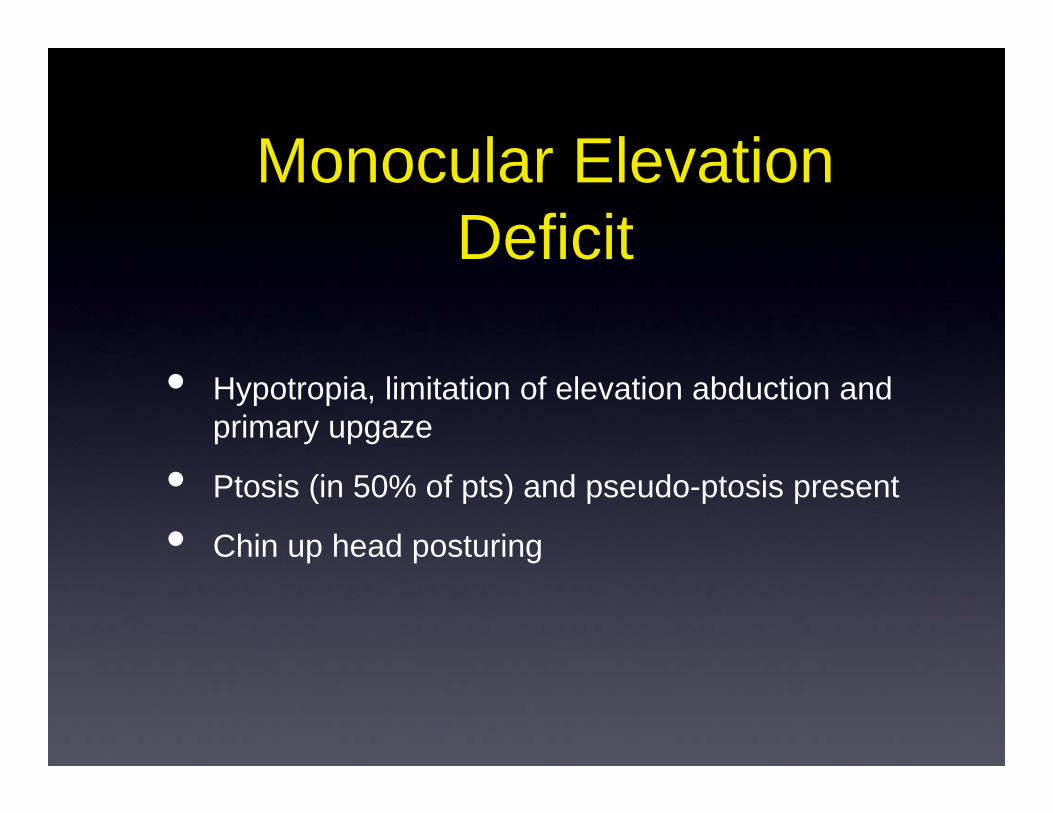
Monocular Elevation Deficit
• Hypotropia, limitation of elevation abduction and primary upgaze
• Ptosis (in 50% of pts) and pseudo-ptosis present
• Chin up head posturing

KNAPP Procedure Transposition of rectus muscles
• If IR tight, recess IR
• If IR not tight, transpose MR and LR to SR (re-create supraduction vector) - KNAPP
• Can be half tendon width or full tendon

Orbital floor fracture with entrapped inferior
rectus• Inferior Rectus becomes incarcerated in
the fracture, or tethered on a bony fragment and restricted in a hypotropic position
• limitation in upgaze, + forced duction test


Strabismus Syndromes

Congenital Fibrosis SyndromeGroup of congenital anomalies with variable restriction of EOM
+ FDT due to restriction
Nonprogressive
Congential fibrosis - most severe, ADcongenital fibrosis of inferior rectusStrabismus fixusVertical retracton syndromeCongenital unilateral fibrosis

Chronic Progressive External Ophthalmoplegia (CPEO)
sporadic, or mitochondrial
may present at any age
severe ptotis with complete ophthalmoplegia, no restriction on FDT, no Bell’s (because not supranuclear)
association: Kearns-Sayre Syndrome


Congenital Myasthenia Gravis
• Presentation from birth to early adulthood - most commonly in first 2 years
• Sporadic or AD inheritance, not autoimmune
• Presentation of bilateral ptosis, facial weakness, variable limitation in eye movements, possible limb weakness, and reduced muscle mass
• May show some response to anti-cholinesterase tx

Juvenile Autoimmune Myasthenia Gravis
• Age of onset < 15 years• Female to male ratio - 3:1• Presentation with ocular MG, 50% progress to
generalized MG• at greater risk than adult disease for
aspiration and ventilator dependency• Associations: Hyperthyroidism, Juvenile onset
DM, Rheumatoid Arthritis• Treatment with anti-cholinesterase drugs,
immunosuppression, plasmapheresis

Craniosynostosis• Premature closure of a cranial suture which
results in deformities of skull shape
• Orbital and ocular extorsion,
–Causes varying patterns of strabismus• MR – adducts and elevates (simulates IOOA, SOUA)
• SR – elevates and abducts
• LR – abducts and depresses
• IR – depresses and adducts

Apert Syndrome
Crouzon Syndrome

Excision of MuscleMarking of inferior and superior pole, checking of excised muscle

Rectus Muscle RecessionMuscle belly is pulled up to position at its new insertion site
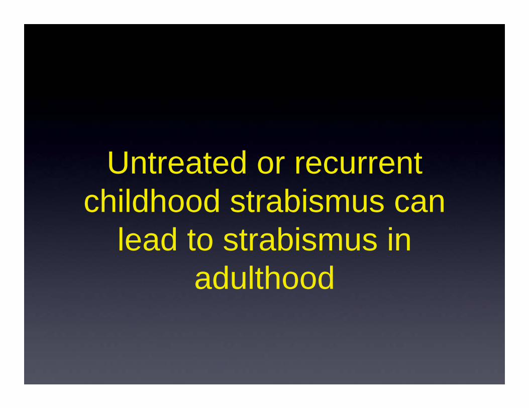
Untreated or recurrent childhood strabismus can
lead to strabismus in adulthood

Adult vs. Pediatric Strabismus
• Onset before visual maturation• Adapt to misalignment with SUPRESSION or abnormal
retinal correspondence (ARC)• No diplopia!• Untreated or recurrent childhood strabismus makes up
majority of adult strabismus patients
• Onset after visual maturation• Onset after age 8-10 years• Cranial nerve palsy, thyroid related eye disease,
traumatic• Misalignment causes diplopia, patients may adopt
abnormal head posture

Why treat adult strabismus?
• Resolution of diplopia• Improved depth perception• Expand peripheral field of vision
• Psychosocial aspects of reconstructed ocular alignment
• Emotional, social and economic benefits
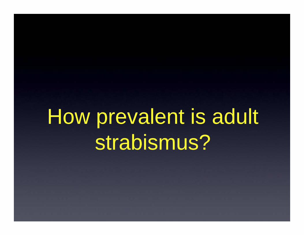
How prevalent is adult strabismus?

2012 review of random Medicare Part B physician claims from 2002-2010 performed to
determine the prevalence of strabismus and strabismus surgery in the
aged medicare fee-for service population.
0.68 % of aged Medicare beneficiaries were diagnosed with strabismus in 2010.
Strabismus surgery was performed on 0.016% of beneficiaries,
or in 2.3% of those patients diagnosed with strabismus.
Why are so few patients undergoing strabismus surgery?
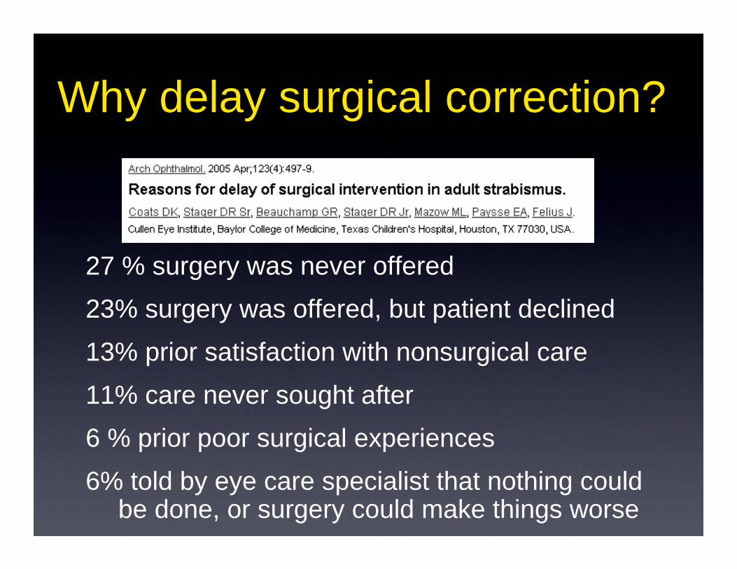
Why delay surgical correction?
27 % surgery was never offered23% surgery was offered, but patient declined13% prior satisfaction with nonsurgical care11% care never sought after6 % prior poor surgical experiences6% told by eye care specialist that nothing could
be done, or surgery could make things worse

Thank youSpecial thank you to Vesna Jurisic Friberg
Ophthalmic Photographer Children’s Hospital of Pittsburgh
References1. Nelson B, Gunton K, Lasker J, Nelson L, Drohan L. The psychosocial aspects of strabismus in teenagers and adults
and the impact of surgical correction. J AAPOS 2008; 12: 72-76)
2. Satterfield D, Keltner JL, Morrison TL. Psychosocial Aspects of Strabismus Study. Arch Ophthalmol. 1993; 111:1100-1105.
3. Coats DK, Stager DRS, Beauchamp GR, et al. Reasons for delay of surgical intervention in ault strabismus. Arch Ophthalmol 2005; 123: 497-9.
4. Repka MX. Strabismus surgery among aged Medicare beneficiaries. J AAPOS 1997; 1: 237-41.
5. Repka MX, Coleman A. Strabismus among aged fee-for-service medicare beneficiaries. J AAPOS 2012; 16: 495-500.
6. Beauchmap G, Felius J, Stager D, Beauchamp C. The Utility of Strabismus in Adults. Trans Am Ophthalmol Soc 2005; 103: 164-172.
7. Greenberg AE, Mohney BG, Diehl NN, Burke JP. Incidence and Types of Childhood Esotopia. Ophthalmology 2007; 114:170-174.
8. Friedman DS, Repka MX, Katz J, Giordano L, Ibironke J, Hawse P, Tielsch JM. Prevalence of Amlbyopia and Strabismus in White and African American Children Aged 6 thought 71 months . Ophthalmology 2009; 116:2128-2134.
• Multi-Ethnic Pediatric Eye Disease Study Group. Prevalence of Amlbyopia and Strabismus in African American and Hispanic Children Ages 7 to 72 Months. Ophthalmology 2008; 115: 1229-1236.
1. Donahue, SP. Pediatric Strabismus. N Eng J Med 2007; 356:1040-7.
2. Robaei D, Roase K, Ojaimi E, Kifley A, Huynk S, Mitchel P. Visual Acuity and the Causes of Visual Loss in a Population -Based Sample of 6 year Old Ausralian Children. Ophthalmology 2005; 112: 1275-1282.
3. Graham, PA. Epidemiology of strabismus. Brit.J.Ophthal. 1974;58, 224.
4. Kvarnstom G, Jakobsson P, Lennerstrand G. Visual screening of Swedish children: An ophthalmolgical evaluation. Acta Ophthalmol. Scand. 2001: 79; 240-244.
5. Taylor D and Hoyt G. Pediatric Ophthalmology and Strabismus - Third Edition. Elsevier Saunders, 2005.