pediatric pearls a to b - cascademedical.org · secretions, ngt placement, nasal congestion ....
TRANSCRIPT

Pediatric Pearls A to B
Brian Rogge, RN, EMT-P Life Flight Network

A Pediatric Airway
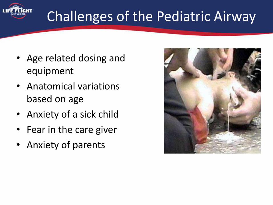
Challenges of the Pediatric Airway
• Age related dosing and equipment
• Anatomical variations based on age
• Anxiety of a sick child
• Fear in the care giver
• Anxiety of parents

Pediatric Airway Anatomy
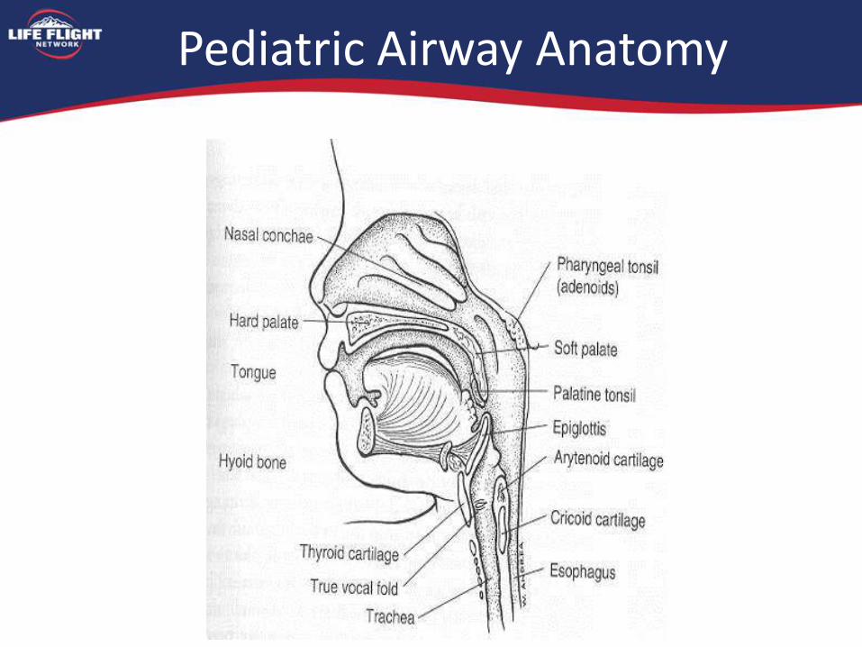
Pediatric Airway Anatomy
Tongue
• Potential site of airway obstruction
– Difficult ventilation
– Loss of tone with sleep, sedation or CNS dysfunction
– Posterior displacement of the tongue may cause severe airway obstruction

Occiput
• A child’s head/occiput are proportionately larger than an adult’s
• Neck flexion while supine – Leads to obstruction
• Overcome with the sniffing position – Roll placed under back
(infant) for BVM
– None – small child
– Roll placed under occiput in older children
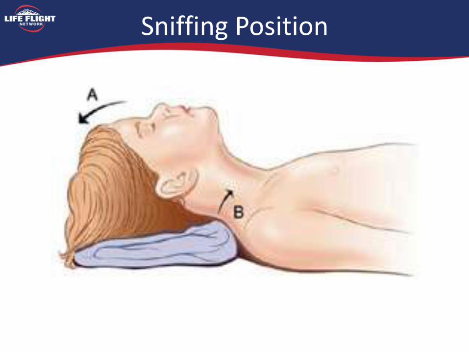
Sniffing Position

Nasal Passage
• Increased mucosa and lymphoid tissue
• Nasal airway is primary pathway for normal breathing in the infant
– Warming, humidification, particle filtration
• Compromised breathing with increased secretions, NGT placement, nasal congestion

Larynx
• Newborns
– Larynx at the base of the occiput/C1 to C4
• Enables epiglottis to lock the larynx into the nasopharynx by passing up behind the soft palate
• Provides a direct air channel from the nares to the lungs, allowing liquids to pass on the sides into the esophagus

Larynx
• Two separate anatomic pathways – Respiratory tract from the nose to the lungs
– Digestive tract from the mouth to the stomach
• Large Tongue – Entirely within the oral cavity
• High Glottis
• Difficult line of vision from mouth to the larynx during laryngoscopy – Anterior Airway

Anatomic Changes in Childhood
• Occurs after the second year of life
• Posterior 1/3 of tongue descends into the neck, forming upper anterior pharyngeal wall
• By 7 years, the larynx lies between C3 and C6
• In adulthood, the larynx lies between C4 – C7

Anatomy
• In adults, the vocal cords and trachea are of equal dimensions
• In newborns, the narrowest portion of the airway is the cricoid ring
– Tight ET tubes may lead to cricoid damage, subglottic stenosis (more on this later)

Preoxygenate
• 5 minutes of 100% oxygen or 8 deep breaths over 60 seconds
– Time before sats < 90%
• 70 kg pt -- 8 minutes
• 120 kg pt -- 3 minutes
• 10 kg child -- 4 minutes
• Time for sats 90% 0%
• 70 kg pt -- 120 seconds
• 10 kg child -- 45 seconds
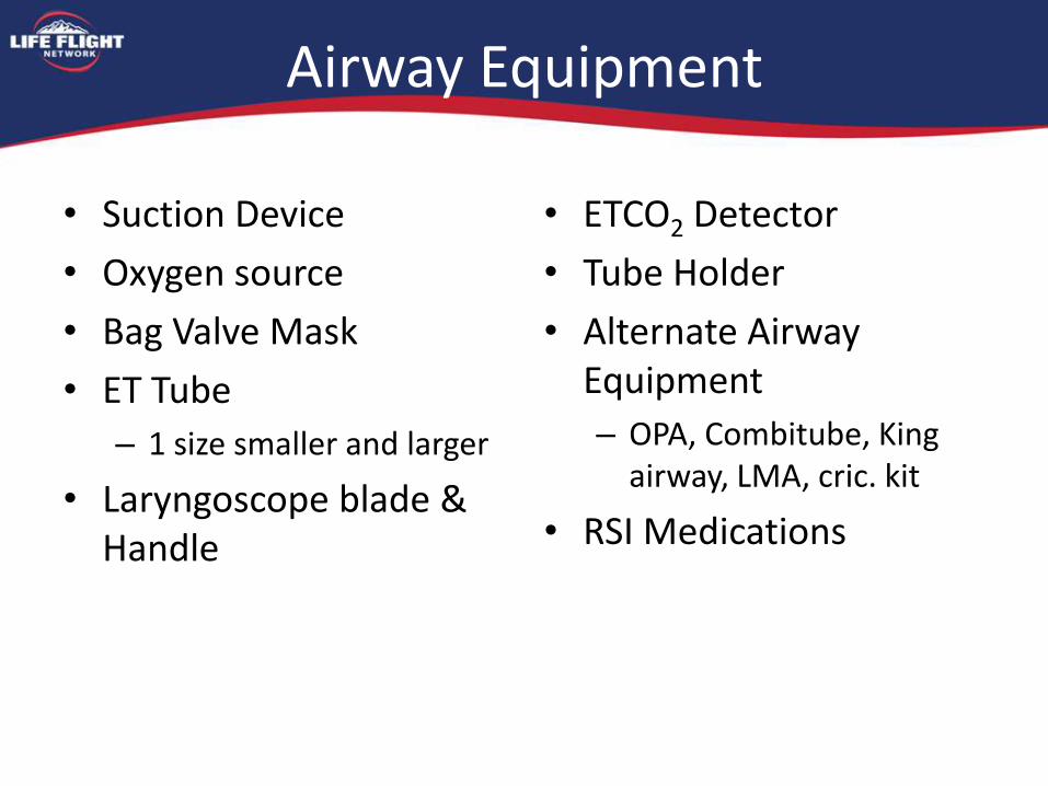
Airway Equipment
• Suction Device
• Oxygen source
• Bag Valve Mask
• ET Tube
– 1 size smaller and larger
• Laryngoscope blade & Handle
• ETCO2 Detector
• Tube Holder
• Alternate Airway Equipment
– OPA, Combitube, King airway, LMA, cric. kit
• RSI Medications
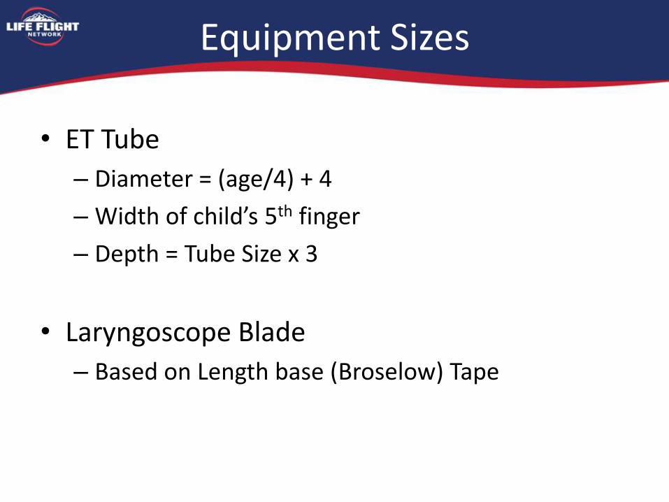
Equipment Sizes
• ET Tube
– Diameter = (age/4) + 4
– Width of child’s 5th finger
– Depth = Tube Size x 3
• Laryngoscope Blade
– Based on Length base (Broselow) Tape
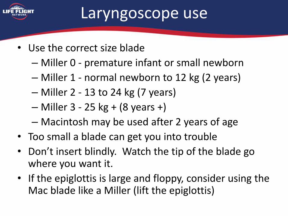
Laryngoscope use
• Use the correct size blade
– Miller 0 - premature infant or small newborn
– Miller 1 - normal newborn to 12 kg (2 years)
– Miller 2 - 13 to 24 kg (7 years)
– Miller 3 - 25 kg + (8 years +)
– Macintosh may be used after 2 years of age
• Too small a blade can get you into trouble
• Don’t insert blindly. Watch the tip of the blade go where you want it.
• If the epiglottis is large and floppy, consider using the Mac blade like a Miller (lift the epiglottis)

Laryngoscope use
• Spend an extra 3-5 seconds sweeping the tongue completely out of the way.
• If you don’t do this, the laryngoscope displaces the tongue posteriorly, and it occludes the view
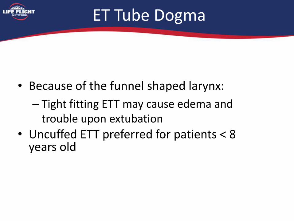
ET Tube Dogma
• Because of the funnel shaped larynx:
– Tight fitting ETT may cause edema and trouble upon extubation
• Uncuffed ETT preferred for patients < 8 years old

What PALS says
• Cuffed tubes may be preferred in certain circumstances …poor lung compliance, high airway resistance, or large glottic air leak - really any sick kid
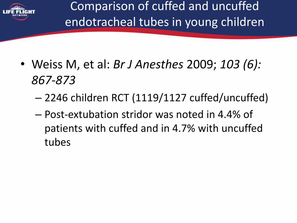
Comparison of cuffed and uncuffed endotracheal tubes in young children
• Weiss M, et al: Br J Anesthes 2009; 103 (6): 867-873
– 2246 children RCT (1119/1127 cuffed/uncuffed)
– Post-extubation stridor was noted in 4.4% of patients with cuffed and in 4.7% with uncuffed tubes

Cuffed vs. uncuffed endotracheal tubes
• Using the standard formula for tube size (age/4) + 4, uncuffed tubes were incorrectly sized in 23% of cases.
• In another study, tube changes due to significant air leaks occurred in 28-30% of patients with uncuffed tubes.
• These problems did not occur with cuffed tubes.

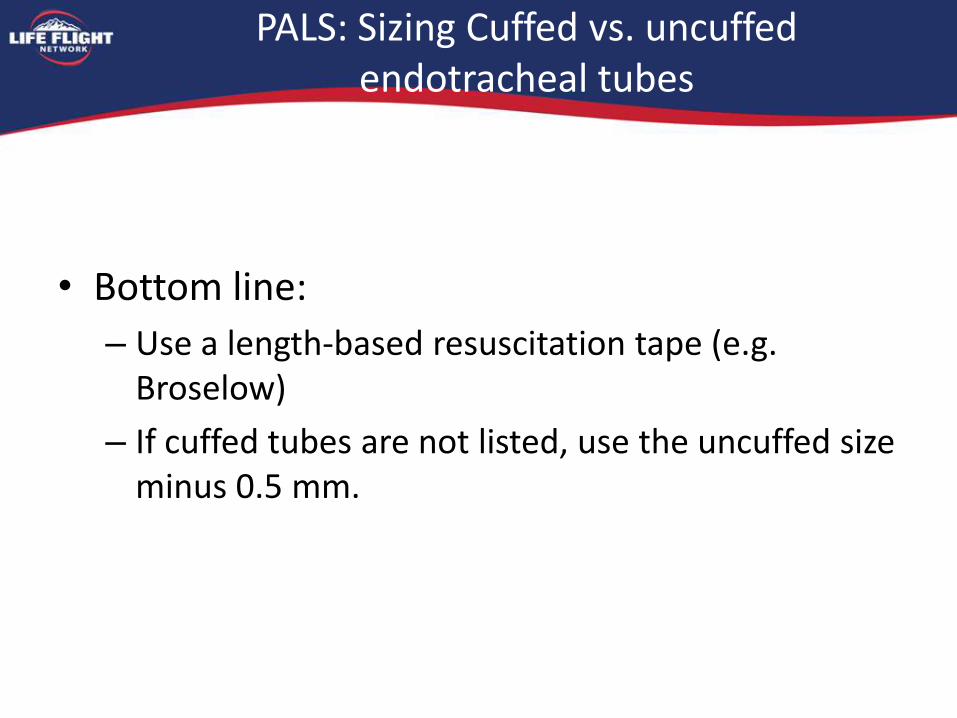
PALS: Sizing Cuffed vs. uncuffed endotracheal tubes
• Uncuffed
– < 1 year old: 3.5 mm ET tube
– 1-2 year old: 4.0 mm ET tube
– > age 2: (yrs/4) + 4 = mm ET tube
• Cuffed
– < 1 year old: 3.0 mm ET tube
– 1-2 year old: 3.5 mm ET tube
– > age 2: (yrs/4) + 3.5 = mm ET tube
PALS: Sizing Cuffed vs. uncuffed endotracheal tubes
• Bottom line:
– Use a length-based resuscitation tape (e.g. Broselow)
– If cuffed tubes are not listed, use the uncuffed size minus 0.5 mm.

Post Intubation Management
• Verification of Tube Placement
– Visualization
– ETC02
– Auscultation
• Secure the tube with tape or commercial device
– Head/neck immobilization in small children to avoid neck movement and dislodgement

Other Airways
• Combitube®
– Small Adult is smallest size
– Must be 4 foot tall.
• King airway
– Small Pediatric sizes available now
• i-Gels
– 4 pediatric sizes
– Smallest is 2-5kg
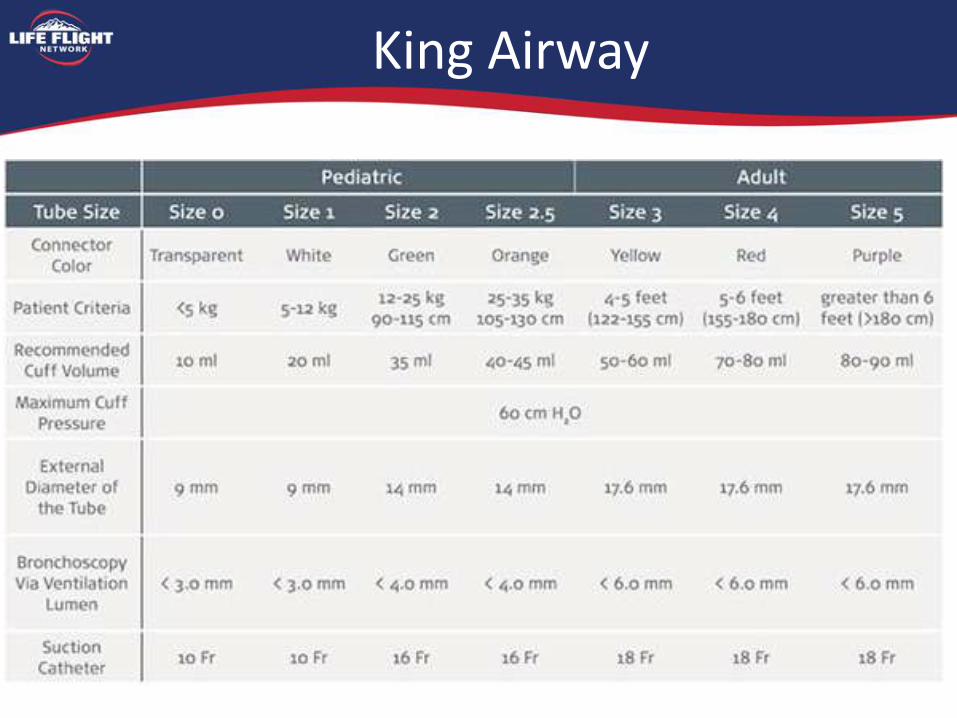
King Airway
B Pediatric Breathing (and ventilation)

Hypoxia
• Hypoxia in Children
– First sign: Anxiety/ Fear/ Irritable
– Second sign: Lack of engagement
– Third sign: Bradycardia
– Fourth sign: Loss of consciousness
– Develops quickly in children
• Higher metabolic rate increases consumption
• Minimal reserve capacity
Signs of Respiratory Distress
• Rapid breathing
• Grunting
• Inability to lie down
• Agitation
• Accessory muscle usage
• Retractions
• Tachycardia
• Apnea

Basic Airway
• Positioning – Jaw thrust vs. Chin lift
– Oral airway • Sizing
• Insertion techniques
• Contraindications
– Nasal airway • Sizing
• Insertion techniques
• Contraindications
Bag Valve Mask Ventilation
• Must fit over the nose, cheeks, mouth, and chin
• Place in sniffing position
– In line stabilization
– Jaw thrust
• OPA – from corner of mouth to angle of jaw
• NPA – from nares to tragus of ear
• Inspect for foreign body
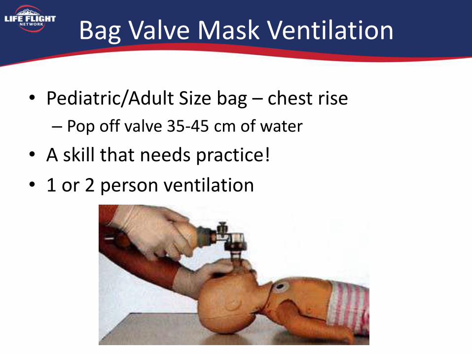
Bag Valve Mask Ventilation
• Pediatric/Adult Size bag – chest rise
– Pop off valve 35-45 cm of water
• A skill that needs practice!
• 1 or 2 person ventilation

Reasons to Intubate
• Failure to Oxygenate – low SpO2
• Failure to Ventilate – high CO2
• Expected Clinical Course

Primum non nocere….
• You can cause harm or death with intubation and a ventilator.
– Doesn’t mean you shouldn’t use the tools
– Why you are here today
– Practice doesn’t make perfect, but it helps
Rate Matters
• Look at the patient before you intubate
– Rate
– Effort
– Why are they in tachypneic
• Hint: it may have nothing to do with airway or lungs.
• Minute Ventilation (?)
• Disease Process
• Metabolic demands
I:E Ratio
• Inspiratory Rate can be too fast
• Expiratory phase can be too short
• Anatomy versus disease process
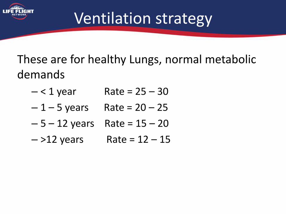
Ventilation strategy
These are for healthy Lungs, normal metabolic demands
– < 1 year Rate = 25 – 30
– 1 – 5 years Rate = 20 – 25
– 5 – 12 years Rate = 15 – 20
– >12 years Rate = 12 – 15

Ventilation strategy
• Tidal Volume should be appropriate – Be aware of Peak inspiratory pressure, but not afraid
of it.
– Ok if needed up to 25-30 cm/H2O
– Healthy lungs 16-20 cm/H20
• Remember PEEP. Start with 5 cm/H2O – If you are bagging place a peep valve
– Asthma patients are the exception that may do better without peep.
Asthma Patients
• Very high threshold to intubate
• Peep may not be beneficial
• Lower Respiratory Rates
• Very long expiratory phase
• May require high peak inspiratory Pressures
• Measure Pplat

Asthma Patients
• The recommended method to monitor patients for hyperinflation and injurious airway pressures is Pplat, the average end-inspiratory alveolar pressure.
• Pplat is measured using an end-inspiratory pause. Values > 30 cm H2O indicate hyperinflation and excessive airway pressures.
• Of note, peak airway pressure (Ppeak) measurements do not correlate with patient outcomes and therefore are not useful for assessing hyperinflation. – Ventilator Management of the Intubated Patient With Asthma, Michael E. Winters, MD
December 13, 2010

Slide Title

Thank You