pediatric ocular diseases
TRANSCRIPT

Pediatric ocular diseases with A systemic approach
Presenter : Surendra Prasad SahM.Optom
(10/11/2014)

Alport’s syndrome Hereditary nephritis syndrome
Caused by a genetic defect in the type iv collagen
Worsen as renal function deteriorates
Males and females are equally affected
Hearing loss in patients sensorineural hearing loss
Tubular atrophy , interstitial inflammation and fibrosis and foam cell developed ,if the disease progress

Ocular Clinical features Bilateral anterior lenticonus and perimacular retinal flecks
Thinned basal lamina with basement membrane disruptions
Spontaneous and traumatic ruptures of the anterior lens capsule
Posterior lenticonus
Perimacular yellow flecks
Corneal finding include posterior polymorphous dystrophy , corneal arcus and recurrent nontraumatic corneal erosions

Cont…

Cont…

Systemic features
Thrombocytopenia
Macrothromocytopathia
Hypoparathyroidism
Polyneuropathy
Ichthyosis
Thyroid abnormalities

Treatment
There is no effective treatment
Renal plantation is usually very successful
Dual sensory loss in patients creates an urgent need for appropriate vision and care and rehabilitation

Alstrom syndrome
A syndrome of retinal degeneration combined with obesity ,diabetes mellitus and sensorineural hearing loss

Ocular clinical features
Posterior subcapsular cataracts
Iris and cilary body lacy vacuolization and asteriod hyalosis
Retinitis pigmentosa and large, superficial optic nerve drusen are found
Infantile cone and rod retinal dystrophy

Cont…

Treatment
There is no known treatment for vision loss secondary
Management of the endocrine ,cardiac and kidney disease is critical ,although a shortened lifespan is expected

Prognosis
There is a rapid and progressive loss of visual function to less then 20/200 by 10 years of age and no light perception

CHARGE association syndrome
Choanal atresia is present in about 58% of patients
Growth deficiency is usually postnatal and affects all the body in a symmetrical fashion
Mental development is affected
Genital hypoplasia is more prone to recognized

Ocular clinical features Iris colobomas , posterior colobomas of choroid or optic
nerve with no visual impairments
Microphthalmia
Optic nerve hypoplasia
Persistent hypoplastic primary vitreous
Congenital glaucoma
Bilateral Marcus-Gunn jaw winking phenomenon

Cont..
Individuals with CHARGE syndrome who survive the initial neonatal and infantile period merit vigorous rehabilitation of the sensory function to enable proper psychomotor development.
Nasogastric feeding is indicated in individuals with swallowing difficulty.
In the presence of facial palsy, avoid corneal scarring by using artificial tears.
In males with CHARGE syndrome, androgen therapy has been tried for penile growth.

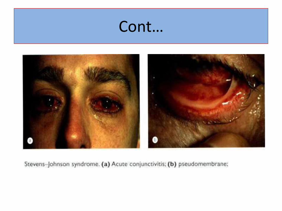
Stevens –johnson syndrome
To severe form of erythema multiform with extensive skin and mucous membrane involvement along with fever and affection of the kidneys, gastrointestinal tract and central nervous system
ocular involvement is much less common in toxic epidermal necrolysis
Males are affected more than females

Ocular and clinical features
Crusty eyelids associated with a transient, self-limiting papillary conjunctivitis
Severe membranous or pseudo membranous conjunctivitis with patchy Conjunctival infarction is less common
Reticular scarring of the upper tarsal plate
Conjunctival keratinization and forniceal shortening
Cont…

Cont….
Dry eye resulting from loss of the goblet cells and destruction of lacrimal gland ductules
Corneal keratinization
Keratopathy secondary to cicatricial entropion, aberrant lashes and infection
Posterior lid margin disease with opening of meibomian orifices onto the ocular surface
Conjunctival scarring and symblepharon formation

Cont..
Treatment
Systemic disease is treated by maintaining hydration
debridement and replacement of sloughing skin.
Systemic steroids are occasionally prescribed
Lubrication and prevention of exposure
Topical steroids and antibiotics
Lysis of Conjunctival adhesions

Cont…..
A scleral ring, consisting of a large haptic lens with the central zone removed may prevent symblepharon formation
Lubrication and punctual occlusion for mild disease
Topical retinoic acid for keratinization
Lubrication and punctual occlusion for mild disease
Topical retinoic acid for keratinization

Cont..
Bandage contact lenses to maintain surface moisture and overcome irregular astigmatism. Gas permeable scleral contact lens for trichiasis and visual rehabilitation
Mucous membrane grafting and limbal cell transplantation
Lamellar corneal grafting is preferred to penetrating keratoplasty
Keratoprosthesis is end stage disease.

Sturge-weber syndrome
Sws is a neurocutaneous disorder characterized by cutaneous facial angiomas, liptomeningeal angiomas, and siezures and other neurological complications, including mental retardation and glaucoma.

Ocular Clinical features
1. Orbital
General:-
Exophthalmos
Lids
Ptosis
Port-wine stain of eyelid
2. Extraocular
Sclera
Nevoid marks or vascular dilation of the episclera
Telangiectasia of the episclera

Cont..

Contd..
Large, anomalous vessels in the conjunctiva
Oculocutaneous melanosis
3. Intraocular:-
Anterior segment
Increased corneal diameter
Reduced corneal reflex
Iris discoloration
Telangiectasia of the iris with heterochromia
Dilation and tortuosity of iris vessels

Contd..
Sluggish pupils
Anisocoria or other disturbances in pupil reaction
Coloboma of the iris
Deep anterior chamber angle
Buphthalmos
Media
Ectopia lentis
Spontaneous dislocation of the lenses

Contd..
ChoroidChoroidal angiomataChoroidal hemangioma
RetinaDilation and tortuosity of retinal vesselsRetinal arteriovenous aneurysmVaricosity of retinal veinsRetinitis pigmentosaRetinal detachmentCentral retinal vein occlusion

Contd..
Optic nerve
Arteriovenous angiomas
Papilledema
Optic atrophy
Optic nerve cupping
Optic nerve coloboma
Optic nerve drusen

Cont…

Cont….
4. Other
Strabismus
Nystagmus
Loss of vision (any degree)
Cortical blindness
Cortical blindness
Abnormal visual field caused by lesion in optic tract
Hemianopia
Secondary glaucoma (late)
Anisometropia

Treatment
Surgical resection with split- thickness skin grafting
Laser therapy
Drug therapy
Laser photocoagulation
Marfan’s syndrome
Characterized by the presence of abnormalities of the eye, aorta and skeleton.
About 355 of patients, do not develop lens subluxation
Cause due to mutation of the fibrill in gene on chromosome.

Ocular clinical features
Subluxation of the crystalline lens
Myopia, microcornea, keratoconus and occasionally retinal detachment and glaucoma.
Stretched zonular fibers can be seen through the dilated pupil.
Coloboma of the lens
Microspherophakia
iridodonesis results from lens subluxation
strabismus

Cont…

Cont….

Down syndrome
Systemic features :- Mental handicap
Upward slanting palpebral fissures
Epicanthic folds
Broad short hands and a protruding tongue
Ocular features :- Cataract of varying morphology occurs in about 75% of
patients .The opacities are usually symmetrical and often develop in late childhood

Cont…

Cat scratch disease
Self-limited illness caused by the fastidious gram –negative bacillus
Transmitted via the scratch or bite of a cat or kitten
Characterized by lymphadenopathy of local lymph nodes drainining the site of infection

0cular clinical features
VA is impaired to a variable degree
Papillitis associated with peripapillary and macular oedema
A macular star composed of hard exudates
After several months VA improve
The fellow eye occasionally becomes involved but recurrences in the same eye are uncommon

Cont…

Cont…..
Parinaud oculoglandular syndrome
Focal choroiditis
Intermediate uveitis
Exudative maculopathy
Retinal vascular occlusion
panuveitis

Treatment
The most effective antbiotics are rifampin ,ciprofloxacin,intramuscular Gentamicin and Trimethoprim -sulfamethoxazole

Herpes simplex virus
A double –stranded DNA virus
HSV-1 is most commonly found in lesion of oral ,the eye and on skin
HSV-2 is found in lesion on the genitalia and the skin of the thighs
Transmitted during periods of asymptomatic shedding of virus by infected persons

Ocular clinical features
Conjunctivitis ,karatitis and chorioretinitis
Lateral blepharoconjunctivitis
Follicular conjunctivitis
Recurrent HSV karatitis such as stromal scar formation and induced astigmatism may lead to amblyopia

Cont…

Cont..

Cont..

Treatment
For systemic or posterior ocular disease ,the mainstay of therapy is intravenous acyclovir
Conjunctival or corneal involvement should be managed with the use of topical trifluridine 1%
Epithelial keratis is treated with topical antiviral therapy ,trifluridine solution every 2h or vidarabine

Rubella
Acute viral infection of children
Characterized by low-grade fever ,rash and lymphadenopathy
Infection during pregnancy can result in fetal infection with severe congenital defects
Transmission occurs by droplets from respiratory secretions

Ocular clinical features
Conjunctivitis ,karatitis and rarely retinitis
Retinitis occurs rarely and presents with atrophy ,hypoplasia of the ciliary body and hypoplasia of the iris dilator muscle
The characteristic “salt-and –pepper “retinopathy typically appears in the macula and periphery of both eye
Cataract ,microphthalmia ,glaucoma ,anterior uveitis and corneal haze

Cont…

Treatments
Cataract surgery can be performed

Varicella –Zoster
Resulting from primary infection with varicella-zoster virus(VHV)
Spread by airborne droplets and direct contact with infected lesions
Congenital varicella results from transplacental spread of the virus by an infected mother
Ocular clinical features
Papillary conjunctivitis and Conjunctival vesicle formation
Epithelial karatitis or mild nongranulomatous anterior uveitis
Internuclear ophthalmoplegia and oculomotor palsy
Cranial nerve palsies

Cont……
Dermatitis of eyelids leads to secondary bacterial infections
Follicular conjunctivitis ,episcleritis and scleritis
Elevated intraocular pressure
Optic neuritis and the retinal necrosis

Cont…

Treatment
Anterior uveitis 0r stromal karatitis with decreased VA may require therapy with topical corticosteroids
Systemic acyclovir
The cutaneous lesion can be treated with moist compress and antibiotic ointments

References
Clinical ophthalmology ,Jack J Kanski(sixth edition )
Handbook of pediatric eye and systemic disease ,Kenneth W. Wright , Peter H .Spiegel ,Lisa H. Thomp
Pediatric ophthalmology ,Dr. P.K. Mukherjee
Pediatric ophthalmology ,M. Edward Wilson ,Rupal H. Trivedi