pediatric lung isolation techniques
TRANSCRIPT

University of North DakotaUND Scholarly Commons
Nursing Capstones Department of Nursing
7-18-2018
Pediatric Lung Isolation TechniquesKasey J. Trontvet
Follow this and additional works at: https://commons.und.edu/nurs-capstones
This Independent Study is brought to you for free and open access by the Department of Nursing at UND Scholarly Commons. It has been accepted forinclusion in Nursing Capstones by an authorized administrator of UND Scholarly Commons. For more information, please [email protected].
Recommended CitationTrontvet, Kasey J., "Pediatric Lung Isolation Techniques" (2018). Nursing Capstones. 210.https://commons.und.edu/nurs-capstones/210

Running head: Pediatric Lung Isolation Techniques
PEDIATRIC LUNG ISOLATION TECHNIQUES
by
Kasey J. Trontvet
Bachelor of Science in Nursing, University of North Dakota, 2012
An Independent Study
Submitted to the Graduate Faculty
of the
University of North Dakota
in partial fulfillment of the requirements
for the degree of
Master of Science
Grand Forks, North Dakota
December 2018
Pediatric Lung Isolation Techniques 2
PERMISSION
Title PEDIATRIC LUNG ISOLATION TECHNIQUES
Department Nursing
Degree Master of Science
In presenting this independent study in partial fulfillment of the requirements for a graduate
degree from the University of North Dakota, I agree that the College of Nursing and Professional
Disciplines of this University shall make it freely available for inspection. I further agree that
permission for extensive copying or electronic access for scholarly purposes may be granted by
the professor who supervised my independent study work or, in her absence, by the chairperson
of the department or the dean of the School of Graduate Studies. It is understood that any
copying or publication or other use of this independent study or part thereof for financial gain
shall not be allowed without my written permission. It is also understood that due recognition
shall be given to me and to the University of North Dakota in any scholarly use which may be
made of any material in my independent study.
Signature____________________
Date________________________

Pediatric Lung Isolation Techniques 3
Abstract
Title: Pediatric Lung Isolation Techniques
Background: Anesthetic care of pediatric patients during thoracic procedures proves to be quite
difficult due to anatomical and physiological challenges and limited equipment availability.
Purpose: The purpose of this literature search is to provide a review of literature regarding
perioperative lung isolation techniques and clinical management, particularly in the pediatric
population.
Process: A literature review was conducting using the Cochrane Library, PubMed, and CINAHL
databases, which were accessed through the University of North Dakota’s Harley E. French
Library of the Health Sciences. Other relevant literature was found through a search of reference
lists of the acquired articles. All referenced material was closely evaluated for accuracy.
Results: Upon review of available literature, pediatric lung isolation is best accomplished
through age specific methods. Selective mainstem intubation is reserved for emergencies and
children under six months of age. Endobronchial blockers are the preferred technique for
children between six months and six years old. The Univent tube has been shown to be ideal for
six to eight-year-old patients. Lastly, double-lumen endobronchial tubes are limited to children
greater than eight years of age and/or 30 kg.
Implications: Anesthesia providers may utilize suboptimal equipment and techniques when
providing perioperative care for patients requiring lung isolation for thoracic procedures. This is
especially true in a more difficult pediatric population with limited airway equipment availability
due to small size.
Keywords: Lung Isolation, Pediatrics, Anesthesia, Single lung ventilation, One-lung ventilation

Pediatric Lung Isolation Techniques 4
Pediatric Lung Isolation Techniques
Anesthetic care of children during thoracic surgery requires extensive knowledge of both
pediatric and thoracic anesthetic techniques. While a variety of techniques may be used for
thoracoscopic surgery, more specialized techniques are required for smaller children less than 30
kilograms (kg). Older children greater than eight years of age or larger than 30 kg, may often be
managed using typical adult techniques. Standard methods to attain lung isolation in the general
population include intubation with a double lumen endobronchial tube (DLT) or placement of
endobronchial blockers. However, selective mainstem intubation with a single-lumen
endotracheal or endobronchial tube is also a strategy that may be used in emergent situations
and/or with pediatric patients (Purohit, Bhargava, Mangal, & Parashar, 2015). While a variety of
approaches to one-lung ventilation exist, there are many advantages and disadvantages to each.
In order to find the ideal method for lung isolation in each patient, it is important to
consider a variety of factors including the following: indication for lung isolation, anatomy of the
upper and lower airway, availability of airway and visualization equipment, and the anesthesia
provider’s proficiency level with each technique (Collins, Titus, Campos, & Blank, 2017)
Purpose
The purpose of this independent project is to discuss lung isolation strategies for pediatric
patients. A case report is described concerning a pediatric patient undergoing a left lower lung
lobectomy, requiring lung isolation and alternative ventilatory strategies.
Case Report
A seven-year-old, 27.9 kg, 122 cm male was admitted to the Pediatric Intensive Care
Unit (PICU) with non-neutropenic fever and hypoxia secondary to left lower lobe pneumonia.
Past medical history was significant for recent acute lymphoblastic leukemia, encephalopathy,
Pediatric Lung Isolation Techniques 5
seizures, methotrexate toxicity, and acute renal failure (ARF). Past surgical history was limited
to the above abscess drainages. No significant allergies were noted.
On admission, the patient was aggressively treated with intravenous (IV) antibiotics and
subsequently developed necrosis of a part of the left lower lobe (LLL), in addition to a pleural
effusion and fluid collection. Patient underwent interventional radiology (IR) drainage of the
lung abscess and effusion, achieving good expansion of the left lower lobe. Upon a follow-up
computed tomography (CT) scan, there was again evidence of fluid collection within the left
lower lobe. This particular fluid collection was treated with broad spectrum antibiotics in hopes
of resolution, however, was resistant to antibiotics. Upon attempted IR drainage of the fluid,
there was no significant cavity decompression due to the thick nature of the fluid. The decision
was made to pursue surgical correction of the left lower lobe issue, with a plan to undergo a
wedge resection of the area in an attempt to avoid lobectomy. A video assisted thoracoscopic
surgical (VATS) approach was designated, with open thoracotomy as a secondary plan.
Pre-operatively the patient was assigned an American Society of Anesthesiologists
(ASA) physical status classification of III. Upon assessing the patient’s airway, the soft palate,
uvula, and faucial pillars were easily visualized and the patient was given a class 1 Mallampati
score. Pre-operative vital signs included: blood pressure 95/35, pulse 115, respirations 20,
temperature 36.8 degrees Celsius, and oxygen saturation 98%.
The patient was brought to the operating suite, helped into a supine position on the OR
table, and standard monitors were applied, including a non-invasive blood pressure cuff, 5-lead
EKG, and pulse oximetry. He was pre-oxygenated with 100% oxygen via mask and sevoflurane
was slowly added for a smooth and cooperative inhalational induction. Upon achieving an
adequate depth of anesthesia, the anesthesia team started an 18-gauge IV line in the left forearm
Pediatric Lung Isolation Techniques 6
and proceeded to administer 30 mcg of Fentanyl, 20 mg Rocuronium, and 2.5 mg Decadron.
Direct laryngoscopy was performed utilizing a Miller 2 blade and a size 6 mm endotracheal tube
(ETT). A grade 1 view was attained and the ETT was advanced to a depth of 16 cm. ETT
placement was confirmed with symmetrical chest rise, positive end-tidal CO2 (ETCO2), and
bilateral breath sounds. It was decided to utilize a bronchial blocker in the left main bronchus to
isolate the left lung and maintain ventilation to the right lung. A wire-guided endobronchial
blocker (Arndt blocker) was advanced through the ETT, coupled with a small diameter fiberoptic
bronchoscope (FOB) via guide loop, to assist with placement. Correct placement of the bronchial
blocker was visually confirmed with the FOB. Auscultation of lung sounds revealed absence of
air movement to the left lung. Following lung isolation, an arterial line was placed in the right
radial artery under sterile conditions. The patient was then positioned into the right side lateral
decubitus position, utilizing a positioning sand bag.
The pediatric surgeon proceeded with the VATS approach to wedge resection of the left
lower lobe. He was forced to convert to an open lobectomy procedure due to an inability to
adequately visualize the surgical field. During open lobectomy, the patient became hypotensive
and acidotic over the course of multiple hours. In addition to 450 mL Lactated Ringers, the
patient received 60 mL 5% Albumin and 2 units (700 mL) of packed red blood cells (PRBCs) in
an attempt to replace 400 mL of blood loss and ongoing insensible loss. These interventions
improved hypotension and acid/base balance, leading to stability throughout the final minutes of
the case. Over the course of the 240-minute procedure, the patient was given 330 mg Ofirmev
and another 55 mcg Fentanyl.
Pressure control ventilation was utilized throughout the intraoperative period, titrated to
maintain tidal volumes (VT) between 6 and 8 mL/kg to the ventilated lung. Peak Inspiratory

Pediatric Lung Isolation Techniques 7
Pressure (PIP) was maintained less than 35 cmH2O. Upon request of the surgeon, the bronchial
blocker was deflated and removed while actively re-inflating the left lung with positive pressure
ventilation. Vital signs remained stable throughout this process. The patient remained intubated
throughout transport to PICU and was later weaned and extubated to room air approximately one
hour following the procedure.
Discussion
Lung isolation and one-lung ventilation (OLV) refer to the act of separating each lung
into an individual unit through airway instrumentation and manipulation. The lungs typically act
as a single functional unit, working in unison to inflate and deflate, providing oxygenation and
the maintenance of appropriate CO2 levels in the blood. However, there are surgical scenarios
that call for the isolation of a lung field to create sufficient operative conditions. In these
situations, anesthesia personnel must utilize airway equipment such as endobronchial tubes or
endobronchial blockers to ventilate the non-operative lung while increasing surgical exposure
through maintenance of a collapsed and quiet operative lung. Although necessary for various
thoracic procedures, these airway techniques do not come without a significant risk profile,
including airway damage, ventilation / perfusion mismatching, and development of hypoxia
(Purohit et al., 2015).
Indications for One-Lung Ventilation
Typical indications for OLV include thoracic surgical procedures related to the
respiratory system such as: lung resection procedures, bullectomy, pneumonectomy, lobectomy,
wedge resection, video-assisted thoracoscopic surgery (VATS), decortication, diaphragmatic
hernia repair (thoracic approach), and single-lung transplant post-operative complications.

Pediatric Lung Isolation Techniques 8
Indications related to the cardiovascular system include: minimally invasive cardiac
surgeries, valve repairs/replacements, aortic arch surgeries, dissecting aneurysm of aortic arch,
repair of pericardial window, pericardectomy. Indications related to the esophagus include:
minimally invasive thoraco-laparoscopic esophagectomy. Non-surgical indications include:
pulmonary lavage, unilateral lung hemorrhage, ventilation of bronchopleural fistulae, and
prevention of infectious spillage from one lung to the other (Purohit et al., 2015).
Lung Isolation Techniques / Tools
Double-Lumen Endobronchial Tube
The most commonly used method of lung isolation includes the placement of a double
lumen endobronchial tube (DLT). This technique has been in use since its early stages of
development in the 1930’s, in which Gale and Waters (1932) used a cuffed rubber ETT advanced
into a desired bronchus, eliminating ventilation to the opposite lung. Currently, DLTs are
basically made up of two tubes of unequal length, joined together to form a single unit, yet
separated at their proximal end to allow for independent connections. They may be attached to a
y-connector on the same circuit or to two separate breathing circuits. At the distal end of modern
DLTs, the shorter tube is designed to lie mid-trachea, while the longer tube should sit within the
main-stem bronchus of the desired side. DLTs are created side specific and have unique
structural components based on typical airway anatomy. Thus, a right-sided DLT will have a less
oblique angle at its distal end and will include an opening for the right upper lobe bronchus
(RUL) due to the close proximity of the RUL and carina. Due to the more precise requirements
of placing a right-sided DLT, it is more common practice to use a left-sided DLT for cases of
either lung needing surgical isolation (Purohit et al., 2015).
Pediatric Lung Isolation Techniques 9
At present, the most commonly used DLTs are plastic-cuffed and disposable. These come
in both left and right-sided versions in a size range of 30 to 41 Fr for adults. Children between
the ages of 8 and 12 have only a left-sided option in a size range of 26-28 Fr. The tracheal
component is color coded white, including the tracheal cuff. When inflated, this element allows
for dual lung positive-pressure ventilation. The bronchial component is blue, including the
bronchial cuff. This element, when inflated, allows for lung isolation/separation from the
opposite lung (Purohit et al., 2015).
DLT Placement
Under direct laryngoscopy, the DLT (stylet in the bronchial lumen) is introduced into the
oral cavity with its distal tip facing anteriorly. Upon the bronchial cuff passing through the
glottic opening, the stylet should be removed and the DLT should be rotated 90 degrees toward
the desired bronchus and advanced until resistance is met. This may mean the tube has reached
its desired depth. Blind confirmation may be done by inflating the respective cuffs and using a
clamp to block an individual component, thus allowing passage of air and visible condensation
through the unblocked component. Additionally, the observation of unilateral lung expansion
and auscultation of lung fields can assist in the confirmation of lung isolation. A second option
includes the use of a flexible fiber-optic bronchoscope inserted through the tracheal lumen to
visually confirm placement. Upon passage through the distal tip of the tracheal lumen, the carina
should be immediately visible along with the blue bronchial cuff occupying the entire main
bronchial lumen of the desired lung, without the presence of an air leak or blockage of the
opposite side main bronchus due to herniation of the cuff (Purohit et al., 2015).
Selection of DLT should be based on side selection, size, and depth of insertion. Left
DLTs are almost solely used in clinical practice outside of patient cases with anatomical

Pediatric Lung Isolation Techniques 10
abnormality. Left DLT are widely considered a safer choice due to the wider margin of
positioning error allowed anatomically. A right DLT is more easily displaced and may need
more frequent positioning to avoid the blockage of the RUL. The ideal size of a DLT is the
largest that may atraumatically pass through the glottic opening and seat in the bronchus with the
bronchial tip allowing a small leak around its cuff. Typically, age, sex, and height are used to
estimate the correct DLT size, however, research by Brodsky, Macario, and Mark (1996) showed
the use of tracheal diameter measurements via x-ray can provide an accurate prediction of
bronchial size in men, utilizing a 0.68 bronchus tracheal cross section diameter ratio. They found
that regardless of age and height, a 41 Fr DLT should the appropriate size for all adult male
patients with typical anatomy. No similar specifications were identified for females. Finally,
correct depth in 170 cm individuals of either gender is estimated at approximately 29 cm on the
DLT. It is expected that with every 10 cm change in height, there is a correlated 1 cm change in
correct placement depth of DLT (Purohit et al., 2015).
Bronchial Blockers
Endobronchial blockers (EBBs) are another tool used to isolate a lung through the
inflation of a balloon at the distal end of a catheter. There are multiple commercial devices used
for bronchial blockade, however, those that are most commonly used include: Fogarty’s vascular
embolectomy catheter, wire-guided endobronchial blocker (Arndt blocker), and EZ-blocker
(Purohit et al., 2015).
Fogarty’s catheter comes in sizes 6 to 8 Fr, with a length of 80 cm. These are guided into
place with direct visualization via FOB, either coaxially or parallel to the ETT. Arndt blockers
come in sizes 5, 7, and 9 Fr with the smallest recommended single-lumen ETT (SLETT) for
coaxial use 4.5, 7, and 8 mm respectively. Length options for Arndt blockers include 68 and 75

Pediatric Lung Isolation Techniques 11
cm. These are guided via FOB guidance through a Cook’s multiport adapter to allow for
uninterrupted ventilation throughout the placement of the device. This adapter connects the ETT
to the breathing circuit at a 90-degree angle, leaving two additional ports for insertion of FOB
and the Arndt blocker coaxially through the ETT. The Arndt blocker has a nylon loop that can
clinch to the FOB while advancing down into the airway. EZ-blocker (EZB) is a y-shaped
bronchial blocker that has dual balloons on each distal tip. This bronchial blocker comes in one
size (7 Fr) and combines some advantages of both DLT and bronchial blockers through its Y-
shaped design. It is directed into the airway through a SLETT in a coaxial fashion and is seated
at the carina with no definitive need for direct visualization via FOB (Purohit et al., 2015).
In a randomized trial by Mourisse et al. (2013), there was similar quality of lung deflation
between DLTs and EZB, however, placement of the EZB was rated easier by practitioners with a
decreased incidence of sore throat or airway injury. Likewise, in agreement with that study, a
more in depth systematic review and meta-analysis by Clayton-Smith et al. (2015) showed EBBs
to have lower incidence of airway injury and sore throat post-operatively, while DLTs were
shown to be quicker to place and more reliable to stay in position.
Single-Lumen Endobronchial Tube
Single-lumen endobronchial tubes (EBTs) are utilized much less in common anesthesia
practice. These are similar to ETTs, however, are longer in length to achieve the necessary
distance to either mainstem bronchus. Additionally, these EBTs feature a relatively narrow
bronchial cuff and a short distance from the proximal end of the cuff to the distal end of the EBT
lumen. This shortened distance allows for a larger margin of error when placing the EBT, to help
avoid blockage of upper lobe conducting airways. Placement can be assisted with FOB
visualization either coaxially or paraxially to the EBT (Hammer, Fitzmaurice, & Brodsky, 1999).
Pediatric Lung Isolation Techniques 12
Typically, these EBTs are used only in small children as there are fewer options for lung
isolation due to their size. In extreme emergent situations, such as acute tension pneumothorax or
unilateral airway hemorrhage, an available ETT may be utilized to manage the situation in the
short term, although DLTs and EBBs are always considered the better choice for the adult patient
(Purohit et al., 2015).
Pediatric Thoracoscopy
Thoracoscopy in the pediatric population was initially brought forth as a proposed
method to obtain pulmonary biopsies in immunocompromised children. The scope of
thoracoscopic procedures widened immensely as techniques were refined and the development
of appropriate instrumentation came about. Current thoracoscopic procedures include complex
procedures such as PDA ligation, Heller’s myotomy, thymectomy, and video-assisted thoracic
surgery (VATS) lobectomy. In certain circumstances, without the need for major intrathoracic
surgical manipulation, older pediatric patients may tolerate local and/or regional anesthesia with
IV sedation. This technique allows the advantage of spontaneous ventilation and less interference
with surgical exposure, however, some patients with more problematic pulmonary disease may
not tolerate spontaneous breathing with the surgically induced partial lung collapse and
decreased pulmonary surface area. Furthermore, these patients may be put at risk if spontaneous
hemorrhage or other surgical complications occur, calling for emergent airway management and
immediate thoracotomy (Dave & Fernandes, 2005).
Pediatric Respiratory System
OLV for both adults and pediatric patients is challenging due to factors that increase
ventilation and perfusion (V/Q) mismatch including general anesthesia, positioning, surgical
manipulation, and mechanical ventilation. Regardless of age, ventilation and perfusion should be

Pediatric Lung Isolation Techniques 13
well matched and are both highest in the dependent portion of the lung due to gravitational pull
and pressure gradient. During OLV, due to the factors listed above, there is a decrease in
functional residual capacity and tidal volumes, which leads to an increase in V/Q mismatch
(Fabila & Menghraj, 2013). One intrinsic factor that can naturally minimize V/Q mismatch is
hypoxic pulmonary vasoconstriction (HPV). This biological, self-regulated mechanism works to
shunt blood away from an underventilated and atelectatic lung through an increase in pulmonary
arterial pressure, redistributing pulmonary capillary blood flow to areas of high oxygen
availability. While most systemic blood vessels dilate in the presence of hypoxia, pulmonary
vessels constrict. The HPV response is greatest in patients of all ages with normal pulmonary
vascular pressures at baseline and normal partial pressure of oxygen in venous blood (PvO2).
Therefore, the use of inhalational agents, with either high or low fraction of inspired oxygen
(FiO2), and/or vasodilating drugs will decrease HPV response (Sommer et al., 2008).
The physiologic impact of patient positioning differs between adults and infants,
especially when utilizing lateral decubitus position for lung procedures. Placing adults laterally,
with their healthy lung in the dependent position, allows for optimal oxygenation due to
gravitational pull and increased hydrostatic pressure gradient. In contrast, the smaller pediatric
patient has softer, and more compressible lungs, leading to a decrease in the hydrostatic pressure
gradient, decreased lung compliance, and increased airway closure. These negative factors lead
to a loss of much of the advantageous HPV response, therefore, the ability to access the operative
lung for oxygenation and ventilation must be maintained during lung isolation in case of
significant oxygen desaturation or hypoxia (Fabila & Menghraj, 2013). In this scenario, it is best
to first apply continuous positive airway pressure to the nonventilated lung when possible,
followed by the application of positive end expiratory pressure (PEEP) to the ventilated lung.
Pediatric Lung Isolation Techniques 14
Often, the application of PEEP occurs first as it avoids unwanted interference with surgical
exposure (Badner, Goure, Bennett, & Nicolaou, 2011).
Pediatric Ventilation Strategies
Strategies to optimize oxygenation and protect the lungs during OLV are similar between
adults and children. However, recommendations to optimize lung protection and gas exchange
has varied over the years. Recently, strategies for OLV have incorporated a decrease in FiO2 and
VT, addition of CPAP to the operative lung, PEEP to the nonoperative lung, and the use of
recruitment maneuvers (Şentürk, Slinger, & Cohen, 2015). It appears that the most important
factor in causing postoperative pulmonary complications (PPCs) is conventional ventilation with
VT > 7 mL/kg. A meta-analysis was completed by Liu, Liu, Huang, & Zhao (2016), which
compared pressure-controlled ventilation (PCV) with volume-controlled ventilation (VCV), and
protective ventilation (PV) utilizing Vt < 6 ml/kg with conventional ventilation (CV) utilizing Vt
> 7 ml/kg. Upon a review of 22 studies including 1,093 patients, they concluded that PV was
associated with reduced risk of PPCs when compared with CV. Interestingly, PCV and VCV had
similar risk profiles, although PCV was shown to decrease intraoperative plateau pressure.
Historical recommendations for OLV often included an FiO2 of 1.0 throughout the
procedure. However, it is now shown that atelectasis can occur even in preoxygenation with an
FiO2 of 1.0. It is thought that the displacement of nitrogen can cause a level of alveolar collapse,
surprisingly worsening patient oxygenation. It would be prudent to keep FiO2 levels at the lowest
possible level, increasing only as necessary (Şentürk, Slinger, & Cohen, 2015).
Similar to FiO2, traditional recommendations supported high volume OLV with VT > 10
mL/kg. In contrast, a recent meta-analysis showed the use of a conventionally high VT of
approximately 10 mL/kg was harmful for even two-lung ventilation, while a lower incidence of
Pediatric Lung Isolation Techniques 15
PPCs was found in patients ventilated at lower a VT (Hemmes, Neto, & Schultz, 2013). From
this information, one can determine that these conventional VTs applied to only one lung would
be likely to cause extensive damage.
Additional lung protective strategies needing more exploration include: permissive
hypercapnia and routine use of PEEP during OLV. Both of these strategies, when used wisely
and in moderation, have been shown to have positive lung protective effects although specific
guidelines are undetermined (Şentürk, Slinger, & Cohen, 2015).
Options for One-Lung Ventilation in Pediatrics
Lung isolation techniques, although often decided by provider preference and comfort,
are also limited by patient size and airway anatomy, especially in the pediatric population. The
smallest DLTs available on the commercial market are 26 Fr and are not for use in patients less
than 30 kg and/or eight years of age (Dave & Fernandes, 2005). The following review of
literature explores the options for OLV in pediatric patients.
In a systematic review of literature by Hammer, Fitzmaurice, & Brodsky (1999),
published values for airway measurements of pediatric patients were evaluated from sets of
autopsy specimens and CT scans to assess for sagittal diameters of the airway, as the sagittal
dimension is the determining factor of the largest tube that may fit. A discussion of the available
options for single-lung ventilation (SLV) ensued with SLETTs being identified as the simplest
option to attain SLV in pediatric patients. A second option is balloon tipped bronchial blockers,
or EBBs, which have low volume, high pressure balloons that have potential to cause trauma to
the airway. Additionally, these EBBs have been known to be dislodged from the bronchus back
into the trachea, blocking ventilation to both lungs. Univent tubes are described in this article as
an ETT tube that has a small second lumen attached that contains a small tube that is balloon
Pediatric Lung Isolation Techniques 16
tipped. This balloon tipped tube functions as a bronchial blocker and can be advanced under
visualization with a FOB. Finally, DLTs are assessed as being advantageous for use in older
children and adults due to ease of placement and quality of lung isolation. However, these tubes
are only available in sizes as small as 26 Fr which has an outside diameter of 9.6 mm which
proves to be too large for pediatric patients under 30 to 35 kg or eight years of age.
In a study by Tobias (1999), the author describes limitations for use of DLT and Univent
endotracheal tubes with moveable bronchial blockers in pediatric patients due to size. The
smallest commonly available size of DLTs at the time of publication was 28 Fr with the smallest
pediatric Univent tube having an outside diameter of 7.5-8.0 mm which would be equivalent to a
size 5.5-6.0 mm ETT. Therefore, the only options available for OLV in the smaller pediatric
population are cuffed SLETTs or EBTs, and EBBs. When utilizing a single-lumen tube in these
lung isolation scenarios, we must be conscious of the inability to intermittently provide two lung
ventilation as it would require movement of the tube from the bronchus to the trachea and back.
In contrast, the EBB is capable of deflation to allow two-lung ventilation. Another consideration
when placing EBBs is whether to place in a coaxial or paraxial fashion. When placing EBBs
coaxially, they considerably reduce the cross-sectional area of the tube which can cause a
significant reduction in airflow, and an increase in airway pressure.
In an expert review of available literature, Dave & Fernandes (2005) provide an overview
of anesthetic care strategies for pediatric patients during thoracic surgical procedures.
Techniques for OLV are examined, including selective mainstem intubation, use of DLTs,
bronchial blockers, and Univent endotracheal tubes. Selective mainstem intubation with a cuffed
ETT was identified as the simplest means of OLV in patients too small (less than 30-35 kg) for
DLT or Univent tube. DLT placement is considered the most advantageous technique for lung
Pediatric Lung Isolation Techniques 17
isolation, when size permits. This technique allows for quick and easy separation of lungs,
suctioning of both lungs, a fast conversion to two lung ventilation if needed, and the ability to
improve patient oxygenation through application of CPAP to the operative lung and PEEP to the
nonoperative lung. Bronchial blockers (Fogarty embolectomy catheter, Swan-Ganz catheter, and
Arndt bronchial blocker) are thought to provide better operative conditions and predictable lung
deflation in comparison to mainstem intubation. However, there is potential for dislodgement
which could lead to complications including complete blockage of ventilation to either lung.
According to the previously cited, critically-appraised topical study by Fabila &
Menghraj (2013), while single-lumen EBTs and ETTs are much less utilized for OLV in
common anesthesia practice, they provide the easiest method for lung isolation in the pediatric
population. Upon tracheal intubation, this method is accomplished through deliberate
advancement of the ETT into the mainstem bronchus of choice. Obviously, due to airway
anatomy, there is increased difficulty in directing the single-lumen tube into the left main
bronchus. Approaches to accomplishing this task include utilizing a rubber bougie with distally-
curved tip directed to the left after passage through the glottis, followed by railroading the single-
lumen tube into the left bronchus. Additionally, one could intubate the trachea, rotate the single-
lumen tube 180 degrees and turn to the patient’s head to the right, then advance the tube until
breath sounds disappear on the right side. These approaches are preferable in certain situations as
they do not require more advanced equipment, unless placement is confirmed with FOB.
Challenges to this method include difficulty maintaining an adequate seal in the bronchus
leading to partial deflation of the operative lung, or obstruction the RUL which may lead to
hypoxia.

Pediatric Lung Isolation Techniques 18
In an expert literature review conducted by Paranjpe & Kulkarni (2017), EBTs were
described as having a much larger safety margin than uncuffed endotracheal tubes. This is due to
the narrow bronchial cuff and shortened distance from the distal end of the tube to the proximal
edge of the cuff. This shortened distance allows for easier placement without obstruction of the
RUL bronchus. Usage of an ETT for lung isolation should be reserved for emergent situations
such as contralateral tension pneumothorax or airway hemorrhage. Additionally, in urgent or
emergent scenarios requiring lung isolation, and in the acute absence of necessary visualization
equipment for coaxial placement, bronchial blockers may be utilized from a paraxial or
extraluminal approach alongside an ETT. Marraro pediatric biluminal tubes are also described in
this study as two uncuffed tubes of different lengths situated parallel to one another, with the
longer tube intended for bronchial placement and the shorter tube designated for tracheal
placement. With this airway in place, one is able to apply high frequency jet ventilation to the
operative lung, acting similarly to CPAP to assist in oxygenation and reduction of shunt fraction.
This particular biluminal airway has been reported as both safe and effective for use in pediatric
patients up to three years old.
The following expert review of literature by Letal & Theam (2017), published in the
British Journal of Anaesthesia, evaluated the various recommended options for lung isolation
techniques in the pediatric population. The article describes single-lumen tracheal tubes (SLT) as
the preferred method of lung isolation technique for children zero to six months of age due to its
simplicity and lack of other viable options for patients of this size. Common disadvantages to
this method include an inability to apply suction or deliver CPAP to the operative lung. In very
small pediatric patients, an alternative method of parallel SLT placement similar to Marraro
biluminal tube placement, is explained. The preferred method of lung isolation in children
Pediatric Lung Isolation Techniques 19
between the ages of six months and two years was found to be parallel or paraxial EBB
placement. From two to six years of age, coaxial placement of an EBB was preferred. It was
found that a stiffer shafted, angled tip EBBs such as the 5 Fr Fuji Uniblocker, or 5 Fr Fogarty
embolectomy catheter are more compatible with paraxial placement, while the Arndt EBB works
well with coaxial placement. The limiting factor to coaxial placement of EBBs is the tracheal
tube (TT) lumen diameter, therefore, for an EBB and FOB to fit through the TT lumen, the
combined outside diameters (OD) of the EBB and FOB must equal less than 90% of internal
diameter (ID) of the TT. For this reason, it is impossible to coaxially place an EBB through any
TT less than 4.5 mm, which corresponds to a pediatric patient of approximately two years of age.
In pediatric patients between the ages of six to eight years old, the Univent tube is suggested as
the preferred method to obtain lung isolation. This particular airway is a TT including a
bronchial blocker within an attached lumen. It comes in pediatric sizes as small as 3.5 mm ID,
however, the OD is much larger in comparison to the equivalently sized TT. Because of this
larger OD, the Univent tube is only compatible for use in pediatric patients as young as six years
old. Due to the narrow-recommended age range for this particular lung isolation tool, many
facilities do not carry it in stock. Finally, for children ages eight to 18, the gold standard lung
isolation technique is the DLT. As described previously, the DLT is available in sizes ranging
from 26 to 41 Fr and in left or right sided options, with the left DLT being the more common
choice as it avoids RUL obstruction in most cases.
Recommendation
Upon review of available literature, there is consistent evidence recommending age
specific methods for lung isolation, provided the patient is of appropriate physical development.
Current literature endorses selective mainstem intubation for emergent situations or pediatric

Pediatric Lung Isolation Techniques 20
patients under 6 months old due to the limited availability of appropriately sized airway tools.
For patients between six months and six years of age, EBBs are recommended as a safe and
effective technique when placed in combination with an SLT positioned in the trachea. EBBs
deliver sufficient lung isolation while allowing for intermittent two-lung ventilation in situations
of hypoxia. These are to be placed paraxially for children under two years old and coaxially for
children two to six years old. The Univent tube is recommended for children ages six to eight,
however, is often limited in availability due to its narrow age range and current accessibility to
other safe and effective airway tools. Finally, a DLT is suggested for patients over the age of
eight and/or greater than 30 kg. This recommendation is due to its many advantages including:
easy placement, an option to apply suction to either lung, and an ability to deliver CPAP to the
operative lung and PEEP to the nonoperative lung.
These methods for achieving adequate lung isolation must also be supported by
ventilatory strategies to minimize lung injury while optimizing gas exchange and pulmonary
function. This can be accomplished with lung protective VT between 5 and 6 mL/kg and
prevention of atelectasis via maintenance of FiO2 < 1.0. Judicial use of PEEP and permissive
hypercapnia may also provide further lung protection and improve pulmonary mechanics.
Conclusion
In retrospect, the case report described above was effectively managed through the use of
a SLETT placed in the trachea, paired coaxially with an EBB. A Univent tube would have been
an appropriate choice for the patient’s age range, however, was not readily available.
Management of the patient through the case could have been optimized by lowering VT to 6
mL/kg or below and maintaining set FiO2 < 1.0.

Pediatric Lung Isolation Techniques 21
References
Badner, N. H., Goure, C., Bennett, K. E., & Nicolaou, G. (2011). Role of continuous positive
airway pressure to the non-ventilated lung during one-lung ventilation with low tidal
volumes. HSR Proceedings in Intensive Care & Cardiovascular Anesthesia, 3(3), 189–
194.
Brodsky, J. B., Macario, A., & Mark, J. B. (1996). Tracheal Diameter Predicts Double-Lumen
Tube Size. Anesthesia & Analgesia, 82(4), 861-864. doi:10.1213/00000539-199604000-
00032.
Clayton-Smith, A., Bennett, K., Alston, R. P., Adams, G., Brown, G., Hawthorne, T., . . . Tan, J.
(2015). A Comparison of the Efficacy and Adverse Effects of Double-Lumen
Endobronchial Tubes and Bronchial Blockers in Thoracic Surgery: A Systematic Review
and Meta-analysis of Randomized Controlled Trials. Journal of Cardiothoracic and
Vascular Anesthesia, 29(4), 955-966. doi:10.1053/j.jvca.2014.11.017.
Collins, S. R., Titus, B. J., Campos, J. H., & Blank, R. S. (2017). Lung Isolation in the Patient
with a Difficult Airway. Anesthesia & Analgesia, 1.
doi:10.1213/ane.0000000000002637.
Dave, N., & Fernandes, S. (2005). Anaesthetic implications of paediatric thoracoscopy. Journal
of Minimal Access Surgery, 1(1), 8-14.
Fabila, T. S., & Menghraj, S. J. (2013). One lung ventilation strategies for infants and children
undergoing video assisted thoracoscopic surgery. Indian Journal of Anaesthesia, 57(4),
339–344. http://doi.org/10.4103/0019-5049.118539.
Gale, J. W., & Waters, R. M. (1932). Closed Endobronchial Anesthesia in Thoracic
Surgery. Anesthesia & Analgesia, 11(1). doi:10.1213/00000539-193201000-00049.
Pediatric Lung Isolation Techniques 22
Hammer, G. B., Fitzmaurice, B. G., & Brodsky, J. B. (1999). Methods for Single-Lung
Ventilation in Pediatric Patients. Anesthesia & Analgesia, 89(6), 1426.
doi:10.1213/00000539-199912000-00019.
Hemmes, S. N., Neto, A. S., & Schultz, M. J. (2013). Intraoperative ventilatory strategies to
prevent postoperative pulmonary complications. Current Opinion in Anaesthesiology,
26(2), 126-133. doi:10.1097/aco.0b013e32835e1242.
Paranjpe, J., & Kulkarni, R. (2017). One-lung ventilation in pediatric patients. Medical Journal
of Dr. D.Y. Patil University, 10(2), 190. doi:10.4103/0975-2870.202110.
Purohit, A., Bhargava, S., Mangal, V., & Parashar, V. (2015). Lung isolation, one-lung
ventilation and hypoxaemia during lung isolation. Indian Journal of Anaesthesia, 59(9),
606-617.
Letal, M., & Theam, M. (2017). Paediatric lung isolation. BJA Education, 17(2), 57-62.
doi:10.1093/bjaed/mkw047.
Liu, Z., Liu, X., Huang, Y., & Zhao, J. (2016). Intraoperative mechanical ventilation strategies in
patients undergoing one-lung ventilation: a meta-analysis. SpringerPlus, 5(1), 1251.
Mourisse, J., Liesveld, J., Verhagen, A., Rooij, G. V., Heide, S. V., Schuurbiers-Siebers, O., &
Heijden, E. V. (2013). Efficiency, Efficacy, and Safety of EZ-Blocker Compared with
Left-sided Double-lumen Tube for One-lung Ventilation. Anesthesiology, 118(3), 550-
561. doi:10.1097/aln.0b013e3182834f2d.
Şentürk, M., Slinger, P., & Cohen, E. (2015). Intraoperative mechanical ventilation strategies for
one-lung ventilation. Best Practice & Research Clinical Anaesthesiology, 29(3), 357-369.
doi:10.1016/j.bpa.2015.08.001.
Pediatric Lung Isolation Techniques 23
Sommer, N., Dietrich, A., Schermuly, R. T., Ghofrani, H. A., Gudermann, T., Schulz, R., . . .
Weissmann, N. (2008). Regulation of hypoxic pulmonary vasoconstriction: Basic
mechanisms. European Respiratory Journal, 32(6), 1639-1651.
doi:10.1183/09031936.00013908
Tobias, J. D. (1999). Anaesthetic implications of thoracoscopic surgery in children. Pediatric
Anesthesia, 9(2), 103-110. doi:10.1046/j.1460-9592.1999.9220281.
Pediatric Lung Isolation Techniques 24
Appendix A
5/10/18
1
PEDIATRIC LUNG ISOLATION TECHNIQUES
Kasey Trontvet, SRNA
Introduction
• Anesthetic care of children during thoracic surgery requires extensive knowledge of both pediatric and thoracic anesthetic techniques. – Specific techniques are required for smaller children
less than 30 kilograms (kg).
• We must consider a variety of factors including: – Indication for lung isolation– Anatomy of the upper and lower airway, – Availability of airway and visualization equipment – Provider proficiency level with each technique
Case Information
• Pediatric patient undergoing left lower lung lobectomy
• 7 y.o.
• 27.9 kg
• Male
• ASA III
Pre-operative Evaluation
• Medical Hx: Recent acute lymphoblastic leukemia, encephalopathy, seizures, methotrexate toxicity, and acute renal failure(resolved)
• Surgical Hx: Previous abscess drainage (IR)
• Pre-op VS: BP 95/35, Pulse 115, Respirations 20, Temp 36.8o Celsius, and O2 Sat 98%.
• Labs: WNL
• CT scan: LLL fluid accumulation
• Airway Evaluation: Mallampati I
Anesthetic Course
• Inhalational induction => Sevoflurane (cooperative)
– 18 gauge IV placed upon achieving adequate depth of anesthesia. Patient had tunneled port to right chest in place.
– Fentanyl 30 mcg, Rocuronium 20 mg, and Decadron 2.5 mg prior to intubation
• Direct laryngoscopy was performed utilizing a Miller 2 blade and Size 6 mm endotracheal tube (ETT)– Grade I view => ETT advanced to 16 cm
Anesthetic Course
• A wire-guided Arndt blocker was advanced coaxially through the ETT, coupled with a small diameter fiberoptic bronchoscope (FOB)
– Left lung auscultation => absence of air movement
– An arterial line was placed in the right radial artery under sterile conditions
– Patient then positioned into the right side lateral decubitus position, utilizing a positioning sand bag
Pediatric Lung Isolation Techniques 25
5/10/18
2
Intraoperative Management
• Pressure Control Ventilation – Tidal volumes 6 - 8 mL/kg to the ventilated lung
• Peak Inspiratory Pressure (PIP) maintained less than 35 cmH2O
• Upon closing and request of the surgeon, the bronchial blocker was deflated and removed while actively re-inflating the left lung with positive pressure ventilation
Intraoperative Issues
• Surgeon began with the VATS approach to wedge resection.– Inadequate visualization => Converted to open LLL lobectomy
• Patient became hypotensive and acidotic over the course of 240 minute procedure
• Patient received: 450 mL Lactated Ringers,60 mL 5% Albumin and 2 units (700 mL) of packed red blood cells (PRBCs) in an attempt to replace 400 ml of blood loss and ongoing insensible loss.– Hypotension and acid/base balance improved => stability throughout
the final minutes of the case – Ofirmev (330 mg) and additional Fentanyl (55 mcg) for pain
management intraoperatively
Closing / Transport
• Patient remained intubated throughout transport to PICU
• Weaned and extubated to room air approximately one hour following the procedure w/o complication
Discussion
Pediatric Lung Isolation Techniques
• Lung isolation and one-lung ventilation (OLV) refer to the act of separating each lung into an individual unit through airway instrumentation and manipulation. – D ouble Lum en Endob ronchia l Tubes (D LTs)
– Endobronchia l B lockers (EBBs)
– The U nivent Tube
– Single Lum en Endobronchia l Tubes (EBTs) and Endotrachea l Tubes (ET Ts)
(Purohit et al., 2015)
Pediatric Lung Isolation Techniques
• D ouble Lum en Tubes (D LTs)– Most commonly used method of lung isolation
– Created side specific and have unique structural components based on typical airway anatomy
– Adults: Left and right-sided versions => Sizes 30 to 41 Fr
– Children (8 to 12 years old): Left-sided only option => Sizes 26 to 28 Fr
(Purohit et al., 2015)
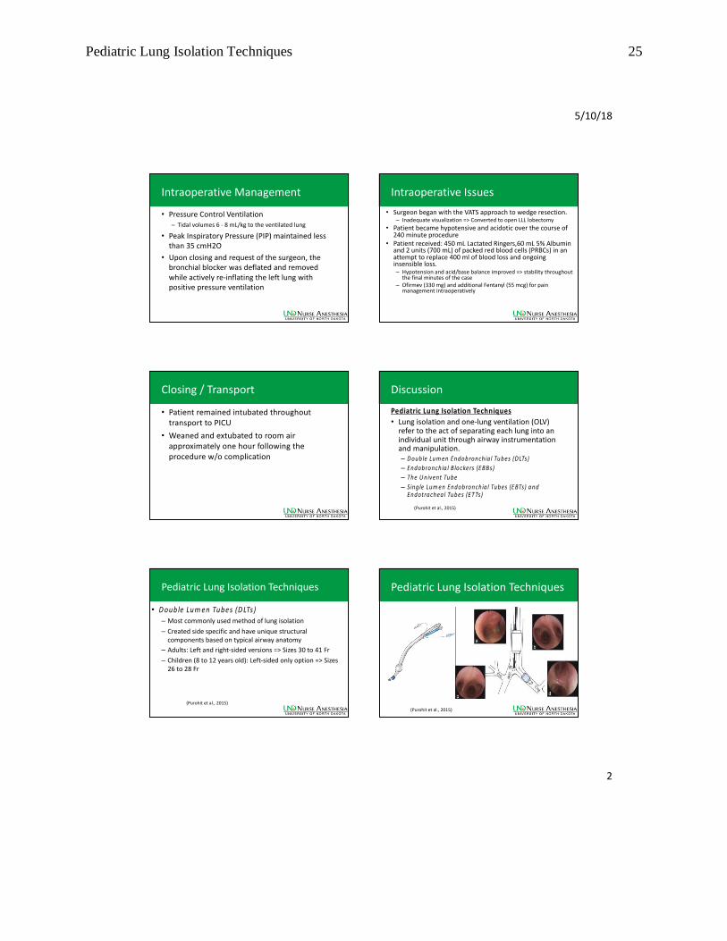
Pediatric Lung Isolation Techniques
(Purohit et al., 2015)
Pediatric Lung Isolation Techniques 26
5/10/18
3
Pediatric Lung Isolation Techniques
• Endobronch ia l b lockers (EB Bs)
– Isolate a lung through the inflation of a balloon at the distal end of a catheter
– Common types Ø Fogarty ’s vascular em bolectom y catheter, w ire-guided
endobronchial b locker (Arndt blocker), and EZ-blocker
(Purohit et al., 2015)
Pediatric Lung Isolation Techniques
• Fogarty ’s Vascu lar Em bolectom y Catheter– Sizes 6-8 Fr, with a length of 80 cm. – Guided into place with direct visualization via FOB,
either coaxially or parallel to the ETT
• A rndt blockers – Sizes 5, 7, and 9 Fr – Smallest recommended single-lumen ETT (SLETT) for
coaxial use 4.5, 7, and 8 mm– Guided via FOB through a Cook’s multiport adapter to
allow for uninterrupted ventilation throughout placement
(Purohit et al., 2015)
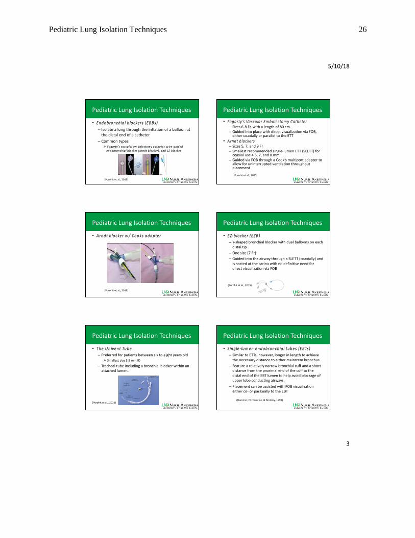
Pediatric Lung Isolation Techniques
• A rndt b locker w / Cooks adapter
(Purohit et al., 2015)
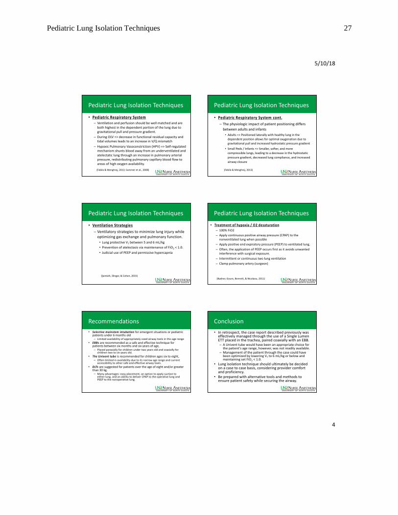
Pediatric Lung Isolation Techniques
• EZ-b locker (EZB ) – Y-shaped bronchial blocker with dual balloons on each
distal tip
– One size (7 Fr)
– Guided into the airway through a SLETT (coaxially) and is seated at the carina with no definitive need for direct visualization via FOB
(Purohit et al., 2015)
Pediatric Lung Isolation Techniques
• The U nivent Tube– Preferred for patients between six to eight years old
Ø Smallest size 3.5 mm ID
– Tracheal tube including a bronchial blocker within an attached lumen.
(Purohit et al., 2015)
Pediatric Lung Isolation Techniques
• Single-lum en endobronchia l tubes (EBTs) – Similar to ETTs, however, longer in length to achieve
the necessary distance to either mainstem bronchus.
– Feature a relatively narrow bronchial cuff and a short distance from the proximal end of the cuff to the distal end of the EBT lumen to help avoid blockage of upper lobe conducting airways.
– Placement can be assisted with FOB visualization either co- or paraxially to the EBT
(Hammer, Fitzmaurice, & Brodsky, 1999).
Pediatric Lung Isolation Techniques 27
5/10/18
4
Pediatric Lung Isolation Techniques
• Pediatric Respiratory System– Ventilation and perfusion should be well matched and are
both highest in the dependent portion of the lung due to gravitational pull and pressure gradient.
– During OLV => decrease in functional residual capacity and tidal volumes leads to an increase in V/Q mismatch
– Hypoxic Pulmonary Vasoconstriction (HPV) => Self-regulated mechanism shunts blood away from an underventilated and atelectatic lung through an increase in pulmonary arterial pressure, redistributing pulmonary capillary blood flow to areas of high oxygen availability.
(Fabila & Menghraj, 2013; Sommer et al., 2008)
Pediatric Lung Isolation Techniques
• Pediatric Respiratory System cont.
– The physiologic impact of patient positioning differs
between adults and infants
• Adults => Positioned laterally with healthy lung in the
dependent position allows for optimal oxygenation due to
gravitational pull and increased hydrostatic pressure gradient
• Small Peds / Infants => Smaller, softer, and more
compressible lungs, leading to a decrease in the hydrostatic
pressure gradient, decreased lung compliance, and increased
airway closure
(Fabila & Menghraj, 2013)
Pediatric Lung Isolation Techniques
• Ventilation Strategies
– Ventilatory strategies to minimize lung injury while optimizing gas exchange and pulmonary function.
• Lung protective VT between 5 and 6 mL/kg
• Prevention of atelectasis via maintenance of FiO2 < 1.0.
• Judicial use of PEEP and permissive hypercapnia
(Şentürk, Slinger, & Cohen, 2015)
Pediatric Lung Isolation Techniques
• Treatment of hypoxia / O2 desaturation
– 100% FiO2
– Apply continuous positive airway pressure (CPAP) to the nonventilated lung when possible
– Apply positive end expiratory pressure (PEEP) to ventilated lung.
– Often, the application of PEEP occurs first as it avoids unwanted interference with surgical exposure.
– Intermittent or continuous two lung ventilation
– Clamp pulmonary artery (surgeon)
(Badner, Goure, Bennett, & Nicolaou, 2011)
Recommendations
• Selective mainstem intubation for emergent situations or pediatric patients under 6 months old – Limited availability of appropriately sized airway tools in this age range
• EBBs are recommended as a safe and effective technique for patients between six months and six years of age, – Placed paraxially for children under two years old and coaxially for
children two to six years old.
• The Univent tube is recommended for children ages six to eight, – Often limited in availability due to its narrow age range and current
accessibility to other safe and effective airway tools.
• DLTs are suggested for patients over the age of eight and/or greater than 30 kg. – Many advantages: easy placement, an option to apply suction to
either lung, and an ability to deliver CPAP to the operative lung and PEEP to the nonoperative lung.
Conclusion
• In retrospect, the case report described previously was effectively managed through the use of a Single Lumen ETT placed in the trachea, paired coaxially with an EBB. – A Univent tube would have been an appropriate choice for
the patient’s age range, however, was not readily available. – Management of the patient through the case could have
been optimized by lowering VT to 6 mL/kg or below and maintaining set FiO2 < 1.0.
• Lung isolation technique should ultimately be decided on a case to case basis, considering provider comfort and proficiency.
• Be prepared with alternative tools and methods to ensure patient safety while securing the airway.

Pediatric Lung Isolation Techniques 28
5/10/18
5
References
• Badner, N. H., Goure, C., Bennett, K. E., & Nicolaou, G. (2011). Role of continuous positive airway pressure to the non-ventilated lung during one-lung ventilation with low tidal volumes. HSR P roceedings in Intensive Care &
Cardiovascular A nesthesia , 3(3), 189–194
• Brodsky, J. B., Macario, A., & Mark, J. B. (1996). Tracheal Diameter Predicts Double-Lumen Tube Size. Anesthesia &
Analgesia, 82(4), 861-864. doi:10.1213/00000539-199604000-00032
• Clayton-Smith, A., Bennett, K., Alston, R. P., Adams, G., Brown, G., Hawthorne, T., . . . Tan, J. (2015). A Comparison of the
Efficacy and Adverse Effects of Double-Lumen Endobronchial Tubes and Bronchial Blockers in Thoracic Surgery: ASystematic Review and Meta-analysis of Randomized Controlled Trials. Journal of Cardiothoracic and VascularAnesthesia, 29(4), 955-966. doi:10.1053/j.jvca.2014.11.017
• Collins, S. R., Titus, B. J., Campos, J. H., & Blank, R. S. (2017). Lung Isolation in the Patient With a Difficult Airway. Anesthesia& Analgesia , 1. doi:10.1213/ane.0000000000002637
• Dave, N., & Fernandes, S. (2005). Anaesthetic implications of paediatric thoracoscopy. Journal of M inim al AccessSurgery, 1(1), 8-14
• Fabila, T. S., & Menghraj, S. J. (2013). One lung ventilation strategies for infants and children undergoing video assistedthoracoscopic surgery. Indian Journal of A naesthesia , 57(4), 339–344. http://doi.org/10.4103/0019-5049.118539
• Gale, J. W., & Waters, R. M. (1932). Closed Endobronchial Anesthesia in Thoracic Surgery. A nesthesia & Analgesia, 11(1).doi:10.1213/00000539-193201000-00049
• Hammer, G. B., Fitzmaurice, B. G., & Brodsky, J. B. (1999). Methods for Single-Lung Ventilation in PediatricPatients. A nesthesia & A nalgesia, 89(6), 1426. doi:10.1213/00000539-199912000-00019
• Hemmes, S. N., Neto, A. S., & Schultz, M. J. (2013). Intraoperative ventilatory strategies to prevent postoperative pulmonarycomplications. Current Opinion in Anaesthesiology, 26(2), 126-133. doi:10.1097/aco.0b013e32835e1242.
References cont.
• Paranjpe, J., & Kulkarni, R. (2017). One-lung ventilation in pediatric patients. M e d ica l Jo u rn a l o f D r. D .Y. P a til
U n ive rs ity, 1 0 (2), 190. doi:10.4103/0975-2870.202110.
• Purohit, A., Bhargava, S., Mangal, V., & Parashar, V. (2015). Lung isolation, one-lung ventilation and hypoxaemiaduring lung isolation. In d ia n Jo u rn a l o f A n a e sth e s ia , 5 9 (9), 606-617
• Letal, M., & Theam, M. (2017). Paediatric lung isolation. B JA E d u ca tio n , 1 7 (2), 57-62.
doi:10.1093/bjaed/mkw047.
• Liu, Z., Liu, X., Huang, Y., & Zhao, J. (2016). Intraoperative mechanical ventilation strategies in patients undergoingone-lung ventilation: a meta-analysis. Sp rin g e rP lu s , 5 (1), 1251.
• Mourisse, J., Liesveld, J., Verhagen, A., Rooij, G. V., Heide, S. V., Schuurbiers-Siebers, O., & Heijden, E. V. (2013).
Efficiency, Efficacy, and Safety of EZ -Blocker Compared with Left-sided Double-lumen Tube for One-lungVentilation. A n e sth e s io lo g y, 1 1 8 (3), 550-561.doi:10.1097/aln.0b013e3182834f2d
• Şentürk, M., Slinger, P., & Cohen, E. (2015). Intraoperative mechanical ventilation strategies for one-lung
ventilation. B e st P ra c t ice & Re se a rc h C lin ica l A n a e sth e sio lo g y, 2 9 (3), 357-369. doi:10.1016/j.bpa.2015.08.001.
• Sommer, N., Dietrich, A., Schermuly, R. T., Ghofrani, H. A., Gudermann, T., Schulz, R., . . . Weissmann, N. (2008).
Regulation of hypoxic pulmonary vasoconstriction: Basic mechanisms. European Respiratory Journal,
32(6), 1639-1651. doi:10.1183/09031936.00013908
• Tobias, J. D. (1999). Anaesthetic Implications of Thoracoscopic Surgery in Children. Pediatric Anesthesia, 9(2), 103
-110. doi:10.1046/j.1460-9592.1999.9220281.
Thank YouAre There Any Questions?
Thank YouAre There Any Questions?