pediatric cardiac anesthesia 2: complex shunts · incidence of common chdz lesions in children •...
TRANSCRIPT
Intensive Review of Pediatric Anesthesia 2015
Pediatric Cardiac Anesthesia 2: Complex Shunts
Susan R Staudt, MD, MSEd With co-authorship attribution to
Dean Andropoulos, MD & Emad B Mossad, MD

Intensive Review of Pediatric Anesthesia 2015
Learning Objectives
• Follows/builds on simple shunts (Dr. Mossad) • Review complex shunts and mixing lesions: Truncus
Arteriosus, d-TGA, TAPVR • Discuss Single ventricle lesion types • Refresh knowledge of anatomy and physiology of the
3 stages of single ventricle palliation • Examine remaining miscellaneous cardiac and
extracardiac congenital heart defects • NOTE: NO DISCLOSURES or COI
Intensive Review of Pediatric Anesthesia 2015
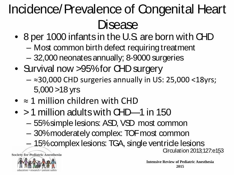
Incidence/Prevalence of Congenital Heart Disease
• 8 per 1000 infants in the U.S. are born with CHD – Most common birth defect requiring treatment – 32,000 neonates annually; 8-9000 surgeries
• Survival now >95% for CHD surgery – ≈30,000 CHD surgeries annually in US: 25,000 <18yrs; 5,000 >18 yrs
• ≈ 1 million children with CHD • > 1 million adults with CHD—1 in 150
– 55% simple lesions: ASD, VSD most common – 30% moderately complex: TOF most common – 15% complex lesions: TGA, single ventricle lesions
Circulation 2013;127:e153 4
Intensive Review of Pediatric Anesthesia 2015
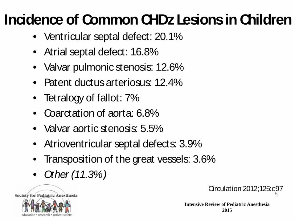
Incidence of Common CHDz Lesions in Children • Ventricular septal defect: 20.1% • Atrial septal defect: 16.8% • Valvar pulmonic stenosis: 12.6% • Patent ductus arteriosus: 12.4% • Tetralogy of fallot: 7% • Coarctation of aorta: 6.8% • Valvar aortic stenosis: 5.5% • Atrioventricular septal defects: 3.9% • Transposition of the great vessels: 3.6% • Other (11.3% )
Circulation 2012;125:e97 5
Intensive Review of Pediatric Anesthesia 2015
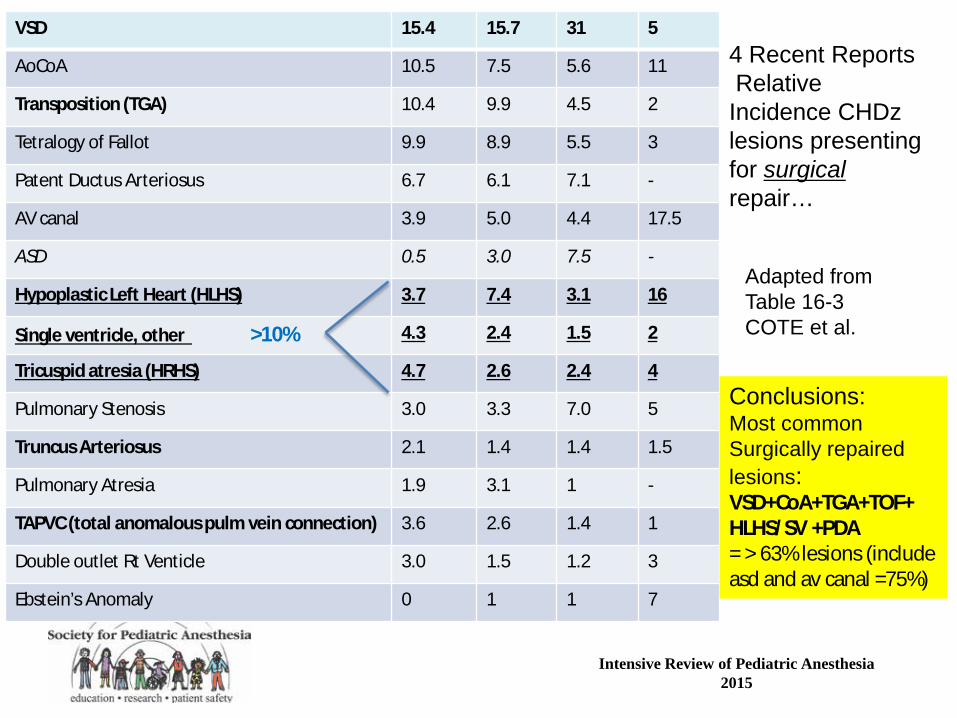
VSD 15.4 15.7 31 5
AoCoA 10.5 7.5 5.6 11
Transposition (TGA) 10.4 9.9 4.5 2
Tetralogy of Fallot 9.9 8.9 5.5 3
Patent Ductus Arteriosus 6.7 6.1 7.1 -
AV canal 3.9 5.0 4.4 17.5
ASD 0.5 3.0 7.5 -
Hypoplastic Left Heart (HLHS) 3.7 7.4 3.1 16
Single ventricle, other >10% 4.3 2.4 1.5 2
Tricuspid atresia (HRHS) 4.7 2.6 2.4 4
Pulmonary Stenosis 3.0 3.3 7.0 5
Truncus Arteriosus 2.1 1.4 1.4 1.5
Pulmonary Atresia 1.9 3.1 1 -
TAPVC (total anomalous pulm vein connection) 3.6 2.6 1.4 1
Double outlet Rt Venticle 3.0 1.5 1.2 3
Ebstein’s Anomaly 0 1 1 7
4 Recent Reports Relative Incidence CHDz lesions presenting for surgical repair…
Adapted from Table 16-3 COTE et al.
Conclusions: Most common Surgically repaired lesions: VSD+CoA+TGA+TOF+ HLHS/SV +PDA = > 63% lesions (include asd and av canal =75%)
Intensive Review of Pediatric Anesthesia 2015
Truncus Arteriosus • Major types are I,II,III and depend on
location/degree of PA branching • Common arterial trunk results in systemic,
pulmonary, and coronary circulations in parallel
• Lowering PVR with excessive FiO2 and hyperventilation creates systemic/coronary steal and myocardial ischemia
7
Intensive Review of Pediatric Anesthesia 2015
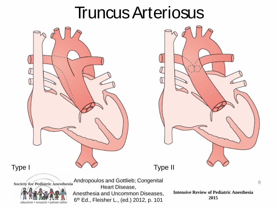
Andropoulos and Gottlieb; Congenital Heart Disease,
Anesthesia and Uncommon Diseases, 6th Ed., Fleisher L., (ed.) 2012, p. 101
Truncus Arteriosus
8
Type I Type II
Intensive Review of Pediatric Anesthesia 2015
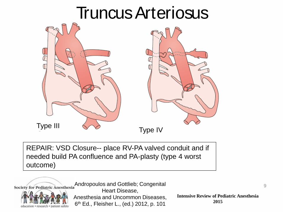
Truncus Arteriosus
9 Andropoulos and Gottlieb; Congenital Heart Disease,
Anesthesia and Uncommon Diseases, 6th Ed., Fleisher L., (ed.) 2012, p. 101
Type III Type IV
REPAIR: VSD Closure-- place RV-PA valved conduit and if needed build PA confluence and PA-plasty (type 4 worst outcome)

Intensive Review of Pediatric Anesthesia 2015
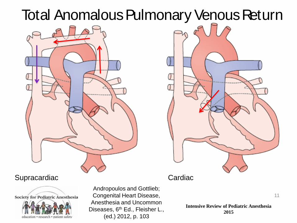
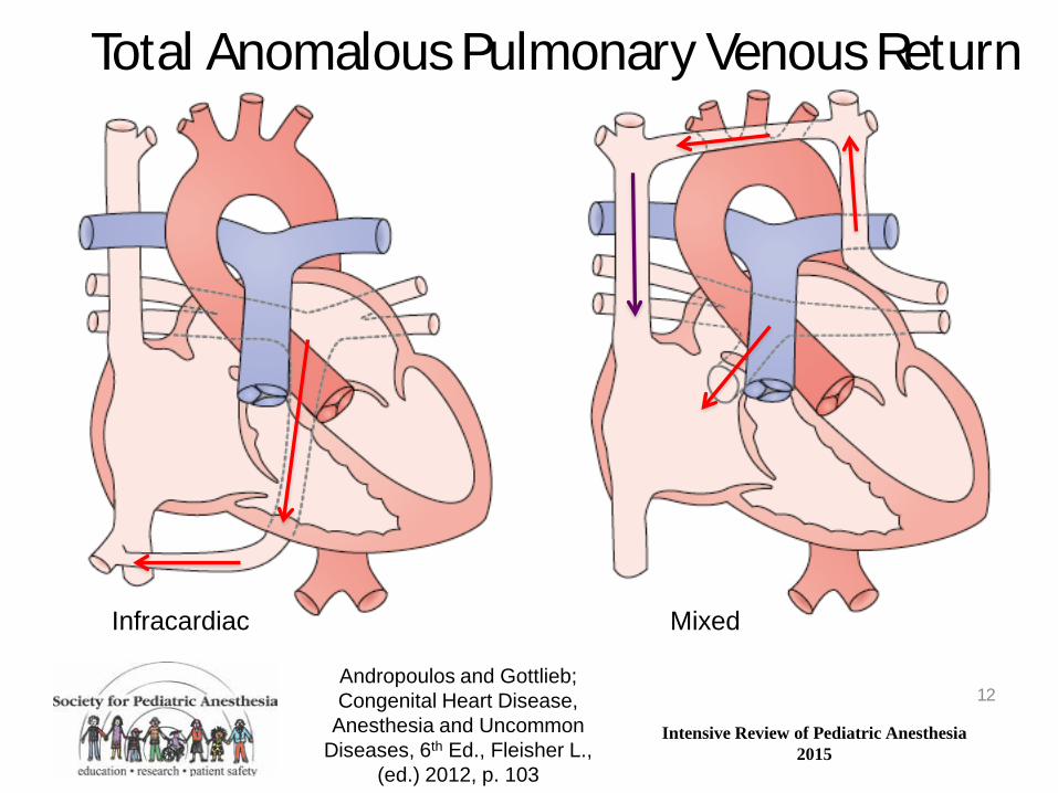
Anomalous Pulmonary Venous Return • Major types are partial (PAPVR) and total (TAPVR) • PAPVR usually has mild symptoms of small left-to-
right shunt and sinus venosus ASD • TAPVR symptomatology depends on degree of
obstruction to pulmonary venous return • Infradiaphragmatic TAPVR prone to severe
obstruction, hypoxia, respiratory failure – Increasing PBF with high FiO2 and iNO may
paradoxically worsen obstruction before repair 10
Intensive Review of Pediatric Anesthesia 2015
Total Anomalous Pulmonary Venous Return
11
Supracardiac Cardiac Andropoulos and Gottlieb; Congenital Heart Disease,
Anesthesia and Uncommon Diseases, 6th Ed., Fleisher L.,
(ed.) 2012, p. 103
Intensive Review of Pediatric Anesthesia 2015
Total Anomalous Pulmonary Venous Return
12 Andropoulos and Gottlieb; Congenital Heart Disease,
Anesthesia and Uncommon Diseases, 6th Ed., Fleisher L.,
(ed.) 2012, p. 103
Infracardiac Mixed

Intensive Review of Pediatric Anesthesia 2015
Left-to-Right Shunt Lesions Left-Sided Obstructive Lesions
Right Sided Obstructive Lesions Transposition of the Great Arteries
Single Ventricle Lesions Miscellaneous
13

Intensive Review of Pediatric Anesthesia 2015
D-Transposition of the Great Arteries
14
Andropoulos and Gottlieb; Congenital Heart Disease,
Anesthesia and Uncommon Diseases, 6th Ed., Fleisher L., (ed.) 2012, p. 116
Intensive Review of Pediatric Anesthesia 2015
D-Transposition of the Great Arteries
• Aorta arises from right ventricle, pulmonary artery from left ventricle , resulting in parallel circulation with “transposition physiology”
• Oxygenation depends on mixing at the atrial level (most important), PDA, or VSD (15-25%)
• Balloon atrial septostomy is often necessary in the neonatal period
15

Intensive Review of Pediatric Anesthesia 2015
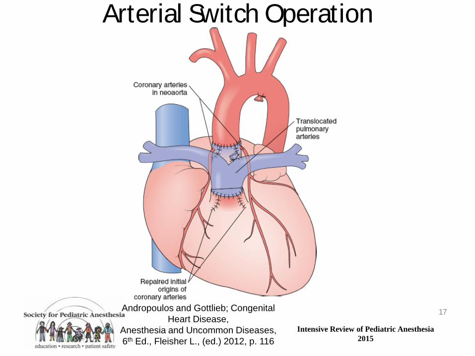
Arterial Switch for D-TGA • Procedure of choice since mid 1980’s • Performed as neonate; CPB without DHCA • 15-25% also have VSD • Beware coronary problems after repair
– Global myocardial dysfunction – ST segment changes – Segmental wall motion abnormalities
• LV can be deconditioned – Intolerant of high preload or afterload – LAP 4-6, systolic BP 60s often desirable
16
Intensive Review of Pediatric Anesthesia 2015
Arterial Switch Operation
17 Andropoulos and Gottlieb; Congenital Heart Disease,
Anesthesia and Uncommon Diseases, 6th Ed., Fleisher L., (ed.) 2012, p. 116
Intensive Review of Pediatric Anesthesia 2015
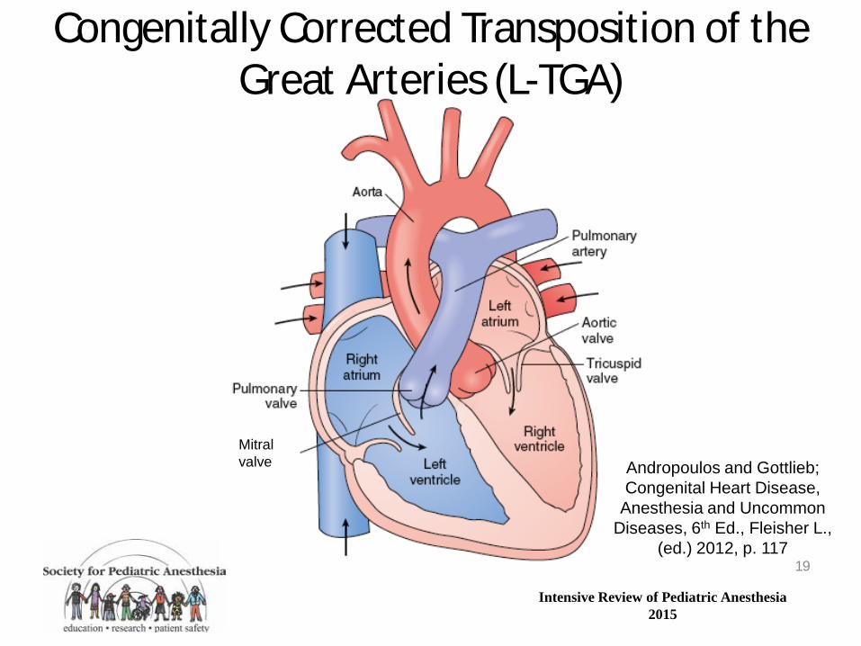
Congenitally Corrected Transposition of the Great Arteries (L-TGA)
• Ventricles are inverted, aorta arises from LV and PA from LV
• RA empties to LV, and LA to RV • Other associated anomalies include VSD and
pulmonic stenosis • Complete atrioventricular block is common in
cc-TGA (2% per year) • Right (systemic) ventricle usually fails over time
18
Intensive Review of Pediatric Anesthesia 2015
Congenitally Corrected Transposition of the Great Arteries (L-TGA)
19
Andropoulos and Gottlieb; Congenital Heart Disease,
Anesthesia and Uncommon Diseases, 6th Ed., Fleisher L.,
(ed.) 2012, p. 117
Mitral valve
Intensive Review of Pediatric Anesthesia 2015
Double Outlet Right Ventricle
• VSD with both aorta and pulmonary artery arising completely or partially from RV
• Pathophysiology depends on degree of RVOTO • Cyanosis and TOF physiology are seen with
significant RVOTO • Acyanosis with CHF can be seen with no
RVOTO; in this case a VSD patch may be only needed surgery
20

Intensive Review of Pediatric Anesthesia 2015
Spectrum of DORV – Taussig Bing
REPAIR: Rastelli procedure-valved RV to PA homogaft, as patching of VSD precludes native PA from use (Also this is used in TGA with PS)
Intensive Review of Pediatric Anesthesia 2015
Left-to-Right Shunt Lesions Left-Sided Obstructive Lesions
Right Sided Obstructive Lesions Transposition of the Great Arteries
Single Ventricle Lesions Miscellaneous
22
Intensive Review of Pediatric Anesthesia 2015
Notes on “single ventricle lesions” • Broad Spectrum of pathology- hence a not uncommon
“type”-40%-50% of all cyanotic congenital heart disease is treated via this path!
• Classic HRHS: (tricuspid atresia, stenosis) • Classic HLHS :(mitral and/or aortic atresia, stenosis) • In addition are other lesions precluding a 2 ventricle repair:
PA/IVS, severe Ebsteins anomaly, unbalanced AV canals, double inlet LV, various “mixed lesions”
• Prognosis is better with: HRHS, more than 1 mass of ventricle, full term, no associated anomalies, absence of lung or airways disease
Intensive Review of Pediatric Anesthesia 2015
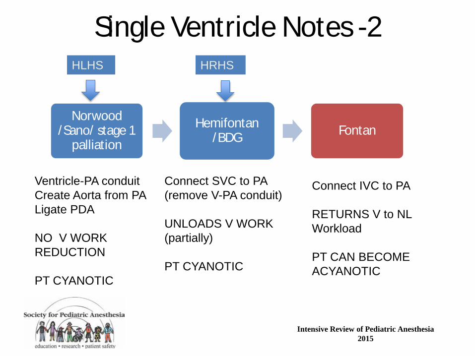
Single Ventricle Notes -2
Norwood /Sano/ stage 1
palliation
Hemifontan /BDG Fontan
HLHS HRHS
Ventricle-PA conduit Create Aorta from PA Ligate PDA NO V WORK REDUCTION PT CYANOTIC
Connect SVC to PA (remove V-PA conduit) UNLOADS V WORK (partially) PT CYANOTIC
Connect IVC to PA RETURNS V to NL Workload PT CAN BECOME ACYANOTIC

Intensive Review of Pediatric Anesthesia 2015
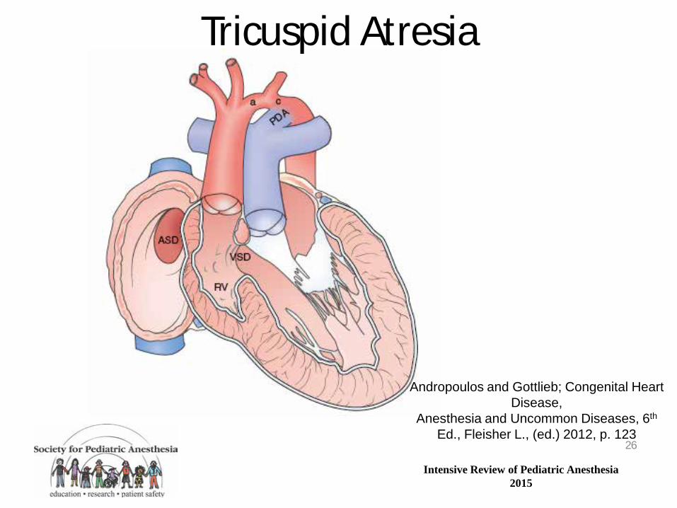
Tricuspid Atresia • Plate-like obstruction instead of tricuspid valve • Neonatal presentation depends on degree of
obstruction to pulmonary blood flow: – Severe: profound cyanosis when PDA closes – Mild/moderate: mild/moderate cyanosis, no CHF – None: mild or no cyanosis, CHF
• Systemic ventricle is the left ventricle – Good long term outcomes despite single ventricle
physiology
25
Intensive Review of Pediatric Anesthesia 2015
Tricuspid Atresia
26
Andropoulos and Gottlieb; Congenital Heart Disease,
Anesthesia and Uncommon Diseases, 6th Ed., Fleisher L., (ed.) 2012, p. 123
Intensive Review of Pediatric Anesthesia 2015
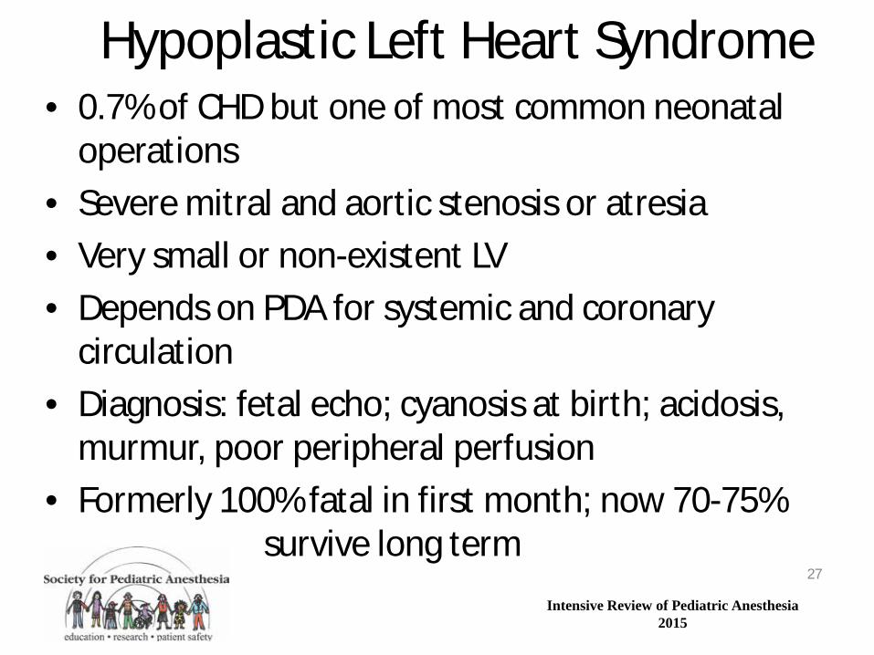
Hypoplastic Left Heart Syndrome • 0.7% of CHD but one of most common neonatal
operations • Severe mitral and aortic stenosis or atresia • Very small or non-existent LV • Depends on PDA for systemic and coronary
circulation • Diagnosis: fetal echo; cyanosis at birth; acidosis,
murmur, poor peripheral perfusion • Formerly 100% fatal in first month; now 70-75%
survive long term 27
Intensive Review of Pediatric Anesthesia 2015
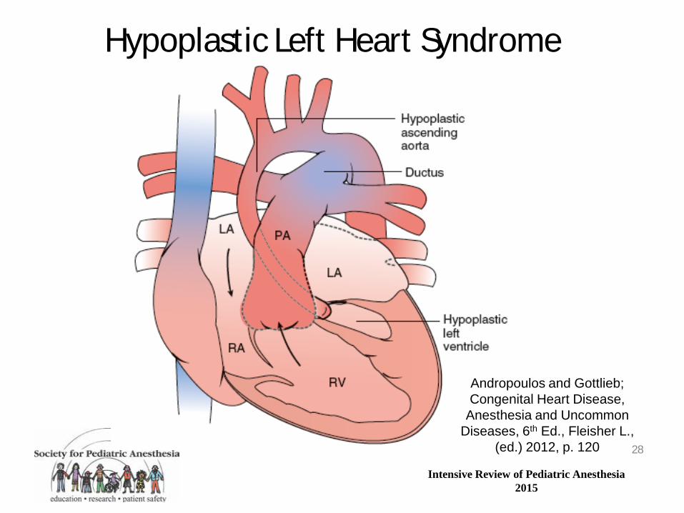
Hypoplastic Left Heart Syndrome
Andropoulos and Gottlieb; Congenital Heart Disease,
Anesthesia and Uncommon Diseases, 6th Ed., Fleisher L.,
(ed.) 2012, p. 120 28
Intensive Review of Pediatric Anesthesia 2015
29

Intensive Review of Pediatric Anesthesia 2015
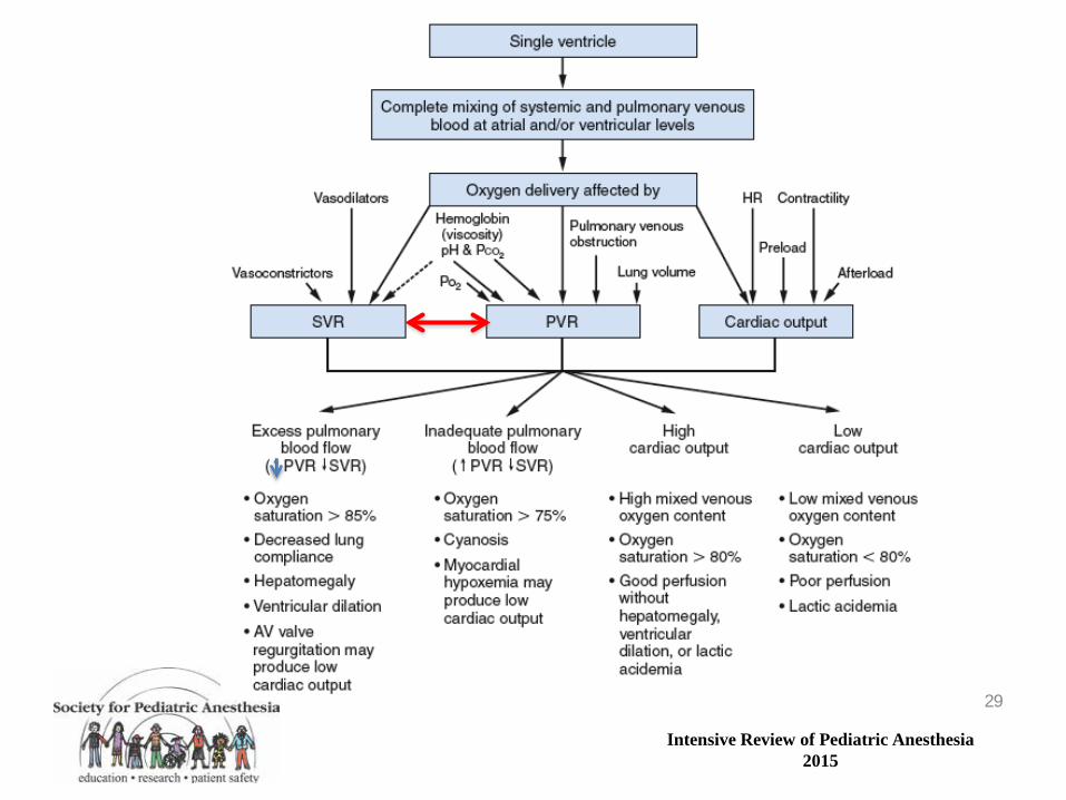
Pathophysiology: Mixing Lesions
30
Andropoulos and Gottlieb; Congenital Heart Disease,
Anesthesia and Uncommon Diseases, 6th
Ed., Fleisher L., (ed.) 2012, p. 78

Intensive Review of Pediatric Anesthesia 2015
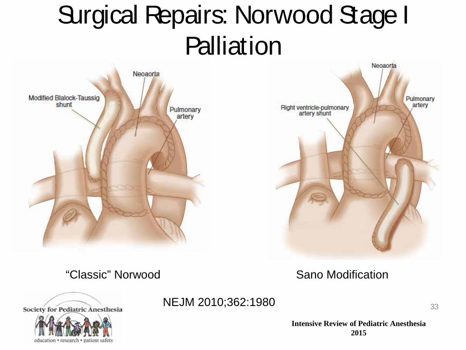
Surgical Repairs: Norwood Stage I Palliation
• Done as neonate: PDA dependent on PGE1
• Delicate balance between systemic and pulmonary circulations – Often FiO2 0.21, PaCO2 40-50 mm Hg
• CPB with DHCA or RCP for aortic arch reconstruction
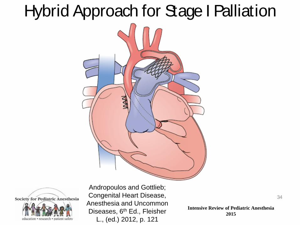
• Can also be done a hybrid procedure in cath lab – Sternotomy, PDA stent, PA banding, ASD stent
31

Intensive Review of Pediatric Anesthesia 2015
Classic Norwood repair or stage 1 palliation
Aortic arch reconstructed from PA, pulm valve and homograft; 3.5 mm B-T shunt placed for pulm blood flow
Shunt can over or undercirculate; can thrombose or kink; runoff aorta to pa lowers diastolic pressure; least risk of distorting PA’s and no ventricular incision
SANO modification
Same aortic arch repair; larger 5.0 mm RV-PA conduit instead of BT shunt
Requires a ventriculotomy (scars ventricle); higher diastolic pressure; ? Less risk of thrombosis, kinking and early complications?
Hybrid procedure
Off CPB- PA band placed via sternotomy and pda is stented (cath)
No CPB required; arch reconstruction occurs at 2nd stage of repair-infant left with retrograde coronary perfusion down tiny aorta until 2nd stage
HLHS-Three Different Approaches to Stage One Palliation
Intensive Review of Pediatric Anesthesia 2015
Surgical Repairs: Norwood Stage I Palliation
33
“Classic” Norwood Sano Modification
NEJM 2010;362:1980
Intensive Review of Pediatric Anesthesia 2015
Hybrid Approach for Stage I Palliation
34
Andropoulos and Gottlieb; Congenital Heart Disease,
Anesthesia and Uncommon Diseases, 6th Ed., Fleisher
L., (ed.) 2012, p. 121
Intensive Review of Pediatric Anesthesia 2015
Bidirectional Cavopulmonary Connection (Glenn, Hemifontan)
• Stage II or first palliation for essentially all single ventricle patients
• Performed at age 2-6 months, following cath, echo possibly MRI to risk stratify
• CPB, often no crossclamp unless intracardiac or aortic repair
• After repair desire transpulmonary pressure gradient of ≈ 10 mm Hg with low atrial pressure
35

Intensive Review of Pediatric Anesthesia 2015
Bidirectional Cavopulmonary Connection (Glenn)
36

Intensive Review of Pediatric Anesthesia 2015
Cardio-Cerebro-Pulmonary Circulation & Glenn SVC anastomosis
37
Andropoulos and Chang. Chapter 20: Pediatric Cardiac Intensive Care. In, Allen H. et al (eds.) Moss & Adams’ Heart
Disease…, 8th ed. p. 514.
êPaCO2èêCBFè êSVC flowèêPBFè êSaO2, CO, and DO2
Systemic ventricle
(both PVR and cerebral autoregulation of SVC flow can impact Qp)
Intensive Review of Pediatric Anesthesia 2015
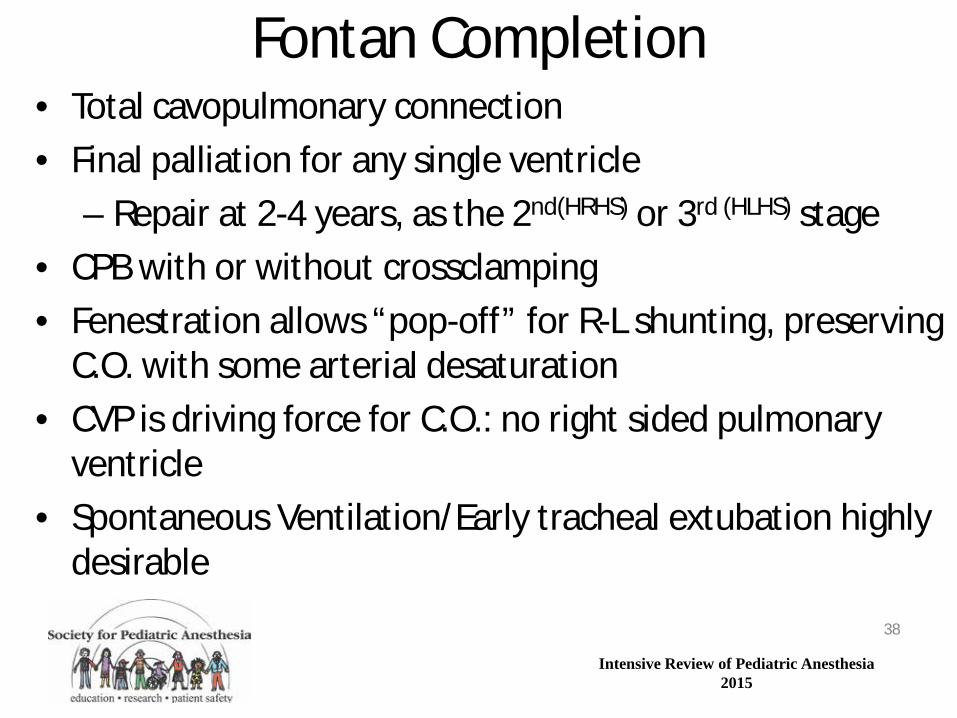
Fontan Completion • Total cavopulmonary connection • Final palliation for any single ventricle
– Repair at 2-4 years, as the 2nd(HRHS) or 3rd (HLHS) stage • CPB with or without crossclamping • Fenestration allows “pop-off” for R-L shunting, preserving
C.O. with some arterial desaturation • CVP is driving force for C.O.: no right sided pulmonary
ventricle • Spontaneous Ventilation/Early tracheal extubation highly
desirable 38
Intensive Review of Pediatric Anesthesia 2015
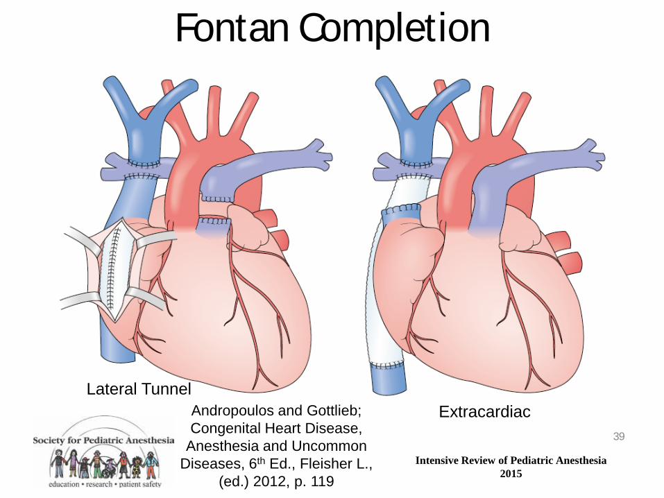
Fontan Completion
39
Lateral Tunnel Extracardiac Andropoulos and Gottlieb;
Congenital Heart Disease, Anesthesia and Uncommon
Diseases, 6th Ed., Fleisher L., (ed.) 2012, p. 119

Intensive Review of Pediatric Anesthesia 2015
Fontan Ten Commandments • Age > 4 years • Sinus Rhythm • Normal systemic venous return • Normal Right Atrial volume • Mean PAP <15 mmHg • PVR <4 Wood units/m2 • PA/Aorta ratio > 0.75 • LVEF (or SVEF)>0.6 • Competent AVV • Absent PA distortion

Intensive Review of Pediatric Anesthesia 2015
Left-to-Right Shunt Lesions Left-Sided Obstructive Lesions
Right Sided Obstructive Lesions Transposition of the Great Arteries
Single Ventricle Lesions Miscellaneous Cardiac Lesions
41
Intensive Review of Pediatric Anesthesia 2015
Anomalous Left Coronary Artery from Pulmonary Artery
• ALCAPA results in myocardial ischemia and infarction in infants as PVR declines and coronary flow to the LV reverses
• Dilated cardiomyopathy ensues; ALCAPA must be ruled out in infants with DCM
• Maintain elevated PVR with low FiO2 and hypercapnea in unrepaired ALCAPA – Elevate PA pressure, increase L coronary pressure,
minimize steal into PA 42
Intensive Review of Pediatric Anesthesia 2015
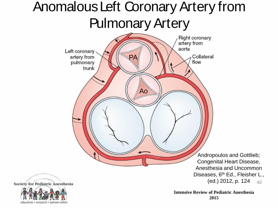
Anomalous Left Coronary Artery from Pulmonary Artery
43
Andropoulos and Gottlieb; Congenital Heart Disease,
Anesthesia and Uncommon Diseases, 6th Ed., Fleisher L.,
(ed.) 2012, p. 124
Ao
PA

Intensive Review of Pediatric Anesthesia 2015
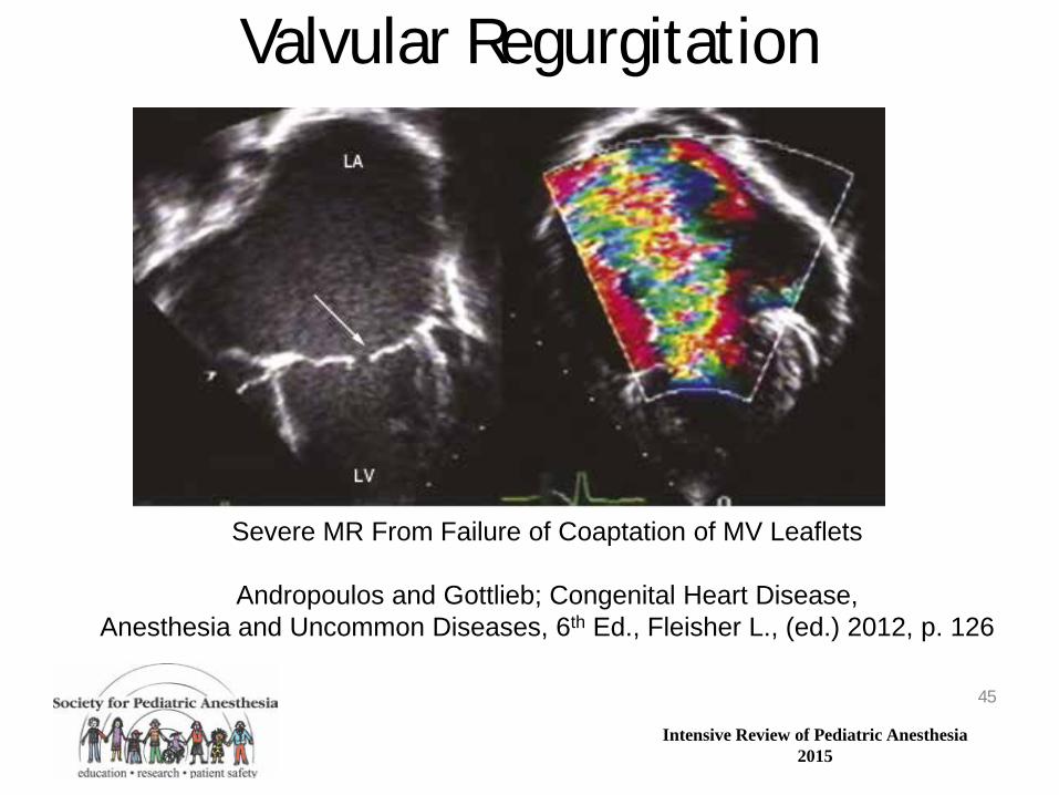
Valvular Regurgitation • Aortic, mitral, pulmonic, or tricuspid regurgitation
can be isolated or component of more complex lesion
• Severe AI most problematic: low diastolic pressure causing coronary ischemia
• MR can lead to pulmonary hypertension if severe • PR and TR usually well tolerated • Hemodynamic goals: reduce afterload, maintain
preload, high-normal heart rate 44
Intensive Review of Pediatric Anesthesia 2015
Valvular Regurgitation
45
Severe MR From Failure of Coaptation of MV Leaflets
Andropoulos and Gottlieb; Congenital Heart Disease, Anesthesia and Uncommon Diseases, 6th Ed., Fleisher L., (ed.) 2012, p. 126
Intensive Review of Pediatric Anesthesia 2015
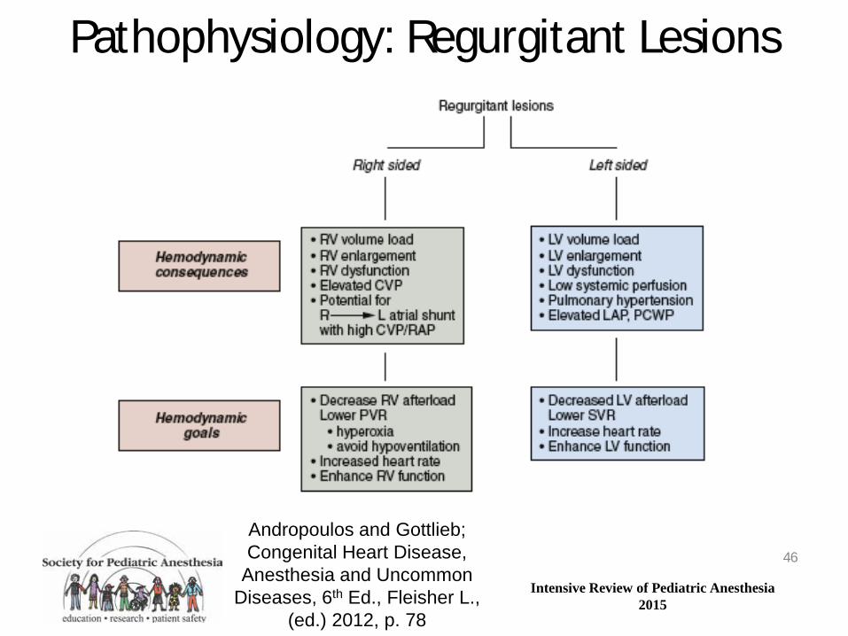
Pathophysiology: Regurgitant Lesions
46
Andropoulos and Gottlieb; Congenital Heart Disease,
Anesthesia and Uncommon Diseases, 6th Ed., Fleisher L.,
(ed.) 2012, p. 78

Intensive Review of Pediatric Anesthesia 2015
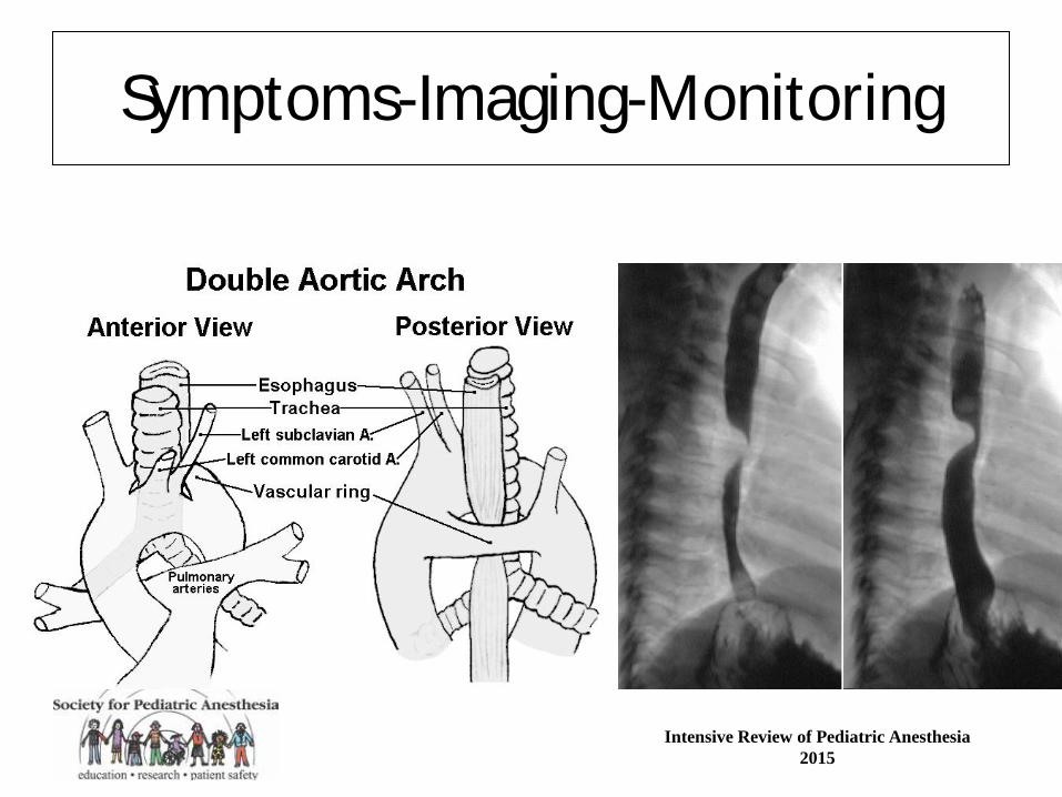
Vascular Rings • Many variations; result from abnormal
regression of the developing aortic arches • Respiratory and/or GI symptoms
– Stridor, “wheezing”, dysphagia
• Double aortic arch (60%), right aortic arch with aberrant L subclavian artery are most common
• CT angio (preferred) or MRI for diagnosis 47
Intensive Review of Pediatric Anesthesia 2015
Symptoms-Imaging-Monitoring

Intensive Review of Pediatric Anesthesia 2015
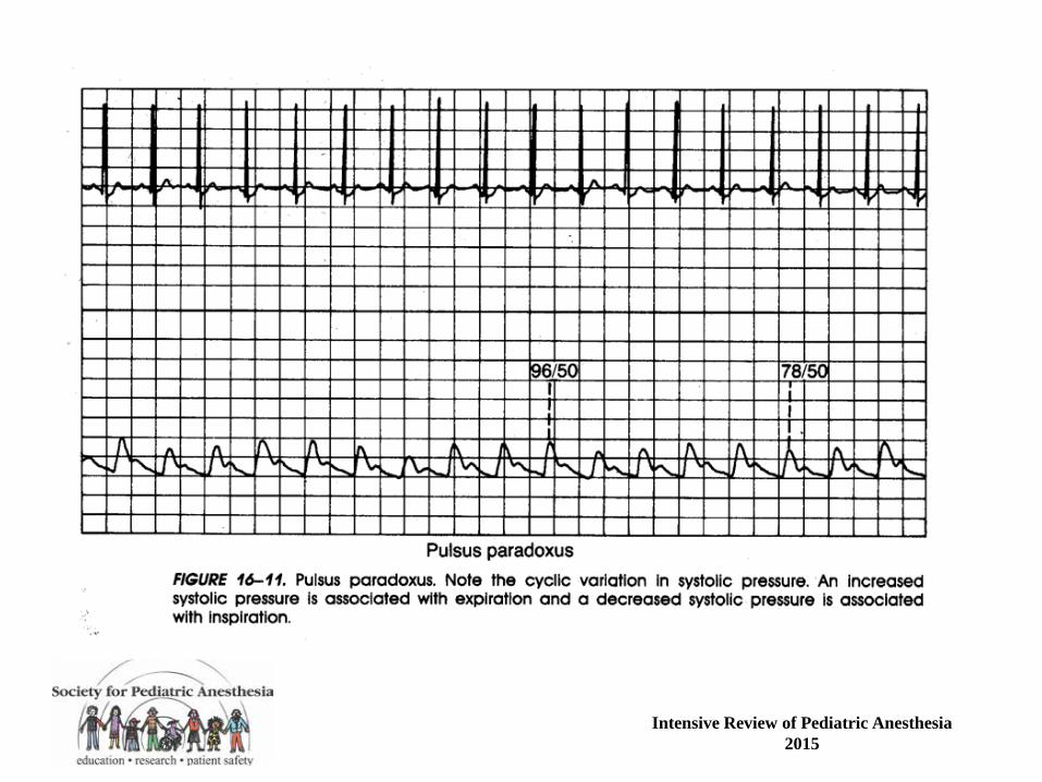
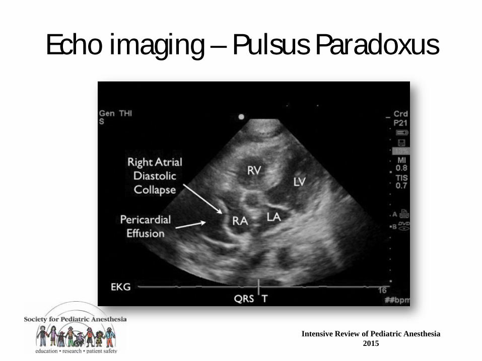
Pericardial Effusion/Tamponade • Increases in intrapericardial pressure cause equalization
of diastolic pressures in all 4 cardiac chambers, resulting in tamponade physiology
• Distended neck veins, pulsus paradoxus, tachycardia, and low cardiac output result
• Induction of anesthesia with PPV can compromise ventricular filling and cause CV collapse
• Sedation, local anesthesia, needle pericardiocentesis; if GA keep SVR up; PPV destabilizing (? Avoid or ino/pressor “bridge”) -team ready to go ie tap then window
49
Intensive Review of Pediatric Anesthesia 2015
Intensive Review of Pediatric Anesthesia 2015
Echo imaging – Pulsus Paradoxus