pediatric ankle fractures - orthopaedic trauma association ... ankle fractures.pdf · pediatric...
TRANSCRIPT
Anthony I. Riccio, MD
Texas Scottish Rite Hospital for Children
Update 07/2016
Pediatric Ankle Fractures
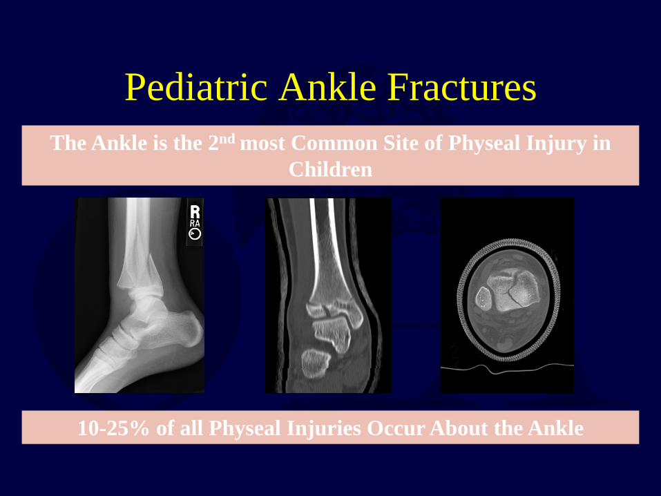
Pediatric Ankle Fractures The Ankle is the 2nd most Common Site of Physeal Injury in
Children
10-25% of all Physeal Injuries Occur About the Ankle
Pediatric Ankle Fractures Primary Concerns Are:
• Anatomic Restoration of Articular Surface
• Restoration of Symmetric Ankle Mortise
• Preservation of Physeal Growth
• Minimize Iatrogenic Physeal Injury
• Avoid Fixation Across Physis in Younger Children
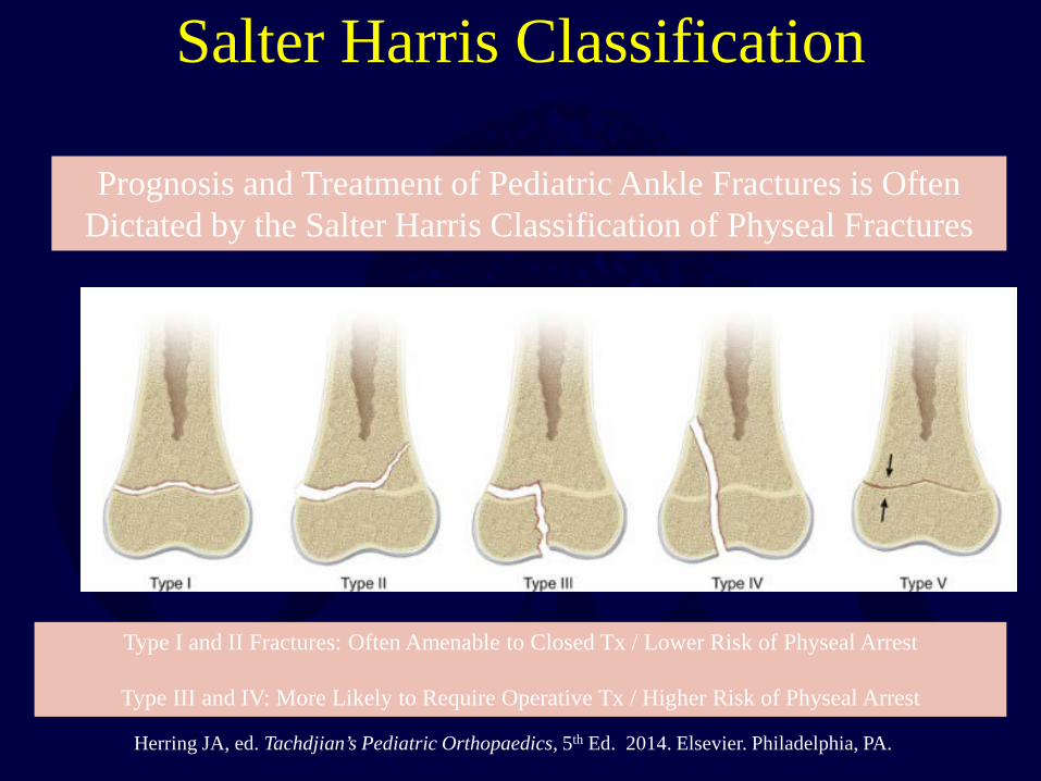
Salter Harris Classification
Prognosis and Treatment of Pediatric Ankle Fractures is Often Dictated by the Salter Harris Classification of Physeal Fractures
Type I and II Fractures: Often Amenable to Closed Tx / Lower Risk of Physeal Arrest
Type III and IV: More Likely to Require Operative Tx / Higher Risk of Physeal Arrest
Herring JA, ed. Tachdjian’s Pediatric Orthopaedics, 5th Ed. 2014. Elsevier. Philadelphia, PA.
ISOLATED DISTAL FIBULA FRACTURES
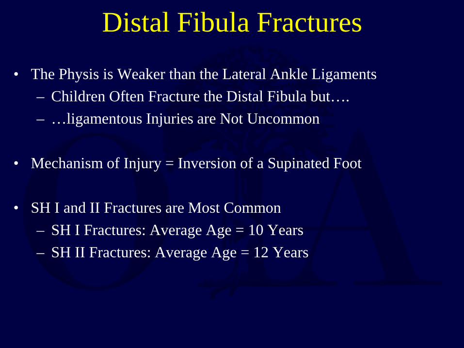
Distal Fibula Fractures
• The Physis is Weaker than the Lateral Ankle Ligaments – Children Often Fracture the Distal Fibula but…. – …ligamentous Injuries are Not Uncommon
• Mechanism of Injury = Inversion of a Supinated Foot
• SH I and II Fractures are Most Common
– SH I Fractures: Average Age = 10 Years – SH II Fractures: Average Age = 12 Years
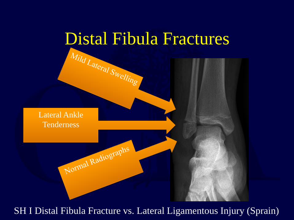
Distal Fibula Fractures
Lateral Ankle Tenderness
SH I Distal Fibula Fracture vs. Lateral Ligamentous Injury (Sprain)
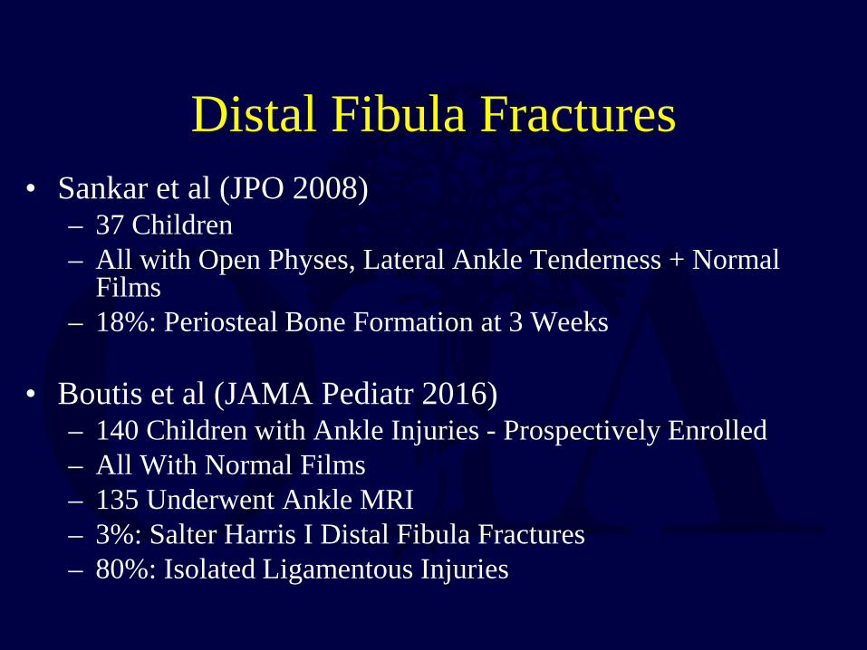
Distal Fibula Fractures • Sankar et al (JPO 2008)
– 37 Children – All with Open Physes, Lateral Ankle Tenderness + Normal
Films – 18%: Periosteal Bone Formation at 3 Weeks
• Boutis et al (JAMA Pediatr 2016)
– 140 Children with Ankle Injuries - Prospectively Enrolled – All With Normal Films – 135 Underwent Ankle MRI – 3%: Salter Harris I Distal Fibula Fractures – 80%: Isolated Ligamentous Injuries
Non-Displaced Distal Fibula Fractures: Treatment
• Removable Walking Boot vs Short Leg Cast (4 Weeks)
• Weight Bearing as Pain Permits
• Boutis et al (Pediatrics 2007): – Randomized Single Blind Study – Short Leg Walking Cast versus Removable Brace – Brace Group:
• Quicker Return to Baseline Activities • More Cost Effective
Displaced SH I and SH II Distal Fibula Fractures: Treatment
• Successful Closed Reduction – Short Leg Cast X 6 Weeks – Non-Weightbearing
• Failed Closed Reduction Open Reduction
– Percutaneous Pin Fixation (>2 Years Growth Remaining) – Internal Fixation (>2 Yeats Growth Remaining)
• SH I and SH II Fractures Are Often Associated with Distal
Tibia Fractures. Treatment is Dictated by Displacement, Ankle Mortise Symmetry and Nature of the Tibia FX
DISTAL TIBIA FRACTURES
SH II Distal Tibia Fractures • Most Common Distal Tibial Physeal Injury • 40% of All Pediatric Ankle Fractures • Associated Fibula Shaft Fracture Present in 20%
• Average Age at Injury = 12.5 Years
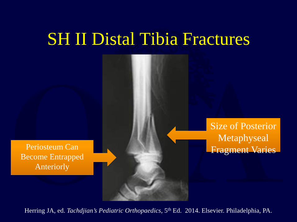
SH II Distal Tibia Fractures
Periosteum Can Become Entrapped
Anteriorly
Size of Posterior Metaphyseal
Fragment Varies
Herring JA, ed. Tachdjian’s Pediatric Orthopaedics, 5th Ed. 2014. Elsevier. Philadelphia, PA.
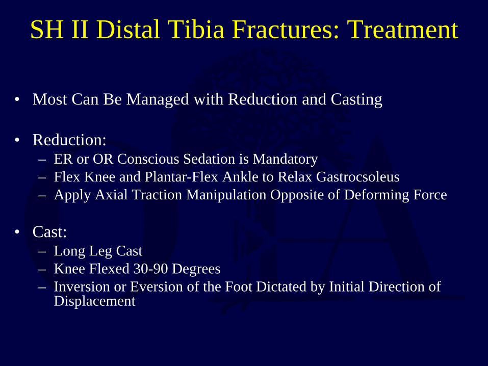
SH II Distal Tibia Fractures: Treatment
• Most Can Be Managed with Reduction and Casting • Reduction:
– ER or OR Conscious Sedation is Mandatory – Flex Knee and Plantar-Flex Ankle to Relax Gastrocsoleus – Apply Axial Traction Manipulation Opposite of Deforming Force
• Cast:
– Long Leg Cast – Knee Flexed 30-90 Degrees – Inversion or Eversion of the Foot Dictated by Initial Direction of
Displacement
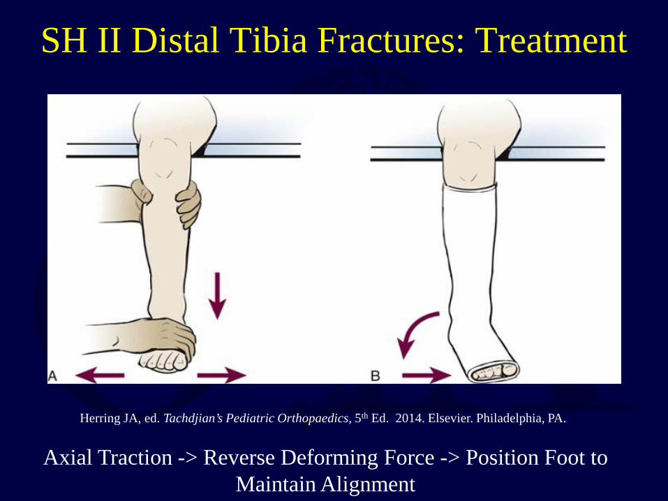
SH II Distal Tibia Fractures: Treatment
Axial Traction -> Reverse Deforming Force -> Position Foot to Maintain Alignment
Herring JA, ed. Tachdjian’s Pediatric Orthopaedics, 5th Ed. 2014. Elsevier. Philadelphia, PA.
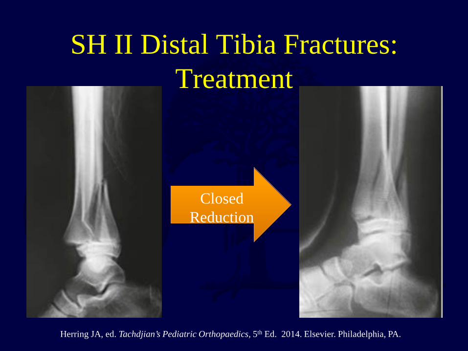
SH II Distal Tibia Fractures: Treatment
Closed Reduction
Herring JA, ed. Tachdjian’s Pediatric Orthopaedics, 5th Ed. 2014. Elsevier. Philadelphia, PA.
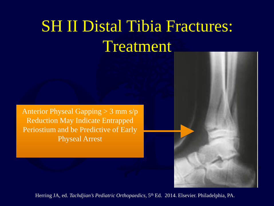
SH II Distal Tibia Fractures: Treatment
Anterior Physeal Gapping > 3 mm s/p Reduction May Indicate Entrapped
Periostium and be Predictive of Early Physeal Arrest
Herring JA, ed. Tachdjian’s Pediatric Orthopaedics, 5th Ed. 2014. Elsevier. Philadelphia, PA.
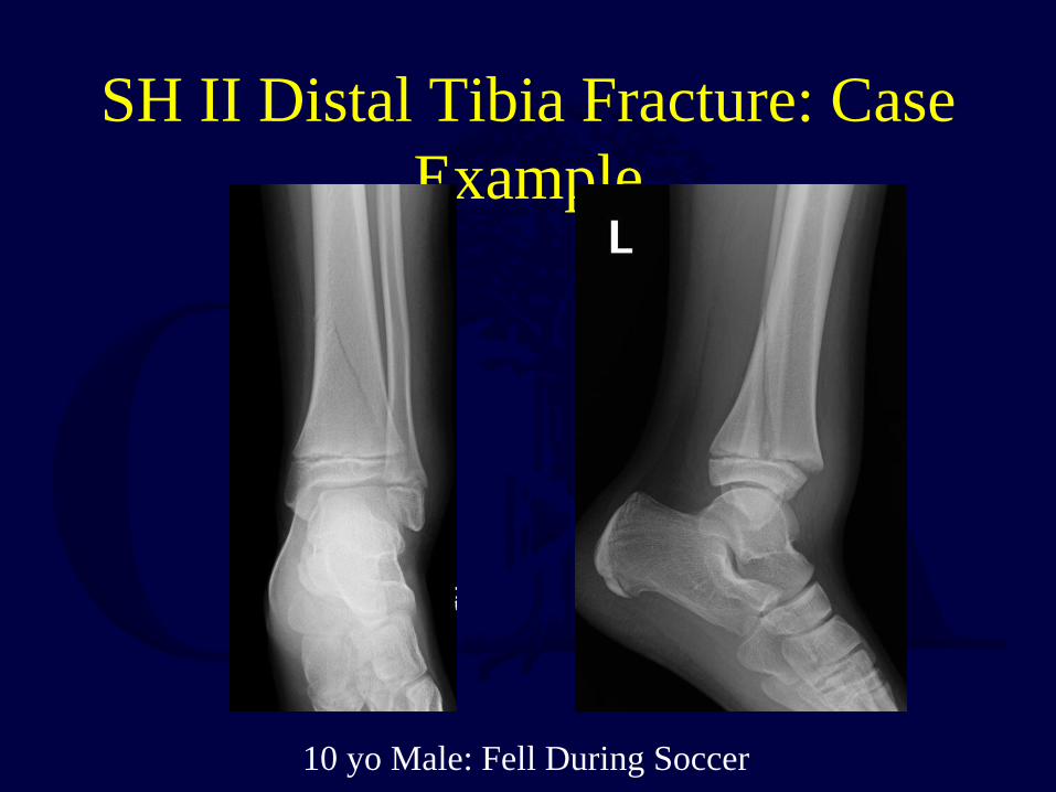
SH II Distal Tibia Fracture: Case Example
10 yo Male: Fell During Soccer
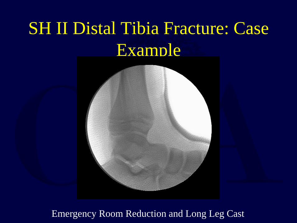
SH II Distal Tibia Fracture: Case Example
Emergency Room Reduction and Long Leg Cast
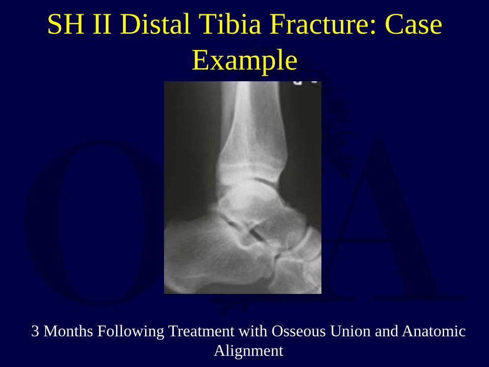
SH II Distal Tibia Fracture: Case Example
3 Months Following Treatment with Osseous Union and Anatomic Alignment

MEDIAL MALLEOLUS FRACTURES
Medial Malleolus Fractures • 20% of All Distal Tibial and Fibula Fractures in Kids • Average Age at Injury = 11 to 12 Years
• SH III Fractures are Most Common
• SH IV and V (Physeal Crush Injuries) Occur as Well • Associated Distal Fibula Fracture in 25%
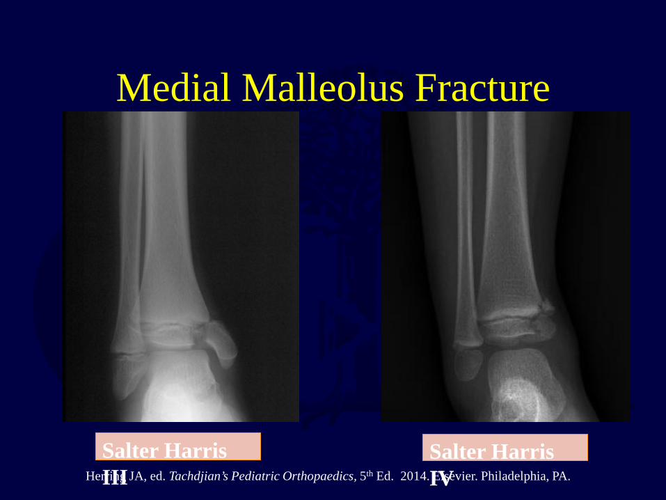
Medial Malleolus Fracture
Salter Harris III
Salter Harris IV Herring JA, ed. Tachdjian’s Pediatric Orthopaedics, 5th Ed. 2014. Elsevier. Philadelphia, PA.
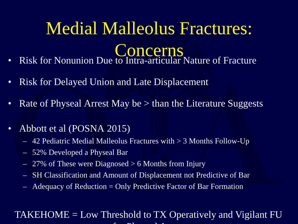
Medial Malleolus Fractures: Concerns
• Risk for Nonunion Due to Intra-articular Nature of Fracture
• Risk for Delayed Union and Late Displacement
• Rate of Physeal Arrest May be > than the Literature Suggests
• Abbott et al (POSNA 2015) – 42 Pediatric Medial Malleolus Fractures with > 3 Months Follow-Up – 52% Developed a Physeal Bar – 27% of These were Diagnosed > 6 Months from Injury – SH Classification and Amount of Displacement not Predictive of Bar – Adequacy of Reduction = Only Predictive Factor of Bar Formation
TAKEHOME = Low Threshold to TX Operatively and Vigilant FU
f Ph l A t
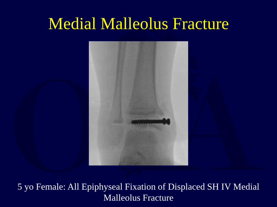
Medial Malleolus Fracture
5 yo Female: All Epiphyseal Fixation of Displaced SH IV Medial Malleolus Fracture
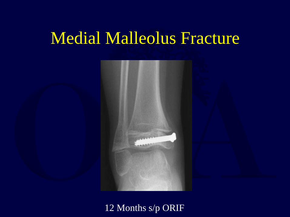
Medial Malleolus Fracture
12 Months s/p ORIF
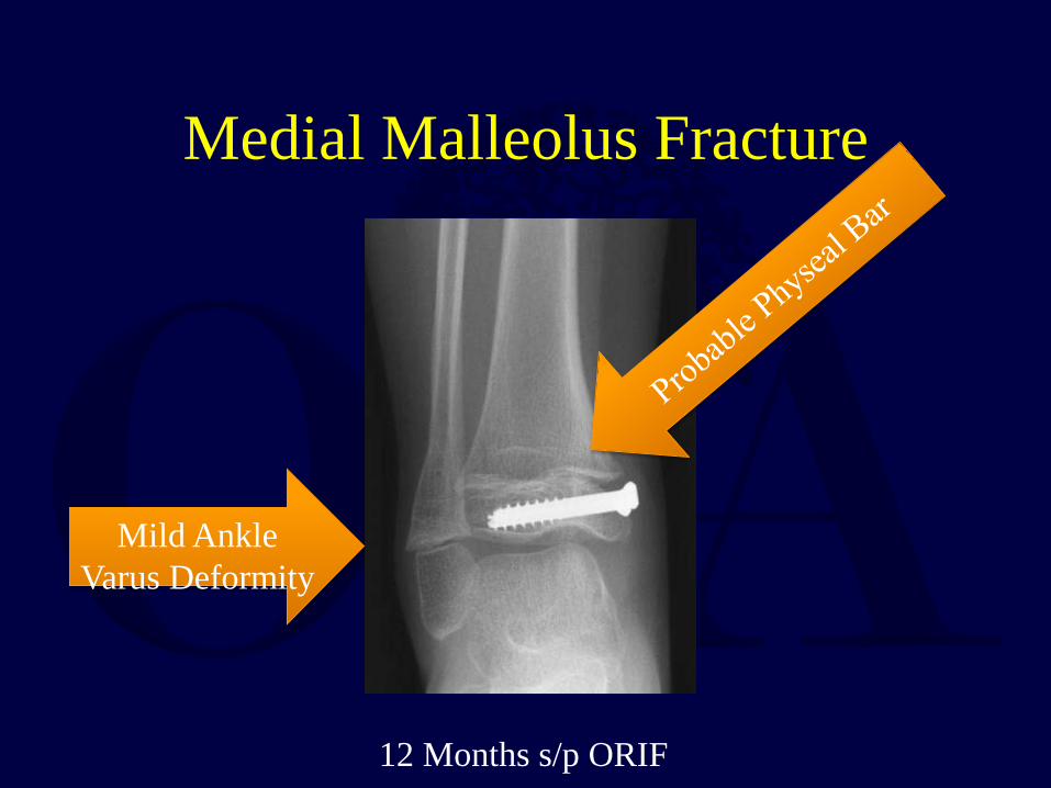
Medial Malleolus Fracture
Mild Ankle Varus Deformity
12 Months s/p ORIF
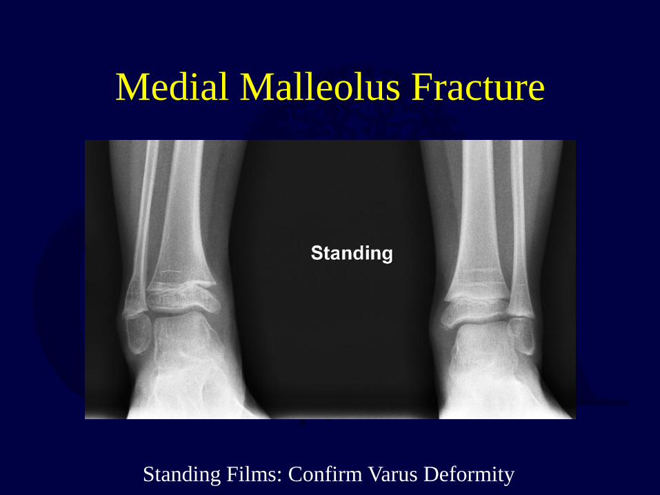
Medial Malleolus Fracture
Standing Films: Confirm Varus Deformity
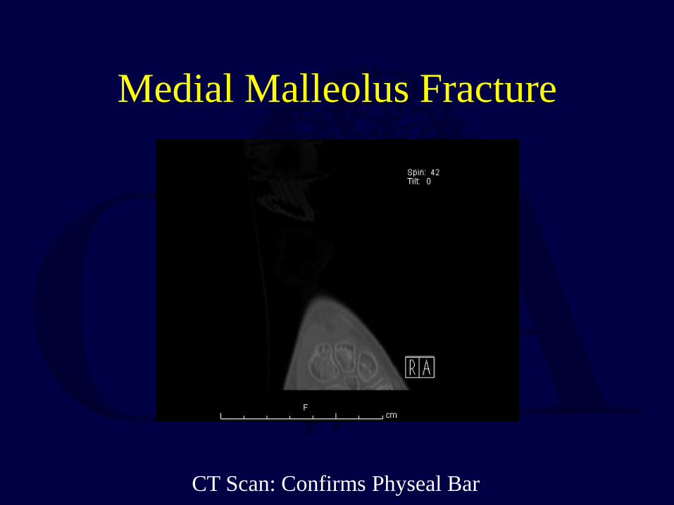
Medial Malleolus Fracture
CT Scan: Confirms Physeal Bar
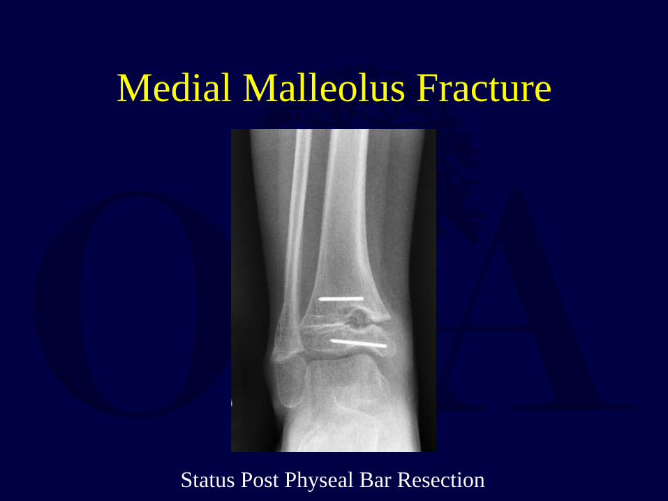
Medial Malleolus Fracture
Status Post Physeal Bar Resection
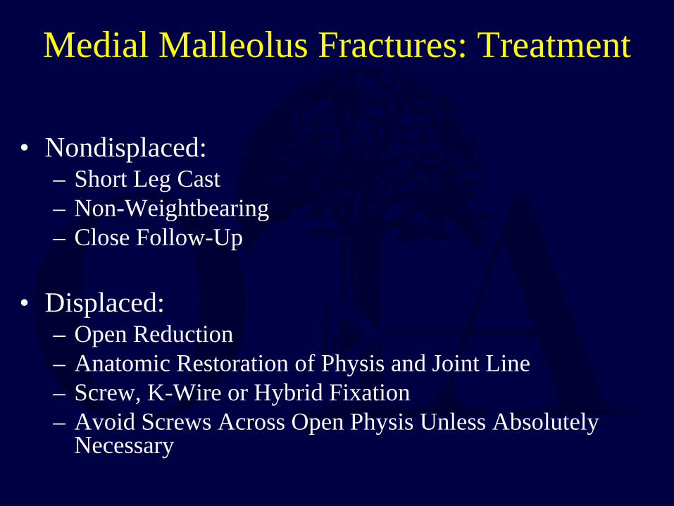
Medial Malleolus Fractures: Treatment
• Nondisplaced: – Short Leg Cast – Non-Weightbearing – Close Follow-Up
• Displaced:
– Open Reduction – Anatomic Restoration of Physis and Joint Line – Screw, K-Wire or Hybrid Fixation – Avoid Screws Across Open Physis Unless Absolutely
Necessary
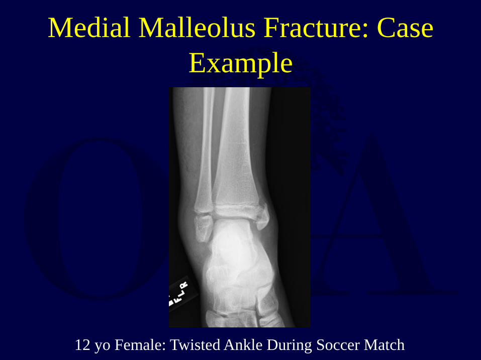
Medial Malleolus Fracture: Case Example
12 yo Female: Twisted Ankle During Soccer Match
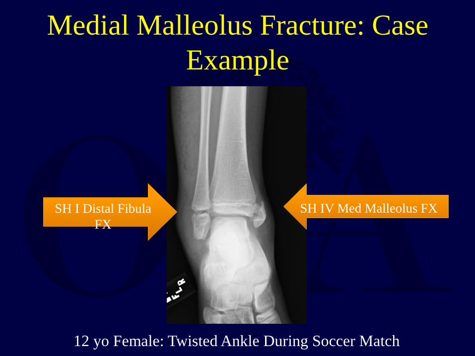
Medial Malleolus Fracture: Case Example
12 yo Female: Twisted Ankle During Soccer Match
SH I Distal Fibula FX
SH IV Med Malleolus FX
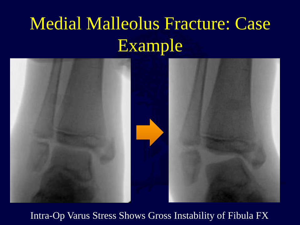
Medial Malleolus Fracture: Case Example
Intra-Op Varus Stress Shows Gross Instability of Fibula FX
Medial Malleolus Fracture: Case Example
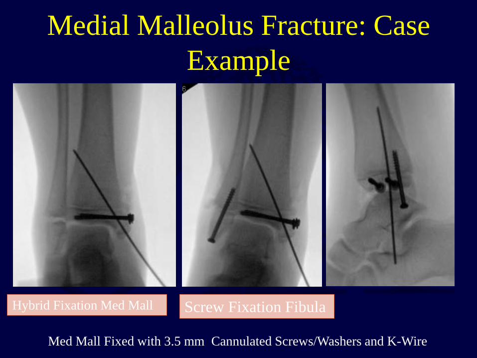
Med Mall Fixed with 3.5 mm Cannulated Screws/Washers and K-Wire
Hybrid Fixation Med Mall Screw Fixation Fibula
Medial Malleolus Fracture: Case Example
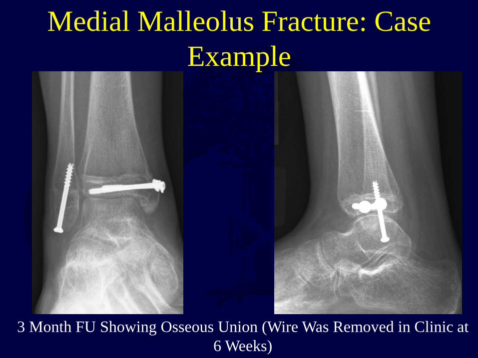
3 Month FU Showing Osseous Union (Wire Was Removed in Clinic at 6 Weeks)
Medial Malleolus Fracture: Case Example
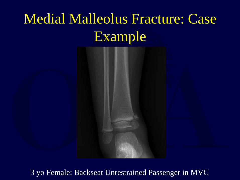
3 yo Female: Backseat Unrestrained Passenger in MVC
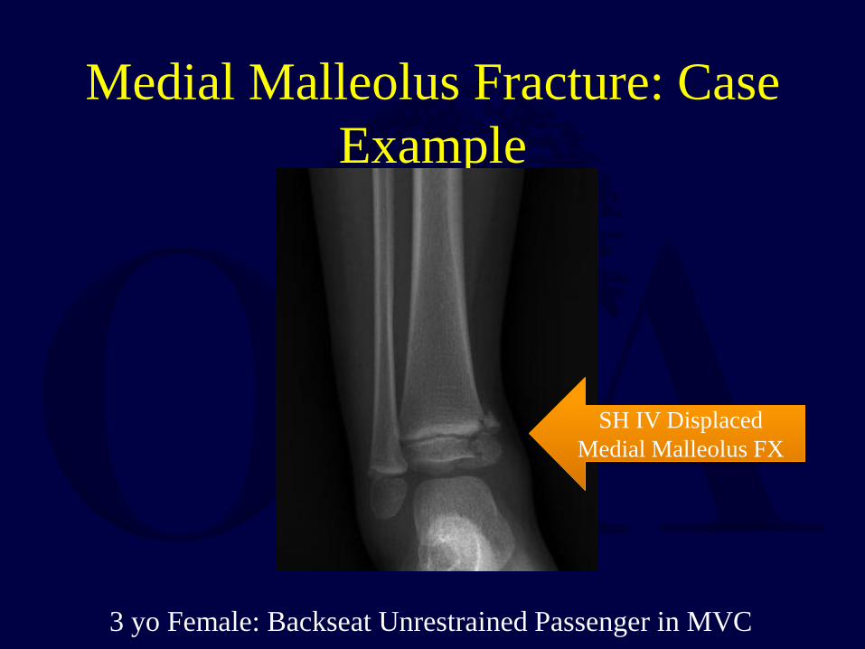
Medial Malleolus Fracture: Case Example
3 yo Female: Backseat Unrestrained Passenger in MVC
SH IV Displaced Medial Malleolus FX
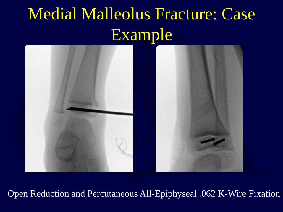
Medial Malleolus Fracture: Case Example
Open Reduction and Percutaneous All-Epiphyseal .062 K-Wire Fixation
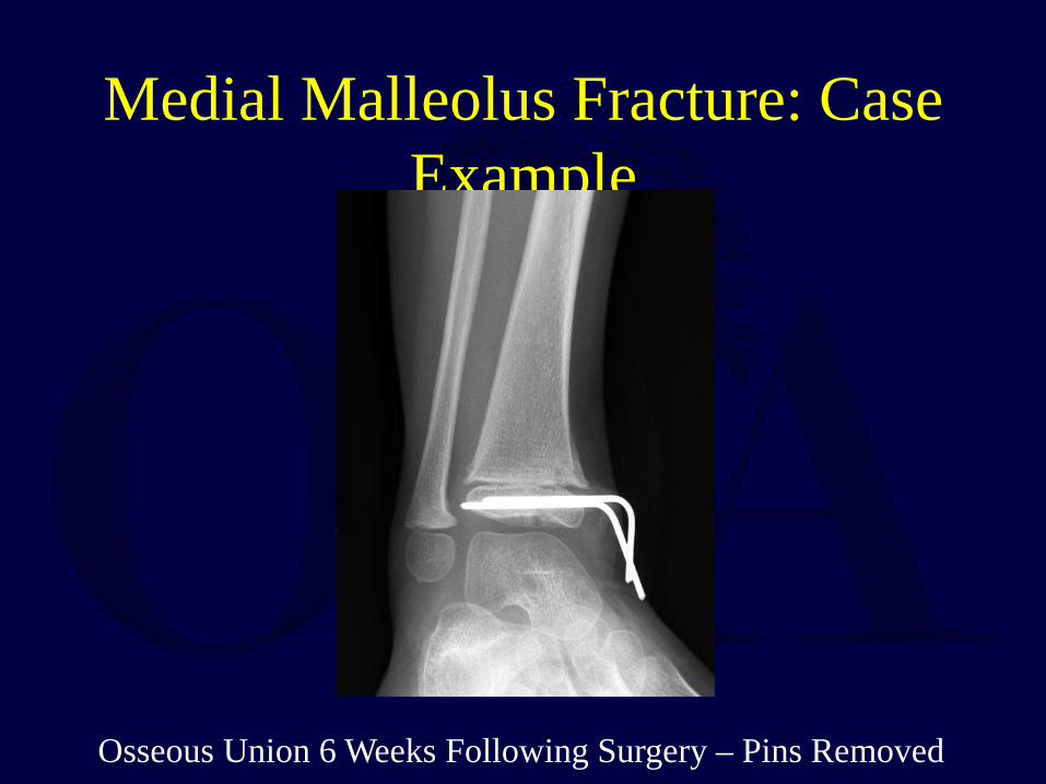
Medial Malleolus Fracture: Case Example
Osseous Union 6 Weeks Following Surgery – Pins Removed
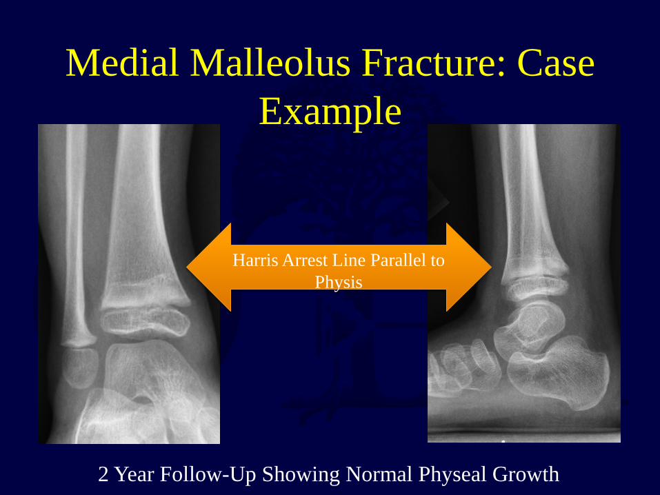
Medial Malleolus Fracture: Case Example
2 Year Follow-Up Showing Normal Physeal Growth
Harris Arrest Line Parallel to Physis
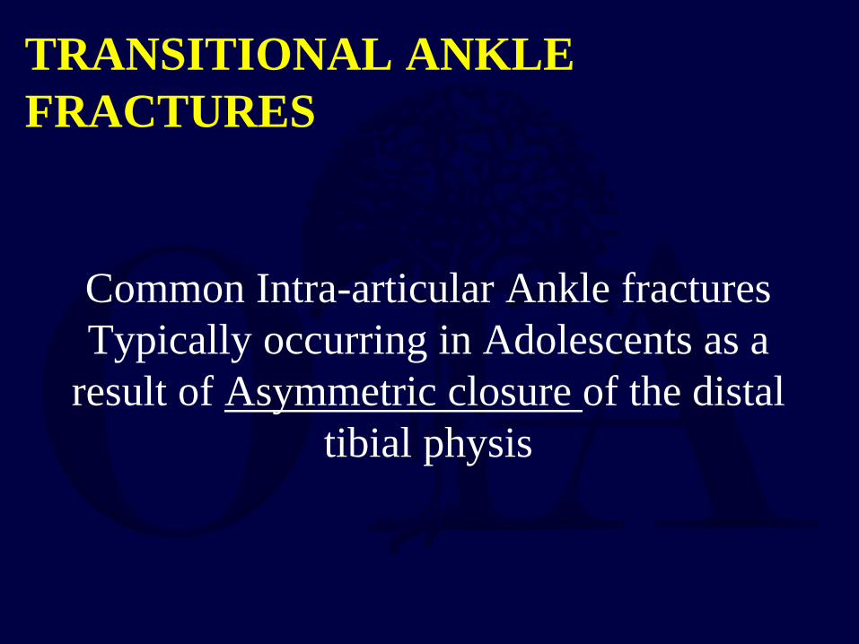
TRANSITIONAL ANKLE FRACTURES
Common Intra-articular Ankle fractures Typically occurring in Adolescents as a
result of Asymmetric closure of the distal tibial physis
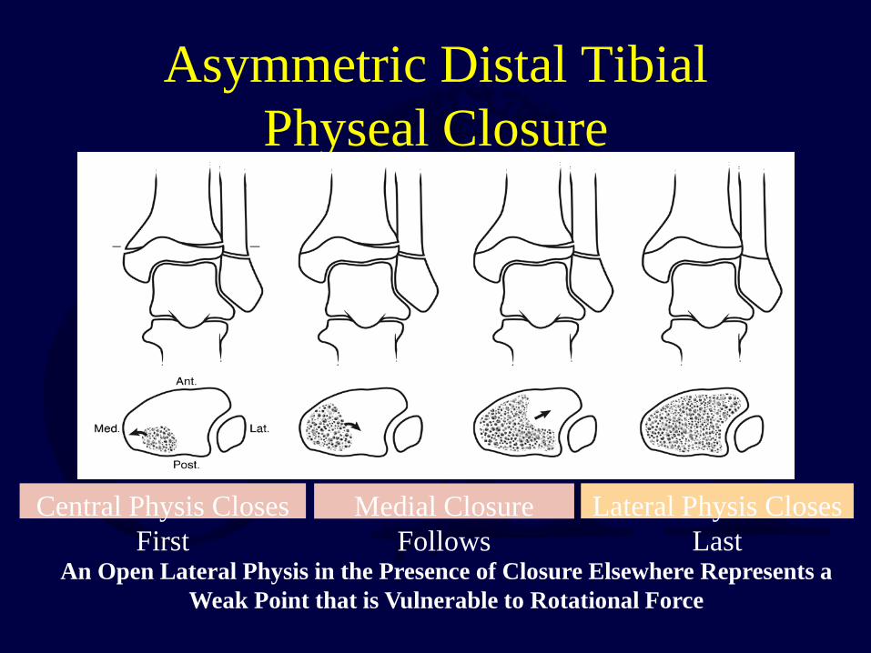
Asymmetric Distal Tibial Physeal Closure
Central Physis Closes First
An Open Lateral Physis in the Presence of Closure Elsewhere Represents a Weak Point that is Vulnerable to Rotational Force
Lateral Physis Closes Last
Medial Closure Follows
12.5 Yrs 13 Yrs 13.5 Yrs 14 Yrs
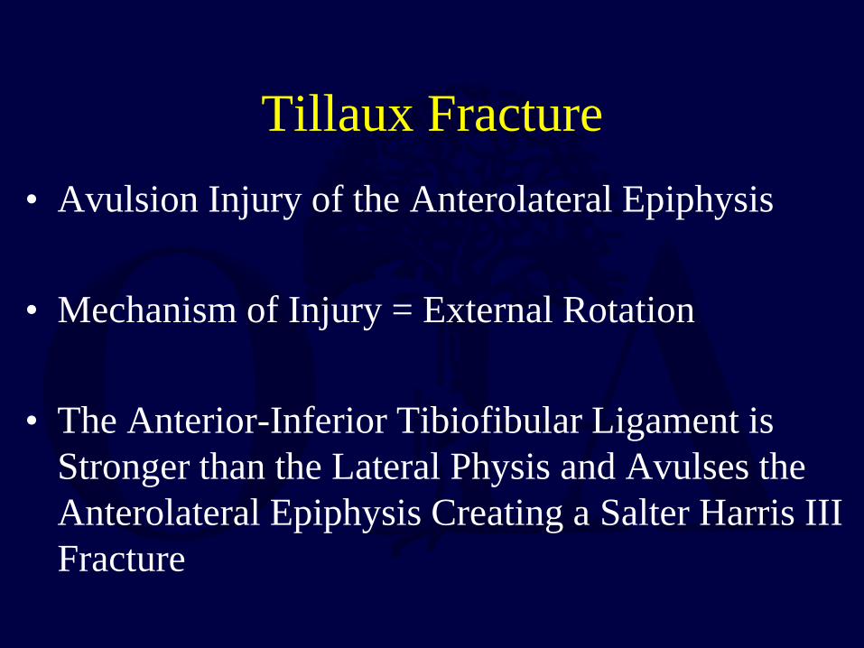
Tillaux Fracture • Avulsion Injury of the Anterolateral Epiphysis
• Mechanism of Injury = External Rotation
• The Anterior-Inferior Tibiofibular Ligament is
Stronger than the Lateral Physis and Avulses the Anterolateral Epiphysis Creating a Salter Harris III Fracture
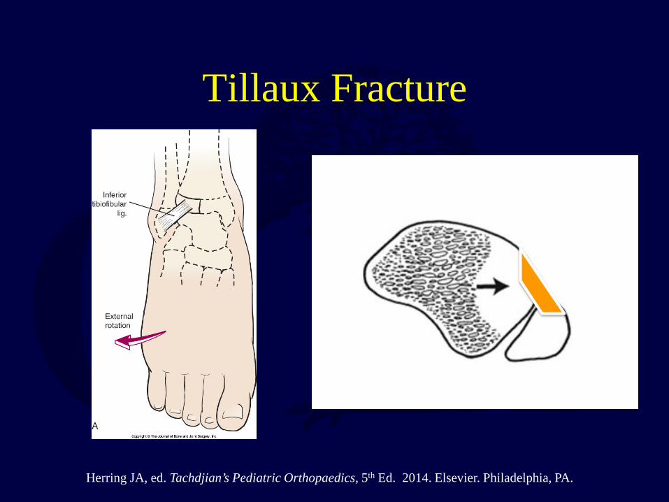
Tillaux Fracture
Inf TFL
Herring JA, ed. Tachdjian’s Pediatric Orthopaedics, 5th Ed. 2014. Elsevier. Philadelphia, PA.
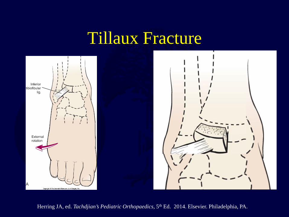
Tillaux Fracture
Herring JA, ed. Tachdjian’s Pediatric Orthopaedics, 5th Ed. 2014. Elsevier. Philadelphia, PA.
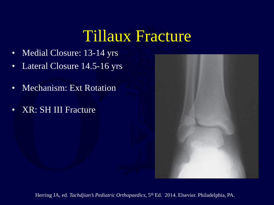
Tillaux Fracture • Medial Closure: 13-14 yrs • Lateral Closure 14.5-16 yrs
• Mechanism: Ext Rotation
• XR: SH III Fracture
Herring JA, ed. Tachdjian’s Pediatric Orthopaedics, 5th Ed. 2014. Elsevier. Philadelphia, PA.
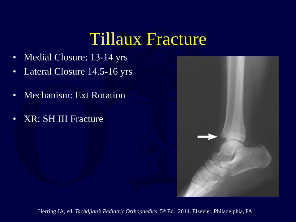
Tillaux Fracture • Medial Closure: 13-14 yrs • Lateral Closure 14.5-16 yrs
• Mechanism: Ext Rotation
• XR: SH III Fracture
Herring JA, ed. Tachdjian’s Pediatric Orthopaedics, 5th Ed. 2014. Elsevier. Philadelphia, PA.
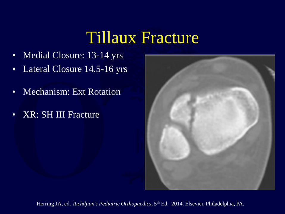
Tillaux Fracture • Medial Closure: 13-14 yrs • Lateral Closure 14.5-16 yrs
• Mechanism: Ext Rotation
• XR: SH III Fracture
Herring JA, ed. Tachdjian’s Pediatric Orthopaedics, 5th Ed. 2014. Elsevier. Philadelphia, PA.
Tillaux Fracture: Treatment Non-displaced (<1-2 mm): Cast and Close Follow-Up
Displaced:
• Closed Reduction: – Internal Rotation – Long Leg Cast – CT Scan to assess reduction
• ORIF: Failed Closed Reduction / Delayed Presentation
Lemburg et al Arch Orthop Trauma Surg 2010
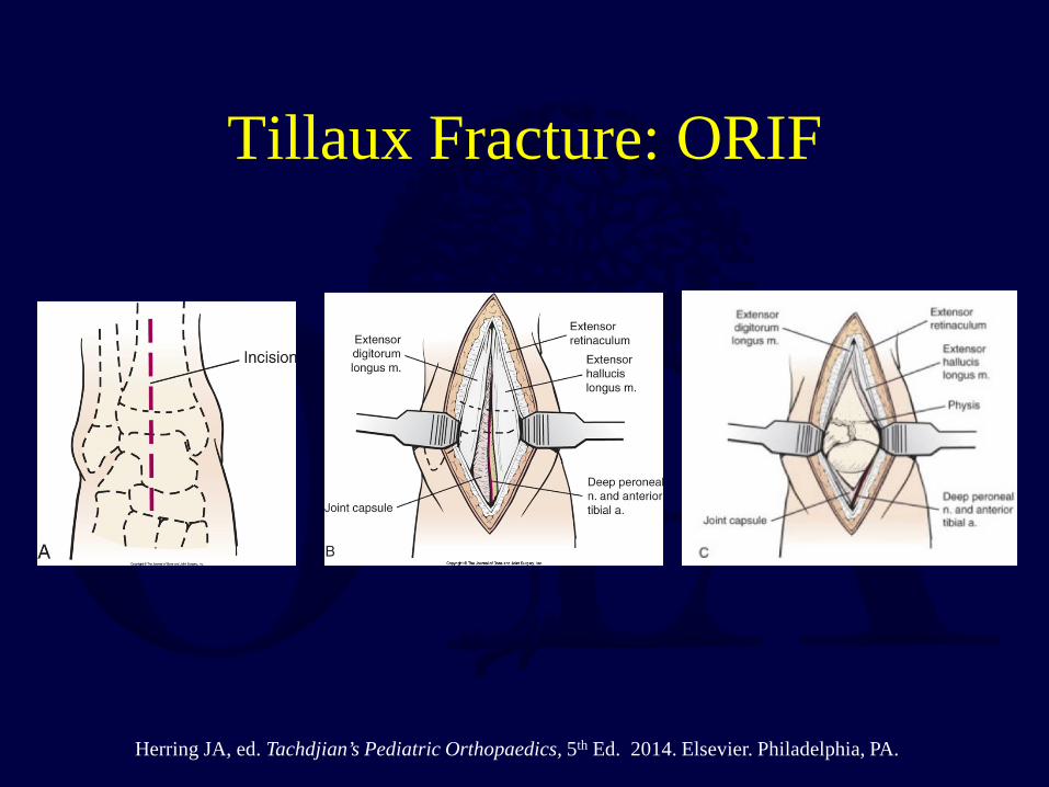
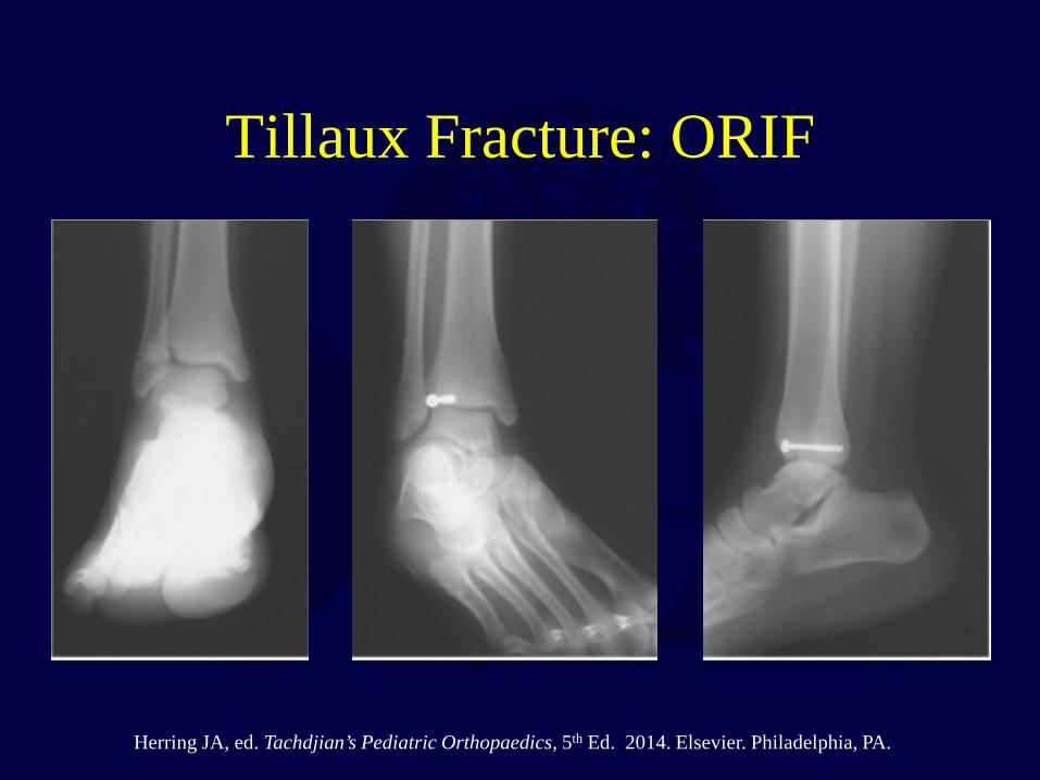
Tillaux Fracture: ORIF
Herring JA, ed. Tachdjian’s Pediatric Orthopaedics, 5th Ed. 2014. Elsevier. Philadelphia, PA.
Tillaux Fracture: ORIF • Exposure: Anterior Approach
• Reduction
– Mobilize Fragment – Reduce Articular Surface Anatomically – Reduction Clamps or Dental Pick to Hold Reduction
• Fixation
– 3.5 mm or 4.0 mm Partially Threaded Cannulated Screw + Washer – Screw Placed Lateral to Medial - Separate Percutaneous Incision – May Cross Physis Due to Eminent Closure – Screw Must Not Violate Joint
Tillaux Fracture: ORIF
Herring JA, ed. Tachdjian’s Pediatric Orthopaedics, 5th Ed. 2014. Elsevier. Philadelphia, PA.
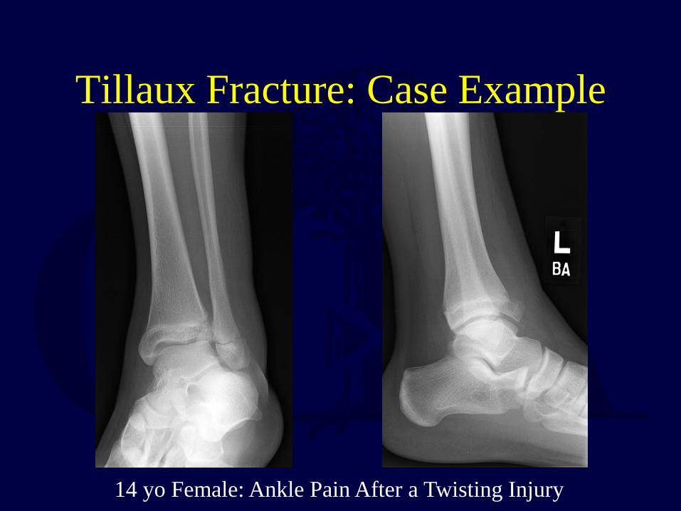
Tillaux Fracture: Case Example
14 yo Female: Ankle Pain After a Twisting Injury
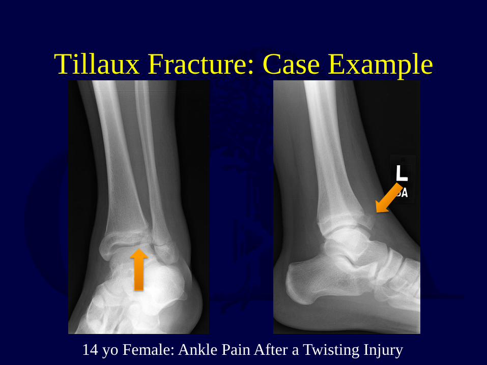
Tillaux Fracture: Case Example
14 yo Female: Ankle Pain After a Twisting Injury
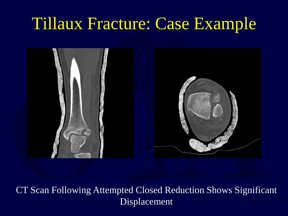
Tillaux Fracture: Case Example
CT Scan Following Attempted Closed Reduction Shows Significant Displacement
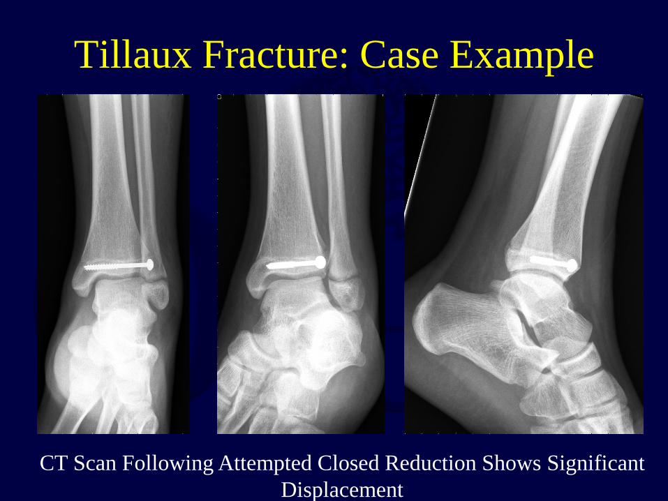
Tillaux Fracture: Case Example
CT Scan Following Attempted Closed Reduction Shows Significant Displacement
Triplane Fracture • 6-8% of Pediatric Distal Tibia Fractures
• Mechanism of Injury = External Rotation w/ Supinated Foot
• Average Age at Injury is 1 to 1.5 Years Younger than Children with
Tillaux Fractures
• Fracture Lines Occur in the Transverse, Coronal and Sagittal Planes
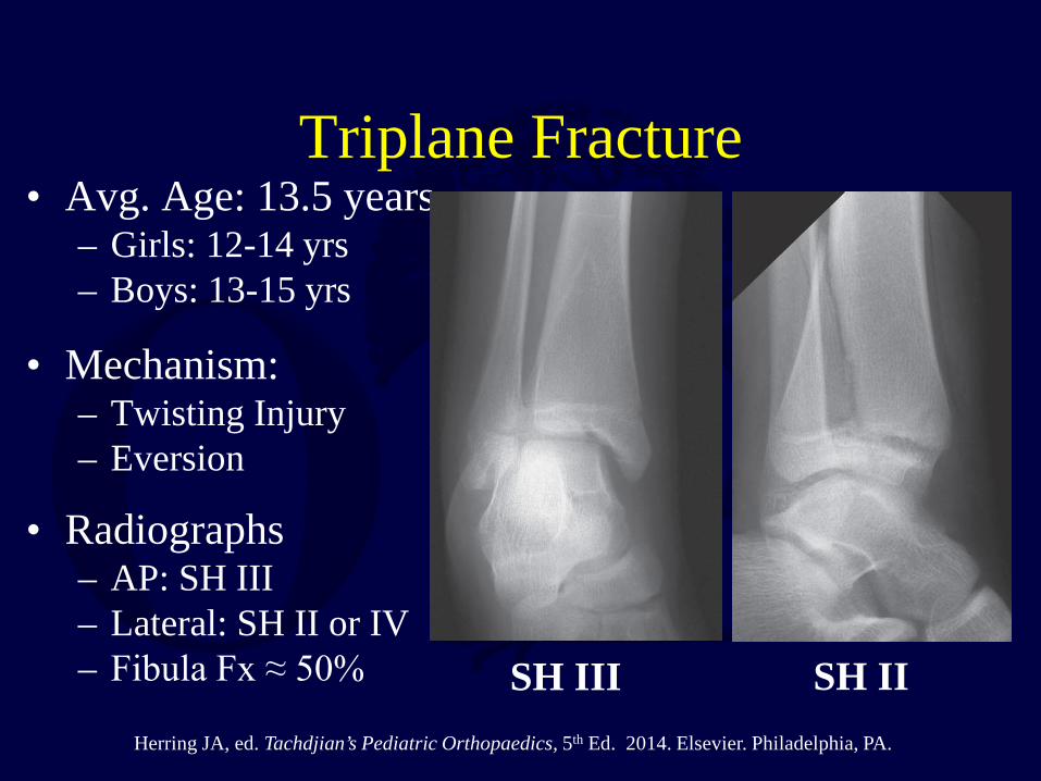
Triplane Fracture • Avg. Age: 13.5 years
– Girls: 12-14 yrs – Boys: 13-15 yrs
• Mechanism: – Twisting Injury – Eversion
• Radiographs – AP: SH III – Lateral: SH II or IV – Fibula Fx ≈ 50% SH III SH II
Herring JA, ed. Tachdjian’s Pediatric Orthopaedics, 5th Ed. 2014. Elsevier. Philadelphia, PA.
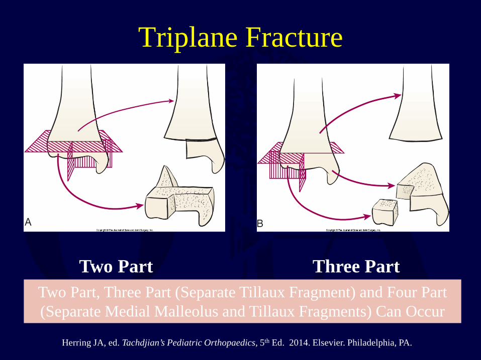
Triplane Fracture
Two Part Three Part Two Part, Three Part (Separate Tillaux Fragment) and Four Part (Separate Medial Malleolus and Tillaux Fragments) Can Occur
Herring JA, ed. Tachdjian’s Pediatric Orthopaedics, 5th Ed. 2014. Elsevier. Philadelphia, PA.
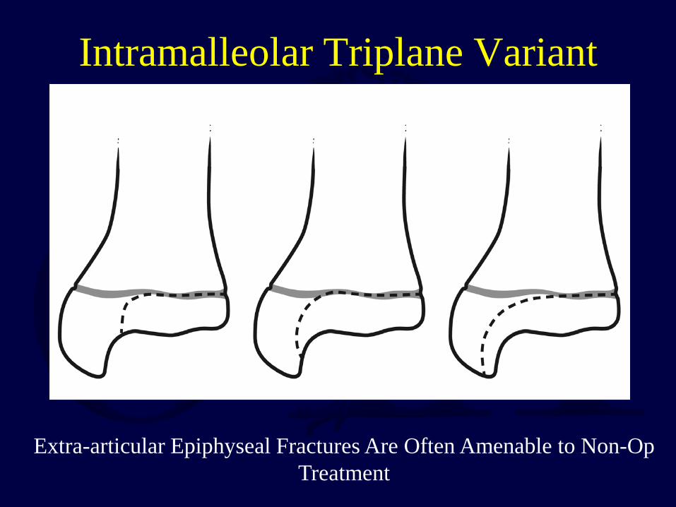
Intramalleolar Triplane Variant
Extra-articular Epiphyseal Fractures Are Often Amenable to Non-Op Treatment
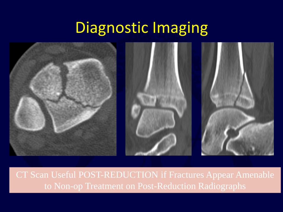
Diagnostic Imaging
CT Scan Useful POST-REDUCTION if Fractures Appear Amenable to Non-op Treatment on Post-Reduction Radiographs
Treatment Non-displaced / Extra-articular Fractures: Long Leg Cast
Displaced (>2mm):
• Closed Reduction: – Anterolateral Fragment: Internal Rotation – Anteromedial Fragment: External Rotation – Long Leg Cast – CT Scan to Assess Reduction if it Appears Well Aligned on Plain Films
• ORIF: Failed Closed Reduction / Wide Displacement
• Ertl (JBJS 1988)
– No successful closed reductions if displaced > 3mm at presentation – This had not been verified with follow-up studies – Many recommend attempted reduction despite amt. of initial displacement
Triplane Fracture: ORIF • Exposure: Anterior Approach • SH II Component Reduction
– Usually Amenable to Manipulative Reduction – Posterolateral Approach and Clamp Reduction if Closed Means Fail (Rare)
• SH III Component Reduction
– Mobilize Fragment – Reduce Articular Surface Anatomically – Reduction Clamps or Dental Pick to Hold Reduction
• Fixation
– 3.5 mm or 4.0 mm Partially Threaded Cannulated Epiphyseal Screw + Washer – Direction of Screw Based on Epiphyseal FX Location - Percutaneous Incision – Do Not Cross Physis in Younger Children – Screw Must Not Violate Joint – Second Anterior to Posterior Screw if Needed to Maintain SH II Reduction
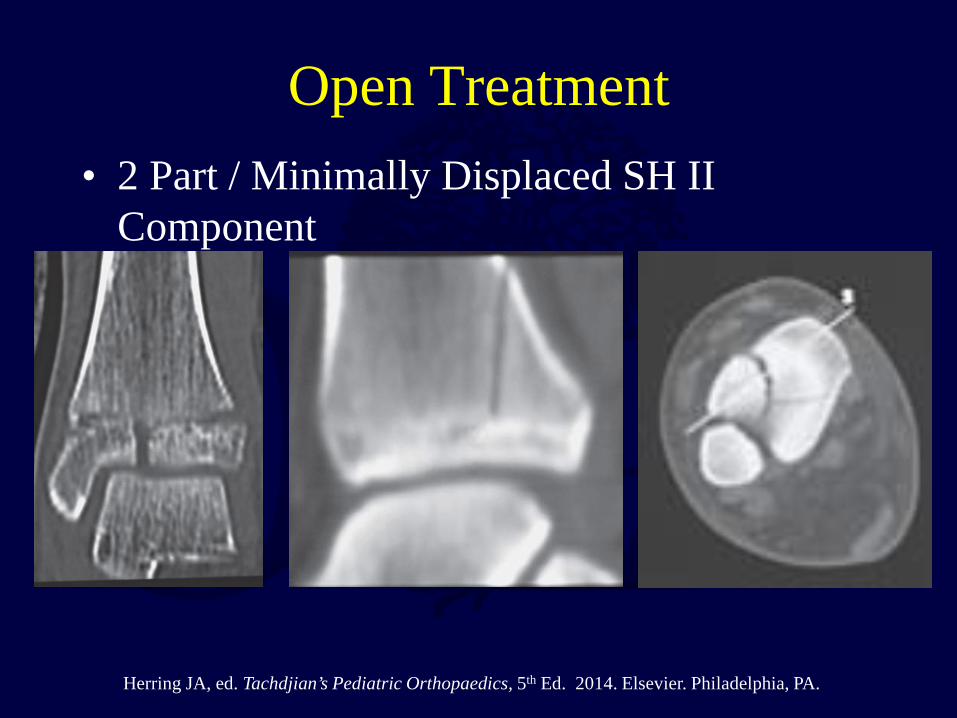
Open Treatment • 2 Part / Minimally Displaced SH II
Component
Herring JA, ed. Tachdjian’s Pediatric Orthopaedics, 5th Ed. 2014. Elsevier. Philadelphia, PA.
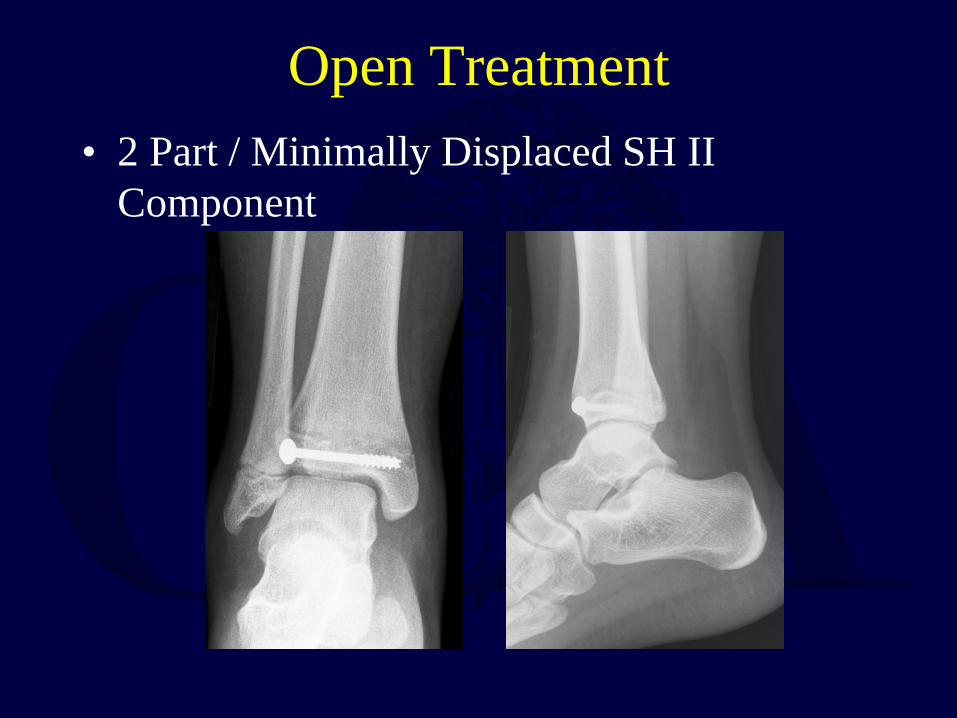
Open Treatment • 2 Part / Minimally Displaced SH II
Component
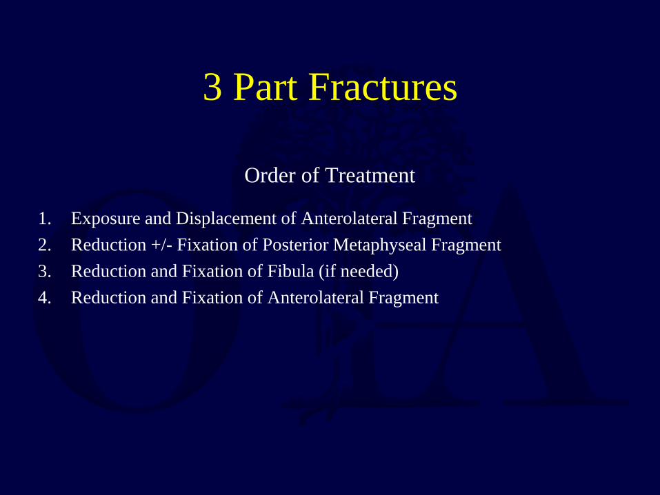
3 Part Fractures
Order of Treatment
1. Exposure and Displacement of Anterolateral Fragment 2. Reduction +/- Fixation of Posterior Metaphyseal Fragment 3. Reduction and Fixation of Fibula (if needed) 4. Reduction and Fixation of Anterolateral Fragment
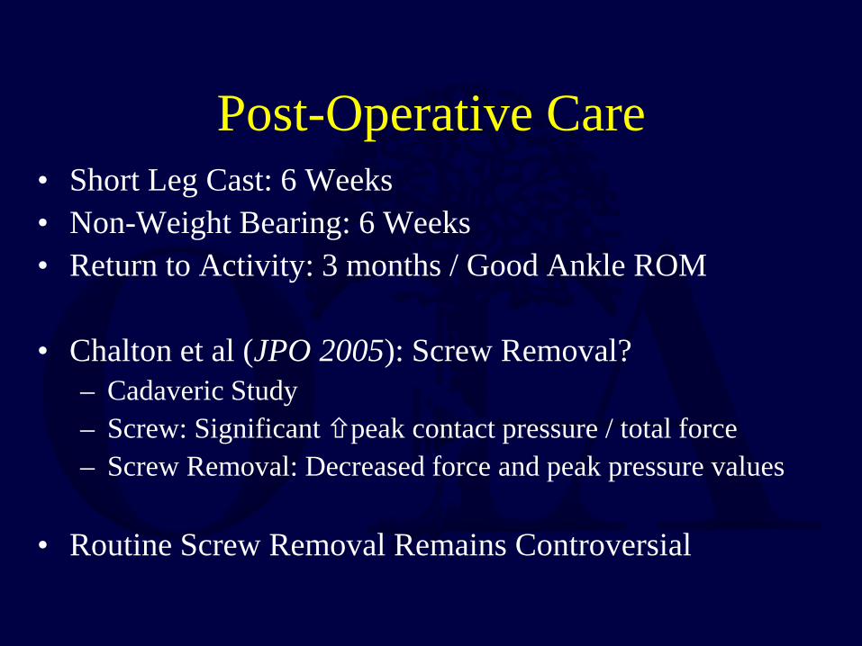
Post-Operative Care • Short Leg Cast: 6 Weeks • Non-Weight Bearing: 6 Weeks • Return to Activity: 3 months / Good Ankle ROM
• Chalton et al (JPO 2005): Screw Removal?
– Cadaveric Study – Screw: Significant peak contact pressure / total force – Screw Removal: Decreased force and peak pressure values
• Routine Screw Removal Remains Controversial
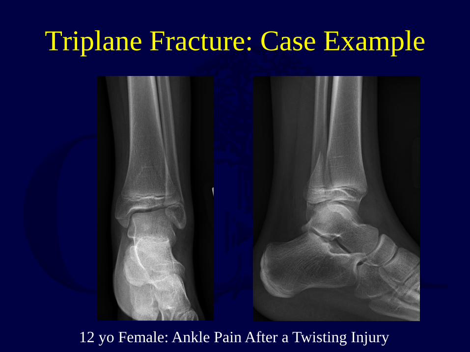
Triplane Fracture: Case Example
12 yo Female: Ankle Pain After a Twisting Injury
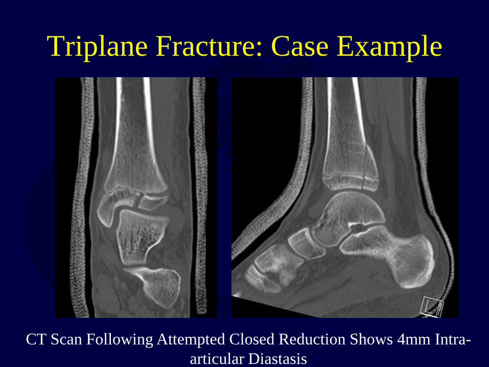
Triplane Fracture: Case Example
CT Scan Following Attempted Closed Reduction Shows 4mm Intra-articular Diastasis
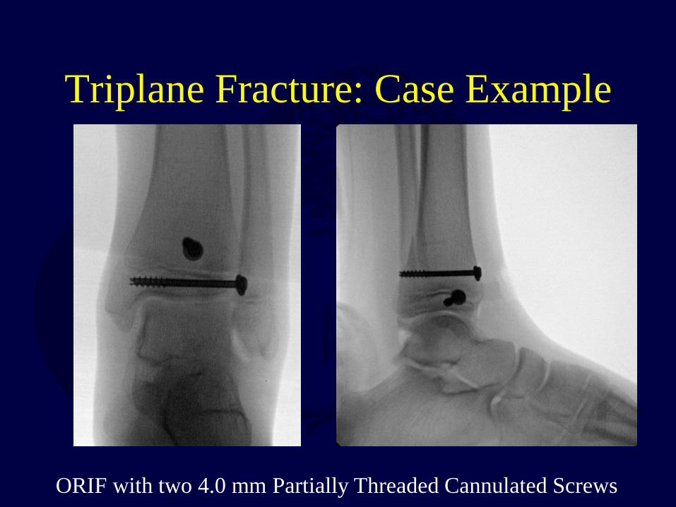
Triplane Fracture: Case Example
ORIF with two 4.0 mm Partially Threaded Cannulated Screws

TSRH