pediatric acute care review course - napnap
TRANSCRIPT

©2020
Pediatric Acute Care Review Course
Hematology, Oncology, and Palliative Care
Jessica L. Spruit, DNP, CPNP-ACAssistant Clinical Professor, College of Nursing
Wayne State University, Detroit, MIPediatric Bone Marrow Transplant Nurse Practitioner
Children’s Hospital of Michigan, Detroit, MIC.S. Mott Children’s Hospital, Ann Arbor, MI

©2020
Disclosures
No relevant financial disclosures

©2020
Learning Objectives
Objectives:1. Describe assessment, evaluation, key diagnostic studies, and
management of acute hematology and oncology disorders2. Identify common presentations of oncologic emergencies and
management3. Introduce palliative care concepts and ethical considerations

©2020
Hematologic Disorders
©2020
Anemia
• Reduction in hemoglobin concentration < 2 standard deviations below the mean for age, gender and race
• Causes (in acute settings):• Excessive red cell destruction• Excessive blood loss• Deficient red blood cell production• Ineffective hematopoiesis

©2020
Anemia
• Clinical Presentation: • Weakness, pallor, confusion, tachycardia, palpitations, flow murmur, diminished pulses, possible jaundice
• Diagnostic Evaluation:• Decreased Hgb, Hct• RBC indices and morphology
• Increased retic and low MCV = hemoglobinopathy• Increased retic and normal MCV = membrane/enzyme/immune disorder, microangiopathicanemia, DIC, infection‐induced hemolysis, or chronic blood loss
• Low MCV (low to sl elevated reticulocyte) = iron deficiency anemia, lead toxicity, Thalassemia trait, Sideroblastic anemia, anemia of chronic disease
• High MCV (low to sl elevated reticulocyte) = congenital hypoplastic or aplastic anemia, acquired hypoplasti or aplastic anemia, aplastic crisis with underlying anemia, megoblasticanemia, immune disorders

©2020
Anemia
• Management:• Treat underlying cause• Maintain oxygenation• RBC transfusion• Dietary counseling• Nutritional supplementation

©2020
Aplastic Anemia
• Rare, life threatening failure of hematopoiesis leading to peripheral pancytopenia and bone marrow aplasia
• May be congenital or acquired• Present with symptoms of pancytopenia
• Bleeding, pallor, tachycardia, petecchiae, purpura, ecchymosis, jaundice
©2020
Aplastic Anemia
• Diagnostic evaluation• Pancytopenia, decreased reticulocytes, no evidence of malignancy on peripheral smear
• Management• Transfusion, supportive care, immunosuppression, bone marrow transplant

©2020
Thalassemia
• Production of abnormal form of hemoglobin resulting in destruction of RBC’s
• Genetic: usually alpha vs beta based on descent (needs genes from both parents)
• Features: Bone deformities in the face, fatigue, growth failure, shortness of breath, jaundice; can cause fetal death
• Diagnosed usually in 1st year of life – present with anemia• Hypochromic, microcytic, decreased MCV, basophilic stippling, Hgb A
• Treatment – blood transfusion and folate supplementation• Complications: iron overload, CHF, early death

©2020
Sickle Cell Anemia
• Deoxygenated hemoglobin S polymerizes and distorts the shape of red blood cells, inherited gene (1 from each parent)
• Sickling causes hemolysis, anemia with acute and chronic complications
• Acute vaso‐occlusive crisis, intra‐pulmonary thrombosis, and infection occur with pain and or fever
• *Diagnosis is with hemoglobin electrophoresis• Can identify hemoglobin S and hemoglobin A • SCD on newborn screen (all 50 states)

©2020
Sickle Cell Disease Acute Infection
• Complications are leading cause of death• Concerning findings and symptoms:
• Anemia, splenomegaly, abdominal distention, shock, respiratory distress, hypoxia
• Management: blood cultures, antibiotics, supportive care• Most common cause of death: Sepsis or meningitis
• Pneumococcus is most likely respiratory organism, requiring penicillin prophylaxis

©2020
Sickle Cell Disease: Acute Chest
• Respiratory illness with new infiltrates• Ischemia/infarction of lung segments due to sickling• May have associated pleural effusions• Lower lobes most likely affected• Pulmonary hypertension
• Treatment• Broad spectrum antibiotics if infiltrates present• Exchange or straight PRBC transfusion• Respiratory support
13

©2020
Sickle Cell Disease: Stroke (CVA)
• Result of occlusions to cerebrovasculature• Occurs in approximately 7‐10% of all patients
• Associated with significant mortality/morbidity• Therapy: exchange transfusions to keep hemoglobin S less than 30%
• No anticoagulation required• Indication for hematopoietic stem cell transplant
14
©2020
Sickle Cell Disease: Splenic Sequestration
• Acute pooling of blood in splenic sinuses• Life‐threatening complication can lead to hypovolemic shock and death• First episode often occurs between age 3 months and 5 years• Acute weakness, pallor, abdominal distention, pain, splenomegaly
• Restore circulating volume and oxygen‐carrying capacity• PRBC transfusion (not to baseline), monitor for recurrence
15

©2020
Other Anemia
• G6PD Deficiency: genetic inborn error of metabolism, x‐linked, results in breakdown of red blood cells
• Can occur with illness, stress, medications and fava beans• Mostly asymptomatic, but symptoms include fatigue, jaundice, SOB, anemia
• PKD Deficiency: autosomal recessive, results in an enzyme deficiency with decreased production of ATP and destruction of red blood cells

©2020
Disseminated Intravascular Coagulation (DIC)
• Disturbance in normal coagulation cascade, intravascular coagulation, consumption of coagulation factors and platelets, which trigger thrombosis and hemorrhage
• Results from underlying condition• Infection, trauma, ARDS, ECMO, hematologic malignancies• High mortality
• Laboratory Findings:• Thrombocytopenia, prolonged PT, PTT, *+D‐dimer
• Management is to treat underlying cause, supportive care

©2020
Hemolytic Uremic Syndrome (HUS)
• Thrombotic microangiopathy from an endothelial injury• Classic triad: thrombocytopenia, hemolytic anemia, organ damage (often kidneys and brain)
• Forms: • Infection‐induced
• *E.coli 0157 is the most common etiology, shiga toxin
• Co‐existing condition• Cobalamin C defect• Atypical
• Most common in children <4 yrs

©2020
Hemolytic Uremic Syndrome (HUS)
• Symptoms: abdominal pain, watery, *bloody diarrhea, fever, lethargy, irritability, vomiting
• Findings: pallor, petechiae, ecchymosis, hematuria, oliguria, azotemia, hypertension
• Supportive therapy• Careful rehydration• Dialysis as indicated• Strep pneumoniae HUS may require antibiotic therapy (amoxicillin, third generation cephalosporin)
• Eculizumab may be indicated for atypical HUS
©2020
Immune Thrombocytopenia Purpura (ITP)
• Immune‐mediated• Isolated thrombocytopenia (<100 x 109/L) in the absence of other causes of thrombocytopenia
• Presentation: • Often history of recent viral illness • Bruising, petechiae, purpura, epistaxis, hematuria, etc.
• Acute: Depends on presentation• Consider IVIG, corticosteroids, WinRhoD for severe bleeds
• *Greatest concern is bleeding in brain

©2020
Henoch‐Schonlein Purpura (HSP)
• Immune mediated small‐vessel vasculitis• Often presents following a respiratory tract infection• Presentation:
• Rash, pain and swelling• Hematuria, proteinuria, hypertension• Kidney is the main origin of chronic disease, can progress to renal failure
©2020
Henoch‐Schonlein Purpura (HSP)
• Diagnostic criteria: palpable purpura plus• Diffuse abdominal pain• Acute arthritis• Arthralgia• Hematuria• Proteinuria• Predominant IgA deposition on tissue biopsy
• Generally self‐limited, although 1/3 of cases will relapse
©2020
Additional Coagulation Disorders
• Thromboembolytic disorders: DVT, Pulmonary Embolus, Renal or Portal vein thrombosus, Cerebral sinus venous thrombosis
• Most sensitive diagnostic study for PE, DVT: CT angiography
• Heparin‐Induced Thrombocytopenia (HIT)• Immune mediated drug reaction, increased thrombosis
• Methemoglobinemia: uncommon cause of cyanosis from heme iron in the ferric vs ferrous state
• Chocolate appearing blood• Treatment is methylene blue IV

©2020
Hemophilia
• Hereditary recessive X‐linked chromosomal disease• Deficiency in Factor VIII (hemophilia A) or Factor IX (hemophilia B)
• Clotting or coagulation disorder• Treatment and prevention:
• Replace missing clotting factor (Factor VIII, IX – usually recombinant)• PRBC, fresh frozen plasma, cryoprecipitate transfusion• Can use DDAVP for mild to moderate bleeding• Focus is on education to prevent bleeding
24

©2020
VonWillebrand Disease
• Deficiency or defect of the vonWillebrand protein• Often diagnosed later in life
• Present with easy bruising, frequent epistaxis, heavy menstrual bleeding, surgical/dental procedure bleeding
• Treatment and prevention with DDAVP, Amicar
25

©2020
Stroke (CVA)
• May be ischemic or hemorrhagic• Dysfunction of brain activity due to interruption of blood flow in brain
• Ischemic strokes: occur with cardiac disease, hematologic disorders (SCD), primary vasculitis, lipid abnormalities, metabolic abnormalities, dehydration/shock

©2020
Stroke (CVA)
• Hemorrhagic strokes: occur with vascular malformation, cavernous malformation, aneurysm, brain tumor, thrombocytopenia or other hematologic problems, coagulopathies, spontaneous dissection
©2020
Stroke
• Presentation: • Acute neurologic symptoms, headache, confusion, lethargy and possible seizures
• Diagnosis: history, neurologic exam, head CT?, MRI • Labs: CBC, coags, LFT’s, electrolytes, ESR• Physical Exam: neurological evaluation, skin assessment, abdominal exam, cardiac evaluation
•Management: • monitoring, treatment is focused on the underlying cause, anticoagulation therapy? (controversial)

©2020
Transfusion Concerns
• Anaphylaxis• Occurs very quickly• Bronchospasm, cough, respiratory distress, hypotension, urticaria, pruritis, vomiting
• Acute hemolytic reaction• Hemolysis secondary to ABO incompatibility• Fever, chills, hypotension, lumbar pain, shock, dyspnea, hemoglobinuria, diaphoresis, anxiety, chest pain, restlessness, DIC
• Nonhemolytic reaction• Antigen or antibody reaction to transfused leukocytes or plasma proteins
• Fever, chills, headache, palpitations, hives, erythema, pruritis

©2020
Transfusion Concerns
• TACO• Circulatory capacity exceeded circulatory overload and pulmonary edema
• Respiratory distress, hypoxia, rales, hypertension• TRALI
• Immune response, involves granulocyte activation/degranulation and alveolar capillary membrane injury
• Onset within 6 hours, acute hypoxemia, noncardiogenic pulmonary edema

©2020
Question
The BEST evidence‐based management of a child with sickle cell disease who is diagnosed with ischemic stroke is:a. Administration of a thrombolytic drug such as retaplaseb. Anticoagulant therapy with aspirinc. Exchange transfusiond. Heparin bolus and continuous heparin infusion
©2020
Answer
The BEST evidence‐based management of a child with sickle cell disease who is diagnosed with ischemic stroke is:
C. Exchange transfusion

©2020
Question
The most appropriate test to confirm the diagnosis of sickle cell anemia in a newborn is:a. Sickledexb. Hemoglobin electrophoresisc. Newborn screend. Mean corpuscular volume (MCV)

©2020
Answer
The most appropriate test to confirm the diagnosis of sickle cell anemia in a newborn is:
B. Hemoglobin electrophoresis

©2020
Question
You suspect a patient with sepsis is developing DIC. The diagnostic test of choice to confirm this diagnosis is:a. D‐dimerb. Prothrombin timec. Erythrocyte sedimentation rated. Fibrinogen
©2020
Answer
You suspect a patient with sepsis is developing DIC. The diagnostic test of choice to confirm this diagnosis is:
A. D‐dimer

©2020
Question
A 16‐year old female presents with obesity and acute shortness of breath. She has no significant past medical history and her only medication is oral contraceptives. What is the most sensitive test for the suspected diagnosis?a. CT angiographyb. Venous ultrasoundc. Chest x‐rayd. MRI of the chest

©2020
Answer
A 16‐year old female presents with obesity and acute shortness of breath. She has no significant past medical history and her only medication is oral contraceptives. What is the most sensitive test for the suspected diagnosis?
A. CT angiography

©2020
Oncologic Disorders
©2020
Pediatric Oncology Diagnoses
Blood Cell Cancers:• Leukemia – ALL, AML, CML• Lymphoma – Hodgkin and non‐HodgkinCentral Nervous System TumorsSolid Tumors: • Neuroblastoma• Osteosarcoma, Ewing sarcoma, Rhabdomyosarcoma• Wilms Tumor, Hepatoblastoma• Retinoblastoma

©2020
Acute Lymphoblastic Leukemia
• Approximately 20% of pediatric cancer diagnoses annually• 75% of pediatric leukemias• Increased risk associated with some inherited conditions
• Trisomy 21• Fanconi’s anemia• Klinefelter Syndrome• Shwachman‐Diamond Syndrome
• Defined as B‐cell or T‐cell

©2020
Acute Lymphoblastic Leukemia
• Pathophysiology:• Dysregulation of hematopoietic development secondary to genetic abnormalities
• Stem cells do not differentiate or mature properly• These immature cells are called “blasts”• Blasts accumulate in the marrow and sometimes, in solid organs
• Thymus, liver, spleen, kidneys, CNS, testes
• Leads to impairment of failure of bone marrow function

©2020
Acute Lymphoblastic Leukemia
• Common presenting symptoms: bone pain, anemia, fatigue, fever, bruising, bleeding
• Diagnosis• Bone marrow aspirate• Lumbar puncture
• Treatment:• Multi‐phase therapy, largely dictated by risk and response to therapy• CNS Therapy

©2020
Acute Myelogenous Leukemia
• 15‐25% of leukemia in children• Slightly increased incidence ages >10• Inherited conditions associated with increased risk• Appear more ill on presentation (cytopenic, chloromas, bleeding)• Overall EVS 50‐70%, lower risk 70‐80%• Diagnosis
• Bone marrow aspirate• Lumbar puncture

©2020
Acute Myelogenous Leukemia
• Treatment:• Shorter, more intense than ALL• Risk stratifications better outcomes• HSCT vs. continued chemo• Targeted therapies (tyrosine kinase inhibitors, etc)
©2020
Lymphoma
• Non‐Hodgkin Lymphoma (NHL)• Malignant proliferation of lymphocytes (T/B/indeterminant cells)• Multiply rapidly, unpredictable, aggressive
• Hodgkin Lymphoma (HL)• Usually spreads to adjacent lymph nodes• Slower, more orderly spread

©2020
Non‐Hodgkin Lymphoma
• 6% of childhood cancers• 3 types:
• Lymphoblastic lymphoma• Mature B‐cell lymphoma
• Burkitt, Diffuse large B Cell, Primary mediastinal B Cell
• Anaplastic Large Cell Lymphoma• Males > Females• Ages 5‐15 years old• Overall EVS ~70‐85%

©2020
Non‐Hodgkin Lymphoma
• Presentation• Dependent on disease site involved• Abdomen: mass, pain, nausea, vomiting, change in bowel habits, hematochezia
• Mediastinum: Dysphagia, SVC syndrome, chest pain• Head/Neck: facial swelling, snoring, rhinorrhea, cervical lymphadenopathy
©2020
Non‐Hodgkin Lymphoma
• Diagnosis:• Imaging: CXR, CT scan, PET • Biopsy & pathology• Staged according to St. Jude Staging System• Often present emergently, require stabilization
• Associated with ATLS and SVC syndrome
• Treatment:• Based on type and extent of involvement• Multiagent, combination chemotherapy
• + IT chemotherapy if CNS involved• Limited role of radiation

©2020
Hodgkin Lymphoma
• 5% of cancers in children & adolescents• 4 sub‐types:
• Lymphocyte predominant• Nodular sclerosing• Mixed cellularity• Lymphocyte depleted
• Males > Females in kids <10 yrs• Males = Females in adolescents• Ages 15‐35 and >50 years old• Overall EVS ~80%
©2020
Hodgkin Lymphoma
• Presentation• Lymphadenopathy• Fatigue, anorexia, pruritis• SVC Syndrome
• Cough, shortness of breath, orthopnea • B Symptoms
• Fevers, wt loss, drenching night sweats

©2020
Hodgkin Lymphoma
• Diagnosis:• Imaging: CXR, CT scan, PET • Biopsy & pathology• Staged according to Ann Arbor Staging• Appropriate consultations
• Treatment:• Stratified into risk categories• Chemotherapy & re‐staging• Involved field radiation when indicated, usually following chemo completion• Autologous/allogeneic HSCT• Immunotherapy
©2020
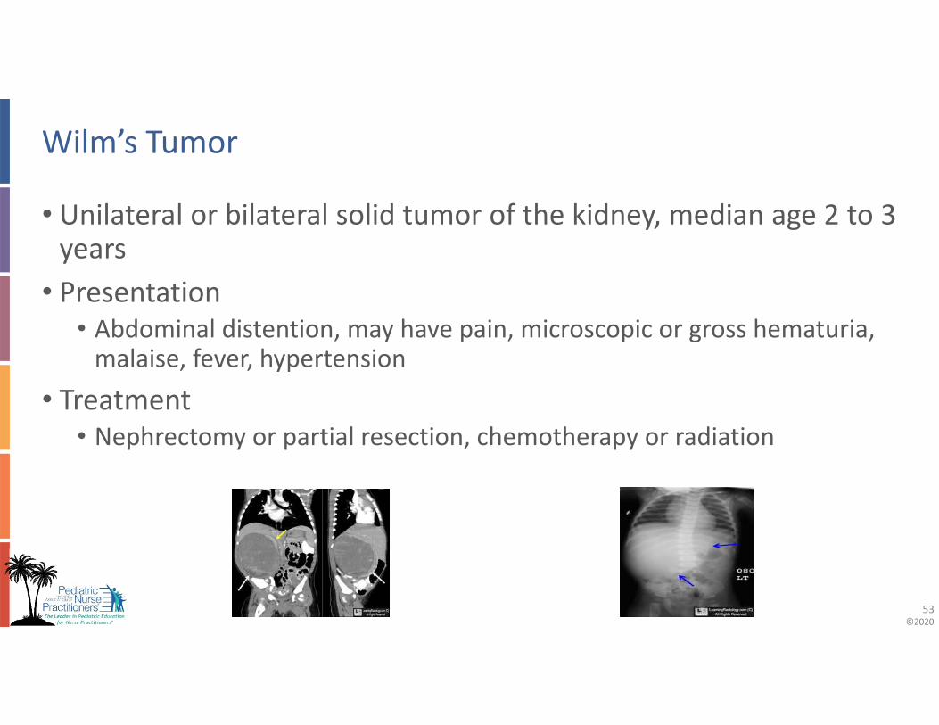
Wilm’s Tumor
• Unilateral or bilateral solid tumor of the kidney, median age 2 to 3 years
• Presentation• Abdominal distention, may have pain, microscopic or gross hematuria, malaise, fever, hypertension
• Treatment• Nephrectomy or partial resection, chemotherapy or radiation
53
©2020
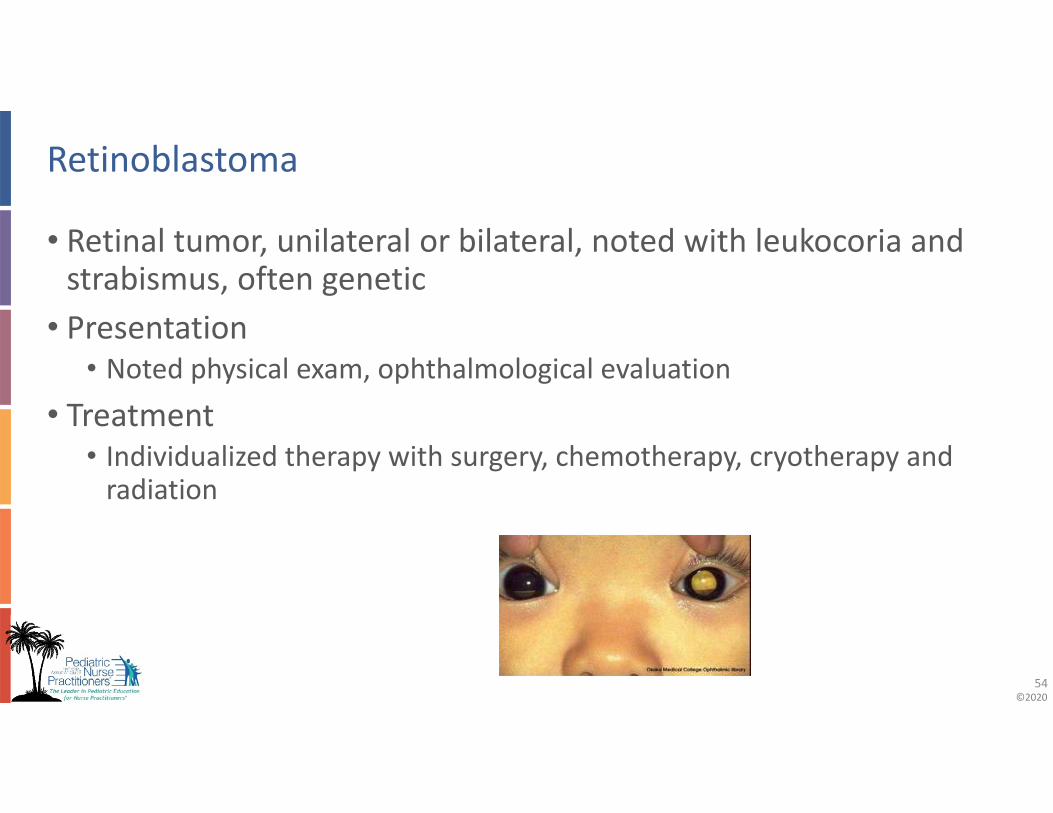
Retinoblastoma
• Retinal tumor, unilateral or bilateral, noted with leukocoria and strabismus, often genetic
• Presentation• Noted physical exam, ophthalmological evaluation
• Treatment• Individualized therapy with surgery, chemotherapy, cryotherapy and radiation
54

©2020
Neuroblastoma
• Neoplasm of the sympathetic nervous system• Most common extracranial solid tumor in childhood cancer• Presentation
• Primary tumors in the chest, abdomen, pelvis• Often present with metastasis• CT, MRI, MIBG
• Treatment• Multi‐modal, requires HSCT and immunotherapy + retinoids
55
©2020
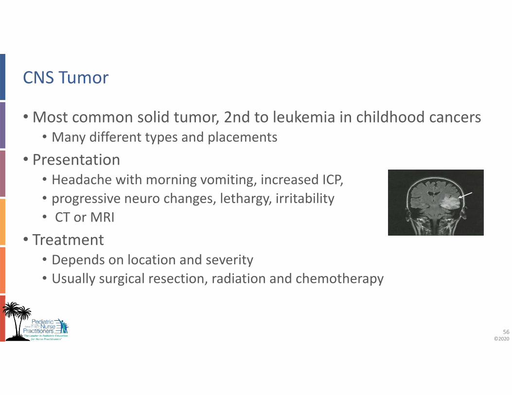
CNS Tumor
• Most common solid tumor, 2nd to leukemia in childhood cancers• Many different types and placements
• Presentation• Headache with morning vomiting, increased ICP, • progressive neuro changes, lethargy, irritability• CT or MRI
• Treatment• Depends on location and severity• Usually surgical resection, radiation and chemotherapy
56

©2020
Bone Tumors
• Various types• Ewings, osteosarcoma
• Presentation• Pain over affected area, fever, weight loss, increased ESR• Imaging – first plain film, than MRI
• Treatment• Treatment usually involves resection, chemotherapy• May have limb salvage or amputation
57

©2020
Oncologic Emergencies

©2020
Mediastinal Mass
• Pathophysiology• Hemodynamic instability, superior vena cava syndrome, hypoxemia, hypoventilation, dysphagia
• Presentation• Cough, dyspnea, hoarseness, stridor, orthopnea, syncope, tachycardia, jugular vein distension, swollen face, cyanosis
• Management• Prone position, caution with anesthesia, positive pressure ventilation if necessary, chest x‐ray, CT
• Emergent radiation or chemotherapy may be required
59

©2020
Tumor Lysis Syndrome
• Metabolic derangements caused by rapid release of intracellular components of lysed malignant cells
• Risks include high turnover rates, large tumor burden, sensitivity to therapy, dehydration, renal dysfunction
• Presentation• Hyperuricemia, hyperphosphatemia, hyperkalemia, hypocalcemia, elevated LDH
• Elevated creatinine, arrhythmia, seizure• Management
• Aggressive hydration, diuretics, correct electrolytes, rasburicase or allopurinol for hyperuricemia, dialysis may be needed
60

©2020
Hyperleukocytosis
• WBC > 100,000/mm3• Increased blood viscosity/leukostasis leads to sludging in the microcirculation of the lungs and CNS
• Presentation• CNS: headache, tinnitus, ataxia, behavioral changes, seizure, hemorrhage, infarct
• Lungs: tachypnea, respiratory distress, hypoxia, hemorrhage, ARDS, respiratory failure
• Other organs: renal artery/vein distention, renal failure, papilledema, dactylitis, priapism, cardiac failure
61

©2020
Hyperleukocytosis
• Management• Prompt chemotherapy, dexamethasone for pulmonary leukostasis, avoid RBC transfusion until WBC reduced
• Leukapheresis and exchange transfusion controversial
62

©2020
Typhlitis / Intra‐abdominal Emergencies
• Inflammation of the cecum• Presentation
• Right lower quadrant abdominal pain, +/‐ fever, mucositis• Management
• Broad spectrum antibiotics (include gram negative coverage), IV fluids, pain management

©2020
Mass Syndromes
• Abdominal, spinal compression via tumor mass• Presentation
• Altered neurologic condition/status, pain, compartment syndrome• Management
• Decrease mass effect via radiation, surgical debulking or chemotherapy
64

©2020
Case Study
• Ten‐year old male presents with a 2‐week history of fever and cough with exertional dyspnea.
• Deny any rhinorrhea, throat pain, diarrhea, vomiting, trauma, or sick contacts
• Lives with grandmother and two siblings. No smoke exposures. No pets.

©2020
Case Study
• Evaluated at primary care PNP office, where cough was present and splenomegaly was palpated
• Immediate referral to the emergency department• Physical Exam:
• VS: T 37.3oC, HR 108, RR 24, BP 117/73mmHg, Ht/Wt 25th %ile• General: pale, tired appearance• Resp: Diminished breath sounds, no wheeze, dry cough noted• Abdominal: spleen tip palpable below left costal margin, liver edge palpable at 3cm below the right costal margin
• Skin: numerous petechiae• Lymph: small anterior cervical and inguinal lymphadenopathy

©2020
Case Study
• Diagnostic Evaluation:
• Differential: 48% atypical lymphocytes• LDH 650, electrolytes WNL, uric acid 24.5 mg/dL
Lab Result
WBC 10600/mm3
Hemoglobin 9.2g/dL
Platelets 103,000/mm3
©2020
Case Study
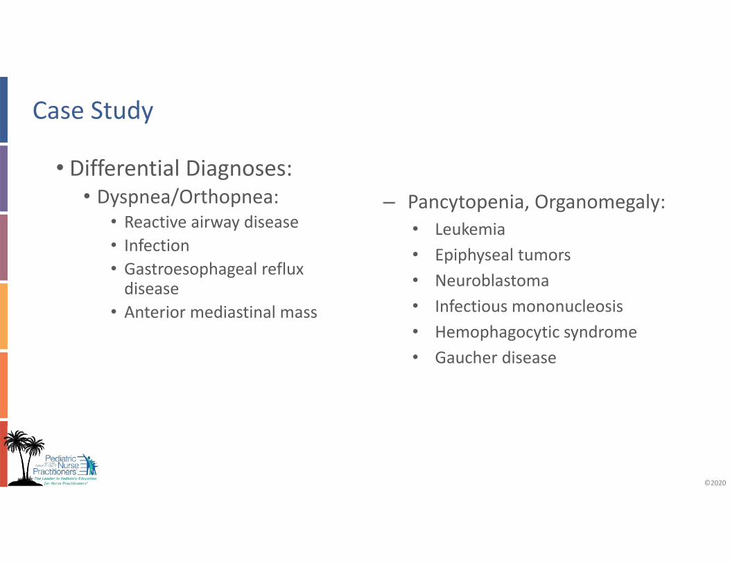
• Differential Diagnoses:• Dyspnea/Orthopnea:
• Reactive airway disease• Infection• Gastroesophageal reflux disease
• Anterior mediastinal mass
– Pancytopenia, Organomegaly:• Leukemia• Epiphyseal tumors• Neuroblastoma• Infectious mononucleosis• Hemophagocytic syndrome• Gaucher disease

©2020
Case Study
• The peripheral smear was evaluated by pathology and hematology and flow cytometry was evaluated: consistent with T cell acute lymphoblastic leukemia.
• What oncologic emergencies are you concerned about and what will your next actions be?
69

©2020
Case Study
• Oncologic emergencies:• Tumor lysis syndrome• Superior mediastinal syndrome
• superior vena cava syndrome + tracheal compression

©2020
Case Study
• Evaluation:• Chest x‐ray• Chest CT• ECHO • Diagnostic/therapeutic LP• Bone marrow aspirate

©2020
Case Study
• What is your plan for this patient?• Consultation to pediatric oncology• Establish central venous access if possible• Frequent laboratory monitoring• Goal of maintaining spontaneous respiration• Emergent initiation of steroids, chemotherapy, or radiation therapy if indicated

©2020
Question
A 2‐year old who is receiving treatment for a neuroblastoma develops fever to 38.9oC, lethargy and has oxygen saturations to 95%. The most important, first‐line management should include:a. Obtain peripheral blood culture and CBCb. Administration of antibiotics to include 3rd generation cephalosporin
along with an antifungalc. Obtain blood cultures from CVL and administer antibiotics to include 4th
generation cephalosporind. Obtain blood culture and lactate level from central line

©2020
Answer A 2‐year old who is receiving treatment for a neuroblastoma develops fever to 38.9oC, lethargy and has oxygen saturations to 95%. The most important, first‐line management should include:
C. Obtain blood cultures from CVL and administer antibiotics to include 4th generation cephalosporin

©2020
Question
A teen with a history of relapsed osteosarcoma presents with difficulty voiding, lacking feeling with bladder fullness. What is the most likely cause?a. Metastatic obstruction of the bladderb. Neurogenic bladder secondary to chemotherapyc. Urinary retention as a side effect of oxycodoned. Metastatic compression of the spinal cord

©2020
Answer
A teen with a history of relapsed osteosarcoma presents with difficulty voiding, lacking feeling with bladder fullness. What is the most likely cause?
D. Metastatic compression of the spinal cord

©2020
Question
Electrolyte disorders noted with a child with non‐Hodgkin lymphoma and tumor lysis syndrome include:a. Hypernatremia, hyperkalemia, and hypoglycemiab. Hypernatremia, hypokalemia, and hyperphosphatemiac. Hyperphosphatemia, hyperkalemia, hyperuricemiad. Hypophosphatemia, hyperuricemia, and hyperglycemia

©2020
Answer
Electrolyte disorders noted with a child with non‐Hodgkin lymphoma and tumor lysis syndrome include:
C. Hyperphosphatemia, hyperkalemia, hyperuricemia

©2020
Question
A 2‐week old infant is noted by his parents to have an asymmetrical red reflex, with one eye producing a normal red reflex while the other eye demonstrates no reflection at all. A probable cause of this asymmetry is:a. Congenital caratactb. Retinoblastomac. Neuroblastomad. This is a normal finding depending on the position of the eyes

©2020
Answer
A 2‐week old infant is noted by his parents to have an asymmetrical red reflex, with one eye producing a normal red reflex while the other eye demonstrates no reflection at all. A probable cause of this asymmetry is:
B. Retinoblastoma

©2020
Question
A 4‐year old girl has had blurry vision, ataxia and has become increasingly confused. She has vomited the past three mornings on arising. What study could be done quickly and effective in diagnosing her illness?a. CT of the brainb. MRI of the brain and spinal cordc. Head ultrasoundd. PET scan of the brain and spinal cord
©2020
Answer A 4‐year old girl has had blurry vision, ataxia and has become increasingly confused. She has vomited the past three mornings on arising. What study could be done quickly and effective in diagnosing her illness?
A. CT of the brain

©2020
Palliative Care Concepts

©2020
Palliative Care
• American Academy of Pediatrics• Relief of suffering• Improvement in quality of life• Facilitation of informed decision‐making• Assistance in coordination of care
84

©2020
Palliative Care
• Commitments (American Academy of Pediatrics)• Patient centered and family engaged• Respect and partnering• Quality, access, equity• Care across age spectrum and life span• Integration into continuum of care
85

©2020
Palliative Care
• Recommendations (American Academy of Pediatrics)• Specialty team composition• Relationships with Hospices• Collaborative, integrated, multimodal care• Communication and decision support• Patient care safety and quality• Family and sibling support• Health care staff support• Education and training• Ethical considerations
86

©2020
Palliative Care
• Life‐sustaining therapy decisions• Guided by the best interest of the patient• Balance benefits and burdens• Involve children and their families• Communication of resuscitation status• Interdisciplinary planning and consultation
87

©2020
Palliative Care
• Life‐sustaining therapy decisions• Special circumstances
• Forgoing nutrition and hydration• Children with developmental disabilities• Infants and children in foster care• Suspected abuse or neglect• Newborns with an uncertain prognosis • Death by neurologic criteria
88
©2020
Test Taking Guideline
• Remember to study with focus on more common diagnoses• Anytime there is an opportunity to note a “gold standard” diagnostic test, remember those!
• Remember primary diagnostic testing and first line treatment