pectic substances prom white mustard nigel j. wight, …
TRANSCRIPT
PECTIC SUBSTANCES PROM WHITE MUSTARD
NIGEL J. WIGHT, B.Sc.
Thesis presented for the Degree of Doctor of Philosophy
University of Edinburgh,
December, 1968.

To my Parents and Catriona
Acknowledgement s
My grateful thanks to Dr. D.A. Rees for his expert
advice and friendly guidance during the course of this
study, and to my colleagues of Lab. 11, past and present,
who gave freely of their time and knowledge.
I am also indebted to Dr. M.M. Yeoman for
preparing the autoradiograms and to Dr. Rees for writing the
computer programmes.
I should like to take this opportunity to thank
Professor Sir Edmund Hirst, F.R.S., for the provision of
laboratory facilities and the Institute of Paper Chemistry,
Appleton, \Visconsin, U.S.A. for financial support.

CONTENTS
PAGE
GENERAL METHODS 1
GENERAL INTRODUCTION 11
CHAPTER 1. STRUCTURAL STUDIES OF THE PECTIC
SUBSTANCES FROM THE COTYLEDONS
OF WHITE MUSTARD SEED.
Introduction 19
Experimental
1.1. Trial purification of the pectic mixture from non-germinated cotyledons.
1.2. Comparison of the f Soluble 1 and'Precipitated 1 fractions of 1 .1 . 35
1 .3* Estimation of the amyloid content of some fractions and extracts of non-germinated and germinated cotyledons. J>6
1.U- Purification of pectic mixtures fromnon-germinated and germinated cotyledons. 38
1 .5» Methylation of the pectic mixtures extracted from non-germinated and germinated cotyledons. 39
1.6. Methylation analysis of pectic mixture extracted at pH 7«5 from non-germinated cotyledons. [4.3
1.6a. Hydrolysis of the methylated pecticmixture. 14.3
1.6b. Reduction of acid fraction. J|J|
1.6c. Separation and identification of thecomponents of the 'reduced 1 fraction. 14.5
1 .6d. Separation and identification of thecomponents of the neutral fraction. 14.9
1.? Comparison of the methylated pectic mixtures from non-germinated and germinated cotyledons. 56
PAGE
Discussion 59
CHAPTER 2. INCORPORATION OF myo-INOSITQL-2-3H
BY GERMINATING MUSTARD SEEDS.
Introduction 72
Experimental
2.1 . Germination of mustard seeds suppliedwith myo-inositol-2-3H and separationof the cotyledons. 82
2.2. Distribution of radioactivity in thefractions obtained "by graded extractionof the cotyledons. 82
2.3. Distribution of radioactivity in the component sugars of the EDTA-soluble and EDTA~insoluble fractions of the cotyledons. 85
2.U. Autoradiography of the cotyledons. 87
Discussion 88
CHAPTER 5. SMITH DEGRADATION OF AN ARABAN
COMPONENT OF THE PECTIG SUBSTANCES
PROM MUSTARD COTYLEDONS.
Introduction 92
Experimental
3.1 . Extraction and purification of arabanfrom germinated cotyledons. 99
3.2. Periodate oxidation of the arabans extracted from non-germinated and germinated cotyledons. 100
3«3. Smith Degradation of araban fromnon-germinated cotyledons. 101
3.14.. Smith Degradation of araban from germinatedcotyledons. 101;
Theory 106
Discussion

PAGE
CHAPTER Uc CHARACTERISATION OF POLYSAC CHAR IDE
STRUCTURES BY GLYCOSIDE STABILISATION
WITH TOLUENE-p-SULPHONATES (TQSYLATES) :
MODEL EXPERIMENTS WITH DEXTRAN,
Introduction 121
Experimental
U.1 Preparation of dextran. 128
U.2. Methylation of dextran 0 128
U«>3« Comparison of the stabilities of the 1,3- and 1,6-glycosidic linkages in dextran and methylated dextran under various solvolysis conditions. 131
Uci|. Comparison of the rate of hydrolysis of0-methyldextran and O-methyl-0-tosyldextranin chloroacetic acid. 135
U.5- Thin Layer Chromatography of the hydrolysisproducts of 0-methyl~0~tosyldextran. 137
U»6. G-el filtration of the hydrolysis productsof 0~methyl~0-tosyldextran c " 138
!;.? Paper Chromatography of the hydrolysisproducts of 0~methyl-0~tosyldextran. 139
U.8. Separation of the hydrolysis products of 0-methyl-(D--tosyldextran by cellulose column Chromatography.
U.9. Identification, after methylation, reductive detosylation and rernethylation, of the methanolysis products of O-methyl-0--tosyldextran.
I4..10. Identification, after methylation, reductive detosylation and remethylation, of the hydrolysis products of 0-methyl-0~-tosyldextran,
U-1^. Relative yields of methyl ethers from0~methyl-0~tosyldextran and O-methyl-0--triphenylmethyldextran.
Discussion 150
REFERENCES ^57

GENERAL METHODS

GENERAL METHODS
1• Paper chromatography
Qualitative work was carried out "by descending
chromatography using Whatman No. 1 paper. Chromatograms
were developed using the following solvent systems (v/v):
A. ethyl acetate : pyridine : water (10 : ij. : 3),
B. butan-1-ol : ethanol : water (£4. : 1 : 5, upper phase),
C. "butan-1-ol : ethanol : water (3:1: 1» upper phase),
D. butan-2-one : water : ammonia (cone. S.G.*880)
(200 : 17 : 1),
E. benzene % ethanol : water (169 : k7 : 15> upper phase)
The symbols used in measuring relative sugar
mobilities are,
~ _ distance moved by sugar______w _ -———————————————————————— ————— ————————————————
distance moved by solvent front
,., distance moved by sugartf-n — ____________________ ___ _____ ______________
distance moved by 2,3,U,6-tetra-0-methyl-D-glucose
The chromatograms, having been run for a
predetermined time, were air dried and the sugars located by
spraying with one of the following reagents.
a) p-anisidine hydrochloride (l), as a solution (~5%) in
butan-1-ol. The chromatograms were heated at 120° for 5
minutes to develop the colours which are highly characteristic,
especially for methylated sugars.

2.
b) alkaline silver nitrate (2). The presence of
polyhydroxy compounds in general may be detected "by this
highly sensitive reagent. Excess background was removed
with thiosulphate (3).
c) tri-phenyltetrazolium hydroxide (k) • The chromatogram
was sprayed with a mixture (1 : 1) of aqueous
triphenyltetrazolium chloride (2%) and sodium hydroxide (N).
The colour was developed by holding the chromatogram over a
boiling water bath for 5 minutes and after washing with water
the chromatogram was air dried. Sugars unsubstituted at
and Ca give deep red colours,
Preparative separations of sugars were carried out
on Whatman 3MM filter sheets, prewashed with methanol. The
positions of the sugars were located by cutting off and
developing narrow side strips. The appropriate bands of
the chromatogram were then cut out and the sugars eluted with
aqueous methanol.
2. Gas- liquid chromatography (g.l.c.) was carried out on a
Pye f Argon Chroma tograph 1 with 90~ detector, or on a Pyet>r «'series ^0k chroma tograph 1 with dual hydrogen flame ionisation
detectors and temperature programming. Sample size varied
but was of the order of 0.1/^1 (chloroform solution).
Retention times (T) were calculated relative to the methyl
2,3»U,6-tetra-0-methyl-p-glucoside peak with the following
3.
notation for peak heights; s = strong, m = medium, w = weak,
sh 9 shoulder. The following stationary liquid phases were
used, as coatings on 60-100 mesh 'Gas Chrom P f unless
otherwise stated*
1» 15$ "by weight polyethylenegiycol adipate,
2. y?o by weight neopentylglycol adipate,
3» 3# by weight SB-30 on 80-100 mesh 'Oas Chrom Z 1 ,
k. 10$ by weight ECNSS (M).
The flow rate of argon with the 'Argon Chromatograph*
was 80 ml per minute. Temperatures of operation were
measured on a glass thermometer and are quoted in the text.
3. Thin layer chromatography (T.L.C.) (5) Most qualitative
T.L.C. was carried out on micro-plates. Preparative work,
and where better separations were required, was carried out
on larger (20 x 20 cm) plates. The plates were coated with
a thin uniform layer of silica gel (Kieselgel G, nach Stahl,
Merck). The solvent systems used are quoted in the text.
Spots or bands were detected by one of the following
reagents,
i) anisaIdehyde-sulphuric acid reagent (6) The plate was
lightly sprayed with a solution made by dissolving
anisaldehyde (1 ml) in ethanol (20 ml) and adding sulphuric
acid (cone. 1 ml, AnalaR). On heating at 120° for 5 minutes
sugars give dark colours on a light background.
ii) iodine vapour. The plate was placed in a closed
container with iodine crystals for several minutes. Sugars
(and other organic material) give brown colours which fade
rapidly on standing.
iii) diphenylamine (7) The plate was lightly sprayed with
a solution of diphenylamine (1?§) in ethanol. After
exposure to ultra-violet radiation for 5 minutes
p-toluenesulphonyl (tosyl) esters gave fluorescent green
spots.
U« Cellulose columns were packed as a thick slurry in
acetone. The packed column was then washed with acetone
containing an increasing amount of the solvent to be used
and finally equilibrated with pure solvent. The solvents
used were similar to those in paper chromatography but
generally contained half the water content.
5« PEAK- Sephadex columns. Diethylaminoethyl-Sephadex
(A25) was allowed to swell in water overnight and all fine
material removed by decantation. The material was then
washed with hydrochloric acid (0.5N) and then sodium
hydroxide (0.5N) twice, before generation in the formate form by

stirring with formic acid (15$« The resin was packed in
a column and washed free with distilled water.
6* Hydrolysis, Cellular material was shaken in sulphuric
acid (12% w/v) for 16 hours. The solution was then
diluted to 2N and heated at 100° for 6 hours. After
neutralisation with "barium carbonate the solution was
chilled, filtered and concentrated to a syrup. More
readily soluble samples were hydrolysed directly in 2N
sulphuric acid as above.
Samples, particularly methylated polysaccharides,
were also hydrolysed in formic acid (U5^)» The solutions
were heated in stoppered tubes at 100° for 16 hours. Formic
acid was removed by repeated distillation of water from the
hydrolysate under reduced pressure. After standing for
2-3 hours (to hydrolyse formate esters) the aqueous solution
was concentrated to a syrup.
Hydrolysis was carried out at 156 w/v.
7. Methanolysis was carried out at 1$ w/v by dissolving
the material in methanolic hydrogen chloride (3$) in a sealed
tube at 100° for 6 hours. After neutralisation with silver
carbonate, filtration and evaporation to dryness, the residue
was dissolved in chloroform for g.l.c, Methanolic hydrogen
chloride (j>% w/v) was prepared "by the careful addition of
acetyl chloride (6 ml) to dry methanol (100 ml).
®» Concentration of solutions was carried out by
evaporation on a rotary film evaporator under reduced
pressure at or below I|0°«
9- Dialysis. Polysaccharide solutions were dialysed in
cellophane tubes suspended in running tap water. Chloroform
was added to prevent bacterial action.
10. Nitrogen determination was carried out by a semi-micro
modification of the Kjeldahl method (8). Protein content
of carbohydrate material was obtained by multiplying the
value above by 6.25.
11. Determination of carbohydrate content (9)• The sugar
solution (1 ml, containing 20 - 100ug sugar) was placed in
a test-tube and aqueous phenol (5% w/v, 1 ml) added.
Sulphuric acid (cone. 5 ml, AnalaR) was added from a fast
delivery pipette. When cool the optical density of the
solution was measured with the EEL colorimeter using filter
no. 623 (maximum transmission at 1+95 nm), and sugar content
calculated by reference to a calibration curve.
12o Determination of methoxyl content. Analyses were
performed by a commercial firm (A.H. Baird, Edinburgh or

7.
Weiler and Strauss, Oxford).
13« Op.tical rotations were measured at room temperature,
using the Perkin-Elmer model 1U1 polarimeter, in 1 dm tube.
^» Infra-red spectra were recorded using the Unicam
S.P. 200 spectrophotometer. Methylated polysaccharides
were examined as films made "by evaporation in a dessicator
of a dry chloroform solution of the sample. Other
methylated samples were examined in dry carbon tetrachloride
solution.
15» U.Vo spectra were recorded using either the Perkin
Elmer 137 or Unicam SP 800 spectrophotometer. Periodate
oxidations were followed spectrophotometrically (l5a) using
the Unicam SP 500 instrument.
16. Removal of protein (10)» Protein contaminating
polysaccharide extracts was partitioned into phenol by making
an aqueous solution (2%) of the extract b£>% w/v with respect
to phenol. The mixture was heated until it became
homogeneous (approx. 75 ) and then left for 16 hours at 2
in a separating funnel. The phenol layer was discarded
and the procedure repeated thrice more. The excess phenol
was finally removed from the aqueous layer by dialysis,
17. Methylation was carried out by one or more of the

8.
following methods,
(a) The method of Haworth et« al. Dimethyl sulphate and
sodium hydroxide were added in portions to an aqueous
solution of the polysaccharide with stirring under an inert
atmosphere.
("b) The carbohydrate was dissolved in N-methy1-2-pyrrolidone
and methylated by addition of methyl iodide and barium
hydroxide octahydrate with shaking (12).
(c) The method of Kuhn and Trischmann (13), involving the
addition of dimethyl sulphate and barium hydroxide octahydrate
to a solution of the carbohydrate in a mixture of
dimethylformamide and dimethyIsulphoxide. This method was
superseded by (d) below.
(d) The method of Kuhn, Trischmann and Low(lU). Methyl
iodide and silver oxide were added to a solution of the
carbohydrate in dimethylformamide and shaken in the dark at
room temperature.
(e) As in (d) but the reaction carried out with stirring at
the boiling point of methyl iodide.
Further details of the procedure and method of
isolating the product are given in the text, where appropriate,
18. Demethylation (11). The methylated sugar (5 - 10 mg)
was dissolved in dry dichloromethane (1 - 2 ml) and cooled
to -80°. Boron trichloride (1 - 2 g), cooled to -80°, was
added and the temperature kept at -80° for 30 minutes then
allowed to warm to room temperature. After 16 hours the

residue was taken up in a drop of water and methanol evaporated
from the solution several times under reduced pressure„ The
product was examined "by paper chromatography in solvent A.
19• Eormation of derivatiyes
N-phenylglycosylamine (anilide) derivative• The
methylated sugar (75 mg) and aniline (I'mole, redistilled)
in ethanol (5 ml) was refluxed in the dark for 1 hour. The
hot solution was filtered through charcoal and evaporated
to dryness. The crystalline product was recrystallised from
the given solvent.
Lactones of the aldonic acids of methylated sugars were
prepared as follows, The methylated sugar (75 mg) was
dissolved in "bromine water (3 ml) and the solution was kept
in a sealed flask in the dark for 3 days. Bromine was
removed "by aeration and the solution neutralised with silver
carbonate. Insoluble silver salts were removed "by filtration
and the filtrate evaporated to dryness. The lactone was
extracted with acetone which on evaporation to dryness gave
a syrup, from which the product crystallised. Recrystallisation
was from the given solvent.
Aldonamides were prepared from the corresponding lactone,
dried in a vacuum oven over phosphorus pentoxide. Thev
lactone (50 mg) was dissoled in dry methanolic ammoniaA
(8?S, 5 ml) and kept at 0° for 2 days in a sealed flask. On
evaporation of the solvent the amide crystallised and was
recrystallised from the given solvent.
Toluene-p-sulphonylhydrazone derivative (15). The sugar

10
(100 mg) was dissolved in methanol (10 ml) and toluene-^)"
sulphonylhydrazide (1 mole) was added* The solution was
heated under reflux for 1 hour. The product crystallised
on standing at 0 for 3 days and was reerystall!sed from
methanol and washed with ice cold methanol.
20o Melting points were obtained on a Kofler hot-stage
apparatus and are uncorrected.
21» Liquid scintiHation counting. All samples were
counted in the Beckman Liquid Scintillation System with
automatic programming. The strips of cellulose acetate or
chromatography.paper were immersed in liquid scintillator
(10 ml) in glass vials (10 ml) with screw-tops in a constant
fashion so as to minimise any orientation effect,
The scintillator was prepared by the addition of
2,5-diphenyloxazole ('PPO', 5-Og) and 1 ,l+-Bis[2-(^-raethyl~5-
-phenyloxazozolyl)]-benzene ('Dimethyl POPOP ? , 0.3g) to
toluene (1 !)<, All chemicals were Scintillation Grade,
supplied by Nuclear Enterprises (G.B.) Ltd. Conditions of
counting are given in the text.

GENERAL INTRODUCTION

11
General Introduction
In "both plant, animal and "bacterial cells the
active protoplasm is "bounded by a lipid membrane known as
the plasmalemma (l6a). This performs a large variety of
functions "but is basically concerned with transport of
materials in and out of the protoplast. In plants and
bacteria however the membrane is itself surrounded by
another structure known as the cell wall which imparts
protection and firmness to the cell.
The osmotic pressure exerted by the protoplast, by
uptake of water from a hypotonic environment, on the cell v/all
can be considerable and thus a fundamental property of the
wall must be a high tensile strength to withstand this
pressure from within 0 Polysaccharides account for by far
the majority of wall material in young plant tissue and these
polymers must be arranged so as to surround the protoplast
with a mesh of the necessary properties.
In the polarising light microscope the cell wall
shows birefringence whereas the layer between adjacent cells
(the middle lamella) appears to be amorphous. In the
electron microscope the cell wall has been shown to be
composed of microfibrils of indefinite length embedded in an
apparently structureless matrix (17). In higher plants the
microfibrils are made up of bundles of highly orientated
cellulose molecules. The staining properties of the wall
and middle lamella with the basic dyes methylene blue and

12.
ruthenium red (18) indicate that the matrix is composed,
in part, of the acidic pectic substances, although
hemicellulosic polysaccharides and in older cells the
non-carbohydrate polymer, lignin, are also present.
Earlier work on the pectic substances has been
reviewed by Hirst and Jones (19) and the constitution of
pectic substances from many different sources have been
recently investigated, e.g. lemon peel (20), apple (21),
soybean cotyledons (22), mustard cotyledons (23) and
amibilis fir bark (2k) .
The parent molecule of pectic substances, known as
pectic acid, is a linear a-1,1± linked polymer of D-galacturonic
acid. The polymer itself seems to be of limited occurrence
but examples do exist (214.). Usually the molecule is
considerably modified in some of the following ways*
(i) by methyl esterification of the uronic acid units, the
highly esterified molecule, which is known as pectin, is
readily extracted with water from the tissues of many fruits
and is important as the gelling agent in jam-making.
(ii) by 0-acetylation of a small proportion of the uronic acid
units. It is not clear whether this occurs at C/ ? \ or C/_\
or both.
(iii) by the co-occurrence of rhamnose units in the main
chain. The disaccharide 2-0-a~D~galacturonosyl-L-rhamnose
has been isolated from many sources, for example lucerne (25).
(iv) by the addition of single unit side chains of xylose units
to the main chain through C/\ of uronic acid residues.
13.
(v) by the addition of linear £-1,1+ linked galactan side
chains. The mode of attachment to the main chain is not
clear.
(vi) by the addition of side chains which are highly
branched and contain L-arabinufuranose units linked through
the C/,\ and C/,-\ positions. Again whether the attachment
is direct to the backbone or through other side chains is
not known.
Other homopolysaccharides, known as pectic
galactan and pectic araban are also found in the pectic
substances of some plants. These are closely related to the
side chains mentioned above although authenticated samples
are comparatively rare; the galactan of white lupin seeds
(26) and the araban of white mustard cotyledons (27,28) are
good examples.
The pectic substances are abundant in the walls of
actively growing cells (29). In mature cells however, the
more typical components are hemicelluloses and lignin. For
example cambial cells may contain up to 25% of the dry
weight as pectic substances as opposed to about 1$ in wood
cells (30a). Hemicelluloses are typified by the xylans, in
which the {3-1 ,Lj. linked xylose main chain carries side chains
of arabinose and / or Lj.-0-methylglucuronic acid.
The most important of the non-carbohydrate
constituents of the cell wall is lignin. This cross-linked
macromolecule is formed by the polymerisation of coniferylic
alcohol to give a poly-(substituted phenylpropane). In

wood a typical analysis of wall material would yield 50$
cellulose, 20$ (soft wood) to 30$ (hard wood) hemicellulose
and 20 to 30$ lignin. Other important polymers are the
highly insoluble cutins and suberins.
The cell wall is first formed at telophase of
mitosis. In the electron microscope (l6b) the initial stage
is seen as an accumulation of semi-liquid droplets, each
with its own membrane, known as vesicles in the central
region of the mitotic spindle. These vesicles appear to
originate from cytoplasmic organelles known as the Golgi
bodies and the organisation of the vesicles into a lateral
arrangement appears to be effected by a system of membranes
joining the daughter nuclei, the endoplasmic reticulum (ER).
The number of vesicles increases until the droplets coalesce
to a semi-solid layer known as the cell plate. The plate
at this stage has the staining characteristics of the cell
wall proper and Northcote and Pickett-Heaps have shown that
a pectic polymer in wheat root-cap cells is secreted in
vesicles formed by the Golgi bodies (31). The pel}, plate
now grows laterally to fuse with the longitudinal wall of
the mother cell by the addition of more vesicles. The
membranes of the vesicles become part of the plasmalemma
and thus it appears that this important membrane also
originates from the Golgi bodies. Pine protoplasmic
connections known as plasmadesmata penetrate the growing
wall and appear to carry profiles of ER. Even before the
cell plate reaches the longitudinal wall it shows some
"birefringence indicating deposition of cellulose fibrils.
In the subsequent development the cell plate "becomes the
middle lamella bounded on either side by the primary cell
walls of the daughter cells. During the rapid growth which
follows mitosis, cellulose microfibrils and matrix
polysaccharides are added to both the cell plate and the
original wall. The latter does not become thicker because
growth is coupled with considerable extension of the
existing longitudinal wall. The wall at this stage, when
the cell is increasing in area, is known as the primary cell
wall and is thin and pliable with a high water content. The
microfibrils are rather disordered and the matrix is supposed
to consist of pectic substances (29).
Expansion of the young cell is frequently more
pronounced in the direction of tissue growth^ this is the so
called 'extension growth' and must involve more stretching of
the side walls than of the end walls. When viewed in the
electron microscope, a side wall of a cell in extension
growth shows the microfibrils of the outermost layer of the
wall to be oriented in the direction of extension while the
inner layers become progressively less orientated in this
direction. The innermost layer (where deposition of new
wall material is thought to occur), although not very
highly orientated, forms a network in which the microfibrils
appear to run more or less transversely to the direction of
growth. These observations form the basis of the theory of
'multinet growth 1 (30b) which is the accepted mechanism of

extension growth. Cellulose microfi"brils are deposited
transversely on the inner surface of the wall, and as each
layer "becomes buried by new layers of wall material the
microfibrils in it become orientated, by cell extension,
into the direction of extension growth. Thus although
turgor pressure is responsible for cell expansion the form
that this will take is determined by the structure of the
cell wall.
As the cell reaches maturity the wall grows in
thickness by the deposition of closely packed layers of
highly orientated microfibrils and hemicellulosic polysaccharides
This secondary cell wall is a rigid structure in comparison
with the primary wall and effectively limits further cell
expansion. Lignin is laid down only in an advanced stage
of growth (30c)•
The process by which cellulose chains are assembled
into partly crystalline microfibrils and deposited with a
particular orientation in the wall is still obscure. The
matrix substances (or their precursors) are transported to
the wall in the Golgi vesicles and in contrast to the
microfibrJls these polysaccharides are deposited at all levels
in the wall (iSc).
Wall extensibility, which must play a ma^or role in
growth and possibly have a controlling influence on it,
might be controlled by the pectic substances of the matrix.
Some of the possible interactions by which this could occur
are discussed later.
17.
The object of the work described here was to
investigate the structures of pectic substances, especially
in relation to the changes that they undergo during cell
growth. The system chosen for the investigation was the
embryo of germinating white mustard (Sinapis alba) seed,
as marked biochemical and morphological changes occur here
during germination.
Mustard is a representative of a family (the
Cruciferae) of important vegetables (e.g. cabbage,turnip)
whose polysaccharides have not been closely investigated.
The seed is easily germinated in the laboratory and as it is
a fatty seed the extraction and fractionation of cell-wall
polysaccharides is not complicated by the presence of reserve
polysaccharides. Examination under the microscope (32)
has shown that as the seeds imbibe water and begin to
germinate the cell walls in the cotyledons both expand and
swell. Cell counts of the cotyledons during germination
indicate that no cell division is taking place at this
stage (33). Another investigation (3U) has shown that
whereas the amount of polysaccharide in the cotlyedons
varies little during germination, experiments in which 1 **C-
sucrose was fed to the seeds at the start of germination
indicate that synthesis of pectic polysaccharides occurs
during this period. This is interpreted as turnover of
pectic substances in existing cell walls.
Much of the work on seed polysaccharides has been
focused on the mucilages obtained by extraction of the whole

seed e.g. mustard (35>36), linseed (37) and plantain (38).
The cold water extract of white mustard seed was shown to
contain a mixture of complex acidic polysaccharides composed
of arabinose, galactose and galacturonic acid and a
considerable proportion of cellulose (35536). Examples of
mucilages of simpler structure are the galactomannans from
seeds of the Leguminosae family (39) and guaran, the
galactomannan from guar seeds (39,^0). More detailed
investigation has, with few exceptions e.g. white mustard
(27*28,3U) and lupin (26), been limited to the cereals, e.g.
the glucans of oats and barley grains (Lj/l) •
The main part of the work described in this thesis
concerns the cell-wall polysaccharides of white mustard seed
cotyledons and the changes which they undergo on germination,
The last chapter is concerned with the development of a
method of polysaccharide structure determination applicable
to polysaccharides in which unsubstituted primary hydroxyls
occur only at end groups, e.g. dextran and pectic araban.

CHAPTER 1
Structural Studies of the Pectic Substances
from the Cotyledons of White Mustard Seed

19
Introduction
As described in the General Introduction,
extension growth of the young cell wall involves progressive
stretching and re-orientation of the cellulose microfibrils.
The physical properties of the embedding matrix must play an
important role in governing wall extensibility and the
biological control of this may function through the matrix
polysaccharides. Growth by the T multinet T mechanism requires
the microfibrils to change their orientation and hence move
relative to each other. If the matric polysaccharides were
present as a weak gel this would not hinder the movement of
microfibrils and might even assist this by a lubricating
effect. Hov/ever if a rigid gel were present severe
restrictions would be imposed on the movement of microfibrils.
It is a safe assumption that the matrix at this stage of
growth is largely composed (in most plants) of pectic
substances and hence the molecular interactions of pectic
gels in vitro might lead to an understanding of the control
of extensibility in vivo.
Polysaccharide gels in vitro, e.g. agar, can be
close to a liquid in composition but still have some of the
properties of a solid. The gel can be melted on heating and
reset on cooling suggesting that the interactions betv/een
molecules in the gel are temporary. If this is the case
in vivo then the plant cell wall presents a quite different
situation to that in the bacterial cell wall. In the latter

20.
the cell is in effect surrounded "by a covalently "bonded
macromolecule (U2), the "bonds presumably "being broken and
reformed as growth occurs. Temporary interactions as
opposed to covalent linkages might be more appropriate to
the plant as growth could occur as a physical response to
turgor pressure.
Gels in vitro are composed of a network of
macromolecules that are cross linked at various points with
the liquid occupying the spaces between polymer chains.
Cross linkage in this context applies to any form of molecular
association or cohesion. The number of such linkages need
not be large provided they are strong enough to withstand
repulsive forces (e.g. coulombic repulsion) which must be
present if the gel is to exist in a swollen state. The
most likely theories of interaction between molecules in
polysaccharide gels are described below.
(a) Entanglement s
Entanglement coupling is a rather loosely defined
term: it is not applied to the short range molecular
entwining which must exist in all polymeric systems but
rather to long range interlooping of polymers of sufficiently
high molecular weight (U3). The presence of bulky side
groups does not markedly inhibit entanglements (ijlj.) . It
is widely accepted that the protein-polysaccharides form
networks in concentrated solution by entanglements. This is
also thought to occur in the natural state, for example in
nasal cartilage the main chains are polypeptides and the
21
side chains the polysaccharide chondroitin U-sulphate
The rigidity is due to electrostatic repulsion between
negatively charged side chains 0
(b) Microcrystallites (U3)
The association of polysaccharide chains can also
occur by different chains coming together in regions of
local crystallinity, the regions usually being short in
relation to chain length. However in regenerated cellulose
the chains bind together so successfully that fibres form (1+6)
By substitution of the accessible (non-crystalline) regions
with carboxymethyl groups in carboxymethyl cellulose a
system with the visco-elastic properties attributable to a
combination of entanglement and crystallite cross linking is
formed .
(c) Hydrophobic Interations
Hydrophobic bonding is thought to account for some
of the tertiary structures of proteins (U7); the three
dimensional structure of lysozyme (from X-ray data) shows
that the molecule in solution (by extrapolation of the
crystal structure) is folded such that contact between
non-polar groups and water is minimised. The formation of
hydrophobia bonds is an entropically favoured effect as the
water molecules around exposed non-polar groups on the
polymer chain become more ordered (U8). Hence on formation
of the hydrophobia bond the degree of order of the system as

22,
a whole decreases, resulting in a favourable free energy of
formation. By addition of compounds which reduce the ability
of water molecules to hydrogen bond with each other e.g.
area and guanidine hydrochloride, the favourability of
hydrophobic bonding is lost, with consequent loss of
tertiary structure of the polymer. As hydrophobic bonding
is an entropy controlled process the strength of the bond
increases with temperature. For example methylcellulose
forms gels which set on heating and melt on cooling. The
cellulose derivative is prepared heterogeneously by
autoclaving cellulose pretreated with sodium hydroxide
(alkali cellulose) with methyl chloride* The degree of
substitution is about 1 «6 to 2.0 methoxyl groups per glucose
residue. The distribution of methoxyl groups within the
molecule is almost certainly governed by accessibility so
that blocks of fully methylated glucose residues occur.
The gelation is seen as the formation of stable cross
linkages of the fully methylated regions of different chains
by hydrophobic bonding, and as such will be favoured by
increasing temperature.
Polyelectrolyte effects
Cross linkages formed in this way usually do not
lead to gel formation but by an aggregation effect assist in
the gelation primarily caused by some other form of
interaction. The uronic acid containing plant gum
Khaya grandifolia in salt solution shows a considerable
23 «
molecular weight increase (as measured by ultracentrifugation)
over that in salt free solution (U9). Previous theories
involved electrostatic attraction "between ionised carboxyl
(or other) groups and a polyvalent cation ( f ionic bridging*).
If the carboxyl groups were in different chains then a cross
linkage was established. Some chelation of mono- or
divalent cations to the sugar hydroxyl groups is also
possible in the polymer system, although the complexes
formed by low molecular weight analogues are weak (50,51)*
The simple ionic bridging of negative charges by
divalent cations such as Ca++ is not compatible with the
nature of electrolyte so3-utions (52). The behaviour of
such systems is most accurately described in terms of the
polyelectrolyte character of the polymer in solution (53)«
The covalent structure of the molecule holds together
anionic groups that would otherwise separate by repulsion.
The sum total of these repulsions is such that from the free
energy point of view of the system as a whole it becomes
favourable to localise the cations present in the domain of
the polymer, a process in itself entropically
unfavourable. It has been shown that a large proportion of
the counterions may be bound in this way amd that di- or
trvivalent cations are more tightly bound than univalent
cations (54)» It follows that chains which pass through
each others domains will share a common atmosphere of
counterions giving junction zones in which any movement of
the chains away from each other will be resisted. The

2k.
effect of coujnterion "binding will be enhanced by
multivalent cations. It seems likely that cations bound
in the domain of the polymer may also take part in chelation
with sugar hydroxyl groups giving some further cross linking
of chains. This is seen as the cation exchanging some of
its water of hydration for sugar hydroxyl groups, although
the extent to which this occurs is not established.
It is stressed that the bound counterions are in
dynamic equilibrium with those in the bulk solution, thus no
individual cation holds the chain together permanently and
hence this theory is fundamentally different from the ionic
bridging and chelation theories put forward previously.
Pectin gels in vitro
Pectins (the highly methyl-esterified molecule)
are readily extracted from the tissues of fruits with boiling
water and form gels on cooling* A substance of low
molecular weight, usually sucrose, must be added in large
quantities before the gel will set. The effect of the
added solute is to make the solvent thermodynamically
poorer by replacing some of the water molecules around the
polymer chains with molecules which react less strongly with
the polymer. The result is that the chains come out of
solution to form points of cross-linkage. The loss of gel
strength which occurs if the methyl ester is replaced by
the ethyl or 2-hydroxyethyl ester establishes that the
backbone of methyl galacturonate units is responsible for
the gelling properties rather than the neutral side chains.
An 0~acetyl group on (on average) every eighth methyl
galacturonate unit prevents gelation and thus the
mechanism would appear to "be due to an intimate association
of chain segments rather than to association "between single
sugar units. In this respect the structural requirements
are similar to those involved in crystallites suggesting
that a local limited crystallisation is responsible for
pectin gels. Structural irregularities in the main chain,
e.g. 0-acetylation, insertion of rhamnose units, would thus
have a weakening effect on the gel "but the presence of
"bulky neutral side chains would have an even more drastic
effect.
Pectic acid (the fully de~esterified molecule) can
"be converted to a gel by addition of a low concentration of
Ga + ions to the solution. However this property is lost on
partial acetylation so that the polyelectrolyte effects
discussed above cannot wholly explain the gelation.
Crystallites might again be involved but as it is unnecessary
to add sugar to set the gel their formation might be more
favourable.
The optimum gelling conditions of low methoxyl gels
(i.e. where only a portion of the uronic acid units are
methyl esterified) has been shown to be a function of the
methoxyl content, the concentration of Ca ions and the
concentration of sucrose (55)•
26*
Pectin gels in vivo
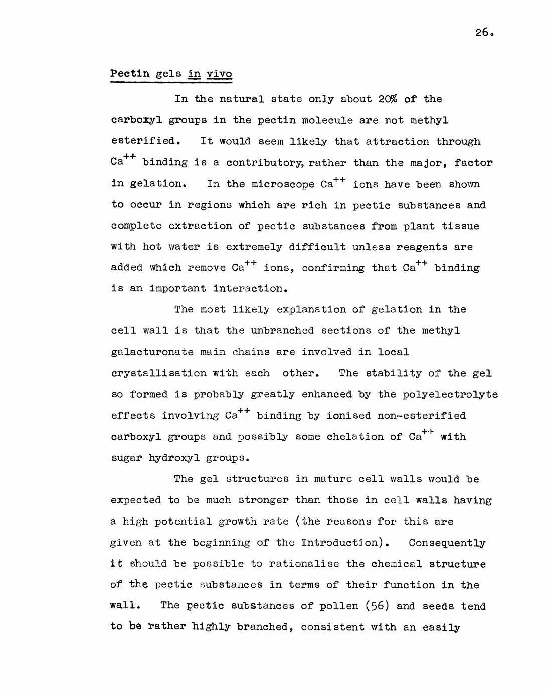
In the natural state only about 2C$ of the
carboxyl groups in the pectin molecule are not methyl
esterified. It would seem likely that attraction through
Ca binding is a contributory, rather than the major, factor
in gelation* In the microscope Ca++ ions have been shown
to occur in regions which are rich in pectic substances and
complete extraction of pectic substances from plant tissue
with hot water is extremely difficult unless reagents are1_ L -J- J
added which remove Ca ions, confirming that Ca binding
is an important interaction.
The most likely explanation of gelation in the
cell wall is that the unbranched sections of the methyl
galacturonate main chains are involved in local
crystallisation with each other. The stability of the gel
so formed is probably greatly enhanced by the polyelectrolyte
effects involving Ca binding by ionised non~esterified
carboxyl groups and possibly some chelation of Ca with
sugar hydroxyl groups.
The gel structures in mature cell walls would be
expected to be much stronger than those in cell walls having
a high potential growth rate (the reasons for this are
given at the beginning of the Introduction). Consequently
it should be possible to rationalise the chemical structure
of the pectic substances in terms of their function in the
walla The pectic substances of pollen (56) and seeds tend
to be rather highly branched, consistent with an easily

27.
deformed, fairly weak gel which would enable rapid growth to
occur. In contrast the strands which are found in mature
celery petioles and seem to have a purely support function
have been shown by X-ray diffraction and electron
microscopy to be mainly composed of short crystallins
bundles of pectic substances (57). The presence of
crystallites suggest that relatively unbranched sections of
galacturonan chains account for the rigidity of the strands.
It is assumed that the effect of side chains and
other structural irregularities on the gel strength would be
similar to that observed for in vitro gels.
Structural studies of pectic substances
Structural analysis, while giving interesting
insight into the constitution of the pectic substances, is
more important in that it may enable the biological role of
these polysaccharides in the cell wall to be more clearly
understood.
The first stage of a structural investigation is
the extraction of the material in a relatively pure state.
The method of extraction adopted in this laboratory for the
extraction of pectic substances involves the use of hot
aqueous ethylenediamine tetra-acetic acid (EDTA) (58)
solution (2$ w/v). The pH must be chosen to maximise yield
but minimise degradation (see fo].lowing Chapter); EDTA
complexes calcium ions most efficiently at a slightly
alkaline pH and thus a pH of 7-5 was chosen for complete

28.
extraction of the pectic substances. However degradation
reactions are more serious under alkaline conditions and in
another experiment extraction was carried out at pH k»9*
Although extraction was shown not to be as efficient at this
pH, it is most unlikely that any degradation had occurred.
The crude pectic mixture was obtained by precipitation from
the EDTA extracts with aqueous ethanol (75%) 9 redissolving
in water, dialysis, concentration and freeze-drying. The
material thus obtained is usually contaminated with
appreciable amounts of protein which must be removed before
proceeding with a structural analysis of the pectic
substances.
The methods available for polysaccharide structure
determination are very powerful. The first consideration
is whether the extracted material is homogeneous. The
heterogeneity, if present, can be of two types, (a) the
extract may contain one or more well known types of
polysaccharide for which efficient and specific fractionation
methods are established, and, (b) within a polysaccharide type
molecules have a common structural pattern but vary in size
and possibly in some details of structure e.g. degree and/or
distribution of branching. If the differences are fairly
small no discontinuity is observed, i.e. one spectrum of
molecules aj?e present, this is known as polymolecularity.
However if the differences are such that discontinuity is
observed, e.g. ultracentrifugation may show some separation
into two fractions, then two (or more) spectra of molecules

29-
are present, this is known as polydispersity.
Electrophoresis and ultracentrifugation give clear evidence
for heterogeneity as do some chemical methods e.g.
fractionation on ion-exchange columns. Before further
analysis it is desirable to fractionate the extract so that
each fraction is as nearly as possibly homogeneous in the
respect that it contains only one type of polysaccharide.
Methylation analysis and Smith Degradation are examples of
elegant, established methods of determining the fine
structure of polysaccharides. Methylation analysis
involves complete methylation of the polysaccharide
followed by hydrolysis and separation and identification of
the hydrolysis products. This is most satisfactorily and
unambiguously achieved by the formation of crystalline
derivatives but as relatively large amounts of material are
required in a pure state for this, identification is
frequently limited to g.l.c. and paper chromatography
evidence (particularly for minor components). Although
giving the nature and relative proportions of the linkages
present in the polymer methylation analysis gives no
information as to sequence or configuration. These are
usually achieved by controlled partial acid hydrolysis
followed by separation and identification of the di- and
oligosaccharicle products.
The pectic substances from both germinated and
non-germinated mustard cotyledons have been the subject of a
considerable amount of work in this laboratory. The

30,
extractions of the pectic substances, the purification and
methylation of which is described in this chapter, were
carried out "by Dr. J. Samuel. The 'non-germinated pectic
mixture* was extracted from commercial 'White Mustard Seed
Germ 1 , (J. and J. Colman, Ltd.), and the 'germinated pectic
mixture 1 from the cotyledons separated from commercial
mustard seeds which had been germinated for four days in the
dark* Obviously the former is available in much larger
quantities. The sugar composition of the non-germinated
pectic mixture was determined by Dr. S. Gould (59); the
main components were galacturonic acid, arabinose and xylose
in the molar ratio of 2 : 3 : 1 respectively, with significant
amounts of galactose and rhamnose and a lesser amount of
glucose. Dr. Gould also fractionated the mixture, on a
column of DEAE cellulose by gradient elution with sodium
phosphate buffer, into a neutral and two partially
separated acidic components 0 The neutral fraction was
composed of mainly galactose and arabinose with small
amounts of glucose and xylose. The acidic components both
contained similar proportions of galacturonic acid,
arabinose, xylose, galactose and rhamnose, and are thus
closely related. Graded hydrolysis of the mixture in
0.01 N sulphuric acid at 100° removed nearly all the
arabinose units from the polymer but liberated only traces
of xylose and galactose. The arabinose units are probably
present in the furanose form and occur as blocks attached
indirectly to the main poly(galacturonic acid) chain. A

preliminary analysis of the hydrolysis products of the
methylated pectic mixture from non-germinated cotyledons,
using g.l.c. and paper chroma tography to identify the
products, indicated the main structural features present.
The average molecule has an extremely high degree of
branching and has "been described in the General Introduction,
but the galactose units were suggested as being present as
bridge units by which other side-chains are attached to
the main chain. This contrasts v/ith the (31 -I± linked
galactan side-chains of other pectic substances. In an
examination in the ultracentrifuge Dr. I. Steele (60)
confirmed that the pectic mixture was heterogeneous, two
rather broad peaks were observed. No significant differences
were apparent between the germinated and non-germinated
pectic mixtures in this study.
Recent investigations of pectic substances from a
variety of sources have shown the presence of a number of
minor structural variations other than those already mentioned
These features, listed below, probably occur as side-chains.
Feature Source Reference
P~D~Galg-(l->2)-D~Xylp Soybean cotyledons (22)Soybean hulls (61 )
Soybean cotyledons (22)Soybean hulls (61 )
p-D-GpA.-(l-*i4-)-D-Galp Soybean cotyledons (22)Soybean hulls (61 )

32.
Feature Source Reference
p-D~GpA-(l-^6)-D~GalTp Lemon peel (20)Soybean cotyledons (22)Soybean hulls (61)
p-D-GpA-(l-4j.)-L-Pucg Lemon peel (20)Lucerne (25)Soybean cotyledons (22)Soybean hulls (61)
The object of the work described in this chapter
was to correlate any changes in molecular structure (as
shown by methylation analysis) of the pectic substances
extracted from germinated and non-germinated mustard
cotyledons with their different function in the cell wall.
The most dramatic effect one might expect would be a decrease
in the degree of branching during germination, enabling more
rigid gels to be formed. This would also result if a
redistribution of branch points occurred during germination
such that blocks of unbranched galacturonic acid units
became available for the formation of junction zones. Of
the two mechanisms only the former would be detected by
methylation analysis.
The structure of the pectic substances from
mustard cotyledons requires to be put on a firmer basis and
to this end the major hydrolysis products of methylated
non-germinated pectic mixture (pH 7-5) were characterised
unambiguously by the formation of crystalline derivatives.
Also as the investigation was carried out on a relatively
large scale the presence of minor features, such as those

33
listed above, should "be detectable (if present). The
comparison of the germinated and non-germinated pectic
mixtures was by g.l.c. and paper chromatography of the
hydrolysis products of the methylated polysaccharides.
The pectic mixture extracted at pH k*9 from non-germinated
cotyledons was also compared v/ith the above by the same
methods.
Another type of polysaccharide was isolated in a
fairly pure state during purification by deproteinisation of
the non-germinated pectic mixture. This belongs to a
family of polysaccharides known as the 'amyloids 1 , a more
detailed description of which are given in the discussion
at the end of this chapter.

3U.
Experimental
Experiment 1 «1 . Trial purification of the pectic mixture
from non-germinated cotyledons 0
The crude pectic mixture was obtained (by Dr, J.
Samuel) by extraction of defatted non-germinated cotyledons
with aqueous EDTA (2% w/v) at 90° and subsequently with
water at 90 . The EDTA and water extracts were combined
and the pectic mixture precipitated with ethanol. The
precipitate was redissolved, dialysed and freeze-dried.
The crude pectic mixture (lUOg) was deproteinised
by four cycles of the treatment described in General
Methods, 16. A white flocculent interfacial precipitate
formed as removal of protein proceeded; both this and the
clear aqueous layer gave positive reactions to the phenol-
sulphuric acid reagents. Trouble was encountered from
interfacial emulsions. The emulsion was retained with the
carbohydrate layer and was found to diminish in amount as
removal of protein reached an advanced state. After the
final treatment the aqueous suspension was allowed to stand
for 2 hr. and the clear supernatant removed by decantation
from the precipitate. The supernatant wasdialysed,
concentrated and freeze-dried, (1.80g; Pound, % N = 1.13).
The precipitate was suspended in water, dialysed, concentrated
and freeze-dried, (50.5g$ Pound, % N = 1.00).
The procedure was repeated on a small scale (lOg)
35
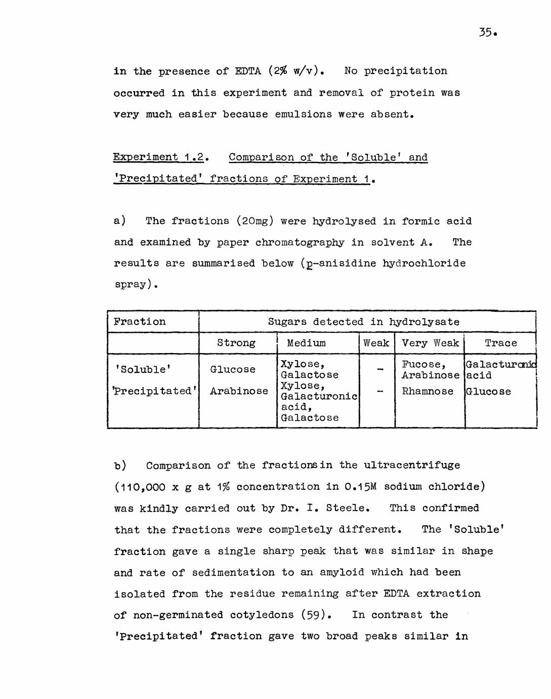
in the presence of KDTA (2% w/v). No precipitation
occurred in this experiment and removal of protein was
very much easier because emulsions were absent.
Experiment 1 .2. Comparison of the 'Soluble* and
f Precipitated* fractions of Experiment 1.
a) The fractions (20mg) were hydrolysed in formic acid
and examined by paper chromatography in solvent A. The
results are summarised below (g-anisidine hydrochloride
spray).
Fraction
1 Soluble '
precipitated '
Sugars detected in hydrolysate
Strong
Glucose
Arabinose
Medium
Xylose, GalactoseXylose, Galacturonicacid,Galactose
Weak
-
—
Very Weak
Pucose, ArabinoseRhamnose
Trace
Galacturonic acidGlucose
b) Comparison of the fractions in the ultracentrifuge
(110,000 x g at 1?£ concentration in 0.15M sodium chloride)
was kindly carried out by Dr. I. Steele. This confirmed
that the fractions were completely different. The 'Soluble*
fraction gave a single sharp peak that was similar in shape
and rate of sedimentation to an amyloid which had been
isolated from the residue remaining after EDTA extraction
of non-germinated cotyledons (59). In contrast the
'Precipitated* fraction gave two broad peaks similar in

36,
pattern and rate of sedimentation to pectic mixtures
studied previously (by Dr. Steele).
c) The 'Soluble* fraction was found to give a strong
blue-green colour ( A mov = 650nm) with potassium triiodidemaxin saturated sodium sulphate (62) very similar to tlvat
given by a sample of Tamarindus amyloid kindly provided by
Dr. P. Kooiman. The 'Precipitated 1 fraction gave only a
weak colour. Quantitative measurements on the colour
reaction are described below.
Experiment 1.3. Estimation of the amyloid content of some
fractions and extracts of non-germinated and germinated
cotyledons.
The analytical method of Kooiman (62) was followed.
To the aqueous polysaccharide solution (1.00 ml, containing
between 0.05 and 0.30 mg of amyloid) was added an aqueous
solution (0.50 ml) containing iodine (0.5 % w/v) and
potassium iodide ("\% w/v, AnalaR). Sodium sulphate
solution (20% w/v, AnalaR, 5»0 ml) was added and the mixture
shaken. After standing for 1 hr. at room temperature to
allow the colour to develop, the extinction at 650nm was
read in 1 cm cells against a reagent blank, (Perkin Elmer
model 137 spectrophotometer).
A calibration curve of extinction against amyloid
concentration (mg per ml) was made using various concentrations
of Tamarindus amyloid up to 0.30 mg per ml. The graph was
linear, and 1.Omg of amyloid gave an extinction of 3.78.

37.
Kooiman's value of 1 «7U for the specific extinction was
obtained using 0.5cm cells.
Various extracts and fractions from "both non-
germinated and germinated cotyledons were analysed for
amyloid content "by the above method. Fractions (e) to (i)
inclusive were obtained by Dr, S. Gould (59).
(a) Crude pectic mixture (see Experiment 1), (2.5 mg per
ml), from non-germinated cotyledons before deproteinisation
gave no colouration and had a specific extinction of zero.
The reason for the apparent absence of amyloid is discussed
later.
(b) The 'Soluble' fraction (see Experiment 1), (0.50 mg
per ml), gave an intense colour. The specific extinction
of 1.25 indicated that the fraction contained ~] J\% amyloid.
(c) The 'Precipitated' fraction (see Experiment 1), (5»0 mg
per ml), gave a weak colour. The specific extinction of
0.13 indicated an amyloid content of 3*&%*
(d) The pectic mixture (2.5 mg per ml) extracted, by a
similar procedure to that outlined in Experiment 1 except
with SDTA adjusted to pH U.9, from non-germinated cotyledons
was analysed. The material was purified by deproteinisation
(Experiment 1.l±b). A weak colour was produced, the specific
extinction of 0.138 indicated an amyloid content of U«Q&.
(e) Another deproteinised pectic mixture (3»0 mg per ml)
extracted with EDTA at pH 7o5 from non-germinated cotyledons
gave a specific extinction of 0.19, 5*5% amyloid.
(f) A similarly extracted deproteinised pectic mixture

38.
(3»0 mg per ml) from germinated cotyledons gave no colour.
The specific extinction of zero showed the absence of amyloid,
(g) The pectic material used in (e) was fractionated on a
DEAE-cellulose column by elution with a sodium dihydrogen
phosphate-urea system into a neutral and two acidic
fractions. The neutral fraction (0*k mg per ml) gave a
strong colour, the specific extinction of 1 .6k indicating an
amyloid content of 29.5$.
(h) Two neutral fractions (3.0 mg per ml and O.k mg per ml)
obtained by a similar fractionation of the material used in
(f) gave no colour. The specific extinction of zero
indicated amyloid to be absent.
(i) The amyloid (0.35 mg per ml) extracted with lithium
thiocyanate from the residue of non-germinated cotyledons
after extraction with EDTA, and purified by complexing with
cupric acetate, gave a strong colour. The specific
extinction of 3«17 indicating an amyloid content of 90.5$.
Experiment 1«U. Purification of pectic material from
non-germinated and germinated cotyledons.
The material purified in Experiment 1»*was found to
have been degraded by bacteria. The organism was identified
as a spore-forming bacillus, probably Bacillus megaterium,
which grew rapidly on pectic material. When an infected
solution was left at room temperature for 2U hr. the
sedimentation diagram in the ultracentrifuge showed complete
disappearance of all peaks.

39
(a) A further batch of material (l20g) was deproteinised
as described in Experiment 1 . 1„ The precipitation was
again observed but complete separation of the two fractions
was not successful (as shown by hydrolysis and paper
chromatography)| (Yield of combined fractions 59»0g;
Pound, % N = 0.55)o
(b) The crude pectic material (23.0g) extracted with 5DTA
at pH L|..9 from non-germinated cotyledons was deproteinised
as above. Very little precipitation or emulsion formation
occurred and fractionation was not effected; (Yield 5«5g;
Pound, % N = 0.53).
(c) The crude pectic material extracted with EDTA at pH
7o5 from germinated cotyledons was deproteinised by Dr.
Gould; Found, % N = 1.9U. This was used without further
purification.
Experiment 1.5» Methylation of the pectic mixtures
extracted from non-germinated and germinated cotyledons.
(a) The purified pectic mixture extracted at pH 7.5 from
non-germinated cotyledons (Experiment 1.Ua) was methylated as
follows.
The polysaccharide (39*1g) was dissolved in water
(1 .0 l) and methylated by the Haworth procedure„ Successive
daily slow additions of sodium hydroxide solution (30$ w/v,
5 x 320 ml) and dimeths'-l sulphate (5 x 110 ml) were made,
with efficient stirring under nitrogen. The temperature
was kept at 0° for the first day. After the final

40.
addition the solution was dialysed, concentrated, and
remethylated "by the above procedure. In all, three such
cycles were carried out. The methylated poly sac char ide
was isolated by freeze-drying, (Yield 37. 5g; Pound % OCH3 =
27.5, Gale. kO.5% from sugar ratios).
The partially-methylated pec tic mixture (l7.5g)
was dissolved in dimethylformamide (200 ml) and methyl
iodide (150 ml). Silver oxide (65g) was added and the
reaction was magnetically stirred in a conical flask fitted
with a double surface condenser, in a water-bath at UO for
U8 hr. Chloroform was added and the mixture was filtered
to remove insoluble silver salts, which were thoroughly
washed with chloroform. The chloroform solution was washed
with aqueous sodium cyanide (0.5% w/v) to remove soluble
silver salts. The first cyanide wash was back-extracted
five times with chloroform and the combined chloroform
solutions were washed with water until neutral. After
drying over sodium sulphate the chloroform layer was
concentrated and the product isolated by precipitation with
light petroleum (b.p. U0-60°). (Yield 13-6g; Found, % OCH3
37.9). The infra-red spectrum of the product showed only
weak hydroxyl adsorption.
The product 03»6g), remethylated by the same
method, gave diminished hydroxyl adsorption (Yield 12.8g;
Pound, % OCH3 = 38.2).
The product (2.0g) was fractionated by dissolving
in chloroform (50 ml) in a large centrifuge bottle and
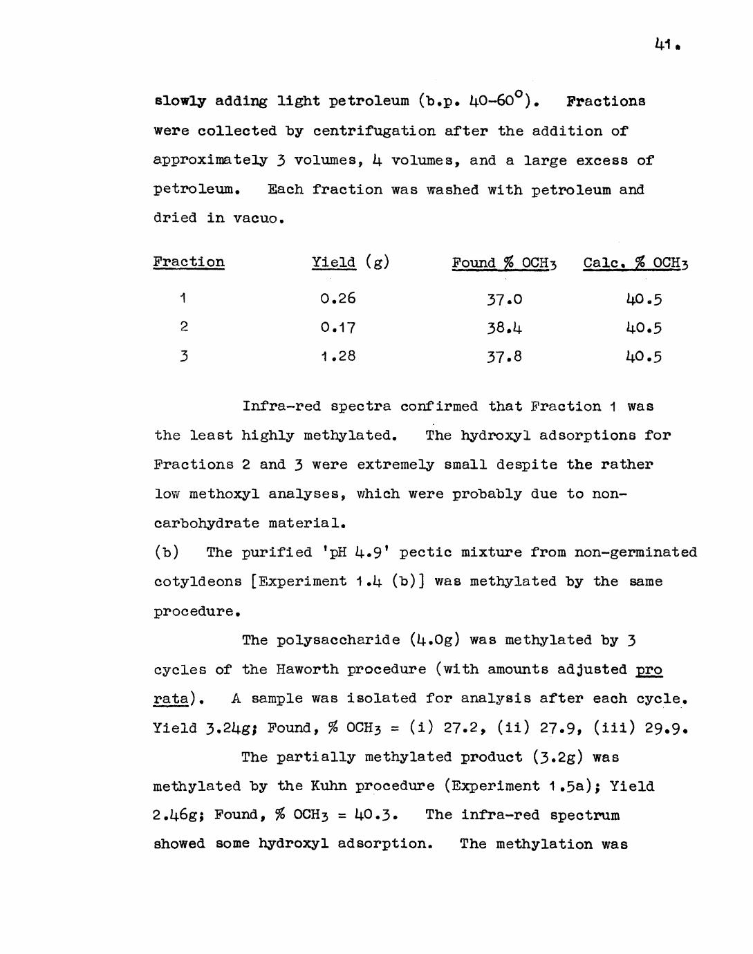
slowly adding light petroleum (b.p. UO-GO). Fractions
were collected by centrifugation after the addition of
approximately 3 volumes, U volumes, and a large excess of
petroleum. Each fraction was washed with petroleum and
dried in vacuo.
Fraction Yield (g) Found % OCH^ Gale, % OGHs
1 0.26 37.0 U0.5
2 0.17 38.U 14-0.5
3 1.28 37.8
Infra-red spectra confirmed that Fraction 1 was
the least highly methylated. The hydro xyl adsorptions for
Fractions 2 and 3 were extremely small despite the rather
low methoxyl analyses, which were probably due to non-
carbohydrate material.
(b) The purified ! pH U«9 f pectic mixture from non-germinated
cotyldeons [Experiment 1 .ij. (b)] was methylated by the same
procedure.
The polysaccharide (U.Og) was methylated by 3
cycles of the Haworth procedure (with amounts adjusted -pro
rata) . A sample was isolated for analysis after each cycle.
Yield 3.2Ug; Found, % OCH3 = (i) 27.2, (ii) 27.9, (ill) 29.9.
The partially methylated product (3.2g) was
methylated by the Kuhn procedure (Experiment 1.5a); Yield
2.U6g; Found, % OCH3 = U0.3. The infra-red spectrum
showed some hydroxyl adsorption. The methylation was

3^.2
35.9
39.0
M.2
U0.5
U0.5
kO.5
U0.5
repeated and gave a product with negligible hydroxyl
absorption; Yield 2.08g; Pound, % OCH3 = 38*2.
The product (2.0g) was fractionally precipitated
with light petroleum from chloroform solution as above.
Fraction 1 (3 volumes of petroleum) was refractionated to
give Fraction 1A and 1B.
Fraction Yield (g) Found, % OCH^ Gale. % OCH^s
1A 0.008
1B O.U13
2 0.085
3 1-3U5
The infra-red spectra showed very slight hydroxyl
absorption for 1A and 1B, and no hydroxyl absorption for 2
and 3. The low analysis of the unfractionated polysaccharide
was presumably due to contamination with non-carbohydrate
material.
(c) The pectic mixture extracted at pH 7»5 from germinated
cotyledons (Experiment U(c)) was methylated by the same
procedure.
The polysaccharide (2.0g) was methylated by 3
cycles of the Haworth procedure; Yield 1.U9g; Found, % OCH3 =
25-6.
The partially-methylated product (1 .Ug) was
remethylated by the Kuhn procedure, Yield 0.80g; Found,

% OCH3 = 38.6. The product had only very slight hydroxyl
absorption in the infra-red spectrum.
The methylated polysaccharide (O.i|0g) was
fractionally precipitated with light petroleum from
chloroform solution as above:
Fraction
1
2
3
Yield (g)
0.115
0.063
0.203
Found, % OCH-s
37.08
U0.3U
37.20
Gale. %
U0.5
The infra-red spectra showed very slight hydroxyl
absorption in Fractions 1 and 3> "but no hydroxyl absorption
in Fraction 2.
Experiment 1.6. Methylation analysis of pectic mixture
extracted at pH 7*5 from non-germinated mustard cotyledons.
Experiment 1.6a. Hydrolysis of methylated pectic mixture.
The methylated pectic mixture (5.0g, % OCH3 = 38.2)
was hydrolysed in formic acid. The residue after removal
of excess acid was heated in 0.1N sulphuric acid (150 ml)
for 6 hr. at 100° to hydrolyse formate eaters, neutralised
with barium carbonate, filtered and an excess of 1R 120
(H+ form) resin added. After standing for 16 hr. the hydrol-
ysate was applied to a column of DEAE Sephadex A25 (30g,
formate form) resin and the neutral sugars eluted with
water (3 l). The neutral fraction was evaporated to
dryness and dried in vacuo over phosphorus pentoxide
(3«75g sugar)o The acidic fraction was eluted with
formic acid (k% 9 51) and on concentration and drying as
a"bove weighed 1 .35g« Paper chromatography in solvent B
confirmed that the fraction eluted with water contained
only neutral sugars and that the fraction eluted with formic
acid contained only acidic material.
Experiment 1.6b. Reduction of acidic fraction.
The acidic material (1.25g) was refluxed in
methanolic hydrogen chloride (~b%, 100 ml) for 6 hr. The
solution was neutralised with silver carbonate, filtered
and evaporated to dryness. The residue was dissolved
in methanol (75 nil) to which 2,2'dimethoxypropane (5 ml)
was added. After standing for 16 hr. a mixture of sodium
"borohydride (l.Og) and sodium borotritiide (2.8 mg, specific
activity 3.5 me per mg) was added. After i|8 hr. excess
reagent was destroyed with dilute formic acid and the
solution passed through a column of 1R 120 (H form) resin.
The solution was evaporated to dryness and "boric acid
removed by repeated distillation of methanol from the
residue under reduced pressure. The residue was hydrolysed
in formic acid followed by 0.1N sulphuric acid as above
(Experiment 1.6a) and dried in vacuo over phosphorus
pentoxide (0.805g). Paper chromatography in solvent B
showed incomplete reduction. Practionation as above
(Experiment 1.6a) on DEAE Sephadex (formate form) gave a
neutral 'reduced 1 fraction (0.620g) and an acidic fraction
(O.l80g). A small amount (0.020g) of the acidic fraction
was completely reduced as above but with a large excess of
sodium horohydride and on examination by paper
chromatography proved to be identical to the original
neutral f reduc ed f fracti on.
Experiment 1»6c. Separation and identification of the
components of'the f reduced * fraction.
The reduced fraction (0.6l5g) was dissolved in
methanol (5.0 ml) and loaded on to a column (55 x 5«5 cm) of
cellulose powder (Whatman, CC31) previously equilibrated
with a solvent consisting of butan-1-ol : ethanol : water
(6 : 2 : 1), and eluted with the same solvent (flow rate
0.6 ml per minute), Fractions (25 ml) were collected
automatically and screened by paper chromatcgraphy in
solvent C. On the basis of the paper chromatography
results, the fractions were combined to give larger fractions
which were filtered, evaporated to dryness, dried in vacuo
and weighed.
Components of the fractions vwsre identified by
paper chroma tography in solvent B and by g. I.e. on column 1
(Pye Argon Chromatograph) at 175° (see Table 1), Major
components were characterised by the formation of crystalline
derivatives. Qualitative assay of the activity of the
methylated sugar or parent sugar given by demethylation
was "by scintillation counting of relevant chromatogram
strips .
Fraction 1 (0.073g)« The major component of this fraction
was 3,U-di-0-methylrhamnose (R& = 0.86; 0? = 1.00). Trace
amounts of 2,3,U~tri-0-methylgalactose (RQ = 0.86$ T = 6.01)
and 3~0-methylrhamnose (R-, = 0.61; T = 3.12s, i|.57w) were— y. —.
also present. Demethylation gave rhamnose as the only
detectable product, a spot of medium intensity (p-anisidine
hydrochloride spray) had a count 130 c.p.m. The sugar was
thus inactive.
Fraction 2 (0.035g). This fraction was composed mainly of
the sugars present in Fraction 1 together with trace amounts
of two unidentified components detectable only by g.l.c.
The first, (T = 1.51s, l.y^w), on the basis of its
mobility in solvent C and retention times was tentatively
identified as a di-0-methyl pentose. The second, (T =
8.82m, 10.9Us), was shown not to be a di~0-methyl galactose
or glucose obtained by reduction of the corresponding
di~0-methyl uronic acid, but nonetheless on the basis of
retention times is probably a di-0-methyl hexose.
Demethylation gave rhamnose and galactose. A
spot of medium intensity of the former had a count of 130
k7.
c.p.m., the sugar thus was inactive. A galactose spot of
medium intensity had a count of 890 c.p.m., the
intermediate activity indicating that 2,3,Lj.-tri-0-
methyIgalactose was only partly derived from galacturonic
acid end-group*
Fraction 3 (0.010g). The fraction was composed of
2,3,U-tri-0-methylgalactose and 3~0-methylrhamnose.
Fraction U (0.11?g). The fraction was composed of
3-0-methylrhamnose and 2,3-di-O-methylgalactose (RQ = 0,50;
T = 8*60s, 11.15m, 13.90w).
Fraction 5 (0.098g). The fraction contained almost pure
2,3-di-O-methylgalactose. The sugar was characterised as
the N-phenylglycosylamine derivative, which after
reerystallisation from ethanol had m.p. = 153-155
undepressed on admixture with authentic 2,3-di-0~methyl-N-
phenyl-D-galactosylamine of m.p. 153-155 • A spot of
medium intensity of the sugar had a count of U750 c.p.m.,
the sugar was thus highly active.
Fraction 6 (0.020g). The fraction was composed mainly of
2,3-di-0-m3thyl-D-galactose with a lesser amount of rhamnose
(RG = O.U3)« A spot of weak intensity of the latter had a
count of 105 c.p.m., the sugar was thus inactive.

Table 1
g.l.c. of standard compounds used in identification of
components of the /reduced fraction'
Column 1 (Pye Argon Chromatograph) at 175°
Methyl glycoslde of - T
2,3 ,U-tri-0-methylgalacto se
2 , 3~di~0-methylgalacto se
2 ,U~di~0~methylgalacto se
3 ?U~di~0-methylgalactose
3 ,U~di-0-methylrhamnos e
3-0-me thy Irhamno s e
i4.-()-methy Irhamno se
2 , 3-di-O-methylgluco se
3 ,l4.-di~0-methylglucose
2 ,U-di-0-methylglucose
2 ,3-di-O-methylfucose
5-98
8.60s 11 .2km 13.90w
0.98
3.10s
3.70
15.37s
1JLj..05s 22.25w
9.03m 11.53s
7.UOs 8.78m
1 .20sh 1 .
Fraction 7 (0.195g). This fraction, which was
chromatographically pure, crystallised on standing and
after recrystallisation from methanol had m.p. = 1U8-150
and mixed m.p. = 114-6-11+8° (with authentic 2-0-methyl-D-
galactose of nup* = 1U6-1U80 ). A spot of medium intensity
had a count of U375 Cop.nu, the sugar was thus highly active.
Fraction 8 (0.030g). The fraction was composed of 2-0-
methyl-D-galactose and 3-0-methylgalactose (R~ = 0.29).r= — (j
Fraction 9 (0.10g). The major component of this fraction
was 3-0-methylgalactose. Demethylation gave galactose as
the only product, a spot of medium intensity of which had a
count of U310 Cep.m. The sugar was thus highly active.
Fraction_10 (<'0.005g). Continued elution yielded a small
fraction, the major component of which was galactose (RG =
0.17). A spot of weak intensity had a count of 2570 c.p.m.,
the sugar was thus highly active.
Experiment 1.6d. Separation and identification of the
components of the neutral fraction.
The neutral material (3*6g) was dissolved in a
mixture (25 ml) of methanol : butan-1-ol half saturated with
water (1 : 1 ) and loaded on to a column (76 cm x 6 cm) of
cellulose powder (Whatman, cc31) previously equilibrated with
solvent (a) below. The sugars were eluted with the solvents

50 o
given below, (k 1 of each), in order.
Solvent Systems;
(a) butan-1-ol half saturated with water : light
petroleum (B.P. 100-120°) (1 : 3, upper phase) and
butan-1-ol : ligh_t petroleum (1 : 3) mixed in ratio 1:1.
(b) as above but using a ratio of butan-1-ol : light
petroleum of 1 : 1 in both cases.
(c) as above but using a ratio of butan-1-ol : light
petroleum of 3 : 1 in both cases.
(d) butan-1-ol half saturated with water.
Fractions (250 x 25 ml, thereafter 50 ml) were
collected automatically and screened by paper chromatography
in solvents B, C, D and E, where appropriate. On the basis
of the paper chromatography results the fractions were
combined to give larger fractions which were filtered,
evaporated to dryness, dried in vacuo over phosphorus
pentoxide and weighed. Evaporation of the earlier fractions
was carried out below 30 to minimise loss of the volatile
tri-0-methyl pentose derivatives.
Components of the fractions were identified by
paper chromatography on column 1 (Pye Argon Chromatograph)
at 125° for Fractions 1 and 2, at 150° for Fraction 3 and
thereafter at 175° (see Table 2). Major components were
characterised by the formation of crystalline derivatives.
Demethylation was also used in identification of sugars.

Table 2
Gas liquid chroma tography of standard compounds-neutral
fraction
Column 1 (Pye Argon Chromatograph)
Temperature methyl glycoside of - T
2,3,I;~tri-0-methylrhamnose O
2,3,Lj.~tri-0-methylxylose O.UOm 0.53s
2,3?5-tri-0-methylarabinose 0.53s O.?1w
150 2,3,1+,6-tetra-O-methylgalactose 1 .76sh 1 ,87s
1 75 2,3 ,U-tri-£-methylara"binose 1
2,3-di-0_-methylarabinose 1 ,U6s 1.6lw 1.78s
2,5-di-0-methylara~binose 1 .62
3,5~di~0-methylarabinose 1.02 2.20
2-0-methylaraMnose 5.07s 9.10W
3,^--di~0>-rnethylrhamnose 0.98
3-0~methylrhamnose 3«10s
U-O-methylrhamnose 3^70
2,3,6-tri-0~methylgalactose 2.83s
2,U>6-tri-£-methylgalactose 3«55ni
2,3,I|~tri-0~methylgalactose 5*95
2, ii-di-0-me thy Iga lactose 13.I;Om 15»37s
2,3>6-tri-0-methylglucose 3»01m U*05s
2,3-di-O-methylglucose 9-07m 11.56s

52.
Fraction 1 (0.005g)« The fraction contained almost pure
2,3,U~tri-0-methylrhamnose (RQ = 1.00; T = O.ij.2) with a trace
of 2,3,5-tri-0-methylara"binose. Demethylation gave rhamnose
as the only product.
Fraction 2 (l.66g). The fraction contained a mixture of
2,3j5-tri-0-methylarabinose (RQ = 0,965 T = 0.53s, O.?1w)
and 2,3,Lj.-tri-0-methylxylose (Rn = 0.9U; T = O.^Om, 0.53s)— (j —with a trace of 2,3,l4. ? 6-tetra-Q-methylgalactose. The
major components were separated "by preparative paper
chromatography in solvent E to give chromatographically pure
syrups, the yields indicating that the fraction contained
60$ of the araMnose derivative. 2,3,5~tri-0-methylara"binose
was characterised as the aldonamide, which after
recrystallisation from ethanol had m.p. = 137 and mixed
m.p. =137 (with an authentic sample of 2,3,5-tri-O-methyl-
-L~ara"bonamide of m.p. = 137 )•
2,3,i4--tri~0-methylxylase crystallised on
standing and was characterised as the aldonolactone, which
after recrystallisation from diethyl ether : light
petroleum (B.P, Ij.0-60 ), (1 : 1) had m.p. = 53 and mixed
m.p. = 53° (with an authentic sample of 2,3,U-tri-0~methyl-
-D-xylonolactone of m.p. = 55 .
Fraction 3 (0.105g). The fraction contained almost pure
2,3,l4.,6-tetra-0~methylgalactose (R^ = 0.88; T = 1 ,73sh,
1.85s) with traces of the major components of Fraction 2.

53.
The sugar was purified "by preparative paper chromatography
in solvent D and characterised as the N-phenylglycosylamine
derivative, which after recrystallisation from ethanol had
m«p. =198 and mixed m.p. =195 (with an authentic sample
of 2,3>^»6-tetra-0-methyl~N-phenyl-D-galactosylamine of
m.p. = 193°).
Fraction U (0.050g). The major component of this fraction
was 3,U~cLi-0~rnethylrhamnose (RQ = 0.88; T = 0.9) with a
smaller amount of 2,3,6-tri-0~methylglucose (R~ = 0.86;
T = 3 •02m, l4..03s) and a trace of tetra-0-methylgalactose.
Fraction 5 (0.055g)« The major component of this fraction
was 2,3y6-tri-0-methylglucose with a small amount of
3,U~di-0-methylrhamnose. 2,5-di~0-methylara~binose (T = 1 .61 )
and 3,5-di-O-methylarabinose (T = 1.02, 2.20) were detected
as minor components "by g.l.c. only. D em e thy la t ion of the
fraction gave glucose and rhamnose.
Fraction 6 (0.290g). The fraction contained mainly
2,3-di-O-methylaraMnose (R^ = 0.7U; T = 1 oij.8s, 1 .62w, 1.80m)
with approximately 5% 2,3,6-tri-O-methylgalactose (RQ = 0.81;
T = 2.82s, other peaks obscured) and 10$ 2,lj.,6-tri~0-
-methylgalactose) (R = 0.?6; T = 3
Fraction 7 (0.565g). The fraction contained almost pure
2,3-di~0-methylarabinose with a trace of 2,U,6-tri-0-

-methylgalactose. The sugar was characterised as the
aldonamide, which after recrystallisation from ethanol had
m»p. =158 and mixed m.p c =158 (with an authentic sample
of 2,3-di~0-methyl~L~ara"bonamide of m.p. = 160°).
Fraction 8 (0.053g)e The fraction contained
methylrhamnose (R = 0.6I4-; T = 3«?6) and 2,3,Lj.-tri-0-
me thy Iga lactose (R» = 0.68; T = 5.95) as the manorvj —
components with a lesser amount of 2,3-di-O-methyl-L-
araMnose. Demethylation gave rhamnose, galactose and
arabinose.
Fraction 9 (0.030g). The fraction contained 3-0-
methylrhamnose (R~ = 0.62; T = 3«10s, i4..6lw) as the major
component with a small amount of 2,3-di-0~methylglucose and
a trace of L|.~O-methylrhamnose. Demethylation gave rhamnose
and a trace of glucose.
Fraction 10 (0.120g). The fraction contained 2,3-di-O-
methylglucose (RG = 0.69; T = 8.88m, 11.55s) as the major
component with a small amount of 3-0-methylrhamnose.
Demethylation gave glucose and a trace of rhamnose.
Fraction 1 1 (0.025g). The fraction contained an
unidentified sugar (RQ = 0.61 ; T = 9.23, 12. 60) as the major
component. Demethylation gave glucose as the only product.
The sugar was tentatively identified as a di-0-methylglucose

55.
on the above evidence, but was neither 2,l4.-di-0-methylglucose
(T = 7.3^-m, 10.U2s) nor 3,U-di-0-raethylglucose (T = 7oUOs,
8.78m).
_Pr action 1 2 (O a 665g). This fraction contained almost pure
2-0-methyl-L~arabinose (RQ = 0.50; T = 5.05s, 9.lOw) with a
trace of 2,Lj.-di~0-methylgalactose (R~ = 0.52: T = 13.30m,—— y. __
15.28s). The sugar was characterised as the toluene-g-
-sulphonylhydrazone, which after recrystallisation from
methanol had m.p. = 1^2-1 UU° and mixed m.p. = 1U2~1UU° (with
authentic 2-(>-me'kfryl-L--arabinose toluene-g-sulphonylhydrazone
of m.p. =
Fraction 13 (O.HOg). The fraction was chromatographically
pure and was identified as 3-0-methylarabinose (R^ = O.U5$
T = 2.93m, 3.81s, 5«86w, 7.88m) by methylation and
demethylation. Methylation followed by g.l.c. gave both
2,3,5- and 2,3,l±-tri-0-methylarabinoses proving the sugar to
be neither U- nor 5~0-methylarabinose. Demethylation gave
only arabinose.
Fraction ^k (0.0ij.0g). The fraction contained pure arabinose
(R - 0.23) which was characterised as the toluene-g"
-sulphonylhydrazone. After recrystallisation from
methanol the derivative had m.p. =155 and mixed m.p. =
153-15U0 (with authentic L-arabinose toluene-jD-
sulphonylhydrazone of m.p. = 153 )•

56.
Continued elution with solvent (d) yielded no
further carbohydrate material„
Experiment 1 •?• Comparison of the methylated pectic
mixtures from germinated and non-germinated cotyledons.
The methylated pectic mixtures (75 nig) (Experiment
1 *5a, b and c) v/ere dissolved in dry tetrahydrofuran (5.0 ml,
freshly distilled from lithium aluminium hydride) and an
excess of lithium aluminium hydride added. The flasks were
tightly stoppered and shaken for 16 hr., followed by
heating under reflux for 2 hr. Excess reagent was destroyed
by careful addition of ethyl acetate, and lithium aluminate
was precipitated with the minimum amount of water. The
contents of the flask were transferred to a centrifuge
bottle (250 ml) with a large volume of acetone and the
precipitate removed by centrifugation. The supernatant
solution was decanted off and the precipitate washed once
with acetone, centrifuged and the supernatant solutions
combined. The precipitate gave a negative reaction to the
phenol sulphuric acid reagents. The supernatant solution
was evaporated to dryness. The products were examined
by paper chromatography in solvent B after hydrolysis, and
by g.I.e. on column 1 (Pye Argon Chromatograph) at 175
after methanolysis. The gas chromatograms are shown in
Diagram 1 and retention times of component sugars are given
in Table 3B. Paper chromatography showed that reduction

57.
was complete; the component sugars were identified using
standards and the following results were obtained ("by
visual examination; g-anisidine hydrochloride spray).
Table 3A
Sugar Relative proportion inhydrolysate of -
2 ,3 ,5-tri~0-methylarabinose
2 ,5-di-O-methylarabinose
2 ,3-di-O-methylarabinose
2-0-methylarabinose
arabinose
2,3>U-tri-0~methylxylose
3 ,U~di-Q-methylrhamnose
3 ~0-m ethy Ir hamno s e
rhamnose
2 , 3 ,U , 6-t etra-0-methylgalactose
2 ,L|. , 6-tri-0~methy Igalactose
2 ,3-di-O-methylgalacto se
2 ,U-di-0-methylgalacto se
2-0~methy Igalactose
s
vw
B
m
w
s
w
vw
vw
w
w
w
vw
w
s
vw
s
m
w
s
w
vw
vw
w
w
m
vw
w
m
vw
w
tr
-
s
w
vw
vw
w
w
m
vw
w
s = strong, m = medium, w = weak, vw = very weak, tr = trace,
a is methylated reduced non-germinated pH 7.5 extracted pectin
b is methylated reduced germinated pH 7«5 extracted pectin
c is methylated reduced non-germinated pH U«9 extracted pectin.

58.
Table 5B
Gas liquid chromatography of methylated, reduced,
methanolysed pectins. (see Diagram 1)
Column 1 (Pye Argon Chromatograph) at 175°
Peak methyl glycoside of -Standard
1 2,3,U-tri-0-methylxylose O.UO 0.14-2 O.U3
2 2,3,5-tri-O-methylaraTDinose/ o 2,3,^-tri-O-methylxylose °
3 2,3,5-tri-O-methylarabinose 0.71 0.7U 0.75 0.7U
U 3,l+-di-0-methylrhamnose 0.98 1.02 1.02 1.02
5 2,3-di~0-methylaraMnose 1 «U6 1.1^-9 1-U9 1 »U9
6 2,3,^,6-tetra-O-methylgalactoseA 2,3-di-O-methylaraMnose 1 '
7 2,3-di-O-metnylarabinose 1.78 1.83 1.83 1.83
7a 2,3,6-tri-O-methylgalactose 3.01 2.98 2.98 2.97
8 3-0-methylrhamnose 3.10 3.21 3.20 3.21
9 2,U,6-tri-0~metnylgalactose 3.55 3.65 3-67 3.67
10 2,3,6-tri~0-methylgalactose/ . 1Q . 1 , ,,, k1 , 2,U,6-tri-0-methylgalactose 4 ' 1 ° U * 13 ^* 13 ^' 13
11 2-0-me thy laraM nose 5.07 5.18 5-20 5.20
12 2,3,i4.-tri~0-methylgalactose 5.98 6.02 6.05 6.03
13 2,3-di-O-metnylgalactose 8.i|8 8.3U 8.3U 8.33
1U \inidentified - 9.U5 9.U7 9-U7
15 2,3-di-O-methylgalactose 11.20 11.16 11.16 11.15
sh unidentified - 12.81+ 12.8U 12.814.
16 2,l4--di-O-methylgalactose 13.UO 13.U7 13.^9 13.U3
17 2,U-di-2-me "U:iy :i-galactose 15.37 15«UO 15-U2 15.U3

I
•of
I
i I
i

59.
Discussion
(A) Previous theories as to the constitution of the
pectic substances have tended to oversimplify the situation.
Until fairly recently it was thought that a mixture of three
homopolysaccharides was present, namely the neutral galactan
and araban and the acidic galacturonan. Although
authenticated samples of each of these polysaccharides have
been isolated, they would seem to occur rarely. Samples of
galacturonan almost invariably contain a substantial
proportion of covalently linked neutral sugars (63). The
arabans from guava (6k) and apple (65) also contain an
appreciable amount of sugars other than arabinose,
particularly galactose and galacturonic acid. Barrett and
Northcote (21) have shown that in alkaline solution apple
pectin undergoes cleavage by an elimination mechanism
(|3 elimination) at methyl esterified galacturonic acid units
to give a fraction composed mainly of galacturonic acid, and
a fraction composed mainly of neutral sugars. In the light
of these results it seems likely that some arabans are
elimination artefacts produced during extraction at
alkaline pH, e.g. with lime water.
The results of fractionation by column
chromatography and ultracentrifugation of mustard cotyledon
pectin mentioned in the Introduction have shown that it is
not homogeneous but that related acidic polysaccharides are
present. The structural features elucidated by experiment
on the whole pectin, although probably common to the
60.
components, may thus be present to a greater or lesser
degree in each component.
The pectic mixtures extracted from non-germinated
cotyledons at pH 7«5 and pH U.9, and from germinated
cotyledons at pH 7-5 have "been satisfactorily purified
(protein content 3 - 1%) and methylated fully.
The main features of the pectin have been
established by the formation of crystalline derivatives of
the sugars obtained by hydrolysis of the methylated
polysaccharide. The acidic sugars were identified after
reduction with sodium borotritiide as the corresponding
galactose derivatives. The high specific activity of the
products confirmed that they had originated from galacturonic
acid derivatives. The other sugars in the reduced acidic
fraction (exclusively rhamnose derivatives) were unlabelled.
The presence of the 1,U linked galacturonan main chain
common to pectic substances was thus confirmed (by the presence
of 2,3-di-O-methyl-D-galactose), the chain being interrupted
by the insertion of rhamnose units linked through C/ 2 \ of
rhamnose (3,^-di-O-methylrhamnose) as has been established
for several other pectins. As rhamnose derivatives were
present in the neutral fraction in different proportions to
those of the acidic reduced fraction (e.g. U-0-methylrhamnose
was found only in the neutral fraction), contiguous rhamnose
units are probably also present. Recent work has shown
that the pectic substances of soybean (22,61) also have
contiguous rhamnose units. Approximately 60$ of the

61.
galacturonic acid and rhamnose residues carry side chains.
The principal mode of branching of galacturonic acid is
through C/,\ (as shown by presence of 2-0-methyl-D-galactose),
although some branching through C/ 2 \ (3-Q~methylgalactose)
and through both C/ 2 \ and C/,\ (galactose) also occurs.
Rhamnose is mainly branched through C/, \ (3-0~methylrhamnose),
occasionally through C/,\ (U-0-methylrhamnose) and rarely
through both C/.,N and C/, % (rhamnose). Derivatives of
glucose (formed by the reduction of glucuronic acid derivatives)
were not observed and hence most of the minor features, e.g.
glucuronosylgalactose and glucuronosylfucose, listed in
the Introduction were not detected in this study. A recent
study of the native pectic mixture by Dr. D. Rees (7k) has,
however, shown that traces of these disaccharides are
present in partial hydrolysates.
The pattern of neutral sugars confirmed that
branched araban side chains occur. Derivatives of arabinose
account for more than 50^ of the pectic mixture, and thus
the side chains are of high molecular weight. The
methylated sugars are typical of arabans (see Chapter 3)»
although 3-0-methylarabinose is comparatively rare.
D-xylose was present in large amounts and exclusively as
end group (2,3,U-tri-0~methyl-D--xylose) confirming the
view (56) that single unit side chains of xylose residues
are attached to the main chain, although other interpretations
are of course possible. Galactose occurred mainly as end
group (2,3,l4.,6-tetra~0~methyl-D-galactose) and as 2,^,6-tri-

62,
-0-methylgalactose, indicating that 1 ,1+ galactan, as side
chain or otherwise, is not a significant structural
feature of the pectic mixture. Hullar and Smith (67) in
an investigation of sugar beet pectin claim that the
galactose units connect the araban side chains to the
galacturonan main chain, and this might well be the case
with the mustard cotyledon pectin. Glucose is not a
typical component of the pectic substances and the results
of Experiment 1.2 show that all glucose is contained in one
discrete fraction, which showed similarities with a family
of polysaccharides known as the amyloids. The glucose
derivatives found in the methylation study of the whole
pectin are thus unlikely to be structural units of the pectic
mixture.
These conclusions are in agreement with the known
chemistry of the pectic substances and in particular with
the general structure of mustard cotyledon pectic mixture
which has been tentatively proposed (23). The average
structure was represented as :-
3t1Z
3t1Z
GalpA1->2 Rhap1->i4. GalgA1-»2 Rhat)1-4j. GalgA- ~ ~ 3 k "
t t1 1Z Z
Where Z = Xylg or araban side chain with or without galg
link unit (s).
The molar ratios of component sugars were found

63.
from the weights of column fractions. For mixed fractions
the proportion of each component was estimated from the peak
areas of gas chromatograms. The results are thus more
valuable as a guide to the relative amounts of methylated
sugars present than as an accurate analysis. The yields of
2,3j5-tri-0-methylarabinose, 2,3,U-tri-0-methylxylose and
2,3>U»6-tetra-0~methylgalactose are probably overestimated,
as, due to the volatility of these sugars, the syrups were
dried only briefly. The ratios (corrected for reduction in
the case of galacturonic acid derivatives and for initial
weight of the neutral and reduced fractions) are given
below :-
2,3-di-O-methylgalacturonic acid 1.00
2-O-methylgalacturonic acid 1 .1U
3-0-methylgalacturonic acid 0.16
galacturonic acid 0.03
2,3,5~tri-0-methylarabinose 2.86
2,3-di-O-methylarabinose 2.50
2,5-di~0-methylarabinose O.OU
2-Q-methylarabinose 2.2Lj.
3-0-methylarabinose O.i±2
arabinose 0.15
2,3,^-tri-O-methylxylose 1.88
2,3,U-tri-0~methylrhamnose 0.03
3,U-di~0-methylrhamnose 0.
3-0-me thyIrhamno se 0.
i±-0-me thy Irhamno s e 0.11

a*.
rhamnose
2,3,4,6-tetra-0~methyIgalacto se
2 ,U, 6-tri-O~methylgalacto se
2,3 ,i4--tri-0-me thy Igalacto se
2 s 3 >6~tEi-0~methyIgalactose
2 ,l±-di-O-methylgalactose
2,3? 6-tri-O-methylgluco se
2,3-di-O-methylgluco se
unidentified components (molecular weight taken as 208)
0.03
0.32
0.15
0.09
0.05
0.0k
0.15
0.29
0.08
These figures maa«y be used to derive an anhydrosugar
content which can be compared with that obtained in a previous
study (59) in which galacturonic acid was determined by
decarboxylation and the neutral sugars colorimetrically
after separation by paper chromatography.
Sugar
Galacturonic acid
Arabinose
Xylose
Rhamnose
Galactose
Glucose
% anhydro sugar
3y methylation analysis
16.2
5U.2
12.U
9-7
U.5
3.0
Directly
23-8
28.8
10.9
2.6
6.1
3.9
The comparison of the non-germinated and
germinated pectic mixtures did not show .the dramatic

65.
differences expected if a decrease in the degree of
branching was responsible for the role of these poly-
saccharides in the developing cell wall. The gas
chromatograms (Diagram 1, a and b) are almost identical and
do not show any major difference in the proportion of
neutral sugar derivatives to 2,3-di-O-methylgalactose
(formed by the reduction of 2,3-di-0~methylgalacturonic
acid), although comparison of spot intensities of paper
chromatograms (Table 3A) does indicate a very slight decrease
in the degree of branching of the germinated pectic mixture
as shown by the ratio of 2-0-methyIgalactose to 2,3-di-O-
-methylgalactose. It would thus appear that if the pectic
substances control wall extensibility through their ability
to form gels via the interactions proposed for pectin
gels in vitro then this must predominantly involve a
redistribution of side-chains. This would most probably
be intramolecular, so that relatively unbranched and highly
branched regions of the galacturonan main chain alternate in
the molecule. A structure of this type has been proposed
for apple fruit pectin (21 ). In a pulse labelling
experiment with 1 **C arabinose, Stoddart and Northcote (68)
have shown that the isolated pectin fractions of actively
growing sycamore cells are metabolically related, the label
passing from the neutral arabinan-galactan fraction to the
acidic fractions as growth occurred. A similar function is
proposed for the pectic araban from mustard cotyledons (28).
Intermolecular redistribution of side-chains would thus

66,
also seem to "be a possibility, although less likely as a
comparison in the ultracentrifuge (60) has not shown a
molecular weight increase of any pectin fraction on
germination.
Alternatively wall extensibility may be controlled
by some other type of interaction involving the pectic
substances.
The non-germinated pectic mixture extracted at
pH k»9 showed significant differences to those extracted at
pH 7-5 (both non-germinated and germinated). Derivatives of
arabinose are present in greatly diminished amounts and are,
with the exception of 2,3 s 5-tri-0-methylarabinose, relatively
minor features of the pectic mixture (Diagram 1c and Table
3A). The degree of branching is thus considerably lower,
the large araban side-chains being of rather limited
occurrence. This result is the opposite to that expected
on the basis of the gel theory. As EDTA is less efficient
at pH U.9 the less strongly bound pectin molecules in the
cell wall will be extracted preferentially. These would
be expected to have a high proportion of side-chains,
particularly the high molecular weight araban type. It
appears that the converse is true and that the species with
a high proportion of araban side-chains are more strongly
bound in the wall. Whether this property is attributable
to the araban side-chains or to some other property of the
molecule is difficult to assess but it seems clear that the
interactions between pectic substances in the wall are more

67,
complicated than those postulated for in vitro gels.
The precipitation of the non-germinated pectin
extracted at pH 7«5> which occurred during deproteinisation
with phenol (Experiment 1.1) would confirm this view. The
stages leading up to deproteinisation are; extraction with
EDTA (pH 7«5) s precipitation of the pectic mixture with
aqueous ethanol (75$)* dialysis against tap water and
freeze-drying. That no precipitation occurred when
deproteinisation was carried out in the presence of EDTA
suggests that Ca*"1" ions are directly involved. A possible
explanation is that a polysaccharide complex involvingJ.J.
"bound Ca ions is present in- the cell wall s and is stabilised
by protein (probably denatured) in the EDTA extract. The
protein apparently confers water solubility on the system,
and on deproteinisation the pectin aggregates, presumably
as calcium pectate. The pectic mixture extracted at pH U»9
showed little tendency to precipitate on deproteinisation
(Experiment 1,J±b), confirming the view that it is the more
highly branched pectin molecules which interact more
strongly, but nevertheless the uronic acid residues must be
involved because of the role of the Ca ions,
(B) The fraction of the pectic mixture (non-germinated,
pH 7.5 extracted) which did not precipitate on deproteinisation
(Experiments 1,1 and 1,2) was shown to be homogeneous in the
ultracentrifuge. The sugars given on hydrolysis, namely
glucose, xylose and galactose are characteristic of a group
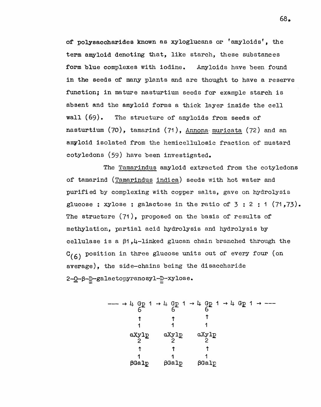
68,
of polysaccharides known as xyloglucans or r amyloids', the
term amyloid denoting that, like starch, these substances
form "blue complexes with iodine. Amyloids have "been found
in the seeds of many plants and are thought to have a reserve
function; in mature nasturtium seeds for example starch is
absent and the amyloid forms a thick layer inside the cell
wall (69). The structure of amyloids from seeds of
nasturtium (70), tamarind (71), Annona- muricata (72) and an
amyloid isolated from the hemicellulosic fraction of mustard
cotyledons (59) have been investigated.
The Tamarindus amyloid extracted from the cotyledons
of tamarind (Tamarindus indica) seeds with hot water and
purified by complexing with copper salts, gave on hydrolysis
glucose : xylose : galactose in the ratio of 3 : 2 : 1 (71*73).
The structure (71 )> proposed on the basis of results of
methylation, partial acid hydrolysis and hydrolysis by
cellulase is a (31 ,L|.-linked glucan chain branched through the
C//-\ position in three glucose units out of every four (on
average), the side-chains being the disaccharide
2-0-p-D-galactopyranosyl-p-xylose.
—— -»Lj.Gp1 -» U Gp 1 -» U Gp 1 -» U Gp 1 -» ——~k Gp 1 -6t 1
aXylj) 2t1
6~
t 1
aXyl£ 2t1
(SGalp
U Gp 1 6
1
2t
1

69.
Tamarindus amyloid, structure proposed by Kooiman (71 )•
The Tamarindus amyloid gives the characteristic
blue-green colour reaction with potassium triiodide in
saturated sodium sulphate (Experiment 1 .2) with an
^extinction maximum at 6UO nm. Amyloids with a less branched
structure, i.e. a more cellulosic character, give colour
reactions in which the absorption maxima are shifted, the
amyloids of the seeds of the Annonaceae for example, for
which a structure identical with Tamarindus amyloid except
that only one in four glucose residues carry side-chains is
proposed, give violet colours (72).
The amyloid from mustard cotyledons which was
insoluble in EDTA (59) (the 'insoluble 1 amyloid) had an
extinction maximum at 650 nm. Hydrolysis of this
polysaccharide gave mainly glucose and xylose with some
fucose and only traces of galactose. A structure related
to that for Tamarindus amyloid but with side-chains in which
xylose units are linked through C/, \, and C/.,\ (instead of
only C/pOancl terminated in xylose, fucose, glucose and,
rarely, galactose was proposed on the evidence of methylation,
which also showed the molecule to be less branched than
Tamarindus amyloid.
The 'soluble' amyloid gave the characteristic
colour with potassium triiodide in saturated sodium
sulphate, with an extinction maximum at 650 nm. In a
methylation study carried out by Miss Ann Peattie the
'soluble' and 'insoluble' amyloids from mustard cotyledons
70 c
were compared "by g.l.c. Several interesting differences
were observed. The ' soluble' amyloid was shown to have a
somewhat less branched (as judged from the ratio of
2,3-di-O-methylglucose to 2,3,6-tri-O-methylglucose) p1 ,U
linked glucan main chain and to have xylose side-chains
linked only through C/, \. The side-chains are terminated
in xylose, glucose, fucose and galactose, the high
proportion of the latter being a major difference between
the two polysaccharides.
The 'soluble 1 amyloid is thus more closely related
to Tamarindus amyloid than is the 'insoluble ' amyloid in
having less complicated side-chains, a higher proportion of
which are terminated in galactose.
It would appear that a parallel situation exists
with the amyloids and the pectins from mustard cotyledons in
that solubility in EDTA is inversely proportional to the
degree of branching.
Quantitative experiments on the iodine-staining
reaction (Experiment 1.3) showed that, though less branched,
the mustard cotyledon amyloids have a lower specific
extinction than the Tamarindus amyloid. In starch-type
polysaccharides the interaction with iodine increases as the
degree of branching decreases and hence the above observation
probably reflects an increased proportion of side-chain sugars.
The 'soluble' amyloid was extracted equally efficiently with
EDTA at pH U«9 and pH 7.5 and occurs to the extent of about
by weight in the deproteinised pectic mixtures from

71.
non-germinated cotyledons. Protein effectively prevents the
colour reaction and deproteinisation is thus a necessary
preliminary to amyloid determination (Experiment 1«3a).
'Soluble* amyloid was found to account for a large
proportion (30$) of the neutral fraction of non-germinated
pectic mixture, "but was completely absent in both the
neutral fraction, and the whole deproteinised pectic mixture
from germinated cotyledons.
The results are consistent with the commonly held
view that amyloids have a reserve function in germinating
seeds. It is interesting to speculate that the amyloids
might serve as a precursor of cellulose and therefore
represent a store of 'soluble cellulose' in the dormant
seed. The structural studies indicate that the amyloids as
a class of polysaccharides are as variable in structure as,
for example, the pectic substances, the differences between
members of both classes arising chiefly from the proportion
and structure of the side-chains.

CHAPTER 2
Incorporation of myo-inositol~2~3H "by
germinating mustard seeds

72.
Introduction
The use of radioactive isotopes as tracers in
"biosynthetic experiments is widespread even though their
availability on a commercial scale is relatively recent.
In most respects the isotopes are chemically identical
the stable isotope of the element but differ physically in
that they emit radiation. The great value of such isotopes
in biological systems is that even in enormous dilution with
non-radioactive material the emitted radiation can be
detected and measured, with the result that only minute
quantities are required. The most commonly used tracers
are compounds containing the isotopes 3H, 1Z*C, 32P and 55S
although many others are also available. Elucidation of
the role of many biologically active substances and the
biosynthetic pathways involved in their metabolism has been
made possible by use of the radioactively labelled compounds
in incorporation experiments. 3H and 1 ^C both decay to a
stable isotope by emission of & particles (the electron).
The energy of the emitted radiation is relatively weak in
3H (0.018 MeV) as compared with 1 kC (0.15 MeV) and the
radiation is classed as f soft f and 'medium 1 (3 emission
respectively. On the laboratory time scale these isotopes
are fairly stable, 3H having a half-life of 12.26 years and
1 **C 5,770 years, so that negligible loss of radioactivity by
decay occurs during experiment.
High energy particles ( ^- and f hard ! p-radiation)
are traditionally counted with the Geiger-Muller counter

73.
(GM tube) in which the radiation produces ion-pairs in a gas
held between two oppositely charged electrodes. The
lonisation Chamber and Gas-flow Counter are also examples of
this approach. Disadvantages of the GM tube are that low
energy particles cannot be counted, high count rates (e.g.
over 5s000 per second) cannot be measured, and efficiency is
low.
The scintillation counter enables particles of all
energies to be counted efficiently even at extremely high
count rates. In this device the emitted particles are
counted via the secondary radiation which they produce in
some systems. Certain fluorescent compounds can be
excited to metastable states by irradiation and on returning
to the ground state emit flashes of light (scintillations).
In the scintillation counter these are detected by
photomultipler tubes whose outputs are amplified, analysed
and recorded.
The most commonly used technique is that of
'liquid scintillation counting 1 . The scintillator in this
case is a liquid which has two main components5 the solvent,
usually toluene, xylene or dioxane of high purity, and the
primary solute, usually a molecule containing several
conjugated aromatic rings, for example anthracene or
2,5-diphenyloxazole. The solvent transfers the energy to
the solute which emits the light. Frequently a secondary
solute is added in small amounts whose function is to shift
the spectrum of emitted light to longer wavelengths at which

7k.
the liquid is more transparent and photomultiplier response
improved. The radioactive material may be dissolved
directly (e.g. aqueous solutions such as tritiated water in
dioxane based scintillator) or indirectly, by the use of a
blending agent, (e.g. plasma with a quaternary amine salt)
in the scirrfcillator» Alternatively chromatogram or
electrophoretogram strips, or solutions spotted onto
cellulose acetate or glass fibre paper may be immersed in
the scintillator and counted with fairly high efficiency.
Severe loss of efficiency can occur through "quenching*,
the term given to energy loss of the emitted radiation by
absorption. This most commonly occurs when coloured
substances, water and other compounds are present in significant
amounts. A method of assessing this is given in the
experimental section.
Gel, plastic and solid scintillation counting are
also widely used. In the latter, which is particularly
useful for ^-radiation due to its high stopping power, the
sample is surrounded by a large inorganic crystal, such as
sodium iodide activated with a trace of thalliums enclosed
in a metal container with perspex windows.
The pulse counting methods described above measure
the sum of the radiation entering the counter from a sample
but give no detail as to the variation in activity within a
heterogeneous sample. The location of radioactivity in such
a sample, e.g. a thin section of a tissue or organ, or a
chromatogram or electrophoretogram, may be determined by

75.
autoradiography (75)• In this technique the property of
radioactive particles to produce a latent image in a nuclear
emulsion held in intimate contact with the source is
utilised. A cumulative record of the radiation incident on
the emulsion is thus produced and is made permanent "by
development and fixation. By responding in a strictly
localised fashion the autoradiograph is ideally suited to
studies of the distribution of activity within a sample, "but
as compared to scintillation counting the process is slow,
complicated and more amenable to qualitative than
quantitative measurements. Low energy £ particles are
counted with extreme efficiency, and coupled with low
background effects, levels of activity as low as one
disintegration per day can be detected. The results of
scintillation counting and autoradiography are frequently
complementary.
The biosynthesis of polysaccharides has been the
object of intensive interest and radiochemical techniques
applied in this field have yielded some dramatic results.
In biological systems,, the interconversion of sugar units
and their polymerisation into oligo- and polysaccharides is
effected through a group of labile compounds known as the
sugar nucleotides, in conjunction with specific enzymes.
The high free energy of these compounds relative to sugar
glycosides (which is associated with the phosphodiester
linkage) and enzyme catalysis account for the ease with
which biologically useful degrees of conversion are obtained
76,
in reactions (e.g. the direct polymerisation of glucose)
which are thermodynamically unfavourable in that the products
have a higher free energy than the reactants.
A series of reactions whereby the commonly
occuring units of plant polysaccharides are interconverted
is shown "below :
epimerisation UDP-D-glucose < • •• A UDP-g-galactose
oxidation
UDP-D-glucuronic acid ^ —> UDP-D-galacturonic acid
decarbox- -ylation
UDP-D-xylose ^ • •• • • r* UDP-L-arabinose
UDP is an abbreviation for uridine diphosphate. Other
nucleosides, namely adenosine (A), guanosine (G), cytosine
(C), and thymidine (T), are also involved in other
interconversions and biosyntheses.
The discovery that phosphorylase catalysed the
polymerisation of glucose from glucose 1-phosphate prompted
the theory that it occupied a key role in starch and glycogen
anabolism (and catabolism). The role of glucose 1-phosphate
as the immediate precursor of starch and glycogen was
disproved by Leloir and his colleagues who showed that both
compounds are synthesised from nucleotide sugars in plants,
animals and bacteria (76). The in vitro syntheses of many
carbohydrates from 1uC-labelled nucleotide sugars by
particulate enzyme systems from mung bean seedlings have

77.
"been reported, e.g. a cellulose-like polymer from GDP-glucose
(77)> glucomannan from GDP-glucose and GDP-mannose (78) and
galactan from UDP-galactose (79). It has also been
demonstrated that UPP-xylose is the precursor of (3-1 ,U xylan
in corn-cobs (80). Much attention has been focussed on the
biosynthesis of the pectic substances; Albersheim and
Bonner (81,82) have shown that pectin in oat coleoptiles is
synthesised as a methyl esterified galacturonan and that
glucose and inositol will serve as uronide precursors.
Methionine and formaldehyde serve as precursors of the
methyl group (81,83). On the basis of these results it was
postulated that the immediate precursor of pectin is the
methyl esterified galacturonic acid derivative of a nucleoside
diphosphate. The work of Hassid and his co-workers (8^,85)
however has shown that UDP-methyl-D-galacturonate was not a
precursor of galacturonan in an enzyme system from mung bean
seedlings, and that of the galacturonic acid nucleotides
only UBP-galacturonic acid is an efficient precursor of
pectin. S-adenosylmethionine has been postulated as a
methyl donor to pectin in this system. Stoddart and Forthcote
(68) in a study of the continuous incorporation of 1/*C-glucose
into the pectin fraction of actively growing cells of sycamore
callus tissue have shown that the first labelled product is
a partly methyl esterified galacturonan from which the
strongly acidic and weakly acidic fractions are formed by
demethylation and addition of neutral side chains respectively.
The authors claim that esterification could take place on

78.
the growing chain and that the reaction may be a function of
chain extension. In an autoradiographic study (in the
electron-microscope) of the root cap cells of wheat seedlings
fed with a pulse of 3H-glucose, Northcote and Pickett-Heaps
(31) have shown that labelled substances, identified as
pectic polymers, are formed within the Golgi bodies and
transported through the cytoplasm, and across the plasmalemma
in vesicles to be deposited in the cell wall.
Inositol is a constituent of most higher plants,
both in the free state and as the hexaphosphate (phytic
acid). Loewus and co-workers (86) have shown that, in
labelling experiments with myo-inositol~2-3H with strawberry
fruit and parsley leaves, inositol is converted primarily to
the galacturonosyl residues of pectin. Incorporation into
arabinose and xylose residues of pectin and xylose residues
of hemicellulose occurs to a much lesser extent. Hexose
constituents are unlabelled. It was suggested that inositol
is metabolised by an oxidative pathway to glucuronic acid,
(such oxidases have been observed in yeast and barley), which
is then converted, via phosphorylation, to UDP-glucuronic
acid. The original carbon sequence of inositol is
conserved (87), C/ ? \ becoming C/(_\ of the uronosyl residues.
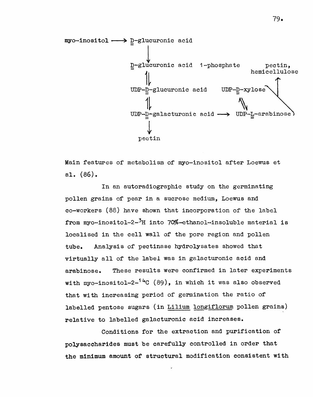
79.
myo-inositol D-glucuronic acid
D-glucuronic acid 1 -phosphate pectin,
I hemicellulosi
UDP-D-glucuronic acid UDP-D-xylose
\UDP-D~galacturonic acid UDP-L-arabinose
pectin
Main features of metabolism of myo-inositol after Loewus et
al. (86).
In an autoradiographic study on the germinating
pollen grains of pear in a sucrose medium, Loewus and
co-workers (88) have shown that incorporation of the label
from myo~inositol-2-3H into 70$-ethanol~insoluble material is
localised in the cell wall of the pore region and pollen
tube. Analysis of pectinase hydrolysates showed that
virtually all of the label was in galacturonic acid and
arabinose. These results were confirmed in later experiments
with myo-inositol-2-1 **C (89), in which it was also observed
that with increasing period of germination the ratio of
labelled pentose sugars (in Lilium longiflorum pollen grains)
relative to labelled galacturonic acid increases.
Conditions for the extraction and purification of
polysaccharides must be carefully controlled in order that
the minimum amount of structural modification consistent with

80
a high yield and degree of purity of the product is attained.
The graded extraction of mustard cotyledons has been
described (27). Oligosaccharides and sugars are extracted
with ethanol and fats with acetone. Cell wall polysaccharides
are not removed by such procedures. Most of the pectic
substances are extracted with hot aqueous EDTA and the
remainder are then extractable with hot water.
Hemicelluloses are extracted from the residue with alkali,
leaving cellulose.
The pectic substances are particularly susceptible
to side-reactions during extraction. Alkaline conditions
provide efficient methods of extracting these polysaccharides
but cause de-esterification and degradation by ^-elimination
and other reactions (66). (3-Elimination can also occur in
hot neutral solution (90). Glycosidic hydrolysis of
furanoside linkages can occur in hot solutions if the pH is
less than 3» Solvents of pH approximately 5 would
therefore seem to involve least risk of modification of the
native pectic substances but it is likely that extraction
would be incomplete at this pH with EDTA. It has recently
been shown (60) that a method involving the use of a
mixture of EDTA and sodium lauryl sulphate at pH 5.0 is
extremely effective, but there are some indications that the
material extracted is modified in some way which is not
attributable to the pH.
Alkaline extraction of hemicelluloses can result
in degradation by a peeling reaction from the reducing end

81.
of chains of 1,3 or 1 ,U linked sugar units (91).
Saponification of acetyl groups known to "be present in some
polysaccharides is also involved, although a mild procedure
involving extraction of acetylated xylans from wood with
dimethyl sulphoxide is reported (92). Although not
relevant to such young tissue as mustard cotyledons, the
side-reactions involved in delignification of wood prior to
the extraction of hemicellulosic polysaccharides may result
in oxidation, depolymerisation and contamination with
degraded cellulose.
The work described in this chapter concerns the
incorporation of myo-inositol-2-^H into germinating mustard
cotyledons. A graded extraction of germinated cotyledons
established the distribution of label between the various
fractions and the composition of the newly synthesised
pectic substances and EDTA-insoluble material was determined
after hydrolysis and separation of the sugars by paper
chromatography. The location of the 70% ethanol-insoluble
labelled material within the cell was determined by
autoradiography of tissue sections.
82
Experimental
Experiment 2.1* Germination of mustard seeds sup-plied with.
myo-inositol-2-3H and separation of the cotyledons,
Mustard seeds (300) were added to a solution of
myo-inositol~2-5H (0.10 me, approx e 2.2 x 10¹⁸ cpm^in 0.80 ml
water; specific activity 0.115 me per mg) and allowed to
stand with occasional shaking to ensure even soaking of the
seeds. After complete absorption (2 hr), more water
(0.80 ml) was added and the seeds were soaked for a further
I|. hr. The apparatus used in the germination of mustard
seeds was identical to that described elsewhere (3^4-) • After
germination in the dark for i± days the cotyledons and part
of the hypocotyls were removed, frozen in liquid nitrogen and
freeze-dried. The cotyledons were separated from the
hypocotyls by hand sorting. Yield, 1 »Ug.
Experiment 2.2. Distribution of radioactivity in the
fractions obtained by graded extraction of the cotyledons.
The cotyledons (1 .Ug) were extracted in the Soxhlet
with aqueous ethanol (80$) (Count A) and subsequently with
acetone (Count B) .
The residue (0.9g) was extracted exhaustively with
EDTA (di sodium salt, 2% w/v, 10 x 50 ml for 3 hr each time)
at 100° and pH 7.5 • The extracts were combined and
concentrated to 500 ml, and a sample (25 ml) removed
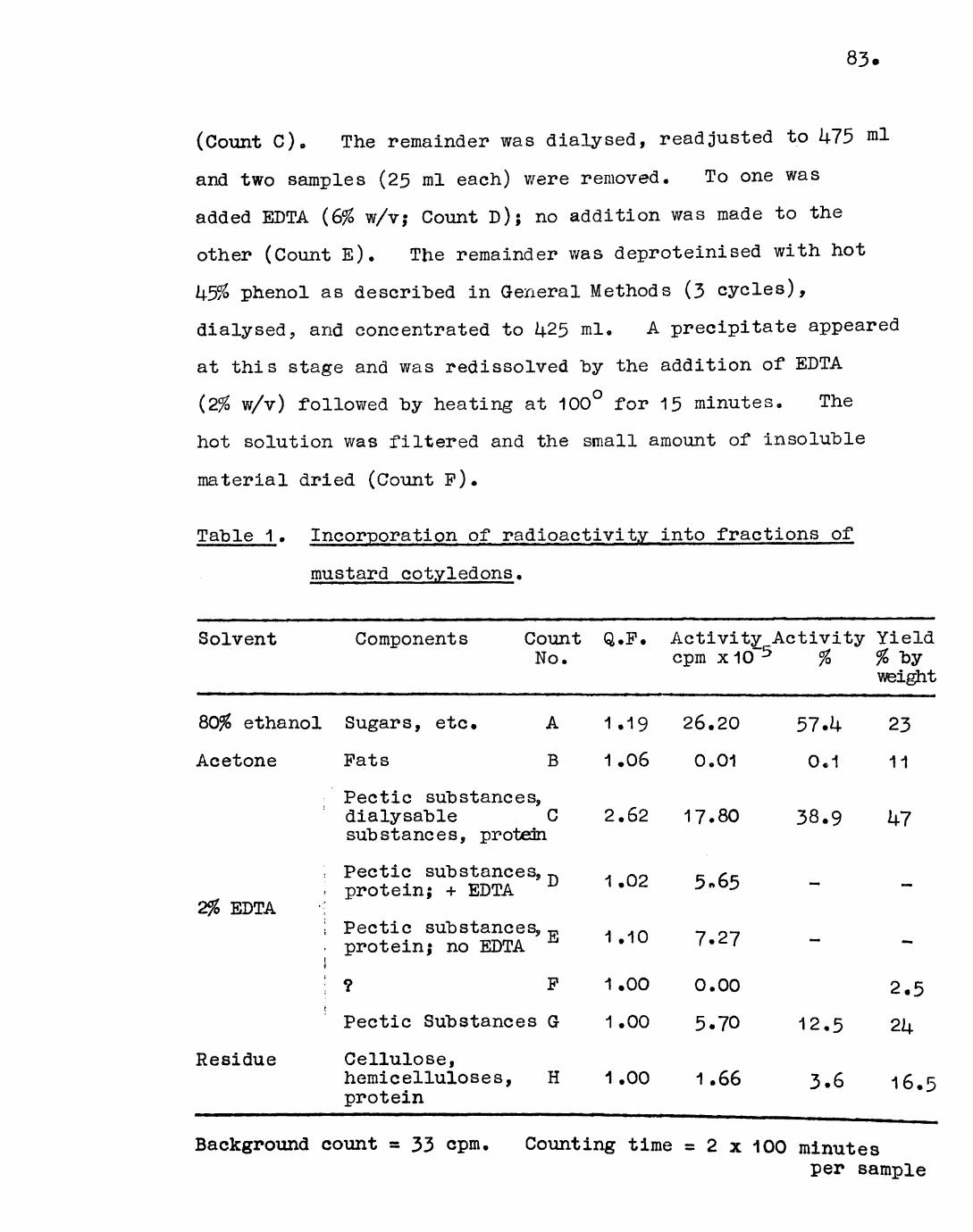
83
(Count C). The remainder was dialysed, readjusted to U75 nil
and two samples (25 ml each) were removed. To one was
added EDTA (6% w/v; Count D); no addition was made to the
other (Count E). The remainder was deproteinised with hot
k5% phenol as described in General Methods (3 cycles),
dialysed, arid concentrated to U25 ml. A precipitate appeared
at this stage and was redissolved by the addition of EDTA
(2% w/v) followed by heating at 100° for 15 minutes. The
hot solution was filtered and the small amount of insoluble
material dried (Count F).
Table 1 . Incorporation of radioactivity into fractions of
mustard cotyledons.
Solvent Components Count Q.P. Activity Activity YieldNo. cpm x10 5 % % by
weight
ethanol Sugars, etc. A 1.19 26.20 57.U 23
Acetone Pats B 1.06 0.01 0*1 11
Pectic substances,dialysable C 2.62 17.80 38.9 U7substances, protein
; Pectic substances, ^ . op R x- '•• protein; + EDTA 10 ^ :>«oi> 2% EDTA •;
! Pectic substances,,, . . n. protein; no EDTA ^ n * 1U r *^'
! ? P 1.00 0.00 2.5
Pectic Substances G 1.00 5.70 12.5 21+
Residue Cellulose,hemicelluloses, H 1.00 1.66 3.6 16.5 protein
Background count = 33 cpm. Counting time = 2 x -\OQ minutesper sample

The filtrate was redialysed, made to U25 ml, and a sample
(25 ml) removed (Count G). The remainder was concentrated
and freeze-dried. Yield, 0.27g; corrected, for removal of
14 x 25 ml samples, 0.3Ug.
The residue after extraction of the cotyledons
with EDTA was freeze-dried. Yield, 0.23g (Count H).
Preparation of samples for counting;
Accurately measured volumes (10 or 25 yul) of
solutions (Count A in ethanol, Count B in acetone, Counts C,
D, E, G- in water or aqueous BDTA) or homogenates (Counts P
and H, in water), of duplicate samples of known concentration
v/ere evaporated to dryness on strips of cellulose acetate
(1.5 cm x 5-0 cm).
Counting of samples;
The strips were immersed in liquid scintillator
for counting as described in General Methods. The "background
ccunt was determined with a cellulose acetate blank and
automatically subtracted from subsequent counts. The true
activity of the sample was obtained after correction for
quenching. The extent to which quenching was effecting the
measured activity (the quenching factor) was obtained by
1 spiking 1 the sample with myo-inositol-2-3H, 720 cpm, and is
given by_ = Spike count (720 cpm) + Sample count (cpm)
[Spike -i- sample] count (cpm)

85.
The true activity was calculated by multiplication of the
observed activity by the quenching factor. The results are
shown in Table 1.
Experiment^.3 * Distribution of radioactivity in the
component sugars of the EDTA-soluble and EDTA-insoluble
fractions of the cotyledons.
The deproteinised freeze-dried EDTA-soluble material
(Experiment 2«2.) (50 mg) was hydrolysed in sulphuric acid
(1 N, 10 ml) at 100° for 16 hr. The residue after KDTA
extraction (50 mg) was dissolved by shaking overnight in
sulphuric acid (12% w/v, 1 ml) at room temperature; after
dilution with water (9 ml), the solution was heated at 100°
for 6 hr. Each hydrolysate was neutralised with barium
carbonate, chilled, filtered and evaporated to dryness.
The residue was taken up in water (1 ml) for paper
chromatography. Each solution was spotted in duplicate on
the chromatogram which was developed in solvent A, marked off
in 1 cm strips on the longitudinal axis and cut in half
between the two spots. One half was sprayed (reagent a) to
locate the sugars; the other was cut into 1 cm strips
numbered from the origin, which were assayed for radioactivity
in the liquid scintillation counter (General Methods). The
background count was determined with a blank strip of
chromatography paper (1 cm x 6 cm) and subtracted
automatically from subsequent counts. The results are given

86.
in Table 2 (EDTA-soluble fraction) and Table 3 (EDTA-insoluble
fraction).
Table 2. Radioactivity in the component sugars of the
EDTA-soluble fraction of mustard cotyledons.
Component Activity
cpm
Oligosaccharides U36
Galacturonic acid 92
Galactose 11
Glucose
Arabinose
Xylose
Rhamnose
8
97
18
5
65 .k
1.6
1.2
1U.5
2.7
0.8
Relative concentration by spray reagent (a)
inestimable
Background = 28.5 cpm (automatically subtracted) Counting time = 3 x 10 minutes each sample
Table 3. Radioactivity in the component sugars of the
EDTA-insoluble fraction of mustard cotyledons.
Component
Oligosaccharides
Galacturonic acid
Galactose
Glucose
Arabinose
Xylose
Activitycpm
29
29
12
31
19
31
%
19
19
8
21
12.5
21
Relative concentration by spray reagent (a)
—
+++++++trace
+Background = 28.5 cpm (automatically subtracted) Counting time =1 x 100 minutes each sample

87 .
Experiment 2.U* Autoradiography of the cotyledons
Mustard seeds (10) were supplied with a solution of
myo-inositol-2-3H and germinated as described in Experiment
2.1.
The cotyledons were removed and washed with a
solution of myo-inositol (2% w/v, 50 ml) at 0° for 1 hr,
followed by a solution of formaldehyde (L$> 9 3 x 30 ml for
30 minutes each time) and then in myo-inositol (2%, 50 ml)
overnight. The cotyledons were thoroughly washed with water
and then taken through a series of aqueous ethanol solutions
(ethanol concentration increasing iron 0 to 7C$ v/v in 10%
intervals, 50 ml for 30 minutes each time) and finally stored
in aqueous ethanol (70$, 10 ml).
Authoradlographs of sections of the cotyledons were
kindly prepared "by Dr. M.M. Yeoman. The method employed is
described "below.
The cotyledons were embedded in wax and thin
sections (a few cells thick) cut from them. The wax was
gradually dissolved from the sections which were finally
washed with water. Slides were prepared to take the
sections as follows: the slide was degreased in chromic acid
before dipping in an aqueous solution of gelatin (5g per l)
and chrome alum (0.5g per 1). The slide was drained and
dried such that an even layer was produced and the section
mounted on the prepared slide. Subsequent stages were
carried out in the dark-room under a Wratten series 1 (red)

88 o
filter. The nuclear emulsion (Kodak K2) was melted and an
equal volume of water was added. The solution was
thoroughly mixed and kept at 55°. The slide was dipped in
the emulsion for 30 sec, removed and drained in an upright
position, and allowed to dry very slowly. The slide was
then stored, in a sealed box containing silica gel, at U
for 14 days. After development (Kodak D19) and fixing, the
slide was washed and dried.
The autoradiographs were examined microscopically
(Leitz 'Orthoplan 1 system); some of the microphotographs
obtained are shown in the Discussion to this chapter.
Discussion
Examination of the results of Experiment 2.2
(Table 1) shows that a large proportion (57$) of the total
activity was contained in the 80$ ethanol-soluble fraction,
presumably mainly in unutilised myo-inositol. Pats were
unlabelled. Of the activity recovered in the 80$ ethanol-
iiisoluble residue, 91 -5$ was contained in the EDTA-soluble
fraction and 8.5$ in the EDTA-insoluble residue. Dialysable
substances accounted for a considerable proportion (nearly
60$) of the activity in the EDTA-soluble fraction. Low
molecular weight carbohydrates which were not extracted by
ethanol for accessibility reasons probably account for most
of this (no break-up of the cotyledons occurred in ethanol),
although polysaccharides of relatively low molecular weight

89.
such as the araban, and degradation artefacts of the pectic
substances will also contribute to the activity. Protein
was unlabelled as shown by the small change in counts caused
by deproteinisation. The pectic substances accounted for
just over 1+0$ of the activity in the EDTA-soluble fraction.
In contrast hemicelluloses and cellulose were weakly
labelled.
On hydrolysis and paper chromatography the
dialysed, deproteinisecl EDTA-soluble fraction Was shown
(Table 2) to have the typical component sugars of pectic
substances. A large proportion (nearly 80$) of the label was
present in galacturonic acid and galacturonic acid-
containing oligosaccharides which are resistant to hydrolysis;
most of the remaining activity v/as contained in arabinose.
Xylose was relatively weakly labelled and only traces of
activity were found in the hexoses.
Hydrolysis and paper chromatography of the
EDTA-insoluble reside (Table 3) showed glucose to be the
major component, although other sugars derived from
hemicelluloses and residual pectin were also present. A
large proportion (over 50$) of the label in this fraction
was contained in the component sugars of the residual pectin
(oligosaccharides, galacturonic acid and arabinose); the
remainder was present in xylose, glucose and galactose -
typical sugars of the hemicellulose and cellulose fraction.
The autoradiographs of the cotyledons (Experiment
2.U. and Figures 1a and 1b) show that the 70$ ethanol-

k - ^ ,- \ >
)'\* •^. > •
m
t t±£' • >PV <r **^
• r ^ \ • '&,"'fVY-U' ' T '&T r-^i /• J<...te&m*:-
Figure 1. Autoradiographs of white mustard cotyledons (x
Below : Figure 1a, parenchyma
Above : Figure 1b, as in 1a but showing vascular tissue

90
insoluble label is contained in the region of the cell wall
and middle lamella (as shown by developed silver grains in
the nuclear emulsion), "but resolution is not high enough to
show whether the middle lamella is labelled, although there
are indications that it is not. The cytoplasm is virtually
unlabelled but it is interesting to note (Figure 1b) that
the vascular tissue is labelled.
The conclusions to be reached from the above
observations are *
(i) Myo-inositol-2-3H is incorporated mainly into the
pectic substances of mustard cotyledons rather than other
cell-wall polysaccharides.
(ii) The incorporation of label within the pectic substances
is mainly into galacturonic acid and arabinose (and to a
lesser extent xylose); very little label is incorporated
into hexose components. The newXv-synthesised pectin has a
ratio of galacturonic acid : arabinose : xylose of 1 .00 :
1.05 : 0.20 compared with that of 1.00 : 3-25 : 0.77 present
in the pectin of non-germinated cotyledons (derived from the
results of methylation analysis). The reduction in neutral
sugars signifies a dramatic decrease in the degree of
branching of the pectin synthesised during germination,
despite the fact that galacturonic acid is underestimated
due to incomplete liberation during hydrolysis,
(iii) The newly synthesised pectin is located in the cell
wall. The vascular tissue is also the site of 70$
ethanol-insoltible labelled material. This could be due to

91.
incomplete extraction of 70$ ethanol-soluble substances from
this tissue, but it is more likely that it reflects the
synthesis of considerable amounts of new cell wall material
as a result of differentiation.
CHAPTER 5
Smith Degradation of an ara~ban component
of the pectic substances from mustard cotyledons

92
Introduction
Many of the existing methods of polysaccharide
structural analysis involve controlled fragmentation of the
polymer e These procedures entail selective cleavage of one
linkage rather than another, followed "by characterisation of
the products. The elucidation of the branching pattern of
amylopectin by Peat et al. (93) using specific enzymes is a
classical example of the highly controlled breakdown of a
polysaccharide. Unfortunately direct chemical methods are
much less specific but, as the selection of pure specific
enzymes is somewhat limited, these are frequently the only
methods available. ^-Elimination of uronic acid-containing
polysaccharides has been employed (21,91+) but elimination
reactions are not yet in such general use as soIvolysis
procedures. The reverse specificities of aqueous acid
hydrolysis and acetolysis has been employed in determination
of the structure of some dextrans (95)• Chemical modification
of sugar units can be used to alter the cleavage pattern of
the polysaccharide either by stabilising a particular linkage
(some such methods, and one in particular, are described in
the following Chapter) or by rendering it extremely labile.
A powerful approach to the characterisation of the
branching pattern in many polysaccharides involves the use
of periodate degradation methods; lability at the oxidised
unit enabling controlled breakdown under mild conditions.
Other structural features may also be detected by this

93.
procedure. In the Barry Degradation the periodate oxidised
polysaccharide is treated with phenylhydrazine 0 The main
drawback is that the work-up is complicated and the osazone
products are difficult to handle. Treatment of the oxidised
polysaccharide with sodium borohydride followed by mild acid
hydrolysis (the Smith Degradation) is more powerful because
the reaction is exceptionally clean and the products
relatively easy to characterise. The nature and relative
yields of the Smith Degradation products, coupled with the
results of methylation and other studies often enables a
considerable narrowing down of the possible structures of a
polysaccharide. In favourable cases where experiment has
determined the nature of the branching pattern, the
distribution of the side-chains (i«e. regular, block or
random) on the main chain may be elucidated. Aspinall and
Ross (96) showed (by methylation, enzymic hydrolysis and
other experiments) that rye flour arabinoxylan has a (3-1- ? lj.
xylan main chain to which single arabinose side-chains are
attached through the C/^\ position. The isolation of
2-0-p-D-xylopyranosylglycerolj 0-(3-D-xylopyranosyl-(l -*Lj.)-
-0-(3~D-xylopyranosyl-(l-»2)-glycerol, and 0-£-D-xylopyranosyl-
-(1 -*i|) -0-|3-D-xylopyranosy 1- (1-k ) -0~p-D-xylopyrano sy 1-(1 -»2 ) -
-glycerol in the ratio of 7.5 : 2.2 : 1 but no products of
higher molecular weight, showed that the distribution of
side-chains is rather random and that no more than 3 adjacent
xylose units are ever branch points. The estimation of
Smith Degradation products in the above study was by elution
from paper chroma to grams followed by determination of
carbohydrate content with the phenol-sulphuric acid reagents.
The accuracy of this procedure is probably low, due to
incomplete elution and possible contamination with cellulose
from the paper. G-lycerol cannot be estimated by the above
method. Estimation by weighing involves a large amount of
starting material with a subsequent lengthy separation of the
products. This chapter describes a method whereby accurate isfcios
of Smith Degradation products may conveniently be obtained on
a micro scale, with particular reference to an araban
component of the pectic substances of mustard cotyledons
(from both germinated and non-germinated seeds).
The araban is readily isolated because it is not
precipitated with 70^ ethanol from the EDTA extract as are
the other pectic substances. Addition of a large excess
of acetone or butan-2-one to the ethanol filtrate precipitates
the araban in a crude state, which is then purified by
precipitation from alkaline solution with cationic detergents
(27>97)« Tne product has an anhydroarabinose content of
98$ or better. Substantial evidence has been put forward
(27,28,98) in support of the view that the polysaccharide is
a genuine homoaraban rather than a 3-elimination artifact of
pectin as may have been the case with several arabans
isolated from other sources. The araban may well be
metabolically related to pectin in some way.
The araban has been shown to be homogeneous in the
ultracentrifuge and on free solution electrophoresis in

95.
borate, and arabans extracted at different pH values,
extracted from different batches of non-germinated seeds and
extracted from cotyledons after germination are identical in
these respects (99)• The molecular weight given by
ultracentrifugation and vapour pressure osmometry (on the
methylated polysaccharide) is about 6,000 corresponding to a
molecule containing approximately U5 anhydroarabinose units.
Other studies by Rees and Richardson (28,98) have established
the principal structural features of the araban0 The
negative rotation (-180°) and the ease of hydrolysis indicate
that the polymer is composed of a-linked arabinofuranose
units. The configuration of the linkages is confirmed by
enzymic hydrolysis with an cc-arabinosidase (100). Sugars
other than arabinose were not detectable by various methods
and the UV spectrum showed no peak at 235 nm due to
unsaturated 4,5-uronic acid (as would be present in a
(3-elimination artifact). Methylation analysis has
established the main structural features of the araban; the
pattern of methylated sugars is closely related to that
obtained from mustard cotyledon pectin (see Chapter 1 ),
namely, 2,3,5-tri-O-methyl-L-arabinose, 2,3~di-0-methyl-L-
-arabinose, 2-0-methyl-L-arabinose and L-arabinose. Trace
amounts of 2,5-di-O-methylarabinose and a second monomethyl
ether (probably 3-0-methylarabinose by analogy with pectin)
were also detected. The furanose ring form is thus
confirmed for end-group at least. Arabinose was only
present in small amounts; the molecule is thus joined by

96.
1,5 linkages with a considerable proportion of 1 ,3 "branch
linkages and a further small proportion of units "branched
through both the C/ 2 ) and C/,\ positions. Further
discrimination betv/een the possible structures which may be
proposed on the basis of methylation analysis was made from
the results of quantitative g.l.c. of the methylated araban
and the methylated Smith Degraded ar-aban. The gas
chromatograph detector response was calibrated with
crystalline compounds and the hydrolysis products of methylated
araban and the methylated degraded araban chromatographed as
the arabitol acetate derivatives. The absolute proportion
of the structural units was thus determined. The results
from the methylated araban showed that, in whole numbers,
the araban molecule contains 16 end units, 11 chain units
and 15 branch units, when the small proportion of 1,2 linkages
and the end units associated with them are ignored. No
2-0-methylarabinose or 2,5-di-O-methylarabinose was detected
(as the alditol acetate derivatives) among the methylation
products of the degraded araban and thus the araban, unlike
beet araban (6?)» contains no contiguous 1,3 linkages. It
was also shown that most of the oxidisable units (i.e. end
units or chain units) occur in nonadjacent positions so that
a structure having a substantial proportion of a linear
componentmay be excluded. Even this considerable evidence
does not lead to a unique .structure for the araban.
Remaining possible structures are given below (after Rees
and Richardson, 28), although hybrids of these are also
possibles

97 =
A
A-A-A-A-A-
1 comb' or 'herriac bone 1
A
tA
A ~ A
A
- A - A - A -
A IA.IA
A
IA
A
A - A -
A
A
A
A I
I
IA A
A-A~A-A-A~A~A~
f tree f
f bush f III
where —— denotes 1,5 linkage
— ̂denotes 1,3 linkage
A denotes arabinofuranosyl residue
The distribution of branch points within the molecule
may be determined by a Smith Degradation in which the ratio
of the products is accurately determined. Diagram 1
describes the products which would be obtained from the
araban and the structural units from v/hich they derive.
The substituted glycerol derivatives obtained would thus be
expected to be : 1~0-a-L-arabinofuranosylglycerol;
0-cc-L-arabinof urano sy 1- ( 1 ->5 ) ~0-a~L-ar ab inofur ano sy 1- ( 1 -+1 ) -
-glycerol and higher homologues (if present). If
arabinosylglycerol is the major product obtained then the
branch units tend to occur in isolated positions, and
similarly, if only higher homologues are observed, then the

ROCI
RO
CH
OH
Cha
in
unit
OHC
C
HO
HCH
O
CH
2OH
RO
CH OR
/ I0
4~
OR
——
——
» N
o e
ffe
cr
OH
Dran
cK u
nib
HO
C
'°4
\ i/
Ov
^
, B
H4
OH
End
u
nit
I^Y
^O
R'
JL
»4
^2'\
/•
H
OC
H^
HO
H2C
HO
CH
2*
OH
C
CH
O
Jcag
ram
1.
Orm
fch
Deg
rada
fcio
a op
fl
raba
n.
CH
jOH
••• R'O
H+
CH
OR
2.
H(
H
CHO
CM
OH
+
CH
2OH
HO-
CM2O
H

98.
branch units occur in contiguous positions. Patterns of
products between these extremes would represent a more
randomly branched structure. For a structure such as I
('Herring Bone f ) the yields of Smith Degradation products
from a randomly branched molecule can be predicted
statistically. The 'randomness 1 of the branching pattern
of the araban may thus be assessed if it is assumed that the
structure approximates to I.
Periodate oxidation of the arabans from
germinated and non-germinated mustard cotyledons was
followed by reduction with sodium borotritiide and mild
hydrolysis. The yields of the labelled products, separated
by paper chromatography, was calculated from the activities
obtained by liquid scintillation counting.

99
Experimental
Experiment 3«.1» Extraction and purification of araban from
germinated cotyledons•
The pectic substances were extracted from the
defatted cotyledons (l37g) of germinated seeds with an
aqueous solution of EDTA (2%) and sodium lauryl sulphate
(0.2M) at 100°, and precipitated at ?C$ ethanol
concentration. The crude araban was precipitated as an oil
by the addition of butan-2-one (15 volumes) to the supernatant
and was isolated by decantation. The product was
reprecipitated twice by dissolving in water and adding
butan-2-one. The final precipitate was a solid which was
removed on the centrifuge, dissolved in water and freeze-dried,
Yield, 2.65g.
Hydrolysis and paper chromatography (solvent A)
showed that, in addition to arabinose, traces of glucose and
galactose were present.
The crude material was purified by precipitation
with cetyltrimethylammonium hydroxide. An aqueous solution
(1C$) of cetyltrimethylammonium bromide was converted to the
hydroxide by passing through, a column of Amberlite 1RA.-UOO
(OH~) resin and part of this solution (20 ml) was added to
the crude araban (2.5g) in water (300 ml). On slow
addition of sodium hydroxide (1 N, 20ml) a flocculent white
precipitate was formed which was removed after 1 hr by

100.
centrifugation. The precipitate was dissolved in water
(300 ml) containing enough, acetic acid to give a neutral
solution, thenreprecipitated with alkali. This procedure
was repeated once. After redissolution in dilute acetic
acid, the detergent was removed by precipitation with excess
potassium iodide solution followed by centrifugation at 0 .
The supernatant solution was filtered and passed through a
mixed bed ion exchange column (IR 120 H+ and IB U5 OH~
resins). Tests showed that the effluent was free from iodide
(silver nitrate test) and potassium ions (flame test). The
solution was concentrated and freeze-dried* Yield, 0.120g.
Pound, 100$ anhydroarabinose (hydrolysis and paper
chromatography, and quantitative phenol-sulphuric acid
determination), [a]p. - 177° (c 0.66 in water). In thejj ~-ultracentrifuge (2UO,OOOg) the araban was identical with
samples isolated before (60).
Experiment 5.2. Periodate oxidation of the arabans
extracted from germinated and non-germinated cotyledons.
The araban from non-germinated cotyledons was
prepared by Dr. N. Richardson (98)* The polysaccharides
were dried exhaustively in vacuo at 60 .
Equal volumes (10.0 ml) of sodium periodate
solution (0.080 M) and each polysaccharide (0.050 M with
respect to anhydroarabinose units) were mixed and left in the
dark at room temperature. The reduction of periodate was
followed spectrophotometrically (I5a) until the reaction was

. motes K>4 conaumad/mole anhydroarablnoaa.
07<
0.6'
diagram 1.
Periodate uptake of arabans.
24
—^— nongerminatad.
^ — germinated.
time (hr)
101.
complete. The final values correspond to the consumption
of 0.6? and 0.71 moles of periodate per mole
anhydroarabinose for the f non-germinated 1 and 'germinated 1
arabans respectively (see Diagram 1). The residual
periodate was destroyed with a small excess of ethylene
glycol.
Experiment 5.3. Smith Degradation of araban from
non-germinated cotyledons.
Sodium borohydride (0.2g) was dissolved in water
(20 ml) and left to stand overnight, then added to sodium
borotritiide (0.2 - 0.5mg, specific activity 19 me per mg)
and the solution was added to the periodate oxidised araban.
After 60 hr at room temperature the excess reagent was
destroyed with the minimum volume of hydrochloric acid (1 N).
The solution was passed through a mixed bed ion exchange
column (IR 120 H~^ and IR U5 OH~" resins), evaporated to
dryness under reduced pressure and boric acid removed as
the volatile methyl borate by repeated distillation of
methanol from the residue. The residue was dissolved in
sulphuric acid (1.0 If, 8 ml) and left at room temperature.
Samples (approx. 2 ml) were removed after 5 mins, 1 hr,
16 hr, and 2k hr; neutralised with barium carbonate, chilled
and filtered. The filtrates were evaporated to dryness and
redissolved in water (0.2 ml).
The products were examined by paper chromatography

102.
in solvent C with glycerol and arabinose as controls.
With reagent b (alkaline silver nitrate reagents) a series
of spots was obtained from the 16 hr and 24 hr hydrolysates,
but glycerol was the only detectable component in the 5
minute and 1 hr hydrolysates. The mobilities of the
components and tentative assignments are given below.
Tentative identity Rf
Glycolaldehyde 0.8 to 1 .0
Glycerol 0.6^
Arabinosylglycerol 0.52
Arabinobiosylglyceroltogether with arabinose 0.39
Arabinotriosylglycerol 0.28
Arabinotetraosylglycerol 0.20
With reagent c (triphenyltetrazolium hydroxide spray) free
arabinose was detected in the 16 hr and 2U hr hydrolysates;
it therefore follows that some non-specific hydrolysis had
occurred under these conditions.
The hydrolysates were double spotted on chromatograms
and developed in solvent C (Whatman no. i paper; 20 hr).
The dried chromatograms were ruled off in numbered 1 cm
strips perpendicular to the longtitudinal axis and cut in
half between the two spots. One half was cut into strips
and counted in the liquid scintillation counter as described
in General Methods; the other was sprayed (reagent b) so as
to correlate components with activities. A typical result

2.0- 1.0
-
Jl
diag
ram
2.
Sm
ith D
egra
datio
n
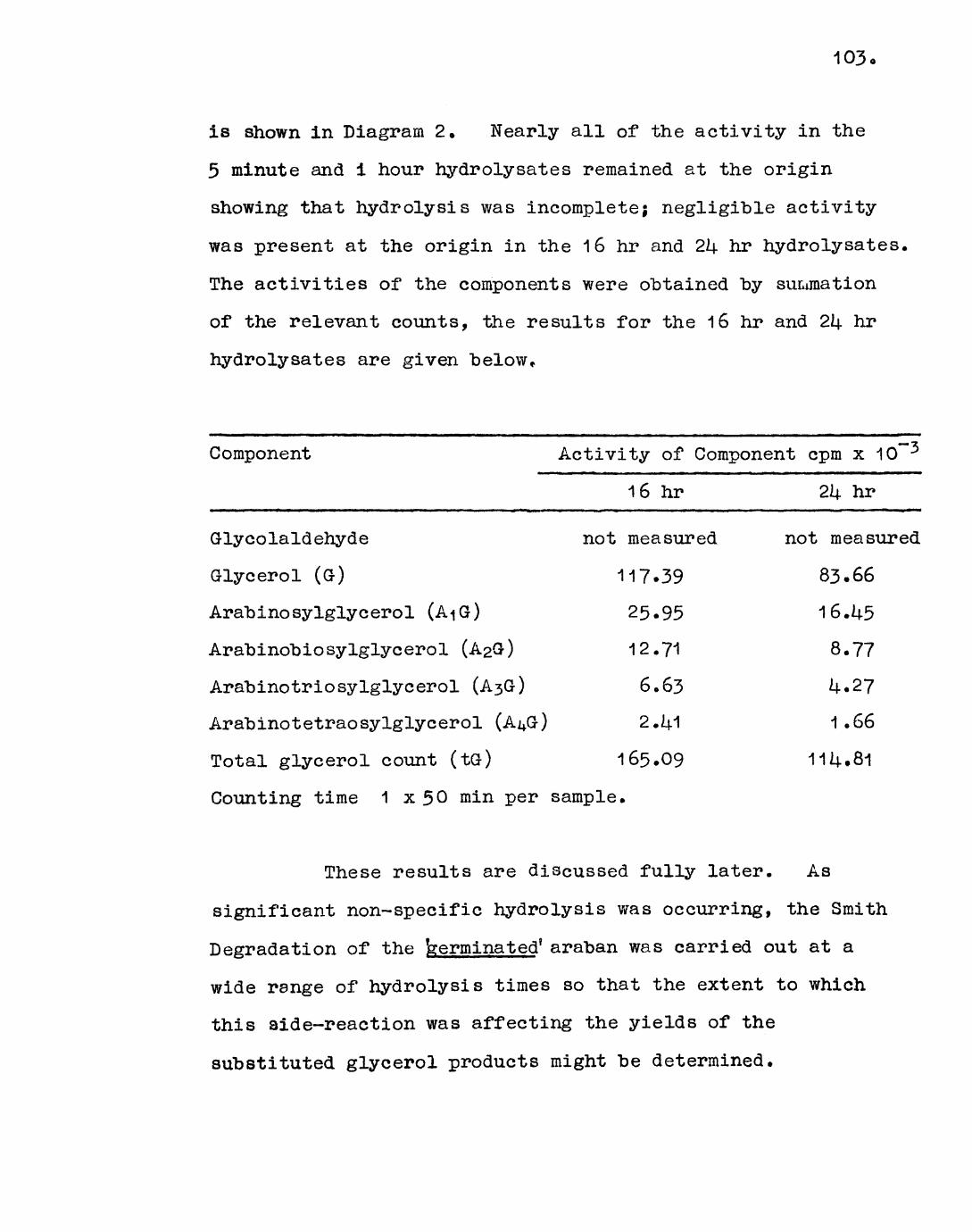
103
is shown in Diagram 2. Nearly all of the activity in the
5 minute and 1 hour hydrolysates remained at the origin
showing that hydrolysis was incomplete; negligible activity
was present at the origin in the 16 hr and 214- hr hydrolysates,
The activities of the components were obtained by summation
of the relevant counts, the results for the 16 hr and 2k hr
hydrolysates are given below*
Component Activity of Component cpm x 10
16 hr 2*4- hr
Glycolaldehyde
Glycerol (G)
Arabinosylglycerol
Arabinobiosylglycerol (A2G)
Arabinotriosylglycerol (A3&)
Arabinotetraosylglycerol (A4G)
Total glycerol count (tG)
not measured
117.39
25-95
12.71
6.63
2.14.1
165.09
not measured
83.66
16.14-5
8.77
U.27
1.66
11*4.81
Counting time 1 x 50 min per sample.
These results are discussed fully later. As
significant non-specific hydrolysis was occurring, the Smith
Degradation of the terminated' araban was carried out at a
wide range of hydrolysis times so that the extent to which
this aide-reaction was affecting the yields of the
substituted glycerol products might be determined.
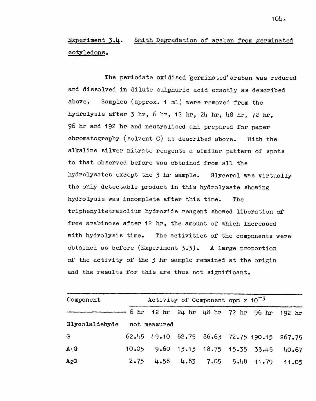
Experiment 3»U. Smith Degradation of araban from germinated
cotyledons.
The periodate oxidised 'germinated* araban was reduced
and dissolved in dilute sulphuric acid exactly as described
above, Samples (approx. 1 ml) were removed from the
hydrolysis after 3 hr, 6 hr, 12 hr, 2k hr, U8 hr, 72 hr,
96 hr and 192 hr and neutralised and prepared for paper
chroma tography (solvent C) as described above. With the
alkaline silver nitrate reagents a similar pattern of spots
to that observed before was obtained from all the
hydroly sates except the 3 hr sample. Glycerol was virtually
the only detectable product in this hydroly sate showing
hydrolysis was incomplete after this time. The
triphenyltetrazolium hydroxide reagent showed liberation of
free arabinose after 12 hr, the amount of which increased
with hydrolysis time. The activities of the components were
obtained as before (Experiment 3-3). A large proportion
of the activity of the 3 hr sample remained at the origin
and the results for this are thus not significant.
Component Activity of Component cpm x 10*"-5
6 to 12 hr 2k hr 1^.8 hr 72 hr 96 hr 192 hr
Glycolaldehyde not measured
G 62.U5 U9.10 62.75 86.63 72.75 190.15 267.75AiG 10.05 9.60 13.15 18.75 15-35 33-U5 U0.67A2G 2.75 U.58 U.83 7-05 5-U8 11.79 11.05

105
6 hr 12 hr 2U hr 14.8 hr 7.2 hr 96 hr 192 hr
A3G 1.65 2.71 2.20 2.35 1.83 5.70 8.61
A^G 1.70 1.60 1.98 1.57 1.30 2.63 3.81
to- 78.60 67.59 8U.91 1l6o35 96.71 2U3.72 331.89
Counting time 1 x 60 min per sample.
106.
Theory
In the experiments described above the label is
contained solely in glycerol and the glycerol moiety of the
other degradation products (if glycolaldehyde is ignored).
Hence the activities of these products accurately reflect
their concentrations on a molar basis. It is stressed that
the identities assigned to the components are at this stage
tentative.
The following abbreviations are used : G = glycerol,
A-jG = arabinosylglycerol, A2G = arabinobiosylglycerol, A$G =
arabinotriosylglycerol, A/J3- = arabinotetraosylglycerol, tG =
total glycerol (i.e. free and combined) = AiG + AaG +
Correction for non-specific hydrolysis obviously
requires values for the concentrations of the products at as
many hydrolysis times as possible. The rate of this
reaction was thus calculated for the 'germinated' araban only
(as several hydrolysis times were employed) and used for both
arabans on the assumption that the rate of non-specific
hydrolysis would be similar.
(A) 'Germinated' araban
The values of the concentrations of the products
(excluding glycolaldehyde), as measured by their activities
(Experiment 3.14..), at different hydrolysis times are shown
below :
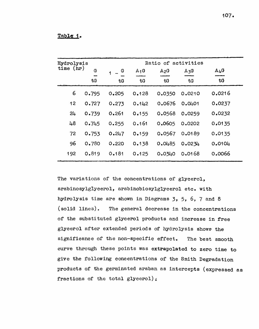
107
Table 1 .
Hydrolysis time (hr)
i (' "i • '
6
12
2k
kQ
72
96
192
0
0
0
0
0
0
0
G
tG
.795
• 727
.739
.745
•753
.780
.819
1
0
0
0
0
0
0
0
__ G
tG
.205
.273
.261
• 255
.247
.220
.181
Ratio of activities A<|G A2G A3G
tG tG tG
0
0
0
0
0
0
0
.128
.142
.155
.161
• 159
.138
.125
0
0
0
0
0
0
0
.0350
.0676
.0568
.0605
.0567
.0485
.0340
0
0
0
0
.0210
.01+01
.0259
.0202
0.0189
0
0
.0234
.0168
A^G
tG
0.0216
0.0237
0.0232
0.0135
0.0135
0.010^
0.0066
The variations of the concentrations of glycerol,
arabinosylglycerol, arabinobiosylglycerol etc. with
hydrolysis time are shown in Diagrams 3, 5, 6, 7 and 8
(solid lines). The general decrease in the concentrations
of the substituted glycerol products and increase in free
glycerol after extended periods of hydrolysis shows the
significance of the non-specific effect. The best smooth
curve through these points was extrapolated to zero time to
give the following concentrations of the Smith Degradation
products of the germinated araban as intercepts (expressed as
fractions of the total glycerol)*

diagram 3.
1-glycsfol/ tots! glycarol ¥ hydrolysis tims.
timsdir.)
0.30-
0.20-
0.10
nHIT.)
First ord«r plot of hydrolysis of

0.17
0.1«
MS-
0.1*
0.12
0.11-
n ndiagram 8.
Ar«bino«ylglyc«fol concentration v». tima.
tim«(hr.)
O experimental. — — - calculated.
0.08-
0.07-
0.06-
0.05-
0.04-
0.03
arabinobmsylglyceroltotal otvcarol.
0 2T
diagram 6
72 96
Ariihinobioawlcilvcerol concentration vs. time.
tima(hr)
o—— wptrimamal — — i Calculated.

arabinotrioeylglycerol
0.05
0.04
0.03
0.02
0.01
total olycerol.
diagram
Arabinotrioaylglycerol concentration vs. time
time(hr)
o experimental. — — calculated.
' arabinotetraoaylglyceroltotal gfycfroL
0.03
0.02'
0.01
24 41 12 96tlnwlkr.)
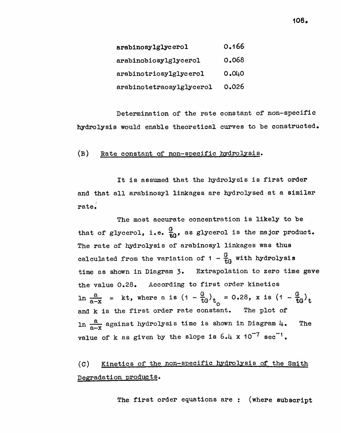
108.
arabinosylglyc erol 0 . 1 66
arabinobiosylglycerol 0.068
arabinotriosylglycerol O.OUO
arabinotetraosylglycerol 0.026
Determination of the rate constant of non-specific
hydrolysis would enable theoretical curves to be constructed,
(B) Rate constant of non- specific hydrolysis.
It is assumed that the hydrolysis is first order
and that all arabinosyl linkages are hydrolysed at a similar
rate.
The most accurate concentration is likely to ber\
that of glycerol, i.e. T, as glycerol is the major product.
The rate of hydrolysis of arabinosyl linkages was thus
calculated from the variation of 1 - T wi"th hydrolysis
time as shown in Diagram 3« Extrapolation to zero time gave
the value 0.28. According to first order kinetics
^ I^E = kt » where a is (1 - to\0 = °'28 ' x ls (1 ~and k is the first order rate constant. The plot of
In — — against hydrolysis time is shown in Diagram U» The a~xvalue of k as given by the slope is 6.U x 10~7 sec"" 1 .
(C) Kinetics of the non-specific hydrolysis of the Smith
Degradation products.
The first order equations are : (where subscript
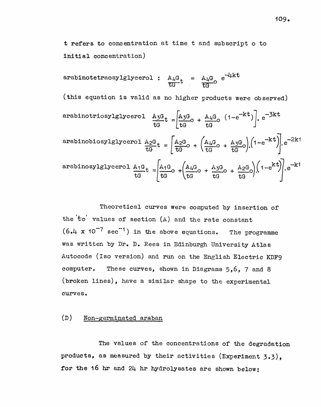
109
t refers to concentration at time t and subscript o to
initial concentration)
arabinotetraosylglycerol : A^G, = A^G e~^"
(this equation is valid as no higher products were observed)
arabinotriosylglycerol A ..[A A (l-e~kt ) .tG I tG tG
arabinobiosylglycerol
arabinosylglycerol
to
. _
.* " to to to
\.e
Theoretical curves were computed by insertion of
the t'o values of section (A) and the rate constant
(6.k x 10~~7 sec"" 1 ) in the above equations. The programme
was written by Dr. D. Rees in Edinburgh University Atlas
Autocode (iso version) and run on the English Electric KDP9
computer. These curves, shown in Diagrams 5>6, 7 and 8
(broken lines), have a similar shape to the experimental
curves.
(D) Non-germinated araban
The values of the concentrations of the degradation
products, as measured by their activities (Experiment 3»3)»
for the 16 hr and 2k hr hydrolysates are shown below:

110
Table 2.
Hydrolysis time (hr)
16
2k
Ratio of Activities
Gto
0.711
0.729
1
0
0
_ 0
tG
.289
.271
A-|Gto"
0.157
0.114.14-
A2G
W
0.0770
0.0765
A3G
tcT
0. OU02
0.0372
A ̂Oto"
0.0114-6
0.0114-5
These values and the value of the rate constant
determined for the germinated araban on insertion into the
above equations give, on averaging, the following
concentrations of the Smith Degradation products of the
f non-germinated 1 araban corrected for non-specific hydrolysis
(expressed as fraction of the total glycerol) :
arabinosylglycerol 0.15?
arabinobiosylglycerol 0.0811$
arabinotriosylglycerol 0.014-3"?
arabinotetraosylglycerol 0.0175
16 hr 2Ij. hr0.158 0.114-5
0.0807 0.0821
O.OU43 O.OU30
0.0169 0.0181
(E) Statistical yield of Smith Degradation products from
a randomly branched araban.
It is assumed, for simplicity, that the araban has
only single unit side-chains, i.e. the structure is of type I
illustrated in the Introduction.
Let the number of 1,3 linked end-groups per

molecule "be E and the number of 1 ,5 linked residues in the
main chain be L. The degree of polymerisation (DP) is thus
E + L. The probability that any arabinose residue is aTjl
branch point is =-, and the probability that it is notJuE branched is 1 - —«Jb
(a) Yield of Glycerol :
(i) General case excluding glycerol from unsubstituted
non-reducing end-groups. The feature required for this is
two or more successive unsubstituted arabinose units, i.e.
- A - A -. The probability that such a pe.ir of units exists
is (1 - =-) 2 and as the molecule contains (L - 1 ) possible LITjl
pairs the yield of glycerol will be (L - 1)0 - ;r) per
molecule. (it is obvious that overlapping eligible pairs,
i.e. where more than two successive unsubstituted arabinose
residues occur, do not interfere with the release of glycerol
from each other)•
(ii) Glycerol released from an unsubstituted end-group of
the 1 ,5 linked main chain. The probability that this existsJUJ i _. __ j *\_ _ __ _ _ i T___ ^ _... .3 ^. "1 J3 _ _C* —^_ T _ _ —^ _ _^. -» "1 J H I A ~~m^
L molecule.
V \ n
is (1 - =•) and hence the yield of glycerol is (1 - —) perL ii
(iii) Glycerol released from the 1,3 linked end-groups.
The yield of glycerol from this is E per molecule.
Total yield of glycerol = (L - 1)(1 - ~) 2+ (1 - fO + E.

112.
(b) Yield of arabinos.ylglycerol :
(i) General case excluding arabinosylglycerol from a
substituted 1 ,5 linked end-group. The feature required for
this is AI
"~" -ri. "~* jft. *"" xi """ o
Tj1 o "R*
The probability that such a fragment exists is (1 - =r) 2 ^jj LIand as the main chain contains (L - 2) trisaccharide segments
TP P1the yield of arabinosylglycerol is (L - 2)(1 - j-) 2 (~) per
molecule. (As before the yield is not affected by
overlapping of eligible fragments.)
(ii) Arabinosylglycerol released from substituted 1,5 A
linked end-groups. The feature required for this is A - A.TJl TP
The probability that this exists is (1 - ^^ and ilence"P "R1
the yield of arabinosylglycerol is (1 - ?/^L' -per molecule «
Total yield of arabinosylglycerol =
(L - 2)(1 -I
j General case; similarly it may be shown that the yield
of any fragment containing N arabinose residues is
(L - 1 - N)(1 ~ f) 2(^)N + (1 - T^T^ P^ molecule.
Molecular weight determinations have shown a DP of
approximately U5 for both the 'germinated' and 'non-
germinated 1 arabans* The statistical yields of the Smith
Degradation products which would be obtained from a

113.
randomly branched molecule having a DP of k5 was computed for
various values of E (programme written by Dr. D. Rees, as
above).
The 'germinated 7 araban has been shown (Experiment
3.1.) to consume 0.71 moles of periodate per mole of
anhydroarabinose. This corresponds to a molecule having,
on average, 13 1*3 linked end-groups, 13 1,5 linked branch
units in the main chain and 19 unsubstituted 1,5 linked chain
units. Similarly the periodate consumption of the
non-germinated araban (0,67 moles) corresponds to a molecule
containing, on average, 15, 1 >3 linked end-groups, 15 branch
units and 15 unsubstituted chain units.
The following yields of Smith Degradation products
were predicted for a randomly branched molecule having a DP
of U5 and 13 and 15 1 >3 linked end-groups (i.e. E = 13 and 15)
respectively (expressed as a fraction of the total glycerol) :
Component E = 13 E = 15
arabino sylglyc erol 0.11+2 0.126
arabinobiosylglycerol 0.056 0.061
arabinotriosylglycerol 0.022 0.029
arabinotetraosylglycerol 0.009 0.01U
higher homologues 0.006 0.013

The above considerations apply only to a 'comb' or
'herring-bone* structure. The yields which would be
obtained from the other allowed structures, II ('tree 1 ) and
III ('bush') are somewhat different.
Tree-type structures :
A 'tree-type' branch can be generated from a
comb-type molecule by splitting a bond and transferring the
reducing end of the chain as shown below.
*A A AA A '
4, | |A-A-AJA-A-A-A-A-A-A
!1 'comb' I
IA A* I J
A-A-A-A-A-A-A
'tree' II
Factors affecting the yields of the degradation products :
(i) The number of end-groups remains the same.
(ii) Splitting the chain means that there is one less
eligible disaccharide fragment, two less trisaccharide
fragments and N + 1 fewer ways of choosing a A - (A)N - A
segment (this is the segment required for a product containing
N arabinose residues, as shown above).
(iii) Despite (i) there is now 1 extra eligible end segment

(from the end of the new branch).j^^
(iv) The 1,3 linked arabinose unit A may now be part of
an oligosaccharide (if the adjacent unit in the branch is"Hi
itself branched), A is always oxidisable and is thus
always cleaved (the absence of contiguous 1,3 linkages showed
this unit is never branched); this means that the branches
behave as separate comb-type molecules and no new products
will be formed. These considerations may be embodied into
a correction factor which when added to the general expression
for the yield of products from a 'comb-type* molecule gives
the expression for a 'tree-type* molecule. If Q * tree-type 1
branches are introduced in the manner described above, then
(using the notations above) :
Correction for (ii) Add - Q(N + 1)(1 - |) 2 (~)N
Correction for (iii) Add Q(1 - )()
Correction for (iv) Add Q(1 - )()N
Correction for glycerol only : Add - Q
* T? Ttf Ti*Total correction = Q(f) (1 - =r) 2 - (N +
plus a further - Q for glycerol only.
As an example when DP = k5 and E = 15 (i.e. L = 30)
the correction factor simplifies to N+2 (3-N) plus a furtner
when N = 0.

If [4. and 8 tree-type "branches (i.e. Q = U,8) for
example are introduced, then the following variations in the
yields (expressed as fractions of the total glycerol) would
"be observed.
Component
Glycerol
Arabinosylglycerol
ArabinobiosyIglycerol
ArabinotriosyIglycero1
Arabinot etrao sylglyo erol
Yield of component
Qs o0.7670.127
0.061
0.030
0.01U
Q = k
0.729
0.159
0.069
0.030
0.12
Q = 8
0.695
0.192
0.077
0.030
0.010
Observed trend
decrease
increase
increase
littlechange
decrease
The decrease in glycerol is easily rationalised :
the number of 1,3 linked end-groups, which always give rise
to glycerol in I, is decreased as branches are added on.
Bush-type structures :
A
A - AtA
A
— A —
f bush ! III
In this case no distinction is made between 1,5
and 1,3 linked end-groups. For a molecule with DP e U5
and 15 end-groups, 7 such branches would be present• These
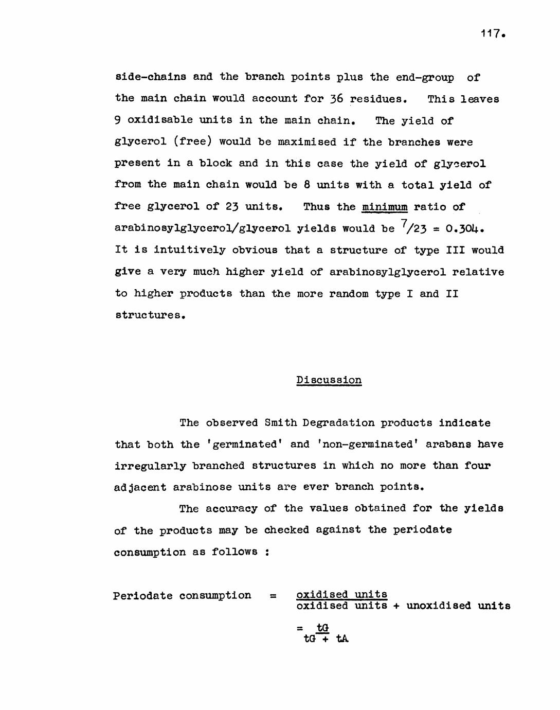
117.
side-chains and the branch points plus the end-group of
the main chain would account for 36 residues. This leaves
9 oxidisable units in the main chain. The yield of
glycerol (free) would be maximised if the branches were
present in a block and in this case the yield of glycerol
from the main chain would be 8 units with a total yield of
free glycerol of 23 units. Thus the minimum ratio of
arabinosylglycerol/glycerol yields would be /23 = 0,301*.
It is intuitively obvious that a structure of type III would
give a very much higher yield of arabinosylglycerol relative
to higher products than the more random type I and II
structures.
Discussion
The observed Smith Degradation products indicate
that both the 'germinated* and 'non-germinated 1 arabans have
irregularly branched structures in which no more than four
adjacent arabinose units are ever branch points.
The accuracy of the values obtained for the yields
of the products may be checked against the periodate
consumption as follows :
Periodate consumption oxidised unitsoxidised units + unoxidised units
= tQ tG + tA
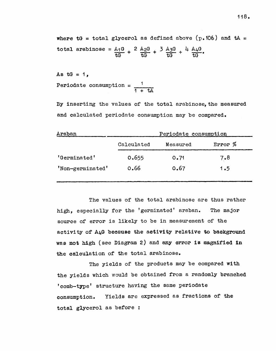
118
where tG = total glycerol as defined above (p. 106) and tA =
total arabinose = AiO 2 A2<3 3 A5& ktG to tO tO *
As tG = 1,
Periodate consumption =1 + tA
By inserting the values of the total arabinose, the measured
and calculated periodate consumption may "be compared.
Araban ______ ___________ Periodate consumption
'Germinated 1
1 Non-germinated'
Calculated
0.655
0.66
Measured
0.71
0.6?
Error %
7.8
1.5
The values of the total arabinose are thus rather
high, especially for the 'germinated' araban. The major
source of error is likely to be in measurement of the
activity of A^O because the activity relative to background
was not high (see Diagram 2) and any error is magnified in
the calculation of the total arabinose.
The yields of the products may be compared with
the yields which would be obtained from a randomly branched
'comb-type* structure having the same periodate
consumption. Yields are expressed as fractions of the
total glycerol as before :
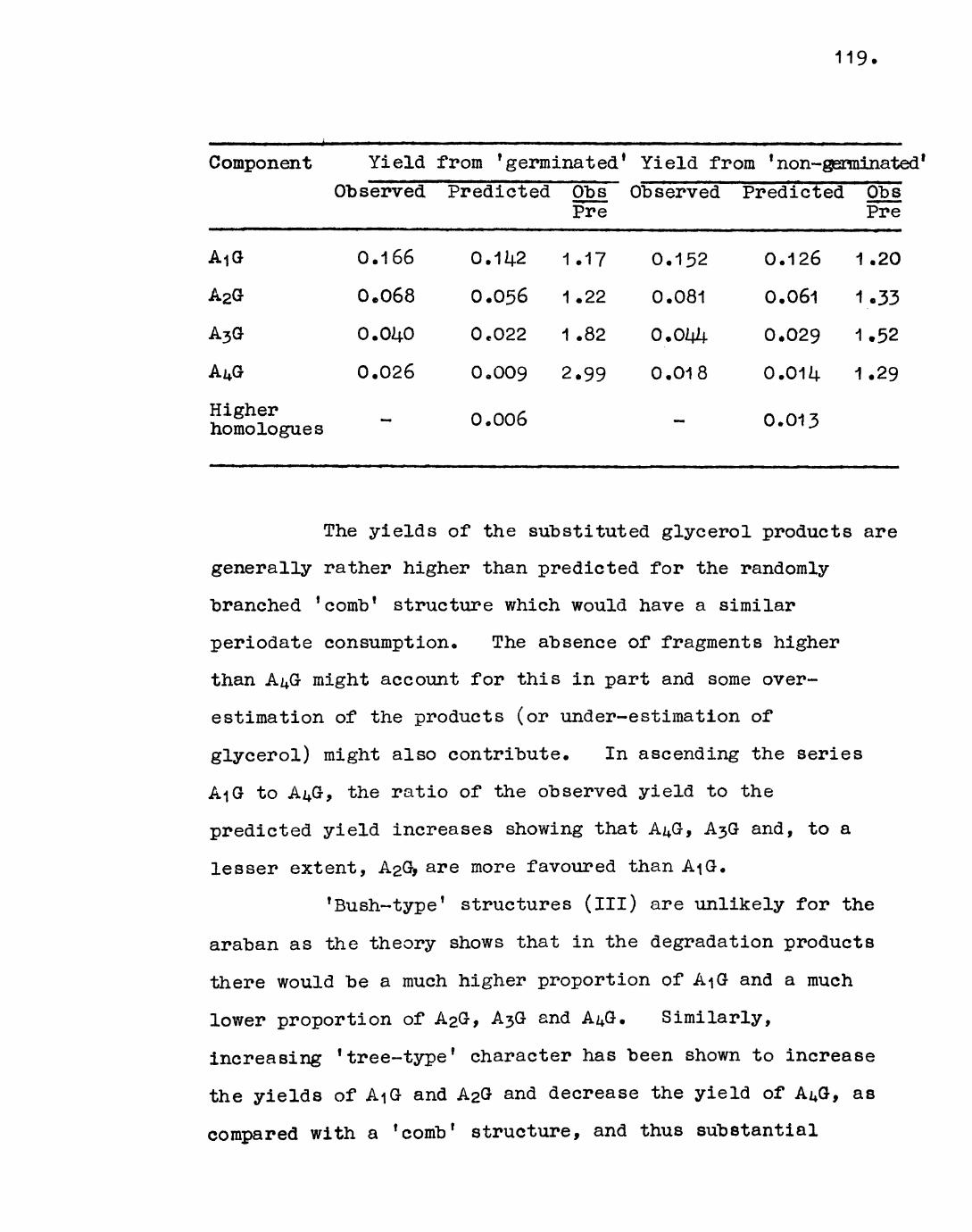
119.
Component
AiG
A2G
A3G
AuG
Higher homologues
YieldObserved
0.166
0.068
0.040
0.026
-
from 'germinatedPredicted
0.1^2
0.056
0.022
0.009
0.006
Obs Pre
1.17
1.22
1 .82
2.99
1 Yield from 'non-gerObserved Predicted
0.152 0.126
0.081 0.061
0.044 0.029
0.018 0.014
0.013
minatoObs Pre
1 .20
1.33
1.52
1.29
The yields of the substituted glycerol products are
generally rather higher than predicted for the randomly
branched f comb f structure which would have a similar
periodate consumption. The absence of fragments higher
than A^G might account for this in part and some over-
estimation of the products (or under-estimation of
glycerol) might also contribute. In ascending the series
AiG to Ai*G, the ratio of the observed yield to the
predicted yield increases showing that A^G, A3G and, to a
lesser extent, AaG, are more favoured than AiG.
'Bush-type* structures (ill) are unlikely for the
araban as the theory shows that in the degradation products
there would be a much higher proportion of A-jG and a much
lower proportion of A2G, A3G and A^G. Similarly,
increasing 'tree-type 1 character has been shown to increase
the yields of AiG and A2G and decrease the yield of A^G, as
compared with a 'comb' structure, and thus substantial
120.
'tree-type' (ll) character is not likely. It is moat
likely that the structure of mustard ara"ban is an irregular
1 comb-type 1 with a "bias in favour of contiguous branch
points. This bias is more pronounced in the 'germinated*
araban and may reflect some redistribution of branches
during germination.
Generally, although the labelling method of
determining the yields of Smith Degradation products was
rather less accurate than was desired it is more accurate
and convenient than other existing methods and may be used
on a micro scale. Correction for non-specific hydrolysis
would seem to be desirable in quantitative work especially
where furanoside linkages are involved.
CHAPTER U
Characterisation of polysaccharide structures
^>y glycoside stabilisation with
toluene~£-sulphona tes (tosylates): mod el
experiments with dextran

121 e
Introduction
Characterisation of polysaccharide structures by
stabilising a particular linkage to acid conditions is an
attractive method of obtaining additional evidence to that
given by the periodate degradation methods discussed in the
previous chapter. The aim of the work described here was
to develop such a method which would be applicable to
polysaccharides in which unsubstituted primary hydroxyls
occur only at the end-groups, such as dextran and araban.
The significance of the evidence which would be obtained by
characterisation of the terminal linkages in the above
polysaccharides will be discussed later. The catalytic
oxidation procedure of Aspinall (101) could be applied to
such polysaccharides as this also depends on selective
reaction of primary hydroxyl groups. These groups are
oxidised to carboxylic acid groups which then confer the
desired stability towards acid hydrolysis. Analysis of the
aldobiuronic acid products characterises the terminal
linkages. Unfortunately the oxidation requires extended
periods and is far from quantitative, probably due to
difficulties in obtaining all the primary hydroxyl groups of
the polymer in the correct orientation on the catalyst
surface, and these are the major drawbacks of the method.
The acid hydrolysis of glycosides, though the
subject of intensive investigation, is not yet fully
understood. Most of the work has been limited to the

122*
pyranosides, the kinetics of the hydrolysis being followed
polar imetrically. In the usual mechanism of hydrolysis of
the pyranosides (102) the initial step is the rapid
equilibrium-controlled protonation of the glycosidic oxygen
to give the conjugate acid. This is followed by the slow
rate-determining heterolysis of the conjugate acid with the
formation of a carbonium-oxonium ion and an alcohol. The
former reacts rapidly with water to give the parent sugar.
If the rate-determining step were bimolecular involving a
molecule of water the transition state, assumed to approximate
to the carbonium-oxonium ion, would be more highly ordered
than the ground state leading to negative entropies of
activation. Positive values have been obtained for a wide
variety of pyranoside glycosides and for glycosides of
pyranosiduronic acids (103) indicating the less ordered
transition state is involved. In contrast negative
entropies of activation have been recorded for some
furanoside glycosides (101^) indicating that a different
mechanism is likely, interpreted as either a bimolecular
displacement involving a molecule of water or a mechanism
involving ring opening.
Formation of the carbonium-oxonium ion intermediate
(i.e. an ion in which the positive charge is stabilised by
sharing between C/ . \ and the ring oxygen) requires that
C/ 2 \, C/,x, the ring oxygen and C/^N become co-planar, and
thus involves a conformational change from the chair to the
half -chair form. The formation is sterically impeded by

123.
partial eclipsing of the substituents on C/ 2 ) and C/-.\ and of
those on C/, \ and C/_x. When an electron-withdrawing
substituent is present in the glycone the inductive effect
will destabilise the carbonium-oxonium ion and hence raise
the activation energy of the hydrolysis reaction. It
follows that both steric and electronic factors affect the
formation and stability of the carbonium-oxonium ion and
hence the rate of hydrolysis.
The nature of the aglycon may also have a marked
effect on the rate of hydrolysis. Feather and Harris (105)
have observed that axially orientated substituents at C/,\
are hydrolysed more slowly than their equatorial
counterparts, presumably because the former are stabilised
by the anomeric effect (106). The methyl glucuronosides
are, surprisingly, hydrolysed at a similar rate to the methyl
glucosides (107), whereas other glycosides of glucuronic
acid (and particularly where the aglycon is another sugar
unit, as in a polymer) are extremely stable (108). This is
a dramatic example of the size of the aglycon affecting the
rate of hydrolysis, although it has been shown that the
entropies of activation of the methyl glycosides of
glucuronic acid are considerably lower than for most
pyranoside glycosides (10?) and this probably reflects that
a somewhat different mechanism is operating. The electron
affinity of the aglycon does not appear to be as important
as the size factor in the glucuronosides and it is thought
that the effect of the carboxyl group on C/\ plus the size

of the C/ \ substituent combine to give the overall result.
The considerable literature on the acid hydrolysis
of glycosides (both pyranoside and furanoside) has recently
been reviewed (109). By analogy with the carboxyl
stabilising effect it might be expected that a large
electron-withdrawing substituent on the primary hydroxyl of
the non-reducing unit of a disaccharide v/ould stabilise the
glycosidic linkage to hydrolysis. Such a substituent in a
1,6 linked hexosan,or possibly a 1,5 linked pentosan^would
enable the terminal linkage to be characterised. The use of
toluene-j>~sulphonates (tosylates) to this end is the subject
of this chapter*
These esters are themselves acid stable and
glycosides of 2~0~toluene~p-sulphonyl pyranoses are known to
be stable to acid hydrolysis (110), Measurements have shown
(111) that tosylates at V/r\ should be sufficiently electron-
withdrawing to stabilise the glycoside to a useful extent,
the inductive substituent constant (C?i ) for
-CH20.S02.C6Hu.CH3 of about 0.37 compares favourably with
0.39 for -COOH and 0.0? for -CH20H. In preliminary
experiments in this laboratory by Dr. N. Richardson (98),
the hydrolysis of methyl 6-0-toluene-p-sulphonyl-a-D-
-glucopyranoside was stabilised by a factor of between 3 and
5 as compared with methyl a-D-glucopyranoside. Although
this is not sufficiently large to be useful as such, it is
greater than for the corresponding uronoside (10?) and by
analogy with the uronic acid series (108) would be expected

125*
to "be very much greater at the d i sac char ide level (i.e. where
the aglycon is another sugar unit). Experiments with
dextran ensued, with the ultimate aim of applying the method
(if successful) to the mustard seed araban.
Dextrans are glucans produced "by strains of
Leuconostoc mesenteroides and other "bacteria on a sucrose
substrate* The polysaccharides have very high molecular
weights (up to several million) and fractions obtained by
controlled partial hydrolysis are used as substitutes for
blood plasma. Determination of the fine structure of
dextran s is thus important in view of the immuno logical
factors involved.
Early work has been reviewed by Neely (112).
Dextrans are essentially a-1 ,6 linked but differ in some
structural details which vary according to origin. The
most common variation is a degree of branching through the
C/^N and/or C/, \ positions of the glucose residues, although
branching through C/ 2 \ is also reported (113). Some
dextrans also have cc-1 ,3 and/or oc-1 ,U linked inter-chain
residues (11L|.)« The principal problem as regards fine
structure concerns the length and structure of side-chains.
The dextrans produced by L. mesenteroides NRRL B-lij.15
(a-1 ,k branched) and NRRL B-1M6 (cc-1 ,3 and cc-1 ,U branched)
have been investigated by enzymatic methods (with
glucamylase) and the dextrans produced by NRRL-1U15 and
NRRL-1375 (oM ,3 branched) by the catalytic oxidation
procedure (115). The results showed that most, if not all
126.
of the side-chains consist of single glucose units and it
was suggested that this structural feature might be common
to other "branched dextrans. Similar conclusions have been
reached on the basis of physical measurements (112).
The particular dextran used in this work was that
from Jr. mesenteroides NCIB 2706 and has been shown by
periodate oxidation studies (116) to contain 1 &%> 1 ,3-type
linkages and about 3% 1,U-type linkages. The molecule is
thus sufficiently highly branched to be a suitable model for
the development of the technique* P.m.r. spectroscopy of
the deuterated dextran (98) confirmed the degree of branching
was of the order of 20^, the interpretation of the spectrumn , x
being based on the assigments of Pasika and Cragg (117) •A
The tosylated derivative of the dextran was prepared by Dr.
Richardson (98). To obtain the most selective substitution
possible the tosyl groups were introduced after
triphenylmethylation (tritylation), methylation and
detritylation. Conditions were chosen to obtain complete
primary substitution in the first stage and the product was
found to be over-tritylated to the extent of 20$.
Examination after the second stage showed tetra-0-methylglucose
to be completely absent proving the location of the majority
of trityl groups. In preliminary hydrolyses of the
0-methyl-0~tosyldextran Dr. Richardson showed that glycoside
stabilisation was apparent under methanolysis conditions and
in 50^ aqueous monochloroacetic acid solution but that
detosylation and other reactions occurred in formic acid

127.
solution* This was attributed to the reducing power of
formic acido The usefulness of the specific tosyl ester
spray of Jackson and Hayward (7 and General Methods) was
confirmed for locating these esters on thin layer
chromatograms•
The aim of the work described in the experimental
section of this chapter was to establish optimum hydrolysis
conditions and to characterise the stabilised linkage(s) 0
The structural features of the native dextran were
established by a methylation analysis, indentification of the
methylated sugars being based on g,l«c. and paper
chromatography 0 A survey of the specificities of some acids
towards the co-occurring linkages in dextran and
0-methyld©xtran gave a background for the hydrolysis of the
tosylated derivative c Various attempts to isolate the
tosylated products of hydrolysis are described.

128
Experimental
Experiment Iu1 • Preparation of dextran.
The dextran was prepared from a culture of
Leuconostpc mesenteroides NCIB 2706 obtained from the Torry
Research Station, Aberdeen. The culture medium contained
sucrose (10$), yeast extract ("\%) , potassium hydrogen
phosphate (0.5%), and peptone (0.25%), in tap water, and was
adjusted to pH 7,0 with dilute sodium hydroxide solution,,
Two 50 ml and two 2 1 conical flasks containing 25 ml and 1 1
of medium respectively were plugged with paper-wrapped cotton
wool and sterilised in an autoclave. The small flasks were
innoculated with the organism and incubated for 2 days at
26 . The resulting viscous solutions were transferred to
the large flasks which were similarly incubated. The
dextran was isolated from solution "by the slow addition of
1 .5 volumes ethanol with stirring. The gummy product was
purified "by redissolving in water (2 l), boiling the
resultant solution for U5 minutes to coagulate proteins and
centrifuging. The supernatant solution was filtered and the
dextran reprecipitated with ethanol. On redissolving the
gum in water and freeze-drying, 100g. of dextran was
obtained. (Found, #N = 0.75)
Experiment \\.2. Methylation of dextran.
Dextran (11 .Og) was dissolved in N-methyl-2-
129*
-pyrrolidone (220 ml). The solution was cooled to 0°, and
methyl iodide (33 ml), dissolved in N-methy1-2-pyrrolidone
(220 ml), and "barium hydroxide octahydrate (77g) were
added. The mixture was shaken at 0° for 2 hr. and then at
room temperature overnight. Methyl iodide (110 ml) and
"barium hydroxide octahydrate (26kg) were added and shaking
was continued for 2Lj. hr. The mixture was transferred to a
separating funnel with water. The product was extracted
with chloroform (3x) and the chloroform layer washed with
sodium thiosulphate solution (1^6), water (3x), and dried over
sodium sulphate. The syrup was diluted with a small volume
of benzene and precipitated with light petroleum (b.p. Lj.0-60 )
The white fibrous product was purified by reprecipitation
(8.98g, 65$). Anal. Gale, for tri-0-methylhexose polymer :
$OCH3 = 1*5.6. Found, % OCH3 = 37.^.
The incompletely methylated material (5«9g) was
dissolved in dimethylformamide (180 ml) and methyl iodide
(60 ml) and silver oxide (30g) were added. The mixture was
magnetically stirred in a conical flask fitted with a double
surface condenser, in a water-bath at UO . After 6 hr
methyl iodide (60 ml) and silver oxide (30g) were added and
the reaction refluxed for Lj.8 hr. The product was obtained
as described in Experiment 1.5. (a) and was purified by the
precipitation procedure above (L|..15g> k&7° overall). Anal.
Pound, % OCH3 = 1|3.5^.
The product (i+.Og) was remethylated by the same
method (3.05g, 35?b overall). Anal. Found, % OCH3 = U3.6.

130 o.
The infra-red spectrum showed negligible hydroxyl absorption.
The product was fractionated, by the addition of light
petroleum (b.p. i±0~60 ) to a chloroform solution of the
methylated polysaccharide, into four subfractions of
approximately egual weight. Anal. Found, % OCH3 = 45«3j
llU.O, U2.9, 38.5-
The fully methylated unfractionated material
(20 mg) was methanolysed and examined by g.l.c. (Pye Argon
Chroma tograph) on column 2 at 160° and 175°. The results
are given below.
Temperature methyl glyco sides of - T (standard) T (dextran
160° 2,3,U,6-tetra-0-methylglucose 1.00, 1.14.6 1.00, 1 .
2,3,U-tri-0~methylglucose 2.32, 3-32 2.33, 3-29
2,U,6-tri-0-methylglucose 2.88, 1±. 48 2.85, U-3&(traces)
175° 2,4-di-0~methylglucose 6.18, 8.54 6*10, 8.54
3,l}.-di-0-methylglucose 7«54 -
2>3-di-0-methylglucose 12.01 -
Hydrolysis and paper chromatography in solvents B
and D respectively confirmed the presence of 2,3,4,6-tetra-
-0-methylglucose (RQ = 0*98, 1*00), 2,3,U~tri-0~methylglucose
(R a 0.89 > 0.80), 2,U,6-tri-0-methylglucose (traces only,
R = 0.82, 0.5i4.) and 2,U-di-0-methylglucose (RQ = 0.65, 0.35).
A small amount of another, unidentified, component having
R =0.80 and 0.90 in solvent B and D respectively and T =

131,
1.92 (160°) may have "been 1 ,6-anhydro~2,3,^-tri-~0-raethylglucose
Separation of the hydrolysis products of fully
methylated dextran (2g) by preparative paper chromatography
in solvent D yielded 2,i|.-di-0~methylglucose (0.168g) and a
mixture of 2 > 3,U-tri-0~methylglucose and 2,3,i|.,6-tetra-0~
methylglucose (l.020g). Both products were exhaustively
dried in vacuo over phosphorus pentoxide at ij.0o . This
corresponds to approximately ^5% branching.
Experiment U*3* Comparison of the stabilities of the 1^,3-
and 1 ,6-glycosidic linkages in dextran and methylated
dextran under various solvolysis conditions.
The general method used is as follows. Samples
(10 - 20 mg) of dextran or methylated dextran were hydrolysed
for varying times under the conditions given and, after
neutralisation or removal of acid, each hydrolysate was
methylated, methanolysed and examined by g.l.c. on column 1
at 175° (Pye Argon Chromatograph). The relative rate of
hydrolysis of the 1,3- and 1,6-glycosidic linkages is given
by the variation of the relative concentration of
2,3,U-tri-0-imethylglucose and 2,U,6-tri~0-methylglucose with
hydrolysis time. This was conveniently measured from the
peak heights of the slower peak of each tri-0-methylglucoside
peaks.
(a) Sulphuric acid (1N) hydrolysis of dextran.
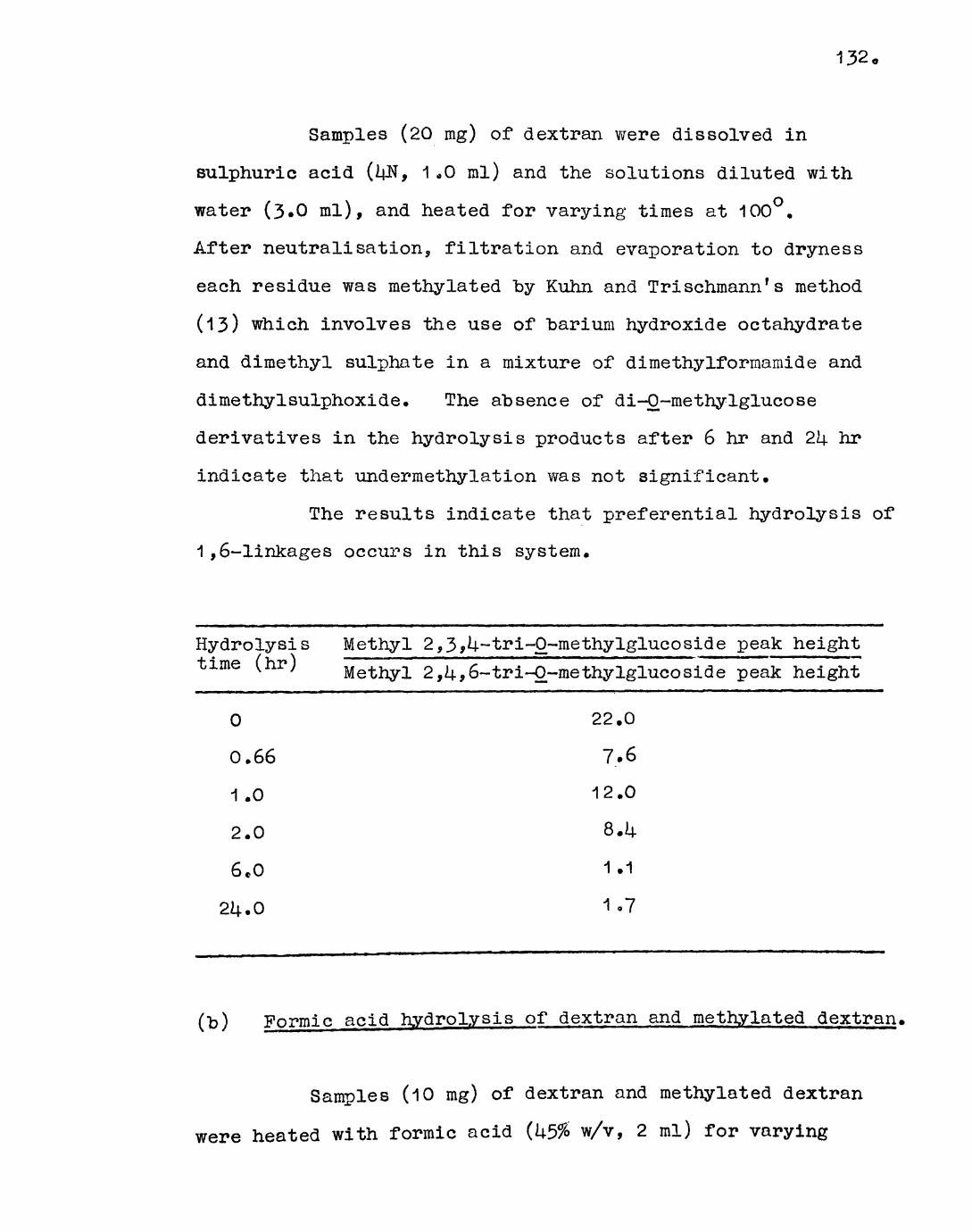
132*
Samples (20 ing) of dextran were dissolved in
sulphuric acid (^N, 1-0 ml) and the solutions diluted with
water (3.0 ml), and heated for varying times at 100°.
After neutralisation, filtration and evaporation to dryness
each residue was methylated by Kuhn and Trischmann's method
(13) which involves the use of "barium hydroxide octahydrate
and dimethyl sulphate in a mixture of dimethylformamide and
dimethyIsulphoxide. The absence of di-0-methylglucose
derivatives in the hydrolysis products after 6 hr and 2L\. hr
indicate that undermethylation was not significant.
The results indicate that preferential hydrolysis of
1 ,6-linkages occurs in this system.
Hydrolysis time (hr)
0
0.66
1 .0
2.0
6.0
2U.O
Methyl 2,3 A-tri-O-methylglucoside peak heightMethyl 2,U,6-tri-O-methylglucoside peak height
22.0
7.6
12.0
8.1+
1.1
1-7
(t>) Formic acid hydrolysis of dextran and methylated dextran.
Samples (10 mg) of dextran and methylated dextran
were heated with formic acid (k5% w/v, 2 ml) for varying

133.
periods at 100°. After removal of acid (General Methods)
and drying, the hydrolysates were methylated, methanolysed
and examined "by g.l.c. as described above.
Hydrolysis time (hr)
0
0,75
1.0
2oO
6.0
2U.O
Methyl 2,3»^-~tri-0~methylglucoside peak height
Methyl 2,U,6-tri-
dextran
22 0 0
12.1
15.3
13.5
1lj..O
5.0
-0-me thy Igluco side peak height
methylated dextran
22,0
36.3
26.6
38.6
22.2
14-0,7
This result was suspect due to slight undermethylation.
The results indicate that "both linkages are hydrolysed
at about the same rate in this system with the methyl ether
groups appearing to confer some stability on the 1,6-linkage.
(c) Chloroacetic acid hydrolysis of dextran and methylated
dextran.
In order to assess the difficulties involved in the
removal of the acid and possible side-reactions (such as
reversion), D-glucose (20 mg) was dissolved in aqueous

13*4-0
chloroacetic acid (50$ w/v, 2 ml) and heated at 100° for
2l4.hr, The acid was removed by evaporation on the rotary
evaporator using an oil-pump and a "bath temperature of 60°
but required repeated distillation of water from the residue
to effect efficient removal. On examination by paper
chromatography in solvent A no traces of disaccharides or
products other than glucose were observed and it was concluded
that reversion was not significant.
(i) Dextran (20 mg) was heated in chloroacetic acid (50$,
2 ml) at 100° for 12 hr. After removal of the acid and
drying, the product was methylated (as in (a) above),
methanolysed and examined by g.l.c. Although slight
undermethylation was observed the results clearly showed
that 1,6-linkages were being hydrolysed at a faster rate
than 1,3-linkages in this system,
(ii) Samples (^2 mg) of methylated dextran were heated in
chloroacetic acid (50$, 2 ml) for varying periods at 100 .
After removal of acid and methylation as above the infra-red
spectra showed slight hydroxyl absorptions. The samples
were rerne thylated in dimethylformamide with methyl iodide
and silver oxide under reflux as in Experiment U.2. After
methanolysis the following results were obtained.
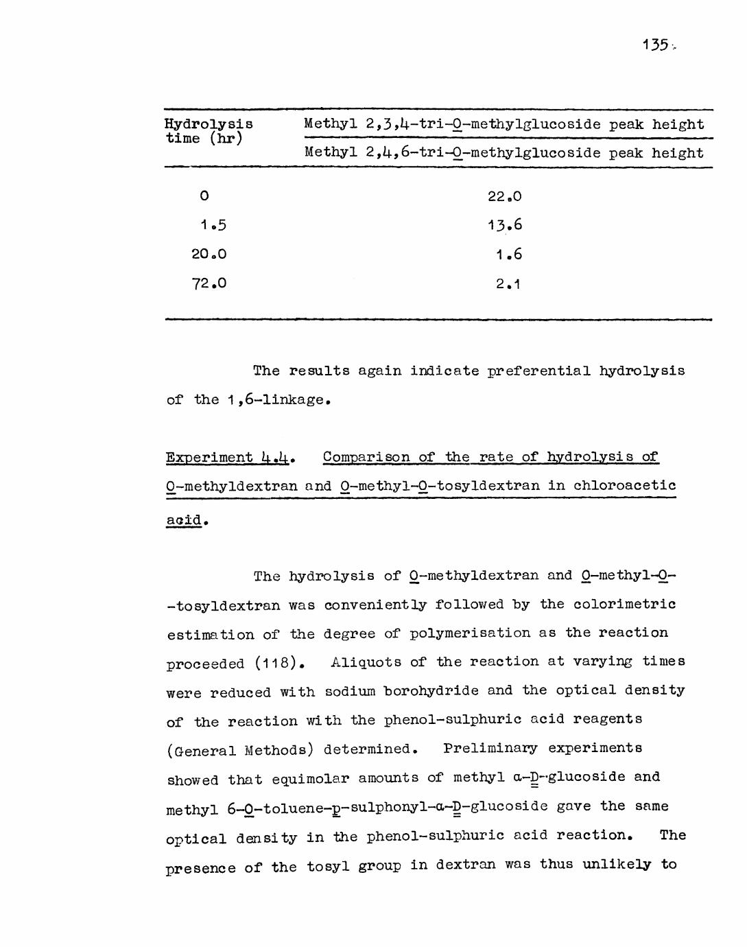
135 -
Hydrolysis Methyl 2,3>U-tri-0-methylglucoside peak heighttime (hr) ————————————————————————————————————
Methyl 2,U,6-tri-0-methylglucoside peak height
0 22.0
1o5 13.6
20oO 1.6
72.0 2.1
The results again indicate preferential hydrolysis
of the 1 ,6-linkage.
Experiment U.U. Comparison of the rate of hydrolysis of
0^-methyldextran and O-methyl-0-tosyldextran in chloroacetic
acid.
The hydrolysis of 0~methyldextran and O-methyl-0-
-tosyldextran was conveniently followed "by the colorimetric
estimation of the degree of polymerisation as the reaction
proceeded (118). Aliquots of the reaction at varying times
were reduced with sodium "borohydride and the optical density
of the reaction with the phenol-sulphuric acid reagents
(General Methods) determined. Preliminary experiments
showed that equimolar amounts of methyl a-D-glucoside and
methyl 6-0-toluene-p-sulphonyl-a~D-glucoside gave the same
optical density in the phenol-sulphuric acid reaction. The
presence of the tosyl group in dextran was thus unlikely to

affect the validity of the results. It was found however
that large amounts of sodium "borohydride supressed the
colour reaction and that addition must "be rigidly controlled.
(Excess borohydride was used to both neutralise the acid and
reduce the sugars),
EcLUimolar samples of 0-methyldextran (20 mg) and
0-methyl~0~tosyldextran (22.8 mg) were heated with
chloroacetic acid (50^, 2.0 ml) in glass stoppered tubes with
mechanical shaking at 100°, 8U.5° and 69° in thermostat
baths. 0~methyldextran dissolved readily but the tosyl
derivative was insoluble for some time (2.5 hr. at 81^.5°),
and this explains the anomalous results for the shorter
hydrolysis times. Samples (0.010 ml) were removed at
intervals, diluted with water (1.00 ml) and a weighed excess
of sodium borohydride (0.1OOg) was added in portions. After
standing for at least 2i| hr with occasional shaking, excess
borohydride was destroyed by the slow addition of
concentrated sulphuric acid (AnalaR, 1.00 ml). Water
(1 .00 ml) was added followed by the phenol-sulphuric acid
reagents and the optical density measured. The optical
density corresponding to 90$ hydrolysis was determined by
tenfold dilution of the hydrolysates after reaction and
addition of 0.010 ml of this, diluted with water (1.00 ml),
to premixed sodium borohydride and concentrated sulphuric
acid in the normal proportions, followed by the
phenol-sulphuric acid reagents.
The plot of optical density against hydrolysis time

3.0-
2.0- 1.0
Opt
ical
dan
aitv
.
12
diag
ram
1.
2448
72ti
me
(hr.
)14
4
Chl
oroa
catic
aci
d hy
drol
ysis
(84
.5)
ofM
O
-met
hyl
-O-t
osy
ldex
tran
•O—
O*m
ethy
ldex
tran

137.
is shown in diagram 1 for the 8Lu5° hydrolysis. (The
broken line corresponds to 9C$ hydrolysis). The second and
slower phase of the hydrolysis of O-methyl-0-tosyldextran
was more pronounced at 8i|.5° and 69° than at 100°, there
"being little difference in this respect between 8U.5° and
69 . However the absolute rate of hydrolysis was
inconveniently slow at 69° (approximately 1 8 days for 80%
hydrolysis) and the most suitable conditions for optimum
yield of stabilised fragments from 0-methyl-0~tosyldextran
was judged to be ?2 hr at
Experiment U.5» Thin Layer Ghromatography of the hydro lysis
products of O-methyl-0-tosyldextran.
The optimum conditions established above were
employed. In addition to a small scale (20 mg) hydrolysis
of O-methyl-Q-tosyldextran, similar samples of 0-methyldextran
and O-methyl-0-triphenylmethyldextran were hydro lysed.
After removal of the acid as above, portions (2 mg) were
dissolved in a small volume of acetone and examined by
T.L.C. on silica gel micro-plates (General Methods). After
double development in benzene-ethanol-water-ammonia
(200 : kl : 15 • 1) tosyl esters were located by use of the
specific spray (iii). Subsequent spraying with the general
reagent (i) located all the carbohydrate components,
Only the O-methyl-0-tosyldextran hydrolysate gave
a positive reaction with the specific spray, the only

significant UV fluorescing spot had the same mobility as
2<,3>U-tri~0~methylglucose and was considerably slower than
2 ?3>U~tri~0-methyl~6~0-tosylglucose a 2,3,L|.,6-tetra-0--
-methylgluco se was only significant in the hydrolysate of
0-methyldextran. 2,3,l4.-tri~0-methylglucose (major component)
and 2,l4.-di-0-methylglucose were present in all the hydrolysates.
Some resolution of the tosylated material and
2,3jU-tri~0-methylglucose was obtained on larger plates
(20 x 5 cm) by development in benzene-ethanol (2 : 1 ) or
chloroform-methanol (25 : 1) but this was not sufficiently
good to allow separation on a preparative scale. In these
solvents the tosylated material did not appear to be entirely
homogeneous. A wide variety of other systems was examined,
with silica, alumina and cellulose plates, but the
results were usually inferior.
Experiment U«6 0 Gel filtration of the hydrolysis products
of 0-methyl~0~tosyldextran.
Attempted separation of the components of the
hydrolysate (20 mg) on a column of Sephadex LH-20 (30 x 2.5
cm) by elution with ethanol was not successful. The column
fractions were screened by UV spectrophotometry
223 nm) but on examination of the combined absorbing material
by T.L.Co the original mixture was obtained.
Separation was similarly unsuccessful when the
mixture (30 mg) was applied to a column (UO x 2 cm) of
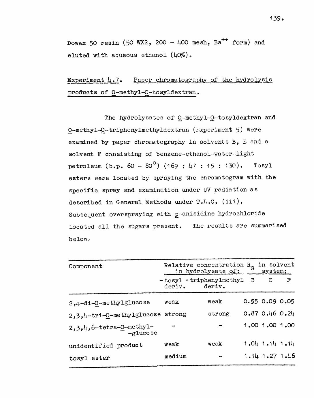
139.
Dowex 50 resin (50 WX2, 200 - ij.00 mesh, Ba*+ form) and
eluted with aqueous ethanol (U0%) «,
Experiment k»7* Paper chroma to gra-phy of the hydrolysis
products of 0-methyl-O-tosyldextran,
The hydrolysates of 0~methyl~0~tosyldextran and
0~methyl~0-triphenylmethyldextran (Experiment 5) were
examined "by paper chromatography in solvents B, E and a
solvent P consisting of benzene-ethanol-water-light
petroleum (b.p. 60 - 80°) (169 : k7 i 15 : 130). Tosyl
esters were located by spraying the chromatogram with the
specific spray and examination under UV radiation as
described in General Methods under T.L.C. (iii).
Subsequent overspraying with £-anisidine hydrochloride
located all the sugars present. The results are summarised
belowo
Component Relative concentration R in solventCrin hydrolysate of; system;
tosyl -triphenylmethyl B E F deriv. deriv.
2,U-di-Qrmethylglucose weak
2,3,U-tri-0-methj'-lglucose strong
2,3,k 9 6-tetra~0~methy1-"* -glucose
unidentified product
tosyl ester
weak
medium
weak 0,55 0.09 0.05
strong 0.87 O.U6 0,2/j.
1 .00 1 .00 1 .00
weak 1 ,0i| 1 .11+ 1 .
1.2? 1 .U6

11*0,
Solvent F gave good separation of the tosyl ester
component from the other sugars. The unidentified component
was shown not to "be residual chloroacetic acid.
Experiment l4«8« Separation of the hydrolysis products of
O-methyl-0-tosyldextran by cellulose column chromatography.
The results of Experiment U.7- suggested that
purification of the tosyl ester fraction might be possible
by cellulose column chroma tography in solvent P.
0~methy1-0-tosyldextran (0.320g) was hydrolysed in aqueous
chloroactic acid under the standard conditions developed
in Experiment l+.k* After exhaustive removal of acid the
hydrolysate was dried in vacuo at 50° (O.UU6g), Paper
chromatography showed a similar composition to the hydrolysate
examined previously. The UV spectrum in ethanol solution
showed a peak at 223 nm identical with that given by methyl
6-0-tosyl-a-D-glucoside. The infra-red spectrum (as a film)
showed bands at 1160 cm" (medium) and 1380 cm (weak)
attributable to the tosyl group, and also a very strong—1
carbonyl absorption at 17UO cm suggesting residual
chloroacetic acid or, less likely, that chloroacetate esters
were present. This would account for the discrepancy
between the weights of starting material and the hydrolysate.
The syrup was treated twice with aqueous
ammonium carbonate (1# w/v, 10 ml) followed, after standing
for 2 hr by evaporation to dryness under reduced pressure
11J.1
at 60°„ Water was distilled from the residue several times
until ammonia was no longer detectable by smell. The—1
infra-red spectrum showed that the band at 1740 cm had
oeen shifted to 1600 cnT confirming that a carboxylic acid
had been present but was now converted to the ammonium salt.
There was a slight residual absorption at 17UO cm which
was not affected by brief (30 minute) de-esterification of
the hydrolysate in methanolic 2,2-dimethoxypropane (5%) with
sodium methoxide. The cleavage of chloroacetate esters may—1
not lead to the disappearance of the residual 1740 cm band
as the product (methyl chloroacetate, b.p. 150°) might not
have been removed by evaporation, After neutralisation
with solid carbon dioxide and evaporation to dryness,
exhaustive extraction of the salty residue with ethanol
(6x10 ml) followed by evaporation gave a clear syrup
(300 mg).
The syrup was dissolved in ethanol (5 ml) and
loaded onto a cellulose column (70 x 3»5 cm) and eluted with
a system essentially similar to solvent F but half-saturated
with water. Fractions (10 ml) were collected automatically
and screened by spotting onto paper and spraying with
p-anisidine hydrochloride. Fractions were combined on the
basis of the results into three larger fractions which were
evaporated to dryness and dried in vacuo.
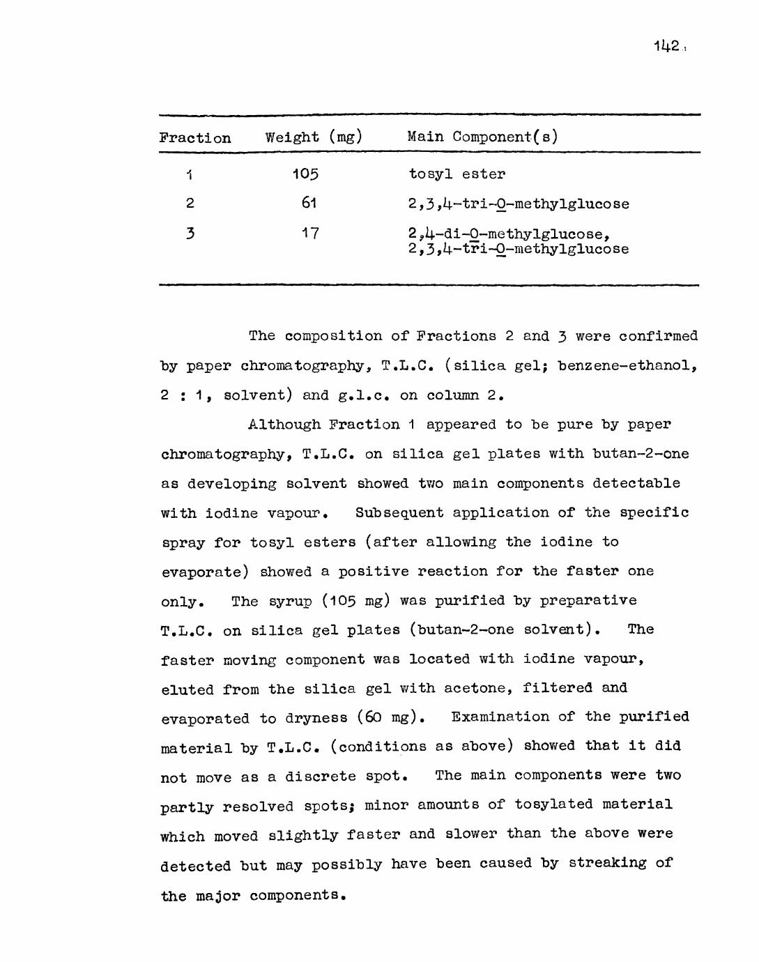
114-2,
Fraction Weight (mg) Main Component(s)
1 105 tosyl ester
2 61 2,3,^~tri~0-methylglucose
3 17 2,0U-di-O-methylglucose,2,3 jU-tri-O-methylglucoSe
The composition of Fractions 2 and 3 were confirmed
"by paper chromatography, T.L.C. (silica gel; benzene-ethanol,
2:1, solvent) and g.l.c. on column 2.
Although Fraction 1 appeared to be pure by paper
chromatography, T.L.C. on silica gel plates with butan-2-one
as developing solvent showed tv/o main components detectable
with iodine vapour. Subsequent application of the specific
spray for tosyl esters (after allowing the iodine to
evaporate) showed a positive reaction for the faster one
only. The syrup (105 mg) was purified "by preparative
T.L.C. on silica gel plates (butan-2-one solvent). The
faster moving component was located with iodine vapour,
eluted from the silica gel with acetone, filtered and
evaporated to dryness (60 mg). Examination of the purified
material "by T.L.C. (conditions as above) showed that it did
not move as a discrete spot. The main components were two
partly resolved spots; minor amounts of tosylated material
which moved slightly faster and slower than the above were
detected but may possibly have been caused by streaking of
the major components.
_>o
Separation of the components of the tosyl ester
fraction appeared to "be "beset with many difficulties, the
streaking mentioned above, which occurred to some extent in
all the systems,examined, "being one» The line of approach
involving direct identification of the tosylated disaccharide(s)
was thus abandoned in favour of an indirect method described
in the next experiment,
Experiment U»9» Identification, after methylation, reductive
detosylation and rernethylation, of the methanolysis products
of Q-methyl-0-~to syIdextran.
Methylation followed "by detosylation ands
remethylation of the methanolysis products of 0~methyl-0~
tosyldextran should yield fully-methylated isomaltose
(6-0-a~D-glucopyranosyl-D-glucose) and/or fully-methylated
nigerose (3-0-a-D-glucopyranosyl-D-glucose), the relative
yields being diagnostic of the "branching pattern of the
dextran. Incomplete methanolysis at non-stabilised linkages
would also yield the methylated disaccharides and correction
for this was made "by addition of an internal standard*
Identification of the fully methylated disaccharide derivatives
was "by g.l.c. and relative yields were calculated from peak
areas*
(a) Preparation of standard compounds, for g.l.c.
Nigerose and isomaltose samples were kindly
provided by Professor D.J. Manners and Dr. J.R. Turvey*
Each disaccharide (10 mg) was dissolved in dimethylformamide
(1.0 ml) and methyl iodide (1.0 ml) and silver oxide (0.1 OOg)
added to the solution at 0°. The reaction was carried out
in a sealed tube with shaking in the dark at room temperature
for U8 hr. The product was obtained by evaporation of the
chloroform solution obtained by the procedure described in
Experiment 1.5. (a). Three such methylations were carried
out.
Octa~0~methylsucrose, kindly provided by Dr. J.C.P.
Schwarz, was used as the internal standard.
The compounds prepared were shown to be separable
from each other and from octa~0-methylsucrose by g.l.c. on
column k (General Methods) (Pye series 10^ chromatograph) at
210 . The retention times (T ) were expressed relative tos
acta-0-methylsucrose, and are in the range to be expected
from the literature (119).
Octa-0-methylsucrose, T 1.00""" S
Methyl hepta~0~methylisomaltosides, T 1 .l|i± 2.02"""* S
Methyl hepta-0-methyInigerosides, T 1.82 2.35""" S
(b) Investigation of the methanolysis products of
0-methy1-0-tosyIdextran.
Samples (25 mg) of O-methyl-0-tosyl dextran were
heated in sealed tubes with methanolic hydrogen chloride
, 2.5 ml) at 100° for 0.5, 1.5, U.5, 13.5 and 1+0.5 hr.

The material dissolved instantly. After neutralisation
with silver carbonate, filtration and evaporation to
dryness, each residue was dissolved in dimethylformamide
(2.0 ml). Methyl iodide (0.5 ml) and silver oxide (0.2g)
were added and the reaction carried out as in (a), above, and
worked up in the usual way. The procedure was repeated
twice. The products were dried .in vacuo and examined "by
infra-red spectroscopy in dry carbon tetrachloride
solution. No trace of hydroxyl absorption was observed for
any sample, showing methylation to be complete. Examination
of the methylated methanolysates by g.l.c. on column k (Pye
series 10ij. chromatograph) at 210 showed isomaltosides as
the only detectable disaccharide products (T , 1.14-8* 2,00,s
2.60 (minor, unidentified). To each sample was added
octa-0-methy1sucrose as the internal standard. The UV
spectra of the samples were recorded in ethanol solution.
After evaporation to dryness, each methanolysate was
dissolved in aqueous isopropanol (80$). Reductive
detosylation (120) was carried out by addition of an excess
of sodium amalgam (2%) to the solution at 60 over 2i\. hr.,
followed by addition of solid carbon dioxide, filtration and
evaporation. The product was extracted from the salty
residue with acetone which was filtered and evaporated to
dryness. The residue was dissolved in chloroform which
was washed with water and evaporated to dryness. The
absorptions at 223 nm of the products in ethanol solution
were virtually undiminished. The treatment with sodium

amalgam was repeated using a very large excess which was
added over Lj.8 hr to a solution of the product in aqueous
ethanol (80$) at 65°. After working up as above, greatly
diminished absorptions at 223 nm were observed in all the
products.
After remethylation and g.l.c. by the same
procedure, the increase in methylated disaccharides was
measured from the peak areas of g.l.c. charts, using the
internal standard* Areas were measured by triangulation.
Both isomaltosides and nigerosides were identified on the
basis of retention times (T , 1 .1+8, 1.98, 2.3U (except ins
product after 0,5 hr), 2.60 (minor)). The faster
nigeroside peak was not observed, probably due to only
trace amounts of this derivative being present. The
assignment of the peaks to disaccharide derivatives was
supported by the gradual build-up of peak areas with
methanolysis time. The maximum increase in the proportion
of isomaltose plus nigerose derivatives relative to the
internal standard, (8.22 fold), was observed after U0.5 hr
methanolysis and it is likely that greater increases may
have resulted from even longer times. The yield of nigerose
derivatives relative to isomaltose derivatives was very small
and the significance of this is discussed later.

Methanolysis Ratio of isomaltose + nigerose Relative increasetime (hr) derivatives g octa~0~methyl- in methylated B
-sucrose disaccharides (A)Before After detosylation (A) detosylation (B)
0.5
1.5
U.5
13.5
U0.5
0.528
0.609
1.1U60.6660.52U
0.833
1.395
3.U732.UOO
U.305
1.5792.2913.031
3.6038.219
The U0.5 hr product was examined further by g.l.c.
using the same apparatus and column as above "but with
temperature programming from 1UO - 210 (increasing "by 6
per min to 210°, followed by 5 min at this temperature) to
estimate the relative amounts of disaccharide derivatives and
tetra-0-methylglucosides (no other methylated glucoside
derivatives were observed). Measurement of peak areas of
the U0«5 h^? methanolysate gave:
Tetra-0-methylglucosides 2.1 6 cm2
Hepta~0-methylisomaltosides 2.10 cm2
Hepta~0~methylnigerosides 0.1? cm2
On the assumption that the flame ionisation
detector gives a response on a weight basis for compounds of
such related structure these values represent a molar ratio
of tetra-0-methylglucosides : hepta-0-methylisomalto sides :

hepta-p-methylnigerosides of 100 ; 5i| : l±. The accuracy of
these values is probably no more than i 10$. The earlier
results have shown that less than one eighth of the total
yield of disaccharides is attributable to incomplete
hydrolysis at non-stabilised linkages.
Experiment U«10«> Identification, after me thy la t ion,
reductive detosylation and rernethylation, of the hydrolysis
products of O-methyl-0-tosyldextrane
The purified tosyl ester fraction (25 mg) isolated
by cellulose column chromatography and T.L.C. from the
chloroacetic acid hydrolysate of O-methyl-0-tosyldextran
(Experiment U.S.) was methylated twice by the procedure
described in the previous experiment. The infra-red
spectrum showed methylation to be complete. The UV spectrum
in ethanol solution showed the usual strong absorption at
223 nm. Detosylation was carried out in aqueous ethanol
(80$) by the addition of a large excess of amalgam to the
solution at 65° over Lj.8 hr. After working up as described
in the previous experiment the UV spectrum showed considerable
de-esterification had been effected. The product was
remethylated and examined by g.l.c. by the methods described
previously. Isomaltosides were detected on the basis of
retention times, expressed relative to octa-0-methylsucrose,
(T , 1.U8, 2.00, (2.60 minor, unidentified)) but not s
nigerosides.
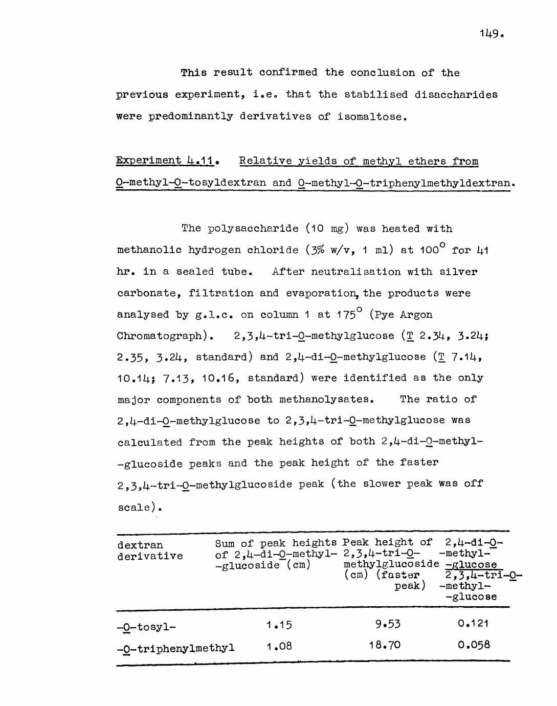
This result confirmed the conclusion of the
previous experiment, i.e 0 that the stabilised disaccharides
were predominantly derivatives of isomaltose.
Experiment U.11o Relative yields of methyl ethers from
O-methyl-O-tosyldextran and 0~methyl-0-triphenylmethyldextran
The polysaccharide (10 mg) was heated with
methanolic hydrogen chloride (~5% w/v, 1 ml) at 100° for M
hr. in a sealed tube. After neutralisation with silver
carbonate, filtration and evaporation, the products were
analysed by g.l.c, on column 1 at 175 (Pye Argon
Chromatograph). 2,3,U-tri-0~methylglucose (T 2.3U, 3*214.;
2.35, 3.214-, standard) and 2,U-di-0-methylglucose (T ?.1l|,
10.1Uj 7.13, 10.16, standard) were identified as the only
major components of both methanolysates. The ratio of
2,l4.~di-0~methylglucose to 2,3,l4--tri-0-methylglucose was
calculated from the peak heights of both 2,U-di-0-methyl-
-glucoside peaks and the peak height of the faster
2,3,U-tri-0-methylglucoside peak (the slower peak was off
scale).
dextran Sum of peak heights Peak height of 2,U-di-0- derivative of 2,U-di-0-methyl~ 2,3,U-tri-0- -methyl-
-glucoside (cm) methylglucoside -glucose(cm) (faster 2,3,U-tri-0-
peak) -methyl- -glucose
-0-tosyl-
-0-triphenylmethyl
1.15
1.08
9.53 18*70
0.121
0.058

150,
The results indicated that the yield of 2,l4.-di-0-
-methylglucose from 0--methyl-0-tosyldextran was approximately
2»1 times that obtained from 0-methyl-0~triphenylmethyldextran
under these conditions 0
Discussion
Analysis of the hydrolysis products of the
methylated poly sac chapide confirmed that the dextran from
L. mesenteroides NCIB 2?06 is of a well-known structural
type in which 1,6-linked glucopyranose residues are branched
through the C/,\ position. Traces of 2,Li.,6~tri-0-methyl~
-glucose were also detected indicating that a small
proportion of 1,3-linked inter-chain residues was also
present, but no traces of 2,3-di-O-methylglucose, 3»U-di-0-
-methylglucose, 2,3,6-tri-O-methylglucose or 3,14.,6-tri-O-
-methylglucose were found. The presence of 1,U-type linkages
which were suggested on the basis of periodate oxidation (116)
was thus not confirmed. The preparative-scale paper
chromatographic separation of the hydrolysate (Experiment
Uo2.) showed that 2,lj.-di-0-methylglucose accounted for 15l/b
of the sugars present (on a molar basis). The accuracy of
the procedure is low and considering this the result is in
fairly good agreement with the degree of branching
(approximately 20$) given by the p.m.r. and periodate
oxidation studies (see Introduction).

151.
The survey of different solvolysis procedures with
"both dextran and 0~methyldextran gave information as to the
specificities of these reagents towards the 1,6- and
1,3-linkages. Goldstein and Whelan (95) have observed that
whereas the 1 ,3-linkages of dextran are more labile than
the 1 ,6-linkages in aqueous acid, the reverse is true under
acetolysis (acetic anhydride and sulphuric acid) conditions.
Mr. Jo Campbell has found that the 1,6-linkages of 0-methyl~
-dextran are preferentially cleaved under both methanolysis
and acetolysis conditions, a similar procedure to Experiment
U»3« being used to determine the relative rate of hydrolysis
of the two linkages. The results of Experiment 1+.3. show
that in dilute sulphuric acid or 50$ aqueous chloroacetic acid,
the 1 ,6-linkages of dextran are hydrolysed more rapidly than
the 1 ,3-linkages. This was also observed in the cliloroacetic
acid hydrolysis of 0-methyldextran (for solubility reasons
hydrolysis of the methylated polysaccharide in dilute
sulphuric acid was not attempted). Formic acid did not
exhibit any marked specificity for either linkage in dextran
or 0-methyldextran. The observation that acid hydrolysis of
dextran leads to preferential cleavage of the 1,6-linkage is
contrary to that observed before.
The hydrolysis of O-methyl-0-tosyldextran, with
the object of isolating the stabilised disaccharide fragments,
was initially carried out in 50% chloroacetic acid.
Methanolysis could also have been used but not dilute
sulphuric acid as the tosylated derivative is even more

insoluble than 0-methyldextran. Formic acid would lead to
side-reactions as mentioned in the Introduction,
Measurement of the average degree of polymerisation for
samples withdrawn from the hydrolysate (Experiment U»U«)
showed that the reaction levelled off at about 80?£
hydrolysis (Diagram 1), consistent with the stabilisation
expected from the degree of branching. The hydrolysis of
0-methyldextran was also incomplete (about 90%) under
similar conditions and hence not all of the stabilisation
observed in 0~methyl-()~tosyldextran is attributable to the
tosyl ester. The conditions for the optimum yield of
stabilised fragments was judged to be 72 hr at 814. .5 •
Possible structures of the dextran are shown
"below. Hybrids of these structures are also possible.
3 t
t 1
r17^ n> 1Gg

153.
6t III
Purification of the tosylated component(s) of the
chloroacetic acid hydrolysate by T.L.C. using a variety of
systems was unsuccessful as satisfactory separation from
2,3jU~tri~0-methylglucose - the other major hydrolysis
product - could not "be obtained. Similarly, attempts at
purification "by gel filtration were unsuccessful. Partition
chromatography on cellulose (paper and column) was a limited
success. Unfortunately the column fraction containing the
tosylated components was heavily contaminated with non-
carbohydrate material (presumably degradation products) due
to the high mobility (R^ 1 .11+ in solvent B). This fraction
after purification by T.L.C. showed two major components.
2,U-di-O-methylglucose was observed in the
hydrolysis products by T.L.C., paper chromatography and in a
fraction from the cellulose column, indicating that single
unit side-chains of the type found in I are not present in
substantial amounts (assuming the terminal linkage is
stable).
After methylation of the 1+0.5 hr methanolysis
product from O-methyl-0-tosyldextran the tosyl esters were
removed by sodium amalgam. Remethylation and analysis by
15U.
g.l.c. then showed the presence of tetra~0-methylglucosides,
hepta-0-methylisomaltosides and hepta-0-methylnigerosides
in the approximate molar ratio of 100 : 5U : U. When the
treatment with amalgam was omitted the proportion os
isomaltosides fell to less than one eighth of the value
above; this arose from the incomplete hydrolysis of
non-tosylated parts of the molecule. The proportion of the
58 disaccharide molecules that are attributable to incomplete
hydrolysis of non~stabilised linkages and / or non-specific
substitution may be calculated as follows. On the basis of
the degree of branching (20$) and if methanolysis at non-
stabilised linkages were complete, tetra-Q-methylglucosides
and disaccharide glycosides would be present in the (molar)
ratio of 100 : 33 • Thus if x molecules of disaccharide have
arisen from incomplete hydrolysis and / or non-specific
substitution then ;
100 + 2x 100—————— = —— , i.e. x = 15-58 - x 33
Thus no more than 15 disaccharide molecules can have arisen
in this way. Therefore 6-0-tosylisomaltosides must be
present in at least tenfold excess over 6-0-tosylnigerosides
and the structure of the dextran thus corresponds more closely
to II or III than I. These conclusions were confirmed by
Experiment 1+.10. in which the tosyl ester fraction obtained
by cellulose column chromatography of the chloroacetic acid
hydrolysate of O-methyl-0-tosyldextran yielded only
155.
isomaltosides, after methylation, detosylation and
remethylation.
A further distinction "between the possible
structures is that only II should give derivatives of
2,l4-~di-0-methylglucose under cleavage conditions which
retain the terminal linkage. This sugar has "been
identified in the hydrolysate* In quantitative terms, the
relative yields of 2,U-di~ and 2,3,U-tri-0-methylglucose
derivatives from II should "be in the ratio of 1 : 2, if the
terminal linkage is retained. To avoid calibration, as
would otherwise be advisable with compounds which contain
different numbers of hydroxyl groups, the ratio was
determined by g.l.c. relatively to O-methyl-0-triphenyl-
-methyldextran for which the ratio should be 1 : U (the
trityl group confers no stability and is rapidly removed
under acid conditions). A tosylated derivative of structure
II should therefore yield twice as much 2,^.~di-0~methyl-
-glucoside as this poly sac claride. The experimental figure
(Experiment U.11.) of 2.1 : 1 is within 5$ of this figure and
it is concluded that the dextran has a structure which
approximates to II.
Shortly after this work was complete Lindberg and
Svensson (121), by application of the catalytic oxidation
procedure, reported side-chains of length greater than one
unit in NRRL B-512 dextran.
It is probably a general drawback of the method
that indirect structure proofs must be used to characterise

156.
the tosylated disaccharide fragments. It would be more
satisfactory to isolate the products in the pure state but
the pronounced tendency of these compounds to streak in
chromatographic systems, plus contamination with small
amounts of products arising from non-specific substitution,
complicate their purification.
The ultimate object of applying the method to the
pectic araban from mustard seeds has not yet been achieved.
The native araban, unlike dextran, is soluble in pyridine
and Mrs. L. Alexander has successfully tosylated the
polysaccharide directly with toluene-g-sulphonyl chloride at
-15° in this solvent. Application of the methods described
above would enable the terminal linkage(s) of the araban to
be characterised, and hence conclusions as to the fine structure
of the molecule to be drawn.
157-
References
1. L. Hough, J.K.N. Jones and W.H. Wadman, J.Chem.Soc., 2511, (1949).
2. R.J. Block, EoL. Durham and G. Zweig, 'A Manual of Paper Chroma tography and Paper Electrp-phoresis' , (Academic Press Inc o s New York), Second Edition.
3. A.A. Benson et. al., J.Biol.Chem., 1 9.6 9 703, (1952).
U. D.J. Bell, in 'Methods of Plant Analysis' (K. Paech and M.V. Tracey, eds.), Volume II, p. 9.
5. E.V. Truter, 'Thin Film Chromatography ' , (Cleaver- Hume Press Ltd., London),
6. E. Stahl and U. Kaltenhach, J.Chromatog. , £, U51 » (1961).
7. M. Jackson and L.D. Hayward, J. Chromatog., ^ 166, (1961 ).
8. R. Belcher and A.L. Godbert, * Semi -micro Quantitative Organic Analysis' , (Longmans Green, London,
9. M. Dubois, K.A. Gilles, J.K. Hamilton, P.A. Refers and P. Smith, Analyt.Chem. , 28, 350 (1956).
10. 0. Westphal, 0. Luderitz and P. Bister, Naturoforsch, 26, 1U8 (1952).
11. T.G. Bonner, E.J. Bourne and S. McNally, J.Chem.Soc., 2929 (1960).
12. N.S. And er son and D.A. Rees, J.Chem.Soc., «£&> 5880, (1965); and B.P. 806,935.
13. R. Kuhn and H. Trischmann, Chem.Ber., ^6, 28U (1963).
R. Kuhn, H. Trischmann and I. Low, Angew.Chem., 6j, 32 (1955).
15. D.G. Easterly, L. Hough and J.K.N. Jones, J.Chem.Soc., 3U1 6 (1 951 ) .
15a. G.O. Aspinall and R.J. Perrier, Chem. and Ind., 1216 (1957).
16. A. Frey-Wyssling and K. Muhlethaler, * Ultra structural Plant Cytology y > (Elsevier, Amsterdam, 1965). a, p. 1UO; t>, p. 275; c, p. 300.

158.
17. K. Wilson, Intern. Rev. Cytol., 17. 1 (196U).
18. W.A. Becker, Acta.Soc.Botan.Polon. , 11 , 139 (193U).
19- E.L. Hirst and J.K.N. Jones, Adv. Carbohydrate Chem., 2, 235 (19U6).
20. G.O. Aspinall, J.W.T. Craig and J.L. Whyte, Carbohydrate Res., "£, UU2 (1968).
21. A.J. Barrett and D.H. Northcote, Biochem.J., 2k, 617 (1965).
22. G.O. Aspinall, I.W. Cottrell, S.V. Egan, I.M.Morrison and J.N.C. Whyte, J.Chem.Soc. (C), 1071 (1967).
23- S.E.B. Gould, D.A. Rees, N.G. Richardson and I.W. Steele, Nature, 208. 876 (1965).
2^. S.S. Bhattacharjee and T.E. Timell, Can. J. Chem., U3(1965).25. G.O. Aspinall and R.S. Fanshawe, J.Chem.Soc., U215
(1961).
26. E.L. Hirst. J.K.N. Jones and W.O. Walder, J.Chem.Soc., 1225 (19U7).
27. E.L. Hirst, D.A. Rees and N.G. Richardson, Biochem.J., 25, ^53 (1965).
28. D.A. Rees and N.G. Richardson, Biochemistry, £, 3099 (1965).
29. D.H. Northcote, Symp.Soc.Exp.Biol. , 17, 157 (1963).
30. P.A. Roelofsen, 'The Plant Cell Wall 1 , (Borntraeger, Berlin, 1959). a, p. 69; b, p. 170; c, p. 76.
31. D.H. Northcote and J.D. Pickett-Heaps, Biochem.J., 28, 159 (1966).
32. Prof. R. Brown, personal communication.
33. Dr. R.F. Lyndon, personal communication.
S.E.B. Gould and D.A. Rees, J.Sci.Fd.Agric. , IjS, 702 (1965).
35. K. Bailey and F.W. Norris, Biochem.J., 26, 1609 (1932).
36. R.J. Skerrett, Ph.D. Thesis, Edinburgh (1968)*

159.
37. K. Hunt and J.K.N. Jones, Canad.J.Chem., UO, 1266 (1962).
38. R.A. Laidlaw and E.G.V. Percival, J.Chem.Soc., 528 (1960).
39. P. Smith and R. Montgomery, 'Chemistry of Plant Gumsand Mucilages '. (Reinhold, New York, 1959), Chapter 12.
R.L. Whistler and C.G. Smith, J.Amer.Chem.Soc., 3795 (1952).
K.G. MacKenzie and I.A. Preece, J. Ins t. Brewing, 353 and 35U (1952).
k2. H. Martin, Ann.Rev.Biochem. , 35, 56^ (1966).
1*3* J*D. Ferry, 'Viscoelastic Properties of Polymers 1 , (John Wiley, New York, 1961).
UU. P.R. Saunders, D.M. Stern, S.P. Kurath, C. Sakoonkin and J.D. Perry, J.Colloid Sci., Ij;, 222 (1959).
D.A. Rees, tfThe Shapes of Molecules; Carbohydrate polymers 1 , (Oliver and Boyd, Edinburgh and London, 196777^
E.V. Bee"be, R.L. Coalson and R.H. Marchessault, J. Polymer Sci., Part C, 1^, 103 (1966).
il7. G. Nemethy and HoA. Scheraga, J.Chem.Phys. , ?6. 3382 and 3^01 (1962).
lj.8. G. Nemethy, Angew.Chem. (English Edition), 6, 195 (1967).
U9. C.T. Greenwood and N.K. Ma the son, Chem. and Ind., 988 (1956).
50. R.K. Cannan and A. Kibrick, J.Am.Chem.Soc. , 60, 231U (1938).
51. J.A. Rendleman, Adv. Carbohydrate Chem., 21 , 209 (1966).
52. H.Deuel, G. Huber and L. Anyas-Weisz, Helv.Chim.Acta,, 563 (1950).
53. D.A. Rees, Adv. Carbohydrate Chem., 2ifc, 1969.
S.A. Rice and M. Nagasawa, 'Polvelectrolyte Solutions*. (Academic Press, New York, 1961).
160.
55. G.L. Baker and M.W. Goodwin, Delaware Agr.Exp.Sta., Bull. 2U6, Techn. No. 31 (19UU).
56. H.O. Bouveng, Acta Chem.Seand., 1j?» 953 (1965).
57. P.A. Roelofsen and D.R. Kreger, J.Exp. Botany, 2, 332 (1951).
58. R.M. McCready and E.A. McComb, Analyt.Chem. , 1986 (1952).
59. S.E.B. Gould, Ph.D. Thesis, Edinburgh (1965).
60. I.W. Steele, Ph.D. Thesis, Edinburgh (1967).
61. G.O.Aspinall, K. Hunt and I.M. Morrison, J.Chem.Soc., 1080 (1967).
62. P. Kooiman, Rec.Trav.Chim. , 22> 6?5 (1960).
63. G-.O. Aspinall, Ann.Rev.Biochem. , 31. > 79 (1962).
6U. U.K. Sengupta, A.K. Mukherjee and C.V.N. Rao, Australian J.Chem., 18, 851 (1965).
65. E.L. Hirst and J.K.N. Jones, Adv. Carbohydrate Chem., 2., 235 (19U6).
66. H. Neukom and H. Deuel, Chem. and Ind. (London), 638 (1958).
67. T.L. Hullar, Ph.D. Thesis, Univ. of Minnesota, St. Paul (1963).
68. R.W. Stoddart and D.H. Northcote, Biochem.J., 105t (1967).
69. A.B. Frank, J.Prakt.Chem. , 2£, U93 (1865).
70. D.S. Hsu and R.E. Reeves, Carbohydrate Res., £, 202 (1967).
71. P. Kooiman, Rec. Trav. Chim., 80, 8U9 (1961).
72. P. Kooiman, Phytochem., 6, 1665 (1967).
73. E.V. White and P.S. Rao, J.Amer.Chem.Soc. , 2%, 2617 (1953).
Jl\.. D.A. Rees, unpublished results.
75. A.W. Rogers, * Techniques of Autoradiography 1 , (Elsevier 1967).

161.
76. L.F. Leloir, Biochem.J., 21» 1
77. G.A. Barber, A.D'. Elbein and W.Z. Hassid, J.Biol.Chem., ', U056 (19614-).
78. A.D. Elbein and W.Z. Hassid, Biochem.Biophys.Res. Commun., 2%, 313 (1966).
79 • P.Albersheim, J.M. McNab and C.L. Villemez, Biochem.J., 106. 355 (1968).
80. R.W. Bailey and W.Z. Hassid, Proc. Nat. Acad.Sci. Wash. , 56, 1586 (1966).
81. P. Albeicheim and J. Bonner, J e Biol,Chem. , 23U, 3105 (1959).
82. P. Albersheim, J.Biol.Chem., 23,8, 1608 (1963).
83 o G.E. Becker, P. A. Hui and P. Albersheim, Plant Physiol., 22, 913 (19&0.
W.Z. Hassid, T. Lin and C.L. Villemez, Proc.Nat.Acad. Sci. U.S.A., ^j., 1626 (1965).
85. W.Z. Hassid, A.L. Swanson and C.L. Villemez, Arch. Biochem.Biophys., 11 6. UU6 (1966).
86. S. Kelly, F.A. Loewus and E.F. Neufeld, Proc. Nat. Acad.Sci. U.S.A., ^8, U21 (1962).
87. S. Kelly and F.A. Loewus, Arch. Biochem.Biophys. , 102, 96 (1963).
88. F.A. Loewus, R.G. Stanley and L.C.T. Young, Nature, 209 , 530 (1966).
89. M. Kroh and F.A. Loewus, Science, 1,60, 1352 (1968).
90. P. Albershein, H, Neukom and H. Deuel, Arch.Biochem. Biophys., 30, U6 (1960).
91. R.L. Whistler and J.N. Bemiller, Adv. Carbohydrate Chem., , 289 (1958).
92. H.O. Bouveng, P.J. Garegg and B. Lindberg, Acta Chem. Scand., 1U, 7i;2 (1960).
93. S. Peat, W.J. Whelan and G.J. Thomas, J.Chem.Soc. , 3025 (1956).
9U. C.W. McCleary, D.A. Rees, J.W.B. Samuel and I.W. Steele, Carbohyd.Res., ^, U92 (1967).

162.
95. I.J. Gbldstein and W.J. Whelan, J.Chem.Soc., 170 (1962).
96. G.O. Aspinall and K.M. Ross, J.Chem.Soc., 1 681 (1963).
97. S.Ac Barker, M. Stacey and G. Zweifel, Chem. and Ind., 330 (1957).
98. N.G. Richardson, Ph.D. Thesis, Edinburgh (1965).
99. D.A. Rees and I.W. Steele, Biochemistry, £, 3108 (1966).
100. H. Gremli and H. Neukom, Carbohyd.Res. , <B, 110 (1968).
101. GoO. Aspinall, I.M. Cairncross and A. Nicholson, Proc.Chem.Soco, 270 (1959).
102. J.T. Edward, Chem. and Ind., 1102 (1955).
103. T.E. Timell, Canad.J.Chem. , lj.2, 1^56 (196U).
B. Capon and D 0 Thacker, J.Chem.Soc., 185 (1967).
105. M.A. Feather and J.P. Harris, J.Org.Chem., 3Pi 153 (1965).
106. R.U. Lemieux, Adv. Carbohydrate Chem., £, 1
107. T.E. Timell, Chem. and Ind., 503 (196U).
108. L.K. Semke, N.S. Thompson and D.G. Williams, J.Org. Chem., 29,, 10M (1?6U;«
109. J.N. BeMiller, Adv. Carbohydrate Chem., 22, 55 (1967).
110. J.K.N. Jones and W.H. Nicholson, J.Chem.Soc., 3050 (1955).
111. J, Hine and O.B. Ramsay, J.Amer.Chem.Soc. , 814., 973 (1962).
112. W.B. Neely, Adv. Carbohydrate Chem., 15» 3U1 (i960).
113. P.Z. Alien and E.A. Kabat, J.Amer.Chem.Soc., 8l_,(1959).
HU. D. Abbot and H. Weigel, J.Chem.Soc., 81 6 (1966).
115. D. Abbot, E.J. Bourne and H. Weigel, J.Chem.Soc., 827 (1966).

163
116. Dr. D.H. Hutson 5 personal communication of results obtained by application of the methods of Jeanes et al«, J.Amer.Chem.Soc., 76 , 501+1 (195U).
117. W.M. Pasika and L.H. Cragg, Canad.J.Chem. , U1 , 293 (1963)<.
118. S. Peat, W.Jo Yifhelan and J.G. Roberts, J.Chem.Soc., 2258 (1956),
119. C.T. Bishop, Adv. Carbohydrate Chenu, i2, 108
120. R.S. Tipson, Adv. Carbohydrate Chem., iB, 1 61 (1953)
121. E. Lindberg and S. Svensson, Acta Chem.Scand., 22, 1907 (1968).