neural and psychophysiological correlates of human ... k. mandrick et al. / biological psychology...
TRANSCRIPT
Nu
KMa
b
a
ARR1AA
KMSPCPF
1
1
ota
3
h0
Biological Psychology 121 (2016) 62–73
Contents lists available at ScienceDirect
Biological Psychology
jo u r n al homep age: www.elsev ier .com/ locate /b iopsycho
eural and psychophysiological correlates of human performancender stress and high mental workload
evin Mandricka, Vsevolod Peysakhovicha, Florence Rémyb, Evelyne Lepronb,ickaël Caussea,∗
ISAE (Institut Supérieur de l’Aéronautique et de l’Espace), Toulouse, FranceCentre de recherche Cerveau et Cognition, Université de Toulouse UPS and CNRS, Toulouse, France
r t i c l e i n f o
rticle history:eceived 12 May 2016eceived in revised form2 September 2016ccepted 6 October 2016vailable online 8 October 2016
eywords:ental effort
tressrefrontal cortexardiovascular activityupil responseunctional near-infrared spectroscopy
a b s t r a c t
In our anxiogenic and stressful world, the maintenance of an optimal cognitive performance is a constantchallenge. It is particularly true in complex working environments (e.g. flight deck, air traffic controltower), where individuals have sometimes to cope with a high mental workload and stressful situations.Several models (i.e. processing efficiency theory, cognitive-energetical framework) have attempted toprovide a conceptual basis on how human performance is modulated by high workload and stress/anxiety.These models predict that stress can reduce human cognitive efficiency, even in the absence of a visibleimpact on the task performance. Performance may be protected under stress thanks to compensatoryeffort, but only at the expense of a cognitive cost. Yet, the psychophysiological cost of this regulationremains unclear. We designed two experiments involving pupil diameter, cardiovascular and prefrontaloxygenation measurements. Participants performed the Toulouse N-back Task that intensively engagedboth working memory and mental calculation processes under the threat (or not) of unpredictable aver-sive sounds. The results revealed that higher task difficulty (higher n level) degraded the performanceand induced an increased tonic pupil diameter, heart rate and activity in the lateral prefrontal cortex,and a decreased phasic pupil response and heart rate variability. Importantly, the condition of stress didnot impact the performance, but at the expense of a psychophysiological cost as demonstrated by lowerphasic pupil response, and greater heart rate and prefrontal activity. Prefrontal cortex seems to be a cen-tral region for mitigating the influence of stress because it subserves crucial functions (e.g. inhibition,
working memory) that can promote the engagement of coping strategies. Overall, findings confirmedthe psychophysiological cost of both mental effort and stress. Stress likely triggered increased moti-vation and the recruitment of additional cognitive resources that minimize its aversive effects on taskperformance (effectiveness), but these compensatory efforts consumed resources that caused a loss ofcognitive efficiency (ratio between performance effectiveness and mental effort).© 2016 Elsevier B.V. All rights reserved.
. Introduction
.1. Mental effort and stress
In our anxiogenic and stressful world, the maintenance of an
ptimal cognitive performance is a constant challenge. It is par-icularly true in complex working environments (e.g. flight deck,ir traffic control tower), where individuals have sometimes to∗ Corresponding author at: ISAE−SUPAERO, DCAS, 10 Avenue Edouard Belin,1055 Toulouse, France.
E-mail address: [email protected] (M. Causse).
ttp://dx.doi.org/10.1016/j.biopsycho.2016.10.002301-0511/© 2016 Elsevier B.V. All rights reserved.
cope with a high mental workload (Borghini, Astolfi, Vecchiato,Mattia, & Babiloni, 2014) and stressful (Causse, Dehais, & Pastor,2011) or unexpected situations (Causse et al., 2013). Accord-ing to several authors, contexts of high mental workload (e.g.,Causse, Peysakhovich, & Fabre, 2016; Durantin, Gagnon, Tremblay,& Dehais, 2014) and/or stress (e.g. Arnsten, 2009; Qin, Hermans,Marle, Luo, & Fernández, 2009; Schoofs, Wolf, & Smeets, 2009)may result in transient impairments of working memory (WM)and executive functions (Starcke, Wiesen, Trotzke, & Brand, 2016).
Also, as suggested by Davis, Walker, Miles and Grillon (2010) theexpectancy of an unpredictable and uncontrollable stressor (in thispaper, “stress” stands for the emotional tension associated witha sustained state of anxiety in response to the threat of a nega-
al Psy
td(aBth(
sdeuTccwoattpoteTtpfeeeprS(cttfw
ophHtotprAsatcfir(r
c(ic
K. Mandrick et al. / Biologic
ive event) is sufficient to create a threatening context and mayeliver an emotional tension associated with a sustained anxietyBreier et al., 1987; Lupien, Maheu, Tu, Fiocco, & Schramek, 2007),t least when the pending stimulus is sufficiently aversive (Grillon,aas, Lissek, Smith, & Milstein, 2004). For example, threat induc-ion using aversive loud sounds has been shown to actually induceigher tonic and phasic stress compared to predictable situationsBarrett & Armony, 2006; Breier et al., 1987).
Literature shows that there is no straightforward effect oftress on cognitive performance, and human brain does not seemefenseless against adverse effects of stressful situations. Forxample, stress can disrupt WM, and, reciprocally, WM can mod-late anxious response (Schmeichel, Volokhov, & Demaree, 2008).wo important models (the processing efficiency model and theognitive-energetical framework) have attempted to provide a con-eptual basis on how human performance is modulated by highorkload and stress/anxiety. Firstly, the processing efficiency the-
ry (Eysenck & Calvo, 1992) proposes that adverse effects of anxietyre not always visible on performance outcome. The authors posithat worries are triggered in stressful situations and that they havewo main effects. The first effect involves cognitive interference byreempting the processing and storage capacity of working mem-ry. The second effect involves increased motivation to minimizehe aversive anxiety state. This latter function promotes enhancedffort and use of auxiliary processing resources and strategies.hus, potential performance impairments caused by the preemp-ion of working memory resources can be compensated if auxiliaryrocessing resources are available. The theory discriminates per-ormance effectiveness (quality of performance) and processingfficiency (ratio between performance effectiveness and mentalffort). Given the triggering of compensatory mechanisms (e.g.nhanced effort; increased use of processing resources), impairederformance effectiveness is less likely to occur but at the cost ofeduced efficiency. Attentional control theory (Eysenck, Derakshan,antos, & Calvo, 2007), a major development of Eysenck and Calvo’s1992) model, assumed that anxiety effects on processing efficiencyoncern primarily two central executive functions involving atten-ional control: inhibition and shifting. However, the authors positedhat anxiety also impairs processing efficiency (and sometimes per-ormance effectiveness) on tasks involving the updating function oforking memory when the conditions are stressful.
The processing efficiency model has similarities with the sec-nd model, the cognitive-energetical framework. This latter modelosits the existence of compensatory control in the regulation ofuman performance under stress and high workload (Robert &ockey, 1997). The cognitive-energetical framework also predicts
hat performance may be protected under stress by the recruitmentf further resources, but only at the expense of increased subjec-ive effort, and behavioral and physiological costs. Even when norimary task decrements are detected, performance may show dis-uption of subsidiary activities or the use of less efficient strategies.dditional costs include increased psychophysiological activation,train, and fatigue after-effects. Otherwise, cost stability can bechieved by reducing performance goals. Beyond their respectiveheoretical constructs, the common idea in the processing effi-iency theory (Eysenck & Calvo, 1992) and the cognitive-energeticalramework (Robert and Hockey, 1997) is that stress likely triggersncreased motivation and the recruitment of additional cognitiveesources that minimize its aversive effects on task performanceeffectiveness). However, these compensatory efforts consumeesources and thus, cause a loss of cognitive efficiency.
The impact of stress seems to also depend on task diffi-
ulty (Robinson, Vytal, Cornwell, & Grillon, 2013). Patel et al.2015) recently showed that the threat of an aversive loud screammpacted WM performance at low and medium but not high loadonditions. Consistently, Clarke and Johnstone (2013) found thatchology 121 (2016) 62–73 63
the threat of shock significantly disrupted the cognitive perfor-mance under low WM load whereas no significant interference (andeven a trend for performance improvement) occurred in the highWM load condition. Such results may be interpreted as a reduceddistractor effect (Hu, Bauer, Padmala, & Pessoa, 2012), when atten-tion is shifted away from the affective stimulus (Pessoa, McKenna,Gutierrez, & Ungerleider, 2002; Van Dillen, Heslenfeld, & Koole,2009), and suggest the existence of a dynamic balance betweenemotion and cognition (Drevets & Raichle, 1998). Such a balanceis also supported by results from Berggren, Richards, Taylor andDerakshan (2013). In their study, emotion was deleterious to per-formance in the low load condition (tones recognition) during amodified emotional antisaccade task (a condition in which gazetowards emotional stimuli should be inhibited and participants hadto look away from the image and move their eyes to the oppo-site end of the screen). On the contrary, emotion did not influenceperformance under the high load condition (recognition of specifictone pitch), supporting recent evidence that more complex cogni-tive processes can reduce emotional influences on attention andcognition.
1.2. Psychophysiological activity related to cognition and emotion
According to Ryu and Myung (2005), physiological measurescan be classified into three major categories as a function ofthe physiological organs involved: brain-related measures, eye-related measures, and heart-related measures. Prefrontal cortex(PFC) related activity is modulated by WM load, for example, Owen,McMillan, Laird and Bullmore (2005) meta-analysis of 24 n-backstudies has shown that higher workload is consistently associ-ated with greater cortical activation, including critical PFC regions.Recent studies also showed that activations of the PFC observedvia functional near infrared spectroscopy (fNIRS) reflect WM loadduring n-back tasks (Fishburn, Norr, Medvedev, & Vaidya, 2014;Gateau, Durantin, Lancelot, Scannella, & Dehais, 2015; Herff et al.,2014; Peck, Afergan, Yuksel, Lalooses, & Jacob, 2014; Sato et al.,2013; Yuksel et al., 2015). It is also clear that the PFC is one ofthe most sensitive brain regions to the detrimental effects of stress(Arnsten, 2009), because it has a critical role in the integrationof cognitive and affective behavior (Cerqueira, Almeida, & Sousa,2008). In particular, the orbital and medial PFC are known to beinvolved in regulation of emotion via extensive connections withlimbic regions (Hänsel & Känel, 2008; Seo and Sinha, 2011(ch9)).Interestingly, dorsolateral PFC is also likely to indirectly contributeto emotion regulation through its interaction with the orbitofrontalcortex and the anterior cingulate cortex, and via these areas, withthe amygdala (Salzman & Fusi, 2010a, 2010b). In addition, the dor-solateral PFC may be also involved in emotion regulation becauseit plays a crucial role in the neural network subserving WM func-tions (Levy and Goldman-Rakic, 2000). As suggested by Schmeichel,Volokhov and Demaree (2008), WM capacity contributes to thecontrol of emotional response as subjects with higher WM capac-ity suppressed expressions of negative and positive emotion betterthan subjects with lower WM capacity did. Several fNIRS studieshave investigated the impact of emotion on the PFC during a stress-ful situation (Doi, Nishitani, & Shinohara, 2013; Morinaga et al.,2007; Tanida, Katsuyama, & Sakatani, 2007; Tupak et al., 2014).Tanida et al. (2007) indicated that mental stress induced a predom-inance of the right PFC activation, revealed by an increase of HbO2signal with a concomitant decrease of the HHb signal. In addition,Morinaga et al. (2007) showed higher activation of the right PFCactivation (an increase of HbO2) during anticipation of shocks.
The pupil diameter is a well-established measure of WM load(Andreassi, 2000; Beatty, 1982; see also Laeng, Sirois, & Gredebäck,2012, for a recent review). First viewed as a simple measure of“intensity” of attention and mental activity (Kahneman & Beatty,

6 al Psy
1i(2Miaaptdum
iStcacaBHBT&tc
ioaTtawtr
1
uvNeTlepemt(thtobupcalrf
4 K. Mandrick et al. / Biologic
966; Kahneman, 1973), it is now admitted that pupil diameters also a relevant indicator of affective and emotional processingBitsios, Szabadi, & Bradshaw, 1998; Bradley, Miccoli, Escrig, & Lang,008; Clarke & Johnstone, 2013; Partala & Surakka, 2003). Cohen,oyal and Henik (2015) used pupil dilation measurements to mon-
tor the emotional response generated by aversive stimuli (picturesnd sounds). They showed that emotional response was attenuated,s indexed by lower pupil dilation, when the aversive stimuli werereceded by incongruent flanker stimulus, providing evidence thathe physiological response associated with emotional stimulationepends on situational demands, such as prior activation of exec-tive control processes. However, the authors did not explicitlyanipulate the mental workload in their paradigm.Also, task-related psychophysiological costs have been stud-
ed with the analysis of the cardiovascular activity (Riese, 1999).ubjects react to sustained heavy task demands by an initial reac-ion called the defense reaction. This reaction is supposed to beaused by increased activation of the sympathetic nervous systemnd inhibition of the vagal system, inducing classical cardiovas-ular reactions: an increase in blood pressure and heart rate (HR)nd a decrease in heart rate variability (HRV) (Riese, 1999; Causse,aracat, Pastor, & Dehais, 2011). It is also well demonstrated thatR and HRV provide sensitive markers to emotional processes (e.g.rosschot & Thayer, 2003; Lane et al., 2009; McCraty, Atkinson,iller, Rein, & Watkins, 1995; Quintana, Guastella, Outhred, Hickie,
Kemp, 2012). For example, Brosschot and Thayer (2003) showedhat moderate increase of HR (approximatively 1 beat per minute)an last up to 5 min after negative events.
Because the relationship between psychology and psychophys-ological reaction is not always one-to-one (e.g. one psychologicalperation associated with one psychophysiological reaction), butlso many-to-one, one-to-many, or many-to-many (Cacioppo &assinary, 1990), as Zhou, Qu, Helander and Jiao (2011) we suggesthat a single psychophysiological measure is not adequate to give
full picture of the ongoing psychological processes, particularlyhen both cognitive and emotional variables are manipulated. In
he present study, combined eye-related, heart-related, and brain-elated measures were conducted.
.3. Present study
We studied the effects of stress (induced by the threat ofnpredictable aversive loud sounds) on WM performance underariations of task difficulty. We designed a new task, the Toulouse-back Task (TNT), combining the classical n-back task with math-matical processing to induce different levels of mental workload.he task allows eliciting both a sustained high WM and processingoad, which mimics the multidimensional high mental workloadxisting in many safety-critical occupations. The purpose of theresent study was to explicitly investigate the impact of mentalffort and stress in a highly demanding task, by assessing perfor-ance as well as the psychophysiological cost involved. In addition
o the well admitted deleterious impact of high mental workloadCausse et al., 2016; Durantin et al., 2014), we may hypothesizehat task performance would be impacted by stress. On the otherand, various models such as processing efficiency theory andhe cognitive-energetical framework also suggest that we may notbserve any behavioral effect of stress: cognitive performance maye protected under stress thanks to compensatory efforts and these of cognitive coping strategies, but only at the expense of asychophysiological cost (Robert & Hockey, 1997). Yet, reducedognitive efficiency and/or the cost of strategies to cope with stress
re generally examined through variation in task performance (eg.onger reaction times), but their psychophysiological correlatesemain unclear. We hypothesized that pupil diameter, HR, and pre-rontal cortex oxygenation would be higher in the condition of highchology 121 (2016) 62–73
mental workload and also in the condition of stress. Finally, assuggested by various authors, we may also observe an interactionbetween mental workload and stress in a way that the high WMload condition could mitigate the impact of stress. This loweredimpact of emotion might be indexed by behavior and psychophys-iological measurements.
2. Materials and methods
2.1. Participants
We recruited fourteen healthy volunteers (5 women, age25.8 ± 3.8 years) for study 1 and twenty healthy volunteers (5women, age 24.6 ± 4.8 years) for study 2. Pupillometry recordingswere performed in Study 1 and fNIRS recordings were performedin Study 2. Cardiovascular activity was recorded in both studies.Participants were split into two separate studies as simultaneousfunctional near-infrared spectroscopy and pupillometry measure-ment is complex because these two techniques overlap in thenear-infrared wavelength band. All were students at National CivilAviation School (ENAC) and Higher Institute of Aeronautics andSpace Engineering School (ISAE) in Toulouse, France. None reportedneither affective or anxiety disorder, nor any neurological or car-diovascular disease. None was under medication that might affectthe brain or autonomic functions. All volunteers reported normalauditory acuity and normal or corrected-to-normal vision. All par-ticipants gave written informed consent in accordance with localethical board committee. The study complied with the Declara-tion of Helsinki for human experimentation and was approved bymedical Committee (CPP du Sud-Ouest et Outre-Mer IV, n◦CPP15-010b/2015-A00458-41).
2.2. Mental arithmetic N-back task (Toulouse N-back task)
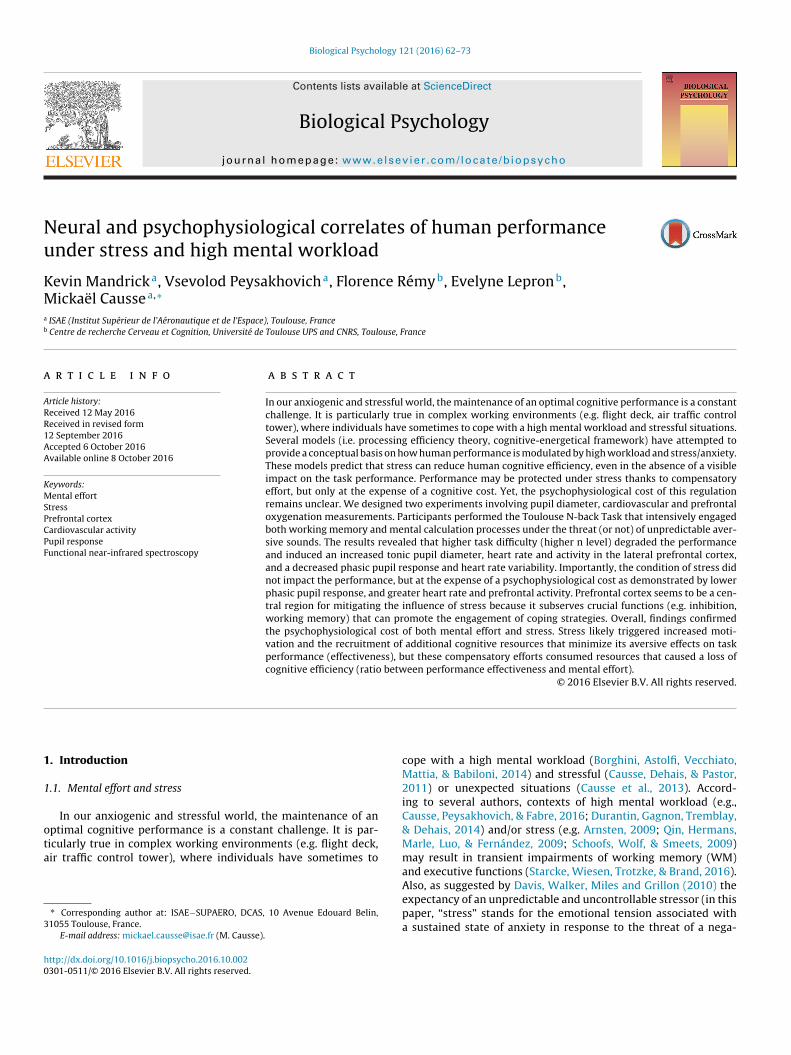
The new n-back task, called TNT, was developed for this studyin order to combine a classical n-back task with mental arithmetic(Fig. 1). Instead of memorizing and comparing unique items, as inthe classical n-back task, the participants had to memorize and tocompare the results of arithmetic operations, computed before-hand. In each trial, volunteers were required to compute the resultand compare it with either a fixed number (0-back) or the resultobtained 1 (1-back) or 2 (2-back) trials before. Arithmetic opera-tions were either additions or subtractions, of which all summandswere a multiple of 5 (e.g., 15 + 40, 90–35). Therefore, WM loadwas variable between conditions, with the 2-back task generatingthe highest load. During the resting periods, screens with “00 + 00”operations were presented and volunteers did not have to give anyresponse.
Operations were displayed in the center of a gray background.Participants were given a 2-button Cedrus response pad (RB-740,Cedrus Corporation, San Pedro, CA) and were asked to press eithera green button if the result matched the target number or a redbutton if not. Participants had to give their response as quickly aspossible. The task was implemented in Matlab (MathWorks) usingthe Psychophysics Toolbox (Psychtoolbox 3, Kleiner, Brainard, &Pelli, 2007).
2.3. Threat using auditory stressors
Stress (i.e. sustained anxiety) was induced with the threat ofunpredictable loud sounds (Patel et al., 2015). The auditory stress-ors occurrence was non-contingent upon the task performance.
We created a set of 34 aversive sounds inspired from the litera-ture (Grillon, Pine, Lissek, Rabin, & Vythilingam, 2009; Hirano et al.,2006; Kumar, Forster, Bailey, & Griffiths, 2008; Zald & Pardo, 2002).The sounds were perceived as mildly stressful, uncomfortable and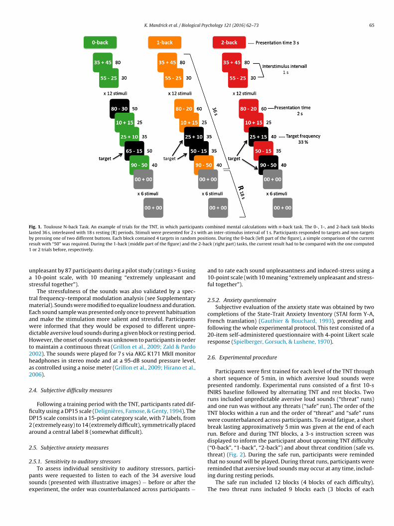
K. Mandrick et al. / Biological Psychology 121 (2016) 62–73 65
Fig. 1. Toulouse N-back Task. An example of trials for the TNT, in which participants combined mental calculations with n-back task. The 0-, 1-, and 2-back task blockslasted 36 s, interleaved with 18 s resting (R) periods. Stimuli were presented for 2 s with an inter-stimulus interval of 1 s. Participants responded to targets and non-targetsby pressing one of two different buttons. Each block contained 4 targets in random positions. During the 0-back (left part of the figure), a simple comparison of the currentr 2-ba1
uas
tmEawdHt2ha2
2
fiD2a
2
2
pse
esult with “50” was required. During the 1-back (middle part of the figure) and the or 2 trials before, respectively.
npleasant by 87 participants during a pilot study (ratings > 6 using 10-point scale, with 10 meaning “extremely unpleasant andtressful together”).
The stressfulness of the sounds was also validated by a spec-ral frequency–temporal modulation analysis (see Supplementary
aterial). Sounds were modified to equalize loudness and duration.ach sound sample was presented only once to prevent habituationnd make the stimulation more salient and stressful. Participantsere informed that they would be exposed to different unpre-ictable aversive loud sounds during a given block or resting period.owever, the onset of sounds was unknown to participants in order
o maintain a continuous threat (Grillon et al., 2009; Zald & Pardo002). The sounds were played for 7 s via AKG K171 MkII monitoreadphones in stereo mode and at a 95-dB sound pressure level,s controlled using a noise meter (Grillon et al., 2009; Hirano et al.,006).
.4. Subjective difficulty measures
Following a training period with the TNT, participants rated dif-culty using a DP15 scale (Delignières, Famose, & Genty, 1994). TheP15 scale consists in a 15-point category scale, with 7 labels, from
(extremely easy) to 14 (extremely difficult), symmetrically placedround a central label 8 (somewhat difficult).
.5. Subjective anxiety measures
.5.1. Sensitivity to auditory stressors
To assess individual sensitivity to auditory stressors, partici-ants were requested to listen to each of the 34 aversive loudounds (presented with illustrative images) − before or after thexperiment, the order was counterbalanced across participants −
ck (right part) tasks, the current result had to be compared with the one computed
and to rate each sound unpleasantness and induced-stress using a10-point scale (with 10 meaning “extremely unpleasant and stress-ful together”).
2.5.2. Anxiety questionnaireSubjective evaluation of the anxiety state was obtained by two
completions of the State-Trait Anxiety Inventory (STAI form Y-A,French translation) (Gauthier & Bouchard, 1993), preceding andfollowing the whole experimental protocol. This test consisted of a20-item self-administered questionnaire with 4-point Likert scaleresponse (Spielberger, Gorsuch, & Lushene, 1970).
2.6. Experimental procedure
Participants were first trained for each level of the TNT througha short sequence of 5 min, in which aversive loud sounds werepresented randomly. Experimental runs consisted of a first 10-sfNIRS baseline followed by alternating TNT and rest blocks. Tworuns included unpredictable aversive loud sounds (“threat” runs)and one run was without any threats (“safe” run). The order of theTNT blocks within a run and the order of “threat” and “safe” runswere counterbalanced across participants. To avoid fatigue, a shortbreak lasting approximatively 5 min was given at the end of eachrun. Before and during TNT blocks, a 3-s instruction screen wasdisplayed to inform the participant about upcoming TNT difficulty(“0-back”, “1-back”, “2-back”) and about threat condition (safe vs.threat) (Fig. 2). During the safe run, participants were remindedthat no sound will be played. During threat runs, participants were
reminded that aversive loud sounds may occur at any time, includ-ing during resting periods.The safe run included 12 blocks (4 blocks of each difficulty).The two threat runs included 9 blocks each (3 blocks of each

66 K. Mandrick et al. / Biological Psychology 121 (2016) 62–73
F uns for threat condition and one run for the safe condition. Run orders and blocks werec e played randomly during a threat run and could occur during resting period.
dbtt2eHT1
2
pnl
2
coteps5pcwifiderstendppotuuwa
Fig. 3. fNIRS headband. Location of the optodes on participant’s forehead with a
ig. 2. Experimental timeline. TNT experimental block design sessions with two rounterbalanced and pseudo-randomized. Unpredictable aversive loud sounds wer
ifficulty). For the analysis of the threat runs, we discarded thelocks containing sounds (6 blocks in total) to exclude any poten-ial effect of sound distraction and to focus on the stress relatedo the expectancy of the unpredictable sounds (Clarke & Johnstone,013). We thus compared the 12 threat blocks in which participantsxpected, but did not actually hear sounds, with the 12 safe blocks.ence, 4 blocks (4 × 12 trials) per condition (0-, 1-, 2-back x Safe,hreat) were included in the analyses. For the pupillary analyses,2 blocks of resting periods (12 × 6 trials) were also included.
.7. Behavioral measurements
TNT accuracy was calculated using the d-prime measure, com-uted as follows: z(hit rate) − z(false alarm rate). Additionally, theumber of misses (no response) was computed, this latter measure
ikely reflecting an exhaustion of cognitive resources.
.8. Study 1: Pupillometry measurements
Participants were seated at approximately 70 cm from a 22′’omputer screen (1680 × 1250 pixels). Ambient luminance wasf 10 lx. During the whole experiment, participants’ gaze posi-ion and pupil diameter were tracked using a remote SMI RED500ye-tracker (SensoMotoric Instruments GmbH, Germany) at a sam-ling rate of 120 Hz. This device allows tracking the pupil despitemall head movements. Before each run, participants performed a-point calibration procedure validated with 4 additional fixationoints. The data acquisition routine used iViewX SDK to communi-ate with Matlab software. Periods of signal loss and blinks (bothere marked as zeros by the eye-tracking system) were linearly
nterpolated. Then the signal was filtered with a two-pass 9-pointlter (low-pass with a cutoff frequency of 5.9 Hz). For tonic pupiliameter analyses, we used the median pupil diameter value forach block. We also conducted (event-related) phasic pupillaryesponse analyses. For this analysis, the pupillary recordings wereegregated in 3-s segments corresponding to each trial. For all par-icipants, an average phasic pupillary response was obtained forach condition. A trial was excluded if the total duration of sig-al loss or blinking exceeded 50% (1.5 s) or if median gaze positionuring the trial deviated from the screen center of more than 150ixels. An average of 44.6 (SD = 8.4) trials out of 48 were validateder experimental condition. An average of 58.1 (SD = 14.6) trials outf 72 were validated for resting periods. The number of validatedrials was non-dependent on the condition (p > 0.05). Median val-
es calculated on the 500-ms period preceding the trial onset weresed as baselines. For statistical analyses of phasic pupil response,e used the maximum pupil diameter in the interval between 1nd 2 s post-stimulus. Resting periods were included in the pupil-
flexible fNIRS sensor pad labeled from channel 1 to channel 16. Emitters are markedas E and D indicates detectors. Areas underlying the channel are approximately overBrodmann’s areas 10 and 46.
lometry analyses to confirm that the pupillary reaction was due tothe psychophysiological phenomenon and not visual factors. Thir-teen participants were included in the pupillary analyses (pupildata from one participant were discarded due to technical issuesduring the recording).
2.9. Study 2: Functional near infrared spectroscopymeasurements
To illuminate the forehead, a CW fNIRS 16-channel headbandmodel 100 fNIRS system (fNIRS Devices LLC, Photomac MD; http://www.fnirdevices.com) was used to obtain raw light intensity byspecific dual wavelengths of 730 nm and 850 nm (Fig. 3). Data wereacquired at a frequency of 2 Hz for all 16 channels.
At the beginning of the experiment, participants were equippedwith the fNIRS headband. The 10 s baseline was taken at rest witheyes closed. fNIRS-PFC activity was recorded through the entireexperiment. COBI Studio software (Drexel University) was usedfor data acquisition and visualization of the 16 channels then theversion 4.0 of fnirSoft software package was used for filtering,converting and analyzing data (Ayaz, 2010). First, the raw opticaldensity signals were converted to concentration changes of oxy-genated hemoglobin (HbO2) and deoxygenated hemoglobin (HHb)using the modified Beer-lambert law. fNIRS data were then band-pass filtered using a FIR filter of order 20 and cutoff frequencies of0.01 and 0.1 Hz. No detrending was applied. Then, available optodeswere averaged in 3 regions of interest for each participant; left pre-
frontal cortex (optodes 1–6), frontopolar prefrontal cortex (optodes7–10), right prefrontal cortex (optodes 11–16). Eight participantshad missing fNIRS data in a few medial optodes for at least oneexperimental condition due to poor contact with the forehead (i.e.
al Psy
otwdwmfic
tsoo2ye
2
rsAwawkoSTce
NKcwafanrsTcar
Fa
K. Mandrick et al. / Biologic
ptodes 5, 7–9, and 10). At most, 3 optodes were missing for 1 par-icipant, thus a measure of the frontopolar prefrontal cortex activityas possible for these 8 participants. To dissociate effects of TNTifficulty (0-back vs. 1-back vs. 2-back) and threat (safe vs. threat),e extracted the fNIRS response from each block. Signals were nor-alized towards zero by subtracting the current signal with the
rst data point. Then, signals were averaged on all trials for eachondition.
We calculated the blood-oxygenation response determined ashe difference between HbO2 and HHb signals. For fNIRS data analy-is, we compared the blood-oxygenation response with the analysisf the response amplitude (the difference between the mean valuef the first and the last 10 s of each block) (Mandrick et al., 2013a,013b). Seventeen participants were included in the fNIRS anal-ses (fNIRS data of three participants were excluded, one due toxcessively noisy signal and two due to recording problems).
.10. Cardiovascular activity
Cardiovascular activity (by 1-lead ECG) was continuouslyecorded in both studies. Signals were sampled at 500 Hz in aynchronous manner using a Biopac MP150 Hardware and BiopaccqKnowledge 4.1.1 Software (Biopac Systems Inc., CA, USA). Signalas derived into RR intervals to assess the HR. Data sets were visu-
lly controlled for outliers and artifacts. Analyses were conductedith Kubios HRV software 2.2 (University of Eastern Finland, http://
ubios.uef.fi). We investigated total HRV via the root mean squaref successive differences (RMSSD) (Task Force of the Europeanociety of Cardiology the North American Society of Pacing, 1996).hirty-three participants were included for the analysis of theardiovascular activity (HR and HRV data of one participant wasxcluded due to excessively noisy signal).
Data analysisResults were analyzed using Statistica software (StatSoft).
ormality and homoscedasticity of data were assessed usingolmogorov-Smirnov and Lilliefors tests, respectively. Pre −postomparisons of STAI scores and prefrontal oxygenation responsesere carried out using Student paired t-test. Other data were
nalyzed using repeated-measures analyses of variance (ANOVA)or normally distributed variables (i.e. pupillometry, HR/HRV,nd fNIRS measurements) and Friedman’s ANOVA test for non-ormally distributed variables (i.e. DP15, d-prime, and% noesponse). In the case of significant main effect or interaction,ignificant differences between conditions were identified using
ukey’s HSD posthoc tests for normal distribution or Wilcoxonomparisons test for non-normal distribution. p-values weredjusted for multiple comparisons with the Holm-Bonferroni cor-ection (Holm, 1979). A significance level of p < 0.05 was used for allig. 4. Behavioral performance. TNT scores for each level of difficulty (0-, 1-, and 2-back)s z(hit rate) − z(false alarm rate). Right- the percentage of “no response”. n = 34.
chology 121 (2016) 62–73 67
comparisons. Effect sizes were reported using partial eta-squared(�p
2).
3. Results
3.1. Subjective difficulty ratings
There was no significant difference between Study 1 and 2(p > 0.05) (Mann-Whitney U test for each variable; p > 0.05) regard-ing the perceived difficulty during the familiarization session.Therefore, perceived difficulty data were pooled across studiesand statistics were also calculated for 34 participants. DP15 rat-ings significantly increased with TNT difficulty (n level) (Friedman’schi-square ANOVA = 65.5; p < 0.001), with ratings being higher for1-back vs. 0-back (Z = 5.03; p < 0.001), for 2-back vs. 0-back (Z = 5.08;p < 0.001), and for 2-back vs. 1-back (Z = 5.01; p < 0.001). The 0-backtask was rated as “very easy” (average DP15 = 4.5 ± 2), the 1-backtask as “somewhat difficult” (average DP15 = 7.8 ± 1.9), and the 2-back task as “very difficult” (average DP15 = 11.9 ± 1.6).
3.2. Subjective anxiety ratings
Subjective ratings of induced stress for the 34 aversive loudsounds are given in Supplementary material. Based on this sub-jective report, a mean rating of 5.4 ± 2.4 was observed, confirmingthat sounds were mildly stressful.
There was no significant difference between Study 1 and 2 (t-test for each variable; p > 0.05), therefore, anxiety ratings data werepooled across studies and statistics were calculated for 34 par-ticipants. Participants’ anxiety score measured by STAI Y-A weresignificantly higher at the end of the protocol, relative to the begin-ning (29.1 ± 5.9 versus 33.3 ± 7.1, respectively; t = 3.85; p < 0.001).This outcome shows that the experiment produced an increasedanxiety.
3.3. Behavioral data
There was no significant difference between Study 1 and 2regarding the behavioral performance (Mann-Whitney U test foreach variable; p > 0.05). Therefore, behavioral data were also pooledacross studies and statistics were calculated for 34 participants.Cognitive scores for each n-back condition were assessed using d-prime and percentage of miss (“no response”) (Fig. 4). Participantsshowed lower d-prime scores with increased task difficulty (Fried-
man’s chi-square ANOVA = 115.7; p < 0.001). Post-hoc comparisonrevealed significant differences (p < 0.001) for all n-back level assummarized in Fig. 4. Similar results were observed for the per-centage of omitted responses, which monotonically increased withand threat condition (safe and threat). Error bars are S.D. Left- d-prime calculated

68 K. Mandrick et al. / Biological Psychology 121 (2016) 62–73
Fig. 5. Cardiovascular activity. Cardiovascular activity for each level of difficulty (0-, 1-, and 2-back) and threat condition (safe and threat). Error bars are S.E. Left- Heart rate.Right- Heart rate variability. n = 33.
FlE
tti
3
(apiaeh(ip(b
3
3
(tv
Fig. 7. Phasic pupil response. Grand-average of phasic pupil response aligned tothe stimulus onset for each level of difficulty (rest, 0-, 1-, and 2-back) and threat
ig. 6. Tonic pupil diameter. Bar height shows mean tonic pupil diameter for eachevel of difficulty (rest, 0-, 1-, and 2-back) and threat condition (safe and threat).rror bars represent S.E. n = 13.
ask difficulty (Friedman’s chi-square ANOVA = 57.9; p < 0.001). Thehreat of unpredictable auditory stressors did not impact behav-oral performance (p > 0.05).
.4. Cardiovascular activity
There was no significant difference between study 1 and 2p > 0.05) regarding cardiovascular activity. Therefore, HR data werelso pooled across studies and statistics were calculated for 33articipants. There was a significant elevation of the HR with
ncreased TNT difficulty (F2,62 = 20.5; p < 0.001; �p2 = 0.40), with
monotonical increased across each level of difficulty (small-st p-value = 0.019). HR was also impacted by stress, it wasigher under the threat of the unpredictable aversive soundsF1,32 = 4.35; p < 0.05; �p
2 = 0.12) (Fig. 5). There was also a signif-cant decrease in HRV (RMSSD) with task difficulty (F2,62 = 12.1;
< 0.001; �p2 = 0.28), HRV was smaller in 0-back vs. 1- and 2-back
smallest p-value < 0.001) (Fig. 5). Finally, HRV was not impactedy stress (p > 0.05).
.5. Pupillometry
.5.1. Tonic pupil diameter
Tonic pupil diameter significantly increased with task difficultyF3,36 = 26.27; p < 0.001; �p2 = 0.69) (Fig. 6). Post-hoc tests showed
hat pupil diameter was significantly higher during 1- and 2-backs. resting period (p < 0.01 and p < 0.001 respectively) and signifi-
condition (safe and threat). Vertical lines indicate the time interval used for peakcomputation. n = 13.
cantly higher during 2-back compared to 0- and 1-back (p < 0.001in both comparisons). There was no effect of threat on tonic pupildiameter (F1,12 = 0.75; �p
2 = 0.06), neither any difficulty x threatinteraction (F3,36 = 1.27; �p
2 = 0.10).
3.5.2. Phasic pupil responsePhasic pupil response amplitudes (Fig. 7) significantly decreased
with n-back task difficulty (F3,36 = 12.05; p < 0.001; �p2 = 0.50).
More precisely, a significantly higher dilation was observed in 0-, 1-, and 2-back tasks compared to rest (p < 0.001, p < 0.01 and p < 0.05respectively) and dilation was greater in 0-back vs. 2-back (p < 0.05)conditions. Smaller amplitudes were observed in threat conditions(F1,12 = 6.38; p < 0.05; �p
2 = 0.35). There was no threat x difficultyinteraction (F3,36 = 0.33; p > 0.05; �p
2 = 0.03). We conducted anadditional analysis in order to assess the potential influence ofthe pre-stimulus baseline (median of pre-stimulus 500 ms, usedto compute the phasic response) on the results. There was an effectof n-back task difficulty (F3,36 = 25.38; p < 0.001; �p
2 = 0.68), but no2
any effect of threat (F1,12 = 0.15; �p = 0.01) or threat x difficultyinteraction (F3,36 = 0.56; �p2 = 0.04) on pupil diameter during the
pre-stimulus baseline.

K. Mandrick et al. / Biological Psychology 121 (2016) 62–73 69
F wherea
3
Ffismbtwdpnvsithtpss02swiie
4
bacgpie
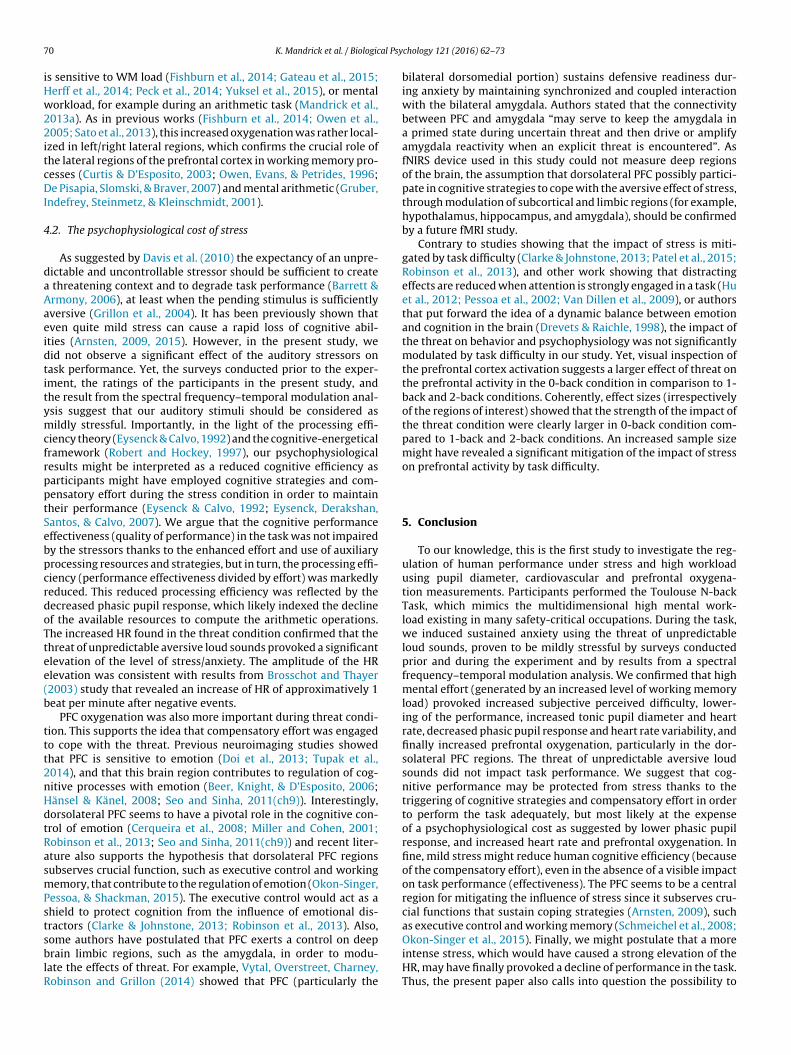
ig. 8. Brain topography maps. Anterior view of the frontal cortex showing regions
nd 2-back) and threat condition (safe and threat). n = 17.
.6. Prefrontal cortex oxygenation changes
Brain topography of PFC blood oxygenation is illustrated inig. 8. PFC blood oxygenation was dependent on n-back task dif-culty (F2,34 = 4.14; p < 0.05; �p
2 = 0.20). More precisely, it wasignificantly higher in 1-back vs. 0-back condition (p < 0.05) andarginally higher in 2-back vs. 0-back condition (p = 0.06). PFC
lood oxygenation was not different in 1-back vs. 2-back condi-ion (p = 0.96). The task difficulty x region of interest interactionas significant (F4,68 = 3.02; p < 0.05; �p
2 = 0.15). HSD revealed thatifficulty had no significant effect on the frontopolar region of therefrontal cortex (p > 0.05); on the contrary, oxygenation was sig-ificantly higher in the left and right prefrontal cortex in 1-backs. 0-back and in 2-back vs. 0-back (smallest p-value = 0.001). Ashowed Fig. 8, PFC blood oxygenation was also significantly highern threat vs. safe conditions (F1,17 = 4.81; p < 0.05; �p
2 = 0.22). Theask difficulty x stress interaction was not significant (p > 0.05),owever, the visual inspection of Fig. 8 highlights a larger effect ofhreat on the prefrontal activity in the 0-back condition in com-arison to the 1-back and 2-back conditions. Coherently, effectizes (independently of the regions of interest) showed that thetrength of the impact of the threat condition was clearly larger in-back condition (�p
2 = 0.39) compared to 1-back (�p2 = 0.04) and
-back conditions (�p2 = 0.04). It suggests that increasing sample
ize might reveal a mitigation of the effect of stress with increasingorking memory load. HR was not correlated with prefrontal activ-
ty in none of the experimental conditions (smallest p-value > 0.05),ncreased prefrontal oxygenation cannot be associated with a gen-ral increase of the systemic cardiovascular activity.
. Discussion
Using a novel mental arithmetic n-back task (i.e. Toulouse N-ack Task), we examined in two separate studies how mental effortnd stress impacted task performance, pupil response (study 1),ardiovascular activity (study 1 and 2), and prefrontal cortex oxy-
enation (study 2). Higher task difficulty (higher n level) increasederceived difficulty, degraded the performance and provoked anncreased tonic pupil diameter, HR and oxygenation in the lat-ral prefrontal cortex, and a decreased phasic pupil response and
PFC blood oxygenation (HbO2 − HHb) increased with n-back task difficulty (0-, 1-,
HRV. Importantly, the condition of stress did not impact the per-formance, but at the expense of a psychophysiological cost asdemonstrated by lower phasic pupil response, increased HR andgreater prefrontal oxygenation. These findings confirmed the psy-chophysiological cost of both mental effort and stress and supportthe common idea of the processing efficiency theory (Eysenck andCalvo, 1992) and the cognitive-energetical framework (Robert andHockey, 1997), namely, the fact that stress can reduce human cog-nitive efficiency, even in the absence of a visible impact on the taskperformance. Stress likely triggered increased motivation and therecruitment of additional cognitive resources that minimized itsaversive effects on task performance, but this was associated witha loss of cognitive efficiency.
4.1. The psychophysiological cost of mental effort
The increase of subjective perceived difficulty, the decreaseof performance (d-prime score) and the increase in the percent-age of missed responses across the three n-back levels confirmedthat mental effort was efficiently modulated. This higher difficultycaused an increased tonic pupil diameter and HR and a decreasedphasic pupil response and HRV. Classically, higher tonic pupil diam-eter (Beatty, 1982; Causse, Sénard, Démonet, & Pastor, 2010; Reiner& Gelfeld, 2014; Peysakhovich, Causse, Scannella, & Dehais, 2015),increased HR, and decreased HRV (Riese, 1999; Causse et al., 2011a)are found under conditions of high load. Interestingly, the phasicpupil response diminished with increased difficulty, in a similarfashion than the commonly observed reduced amplitude of elec-troencephalography event-related potentials when resources areconsumed by a high mental workload (Van Dillen & Derks, 2012;Giraudet, St-Louis, Scannella, & Causse, 2015; Causse et al., 2016).Contrary to tonic pupil diameter that is used to measure men-tal effort throughout the duration of a task, event-related phasicchanges in pupil diameter relate to acute task demands (Brunyéet al., 2016). As tonic load on memory increased with the n level,it is likely that fewer resources were available to process the arith-
metic operations which in turn provoked a reduced phasic pupilresponse to these operations.The TNT increased difficulty also yielded greater PFC oxygena-tion. This is consistent with previous studies showing that fNIRS
7 al Psy
iHw22itcDI
4
daAaeidtitymcfrpptSebpcrdoTtee(b
ttt2nHdtRasmPstsblR
0 K. Mandrick et al. / Biologic
s sensitive to WM load (Fishburn et al., 2014; Gateau et al., 2015;erff et al., 2014; Peck et al., 2014; Yuksel et al., 2015), or mentalorkload, for example during an arithmetic task (Mandrick et al.,
013a). As in previous works (Fishburn et al., 2014; Owen et al.,005; Sato et al., 2013), this increased oxygenation was rather local-
zed in left/right lateral regions, which confirms the crucial role ofhe lateral regions of the prefrontal cortex in working memory pro-esses (Curtis & D’Esposito, 2003; Owen, Evans, & Petrides, 1996;e Pisapia, Slomski, & Braver, 2007) and mental arithmetic (Gruber,
ndefrey, Steinmetz, & Kleinschmidt, 2001).
.2. The psychophysiological cost of stress
As suggested by Davis et al. (2010) the expectancy of an unpre-ictable and uncontrollable stressor should be sufficient to create
threatening context and to degrade task performance (Barrett &rmony, 2006), at least when the pending stimulus is sufficientlyversive (Grillon et al., 2004). It has been previously shown thatven quite mild stress can cause a rapid loss of cognitive abil-ties (Arnsten, 2009, 2015). However, in the present study, weid not observe a significant effect of the auditory stressors onask performance. Yet, the surveys conducted prior to the exper-ment, the ratings of the participants in the present study, andhe result from the spectral frequency–temporal modulation anal-sis suggest that our auditory stimuli should be considered asildly stressful. Importantly, in the light of the processing effi-
iency theory (Eysenck & Calvo, 1992) and the cognitive-energeticalramework (Robert and Hockey, 1997), our psychophysiologicalesults might be interpreted as a reduced cognitive efficiency asarticipants might have employed cognitive strategies and com-ensatory effort during the stress condition in order to maintainheir performance (Eysenck & Calvo, 1992; Eysenck, Derakshan,antos, & Calvo, 2007). We argue that the cognitive performanceffectiveness (quality of performance) in the task was not impairedy the stressors thanks to the enhanced effort and use of auxiliaryrocessing resources and strategies, but in turn, the processing effi-iency (performance effectiveness divided by effort) was markedlyeduced. This reduced processing efficiency was reflected by theecreased phasic pupil response, which likely indexed the declinef the available resources to compute the arithmetic operations.he increased HR found in the threat condition confirmed that thehreat of unpredictable aversive loud sounds provoked a significantlevation of the level of stress/anxiety. The amplitude of the HRlevation was consistent with results from Brosschot and Thayer2003) study that revealed an increase of HR of approximatively 1eat per minute after negative events.
PFC oxygenation was also more important during threat condi-ion. This supports the idea that compensatory effort was engagedo cope with the threat. Previous neuroimaging studies showedhat PFC is sensitive to emotion (Doi et al., 2013; Tupak et al.,014), and that this brain region contributes to regulation of cog-itive processes with emotion (Beer, Knight, & D’Esposito, 2006;änsel & Känel, 2008; Seo and Sinha, 2011(ch9)). Interestingly,orsolateral PFC seems to have a pivotal role in the cognitive con-rol of emotion (Cerqueira et al., 2008; Miller and Cohen, 2001;obinson et al., 2013; Seo and Sinha, 2011(ch9)) and recent liter-ture also supports the hypothesis that dorsolateral PFC regionsubserves crucial function, such as executive control and workingemory, that contribute to the regulation of emotion (Okon-Singer,
essoa, & Shackman, 2015). The executive control would act as ahield to protect cognition from the influence of emotional dis-ractors (Clarke & Johnstone, 2013; Robinson et al., 2013). Also,
ome authors have postulated that PFC exerts a control on deeprain limbic regions, such as the amygdala, in order to modu-ate the effects of threat. For example, Vytal, Overstreet, Charney,obinson and Grillon (2014) showed that PFC (particularly the
chology 121 (2016) 62–73
bilateral dorsomedial portion) sustains defensive readiness dur-ing anxiety by maintaining synchronized and coupled interactionwith the bilateral amygdala. Authors stated that the connectivitybetween PFC and amygdala “may serve to keep the amygdala ina primed state during uncertain threat and then drive or amplifyamygdala reactivity when an explicit threat is encountered”. AsfNIRS device used in this study could not measure deep regionsof the brain, the assumption that dorsolateral PFC possibly partici-pate in cognitive strategies to cope with the aversive effect of stress,through modulation of subcortical and limbic regions (for example,hypothalamus, hippocampus, and amygdala), should be confirmedby a future fMRI study.
Contrary to studies showing that the impact of stress is miti-gated by task difficulty (Clarke & Johnstone, 2013; Patel et al., 2015;Robinson et al., 2013), and other work showing that distractingeffects are reduced when attention is strongly engaged in a task (Huet al., 2012; Pessoa et al., 2002; Van Dillen et al., 2009), or authorsthat put forward the idea of a dynamic balance between emotionand cognition in the brain (Drevets & Raichle, 1998), the impact ofthe threat on behavior and psychophysiology was not significantlymodulated by task difficulty in our study. Yet, visual inspection ofthe prefrontal cortex activation suggests a larger effect of threat onthe prefrontal activity in the 0-back condition in comparison to 1-back and 2-back conditions. Coherently, effect sizes (irrespectivelyof the regions of interest) showed that the strength of the impact ofthe threat condition were clearly larger in 0-back condition com-pared to 1-back and 2-back conditions. An increased sample sizemight have revealed a significant mitigation of the impact of stresson prefrontal activity by task difficulty.
5. Conclusion
To our knowledge, this is the first study to investigate the reg-ulation of human performance under stress and high workloadusing pupil diameter, cardiovascular and prefrontal oxygena-tion measurements. Participants performed the Toulouse N-backTask, which mimics the multidimensional high mental work-load existing in many safety-critical occupations. During the task,we induced sustained anxiety using the threat of unpredictableloud sounds, proven to be mildly stressful by surveys conductedprior and during the experiment and by results from a spectralfrequency–temporal modulation analysis. We confirmed that highmental effort (generated by an increased level of working memoryload) provoked increased subjective perceived difficulty, lower-ing of the performance, increased tonic pupil diameter and heartrate, decreased phasic pupil response and heart rate variability, andfinally increased prefrontal oxygenation, particularly in the dor-solateral PFC regions. The threat of unpredictable aversive loudsounds did not impact task performance. We suggest that cog-nitive performance may be protected from stress thanks to thetriggering of cognitive strategies and compensatory effort in orderto perform the task adequately, but most likely at the expenseof a psychophysiological cost as suggested by lower phasic pupilresponse, and increased heart rate and prefrontal oxygenation. Infine, mild stress might reduce human cognitive efficiency (becauseof the compensatory effort), even in the absence of a visible impacton task performance (effectiveness). The PFC seems to be a centralregion for mitigating the influence of stress since it subserves cru-cial functions that sustain coping strategies (Arnsten, 2009), suchas executive control and working memory (Schmeichel et al., 2008;
Okon-Singer et al., 2015). Finally, we might postulate that a moreintense stress, which would have caused a strong elevation of theHR, may have finally provoked a decline of performance in the task.Thus, the present paper also calls into question the possibility to
al Psy
su
A
pVr
C
ac
A
AAd
A
t1
R
A
A
A
A
B
B
B
B
B
B
B
B
B
B
K. Mandrick et al. / Biologic
tudy visible alteration of the performance under stress with these of the threat of aversive loud sounds.
uthor contributions
MK, VP, LE, RF, and CM designed the research, and MK and VPerformed the experiments. MK and VP analyzed the data. MK, CM,P prepared the figures and wrote the manuscript. LE, RF, and CMeviewed the manuscript.
onflict of interest statement
The authors declare that the research was conducted in thebsence of any commercial or financial relationships that could beonstrued as a potential conflict of interest.
cknowledgements
This work was supported by the French Research Nationalgency and the French Defence Procurement Agency via theccompagnement Spécifique des travaux de Recherches et’Innovation Défense (ASTRID).
ppendix A. Supplementary data
Supplementary data associated with this article can be found, inhe online version, at http://dx.doi.org/10.1016/j.biopsycho.2016.0.002.
eferences
ndreassi, J. L. (2000). Pupillary response and behavior. psychophysiology: Humanbehavior & physiological response. pp. 218–233. Mahwah, NJ: LawrenceErlbaum Associates.
rnsten, A. F. T. (2009). Stress signalling pathways that impair prefrontal cortexstructure and function. Nature Review Neuroscience, 10(6), 410–422. http://dx.doi.org/10.1038/nrn2648
rnsten, A. F. T. (2015). Stress weakens prefrontal networks: Molecular insults tohigher cognition. Nature Neuroscience, 18(10), 1376–1385. http://dx.doi.org/10.1038/nn.4087
yaz, H. (2010). Functional near infrared spectroscopy based brain computer interfacePhD thesis. Philadelphia, PA: Drexel University.
arrett, J., & Armony, J. L. (2006). The influence of trait anxiety on autonomicresponse and cognitive performance during an anticipatory anxiety task.Depression and Anxiety, 23(4), 210–219. http://dx.doi.org/10.1002/da.20143
eatty, J. (1982). Task-evoked pupillary responses, processing load, and thestructure of processing resources. Psychological Bulletin, 91(2), 276–292. http://dx.doi.org/10.1037/0033-2909.91.2.276
eer, J. S., Knight, R. T., & D’Esposito, M. (2006). Controlling the integration ofemotion and cognition the role of frontal cortex in distinguishing helpful fromhurtful emotional information. Psychological Science, 17(5), 448–453. http://dx.doi.org/10.1111/j.1467-9280.2006.01726.x
erggren, N., Richards, A., Taylor, J., & Derakshan, N. (2013). Affective attentionunder cognitive load: Reduced emotional biases but emergent anxiety-relatedcosts to inhibitory control. [Original Research]. Frontiers in HumanNeuroscience, 7(188) http://dx.doi.org/10.3389/fnhum.2013.00188
itsios, P., Szabadi, E., & Bradshaw, C. M. (1998). Sensitivity of the fear-inhibitedlight reflex to diazepam. Psychopharmacology, 135(1), 93–98.
orghini, G., Astolfi, L., Vecchiato, G., Mattia, D., & Babiloni, F. (2014). Measuringneurophysiological signals in aircraft pilots and car drivers for the assessmentof mental workload, fatigue and drowsiness. Neuroscience & BiobehavioralReviews, 44, 58–75. http://dx.doi.org/10.1016/j.neubiorev.2012.10.003
radley, M. M., Miccoli, L., Escrig, M. A., & Lang, P. J. (2008). The pupil as a measureof emotional arousal and autonomic activation. Psychophysiology, 45(4),602–607.
reier, A., Albus, M., Pickar, D., Zahn, T. P., Wolkowitz, O. M., & Paul, S. M. (1987).Controllable and uncontrollable stress in humans: Alterations in mood andneuroendocrine and psychophysiological function. The American Journal ofPsychiatry, 144(11), 1419–1425.
rosschot, J. F., & Thayer, J. F. (2003). Heart rate response is longer after negative
emotions than after positive emotions. International Journal ofPsychophysiology, 50(3), 181–187. http://dx.doi.org/10.1016/S0167-8760(03)00146-6runyé, T. T., Eddy, M. D., Mercan, E., Allison, K. H., Weaver, D. L., & Elmore, J. G.(2016). Pupil diameter changes reflect difficulty and diagnostic accuracy
chology 121 (2016) 62–73 71
during medical image interpretation. BMC Medical Informatics and DecisionMaking, 16(1), 77. http://dx.doi.org/10.1186/s12911-016-0322-3
Cacioppo, J. T., & Tassinary, L. G. (1990). Inferring psychological significance fromphysiological signals. American Psychologist, 45(1), 16. http://dx.doi.org/10.1037/0003-066X.45.1.16
Causse, M., Sénard, J.-M., Démonet, J. F., & Pastor, J. (2010). Monitoring cognitiveand emotional processes through pupil and cardiac response during dynamicversus logical task. Applied Psychophysiology and Biofeedback, 35(2), 115–123.http://dx.doi.org/10.1007/s10484-009-9115-0
Causse, M., Baracat, B., Pastor, J., & Dehais, F. (2011). Reward and uncertainty favorrisky decision-making in pilots: Evidence from cardiovascular and oculometricmeasurements. Applied Psychophysiology and Biofeedback, 36(4), 231–242.http://dx.doi.org/10.1007/s10484-011-9163-0
Causse, M., Dehais, F., & Pastor, J. (2011). Executive functions and pilotcharacteristics predict flight simulator performance in general aviation pilots.The International Journal of Aviation Psychology, 21(3), 217–234. http://dx.doi.org/10.1080/10508414.2011.582441
Causse, M., Péran, P., Dehais, F., Caravasso, C. F., Zeffiro, T., Sabatini, U., & Pastor, J.(2013). Affective decision making under uncertainty during a plausibleaviation task: An fMRI study. Neuroimage, 71, 19–29. http://dx.doi.org/10.1016/j.neuroimage.2012.12.060
Causse, M., Peysakhovich, V., & Fabre, E. F. (2016). High working memory loadimpairs language processing during a simulated piloting task: An ERP andpupillometry study. Frontiers in Human Neuroscience, 10 http://dx.doi.org/10.3389/fnhum.2016.00240
Cerqueira, J. J., Almeida, O. F. X., & Sousa, N. (2008). The stressed prefrontal cortex.left? right!. Brain, Behavior, and Immunity, 22(5), 630–638. http://dx.doi.org/10.1016/j.bbi.2008.01.005
Clarke, R. J., & Johnstone, T. (2013). Prefrontal inhibition of threat processingreduces working memory interference. Frontiers in Human Neuroscience, 7, 228.http://dx.doi.org/10.3389/fnhum.2013.00228
Cohen, N., Moyal, N., & Henik, A. (2015). Executive control suppresses pupillaryresponses to aversive stimuli. Biological Psychology, 112, 1–11. http://dx.doi.org/10.1016/j.biopsycho.2015.09.006
Curtis, C. E., & D’Esposito, M. (2003). Persistent activity in the prefrontal cortexduring working memory. Trends in Cognitive Sciences, 7(9), 415–423. http://dx.doi.org/10.1016/S1364-6613(03)00197-9
Davis, M., Walker, D. L., Miles, L., & Grillon, C. (2010). Phasic vs sustained fear inrats and humans: Role of the extended amygdala in fear vs anxiety.Neuropsychopharmacology, 35(1), 105–135. http://dx.doi.org/10.1038/npp.2009.109
De Pisapia, N., Slomski, J. A., & Braver, T. S. (2007). Functional specializations inlateral prefrontal cortex associated with the integration and segregation ofinformation in working memory. Cerebral Cortex, 17(5), 993–1006. http://dx.doi.org/10.1093/cercor/bhl010
Delignières, D., Famose, J. P., & Genty, J. (1994). Validation of a scale for theassessment of perceived task difficulty. Staps, 34, 77–88.
Doi, H., Nishitani, S., & Shinohara, K. (2013). NIRS as a tool for assaying emotionalfunction in the prefrontal cortex. Frontiers in Human Neuroscience, 7, 770.http://dx.doi.org/10.3389/fnhum.2013.00770
Drevets, W. C., & Raichle, M. E. (1998). Suppression of regional cerebral bloodduring emotional versus higher cognitive implications for interactionsbetween emotion and cognition. Cognition and Emotion, 12(3), 353–385. http://dx.doi.org/10.1080/026999398379646
Durantin, G., Gagnon, J.-F., Tremblay, S., & Dehais, F. (2014). Using near infraredspectroscopy and heart rate variability to detect mental overload. BehaviouralBrain Research, 259, 16–23. http://dx.doi.org/10.1016/j.bbr.2013.10.042
Eysenck, M. W., & Calvo, M. G. (1992). Anxiety and performance: The processingefficiency theory. Cognition & Emotion, 6(6), 409–434. http://dx.doi.org/10.1080/02699939208409696
Eysenck, M. W., Derakshan, N., Santos, R., & Calvo, M. G. (2007). Anxiety andcognitive performance: Attentional control theory. Emotion, 7(2), 336. http://dx.doi.org/10.1037/1528-3542.7.2.336
Fishburn, F. A., Norr, M. E., Medvedev, A. V., & Vaidya, C. J. (2014). Sensitivity offNIRS to cognitive state and load. Frontiers in Human Neuroscience, 8, 76. http://dx.doi.org/10.3389/fnhum.2014.00076
Gateau, T., Durantin, G., Lancelot, F., Scannella, S., & Dehais, F. (2015). Real-Timestate estimation in a flight simulator using fNIRS. PLoS One, 10(3) http://dx.doi.org/10.1371/journal.pone.0121279
Gauthier, J., & Bouchard, S. (1993). Adaptation canadienne-franc aise de la formerévisée du State–Trait Anxiety Inventory de Spielberger. Canadian Journal ofBehavioural Science/Revue Canadienne Des Sciences Du Comportement, 25(4),559.
Giraudet, L., St-Louis, M.-E., Scannella, S., & Causse, M. (2015). P300 event-relatedpotential as an indicator of inattentional deafness? PLoS One, 10(2), e0118556.http://dx.doi.org/10.1371/journal.pone.0118556
Grillon, C., Baas, J. P., Lissek, S., Smith, K., & Milstein, J. (2004). Anxious responses topredictable and unpredictable aversive events. Behavioral Neuroscience, 118(5),916–924. http://dx.doi.org/10.1037/0735-7044.118.5.916
Grillon, C., Pine, D. S., Lissek, S., Rabin, S., & Vythilingam, M. (2009). Increasedanxiety during anticipation of unpredictable aversive stimuli in posttraumatic
stress disorder but not in generalized anxiety disorder. Biological Psychiatry,66(1), 47–53. http://dx.doi.org/10.1016/j.biopsych.2008.12.028Gruber, O., Indefrey, P., Steinmetz, H., & Kleinschmidt, A. (2001). Dissociatingneural correlates of cognitive components in mental calculation. CerebralCortex, 11(4), 350–359. http://dx.doi.org/10.1093/cercor/11.4.350

7 al Psy
H
H
H
H
H
K
KK
K
L
L
L
L
M
M
M
M
M
O
O
O
P
P
P
P
P
human-computer interfaces (Vol. 9319, p. 93190R-93190R-9). http://dx.doi.org/10.1117/12.2075929
2 K. Mandrick et al. / Biologic
änsel, A., & Känel, R. (2008). The ventro-medial prefrontal cortex: A major linkbetween the autonomic nervous system, regulation of emotion, and stressreactivity? BioPsychoSocial Medicine, 2(1), 21. http://dx.doi.org/10.1186/1751-0759-2-21
erff, C., Heger, D., Fortmann, O., Hennrich, J., Putze, F., & Schultz, T. (2014). Mentalworkload during n-back task—quantified in the prefrontal cortex using fNIRS.Frontiers in Human Neuroscience, 7, 935. http://dx.doi.org/10.3389/fnhum.2013.00935
irano, Y., Fujita, M., Watanabe, K., Niwa, M., Takahashi, T., Kanematsu, M., . . . &Onozuka, M. (2006). Effect of unpleasant loud noise on hippocampal activitiesduring picture encoding: An fMRI study. Brain and Cognition, 61(3), 280–285.http://dx.doi.org/10.1016/j.bandc.2006.02.003
olm, S. (1979). A simple sequentially rejective multiple test procedure.Scandinavian Journal of Statistics, 6(2), 65–70.
u, K., Bauer, A., Padmala, S., & Pessoa, L. (2012). Threat of bodily harm hasopposing effects on cognition. Emotion, 12(1), 28–32. http://dx.doi.org/10.1037/a0024345 [Washington, D.C.]
ahneman, D., & Beatty, J. (1966). Pupil diameter and load on memory. Science,154(3756), 1583–1585.
ahneman, D. (1973). Attention and effort. Prentice-Hall.leiner, M., Brainard, D., & Pelli. (2007). What’s new in psychtoolbox-3?
Perception, 36. ECVP Abstract Supplement.umar, S., Forster, H. M., Bailey, P., & Griffiths, T. D. (2008). Mapping
unpleasantness of sounds to their auditory representation. The Journal of theAcoustical Society of America, 124(6), 3810–3817. http://dx.doi.org/10.1121/1.3006380
aeng, B., Sirois, S., & Gredebäck, G. (2012). Pupillometry a window to thepreconscious? Perspectives on Psychological Science, 7(1), 18–27.
ane, R. D., McRae, K., Reiman, E. M., Chen, K., Ahern, G. L., & Thayer, J. F. (2009).Neural correlates of heart rate variability during emotion. Neuroimage, 44(1),213–222. http://dx.doi.org/10.1016/j.neuroimage.2008.07.056
evy, R., & Goldman-Rakic, P. S. (2000). Segregation of working memory functionswithin the dorsolateral prefrontal cortex Executive control and the frontal lobe:Current issues. pp. 23–32. Springer.
upien, S. J., Maheu, F., Tu, M., Fiocco, A., & Schramek, T. E. (2007). The effects ofstress and stress hormones on human cognition: Implications for the field ofbrain and cognition. Brain and Cognition, 65(3), 209–237. http://dx.doi.org/10.1016/j.bandc.2007.02.007
andrick, K., Derosiere, G., Dray, G., Coulon, D., Micallef, J.-P., & Perrey, S. (2013a).Prefrontal cortex activity during motor tasks with additional mental loadrequiring attentional demand: A near-infrared spectroscopy study.Neuroscience Research, 76(3), 156–162. http://dx.doi.org/10.1016/j.neures.2013.04.006
andrick, K., Derosiere, G., Dray, G., Coulon, D., Micallef, J.-P., & Perrey, S. (2013b).Utilizing slope method as an alternative data analysis for functionalnear-infrared spectroscopy-derived cerebral hemodynamic responses.International Journal of Industrial Ergonomics, 43(4), 335–341. http://dx.doi.org/10.1016/j.ergon.2013.05.003
cCraty, R., Atkinson, M., Tiller, W. A., Rein, G., & Watkins, A. D. (1995). The effectsof emotions on short-term power spectrum analysis of heart rate variability.The American Journal of Cardiology, 76(14), 1089–1093. http://dx.doi.org/10.1016/S0002-9149(99)80309-9
iller, E. K., & Cohen, J. D. (2001). An integrative theory of prefrontal cortexfunction. Annual Review of Neuroscience, 24(1), 167–202. http://dx.doi.org/10.1146/annurev.neuro.24.1.167
orinaga, K., Akiyoshi, J., Matsushita, H., Ichioka, S., Tanaka, Y., Tsuru, J., & Hanada,H. (2007). Anticipatory anxiety-induced changes in human lateral prefrontalcortex activity. Biological Psychology, 74(1), 34–38. http://dx.doi.org/10.1016/j.biopsycho.2006.06.005
kon-Singer, H., Hendler, T., Pessoa, L., & Shackman, A. J. (2015). The neurobiologyof emotion-cognition interactions: Fundamental questions and strategies forfuture research. Frontiers in Human Neuroscience, 9(58) http://dx.doi.org/10.3389/fnhum.2015.00058
wen, A. M., Evans, A. C., & Petrides, M. (1996). Evidence for a two-Stage model ofspatial working memory processing within the lateral frontal cortex: Apositron emission tomography study. Cerebral Cortex, 6(1), 31–38.
wen, A. M., McMillan, K. M., Laird, A. R., & Bullmore, E. (2005). N-back workingmemory paradigm: A meta-analysis of normative functional neuroimagingstudies. Human Brain Mapping, 25(1), 46–59. http://dx.doi.org/10.1002/hbm.20131
artala, T., & Surakka, V. (2003). Pupil size variation as an indication of affectiveprocessing. International Journal of Human-computer Studies, 59(1), 185–198.
atel, N., Vytal, K., Pavletic, N., Stoodley, C., Pine, D. S., Grillon, C., & Ernst, M.(2015). Interaction of threat and verbal working memory in adolescents.Psychophysiology, http://dx.doi.org/10.1111/psyp.12582
eck, E. M., Afergan, D., Yuksel, B. F., Lalooses, F., & Jacob, R. J. K. (2014). Using fNIRSto measure mental workload in the real world. In S. H. Fairclough, & K. Gilleade(Eds.), Advances in physiological computing (pp. 117–139). London: Springer[Consulté à l’adresse]. http://link.springer.com/chapter/10.1007/978-1-4471-6392-3 6
essoa, L., McKenna, M., Gutierrez, E., & Ungerleider, L. G. (2002). Neural
processing of emotional faces requires attention. Proceedings of the NationalAcademy of Sciences of the United States of America, 99(17), 11458–11463.http://dx.doi.org/10.1073/pnas.172403899eysakhovich, V., Causse, M., Scannella, S., & Dehais, F. (2015). Frequency analysisof a task-evoked pupillary response: Luminance-independent measure of
chology 121 (2016) 62–73
mental effort. International Journal of Psychophysiology, 97(1), 30–37. http://dx.doi.org/10.1016/j.ijpsycho.2015.04.019
Qin, S., Hermans, E. J., van Marle, H. J. F., Luo, J., & Fernández, G. (2009). Acutepsychological stress reduces working memory-related activity in thedorsolateral prefrontal cortex. Biological Psychiatry, 66(1), 25–32. http://dx.doi.org/10.1016/j.biopsych.2009.03.006
Quintana, D. S., Guastella, A. J., Outhred, T., Hickie, I. B., & Kemp, A. H. (2012). Heartrate variability is associated with emotion recognition: Direct evidence for arelationship between the autonomic nervous system and social cognition.International Journal of Psychophysiology, 86(2), 168–172. http://dx.doi.org/10.1016/j.ijpsycho.2012.08.012
Reiner, M., & Gelfeld, T. M. (2014). Estimating mental workload throughevent-related fluctuations of pupil area during a task in a virtual world.International Journal of Psychophysiology, 93(1), 38–44. http://dx.doi.org/10.1016/j.ijpsycho.2013.11.002
Riese, H. (1999). Mental fatigue after very severe closed head injury: Sustainedperformance, mental effort, and distress at two levels of workload in a drivingsimulator. Neuropsychological Rehabilitation, 9(2), 189–205. http://dx.doi.org/10.1080/713755600
Robert, G., & Hockey, J. (1997). Compensatory control in the regulation of humanperformance under stress and high workload: A cognitive-energeticalframework. Biological Psychology, 45(1), 73–93. http://dx.doi.org/10.1016/S0301-0511(96)05223-4
Robinson, O. J., Vytal, K., Cornwell, B. R., & Grillon, C. (2013). The impact of anxietyupon cognition: Perspectives from human threat of shock studies. Frontiers inHuman Neuroscience, 7, 203. http://dx.doi.org/10.3389/fnhum.2013.00203
Ryu, K., & Myung, R. (2005). Evaluation of mental workload with a combinedmeasure based on physiological indices during a dual task of tracking andmental arithmetic. International Journal of Industrial Ergonomics, 35(11),991–1009. http://dx.doi.org/10.1016/j.ergon.2005.04.005
Salzman, C. D., & Fusi, S. (2010a). Emotion, cognition, and mental staterepresentation in amygdala and prefrontal cortex. Annual Review ofNeuroscience, 33, 173. http://dx.doi.org/10.1146/annurev.neuro.051508.135256
Salzman, C. D., & Fusi, S. (2010b). Emotion, cognition, and mental staterepresentation in amygdala and prefrontal cortex. Annual Review ofNeuroscience, 33, 173–202. http://dx.doi.org/10.1146/annurev.neuro.051508.135256
Sato, H., Yahata, N., Funane, T., Takizawa, R., Katura, T., Atsumori, H., . . . & Kasai, K.(2013). A NIRS?fMRI investigation of prefrontal cortex activity during aworking memory task. Neuroimage, http://dx.doi.org/10.1016/j.neuroimage.2013.06.043
Schmeichel, B. J., Volokhov, R. N., & Demaree, H. A. (2008). Working memorycapacity and the self-regulation of emotional expression and experience.Journal of Personality and Social Psychology, 95(6), 1526. http://dx.doi.org/10.1037/a0013345
Schoofs, D., Wolf, O., & Smeets, T. (2009). Cold pressor stress impairs performanceon working memory tasks requiring executive functions in healthy young men.Behavioral Neuroscience, 123(5), 1066–1075.
Seo, D., & Sinha, R. (2011). Neural mechanisms of stress and addiction. In B. Adinoff,& E. A. Stein (Eds.), Neuroimaging in addiction (pp. 209–233). Chichester, UK:John Wiley & Sons, Ltd. http://dx.doi.org/10.1002/9781119998938
Spielberger, C. D., Gorsuch, R. L., & Lushene, R. E. (1970). State-Trait anxietyinventory. CA: Consulting Psychologists Press.
Starcke, K., Wiesen, C., Trotzke, P., & Brand, M. (2016). Effects of acute laboratorystress on executive functions. [Original research]. Frontiers in Psychology,7(461) http://dx.doi.org/10.3389/fpsyg.2016.00461
Tanida, M., Katsuyama, M., & Sakatani, K. (2007). Relation between mentalstress-induced prefrontal cortex activity and skin conditions: A near-infraredspectroscopy study. Brain Research, 1184, 210–216. http://dx.doi.org/10.1016/j.brainres.2007.09.058
Task Force of the European Society of Cardiology the North American Society ofPacing. (1996). Heart rate variability standards of measurement, physiologicalinterpretation, and clinical use. Circulation, 5, 1043. http://dx.doi.org/10.1161/01. CIR.93.5.1043
Tupak, S. V., Dresler, T., Guhn, A., Ehlis, A.-C., Fallgatter, A. J., Pauli, P., & Herrmann,M. J. (2014). Implicit emotion regulation in the presence of threat: Neural andautonomic correlates. Neuroimage, 85(Part 1), 372–379. http://dx.doi.org/10.1016/j.neuroimage.2013.09.066
Van Dillen, L. F., Heslenfeld, D. J., & Koole, S. L. (2009). Tuning down the emotionalbrain: An fMRI study of the effects of cognitive load on the processing ofaffective images. Neuroimage, 45(4), 1212–1219. http://dx.doi.org/10.1016/j.neuroimage.2009.01.016
Vytal, K. E., Overstreet, C., Charney, D. R., Robinson, O. J., & Grillon, C. (2014).Sustained anxiety increases amygdala-dorsomedial prefrontal coupling: Amechanism for maintaining an anxious state in healthy adults. Journal ofPsychiatry & Neuroscience: JPN, 39(5), 321–329.
Yuksel, B. F., Peck, E. M., Afergan, D., Hincks, S. W., Shibata, T., Kainerstorfer, J., . . . &Jacob, R. J. K. (2015). Functional near-infrared spectroscopy for adaptive
Zald, D. H., & Pardo, J. V. (2002). The neural correlates of aversive auditorystimulation. NeuroImage, 16(3, Part A), 746–753. http://dx.doi.org/10.1006/nimg.2002.1115

al Psy
Z
F
pS
features and valence of aversive sounds, The Journal of Neuroscience,32(41) 14184–14192. doi:10.1523/jneurosci.1759-12.2012.
Shamma, S. 2003. Encoding sound timbre in the auditory system
K. Mandrick et al. / Biologic
hou, F., Qu, X., Helander, M. G., & Jiao, J. R. (2011). Affect prediction fromphysiological measures via visual stimuli. International Journal ofHuman-Computer Studies, 69(12), 801–819. http://dx.doi.org/10.1016/j.ijhcs.2011.07.005
urther reading
Chi, T., Ru, P., & Shamma, S.A., 2005. Multiresolution spectrotem-oral analysis of complex sounds. The Journal of the Acousticalociety of America. 118(2) 887–906. doi: 10.1121/1.1945807.
chology 121 (2016) 62–73 73
Kumar, S., Kriegstein, K., Friston, Von, & Griffiths, K. 2012. Fea-tures versus feelings: Dissociable representations of the acoustic
IETE, Journal of Research, 49(2-3) 145-156